Embed Size (px)
Citation preview

www.bba-direct.com
Biochimica et Biophysica Acta 1677 (2004) 120–128
Review
Multiple pathways for telomere tethering: functional implications of
subnuclear position for heterochromatin formation
Angela Taddei, Susan M. Gasser*
Department of Molecular Biology, University of Geneva, Quai Ernest Ansermet 30, CH-1211 Geneva 4, Switzerland
Received 4 November 2003; accepted 18 November 2003
Abstract
Technical advances in the imaging of GFP derivatives in living cells have improved our ability to determine the position and dynamics of
specific chromatin loci. This approach, combined with genetics and functional assays, has shed new light on how nuclear compartments
facilitate gene repression in yeast.
D 2004 Elsevier B.V. All rights reserved.
Keywords: Nuclear organization; Chromosome anchoring; Telomere; Heterochromatin; Silencing; SIR protein; Ku; Esc1
1. Introduction
It is now well established that chromatin domains can
have specified positions within a eukaryotic nucleus. It is
also clear, however, that even the most precisely localized
chromatin participates in constant albeit spatially con-
strained motion [1–3]. The budding yeast Saccharomyces
cerevisiae has provided an excellent model system in
which to examine the impact of nuclear organization and
chromatin dynamics on the regulation of transcription,
replication or repair. In yeast, the 32 telomeres cluster at
the nuclear periphery in 8 to 10 groups, forming discrete
subcompartments that accumulate a complex of histone-
binding silencing factors (Sir2, Sir3, and Sir4 [4]). Sir
proteins are recruited to telomeres via protein–protein
interactions and can then spread along the chromatin fiber,
presumably through interactions with histone tails, leading
to the variegated and heritable repression of telomere-
proximal genes (called telomere position effect or TPE)
[5–7]. The yKu heterodimer is telomere-associated inde-
pendently of its expression status, and yKu cooperates with
Rap1 to recruit the Sir complex, primarily through the
binding of Sir4 [8–11]. The clustering of telomeres is
thought to facilitate TPE by maintaining a high local
concentration of Sir proteins [12]. On the other hand, this
0167-4781/$ - see front matter D 2004 Elsevier B.V. All rights reserved.
doi:10.1016/j.bbaexp.2003.11.014
* Corresponding author. Tel.: +41-22-379-61-28; fax: +41-22-379-
68-68.
E-mail address: [email protected] (S.M. Gasser).
perinuclear arrangement might also simply result from the
establishment of a repressed state. In other words, the
grouping of repressed domains might ensue from interac-
tions of proteins integrated in the silent chromatin with the
nuclear envelope, and not promote repression per se. Here
we review recent advances describing the mechanisms that
mediate anchoring at the yeast nuclear envelope and
discuss evidence that spatial clustering can promote het-
erochromatin formation or propagation. Many of these
insights rely on the use of high-resolution fluorescence
microscopy, combined with yeast genetics.
2. How to monitor locus position and chromatin
mobility
The notion that chromatin is generally immobile is now
disproven by a large body of data from live imaging of
tagged chromosomal loci in interphase nuclei of fly, mam-
malian and yeast cells [1–3]. Not only is chromatin mobile,
but fairly large nuclear substructures such as PML bodies
are dynamic in living cells [13]. The ability to map the
position and to follow the movement of specific chromo-
somal loci in real time was initially rendered possible with
the development of a GFP-tagged lac repressor–operator
system for site recognition [14]. This system exploits the
high affinity and highly specific interaction of the bacterial
lac-repressor (laci) for a DNA sequence called lac operator
(lacop) [15]. Directed insertion of an array of lac operators
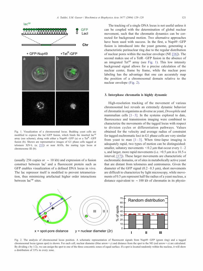
Fig. 1. Visualization of a chromosomal locus. Budding yeast cells are
modified to express the laci-GFP fusion, which binds the inserted lacop
array (see scheme), along with either a Nup49–GFP (a) or a TetR–GFP
fusion (b). Shown are representative images of G1 phase cells tagged at
telomere XIV-L (a; [33]) or near MATa, the mating type locus at
chromosome III (b).
A. Taddei, S.M. Gasser / Biochimica et Biophysica Acta 1677 (2004) 120–128 121
(usually 256 copies or f 10 kb) and expression of a fusion
construct between laci and a fluorescent protein such as
GFP enables visualization of a defined DNA locus in vivo.
The lac repressor itself is modified to prevent tetrameriza-
tion, thus minimizing artefactual higher order interactions
between lacop sites.
Fig. 2. The analysis of chromosomal locus position. A schematic representat
chromosomal locus (green spot) is shown. For each cell, nuclear diameter (blue ar
By dividing x by 1/2y, we can assign the spot to one of the three concentric zones o
a distribution of 33% in every zone.
The tracking of a single DNA locus is not useful unless it
can be coupled with the determination of global nuclear
movement, such that the chromatin dynamics can be cor-
rected for background motion. Two alternative approaches
have been used with success. In the first, a Nup49–GFP
fusion is introduced into the yeast genome, generating a
characteristic perinuclear ring due to the regular distribution
of nuclear pores within the nuclear envelope (NE [16]). The
second makes use of a TetR–GFP fusion in the absence of
an integrated Tetop array (see Fig. 1). This low intensity
background signal allows for a precise calculation of the
nuclear center, frame by frame, while the nuclear pore
labeling has the advantage that one can accurately map
the position of a chromosomal domain relative to the
nuclear envelope (Fig. 2).
3. Interphase chromatin is highly dynamic
High-resolution tracking of the movement of various
chromosomal loci reveals an extremely dynamic behavior
of chromatin in organisms as diverse as yeast,Drosophila and
mammalian cells [1–3]. In the systems explored to date,
fluorescence and transmission imaging were combined to
characterize the movements of the tagged locus with respect
to division cycles or differentiation pathways. Values
obtained for the velocity and average radius of constraint
for tagged euchromatic loci in G1 phase cells are very similar
from yeast to man [1–3]. When time-lapse imaging is
adequately rapid, two types of motion can be distinguished:
smaller, saltatory movements < 0.2 Am that occur every 1–2
s, and larger, more rapid movements (i.e. >0.5 Am in a 10.5-s
interval; [17]). These larger movements are characteristic of
euchromatic domains, or of sites in metabolically active yeast
that are distant from telomeres and centromeres. Given the
diameter of the GFP signal (0.2–0.3 Am), short movements
are difficult to characterize by light microscopy, while move-
ments of 0.5 Am represent half the radius of a yeast nucleus, a
distance equivalent to f100 kb of chromatin in its physio-
ion of fluorescent signals from Nup49–GFP (green ring) and a tagged
row= y) and distance from the spot to the NE (red arrow= x) are calculated.
f equal surface. If a spot is located randomly within the nucleus, it will show

A. Taddei, S.M. Gasser / Biochimica et Biophysica Acta 1677 (2004) 120–128122
logical compaction state, which ranges fromf 51- to 80-fold
in interphase nuclei of yeast and human cells (Bystricky et al.,
submitted for publication, [18]).
In yeast, chromatin movements are not microtubule- or
actin filament-dependent although they are sensitive to
ATP depletion [17]. In Drosophila spermatocytes, individ-
ual step sizes have a roughly Gaussian distribution,alth-
hough the direction of the motion changes every two to
four movements [19]. In other words, large movements in
one direction frequently precede an equally large step
back, consistent with the idea that the contiguous chroma-
tin fiber imposes spatial constraints on locus movement.
Similar patterns are observed for 0.5-Am movements in
yeast nuclei (M. Blaszczyk, personal communication).
Nonetheless, within a limited space, on the other hand,
movement can be modeled as a random walk.
The correlation of interphase dynamics in yeast with
changes in cellular metabolism, notably depletion of glu-
cose, makes it unlikely that chromatin movement results
from simple diffusion. We suggest that the movement
reflects the action of large ATP-dependent enzymes as they
activate transcription or remodel nucleosomes. This would
be consistent with the lack of mobility detected in stationary
phase cells where transcriptional activity drops significantly,
or in cells in which membrane potentials are collapsed,
leading to the depletion of ATP [17].
4. The extent of chromatin movement
When thousands of measurements for a GFP-tagged locus
are compared over time, either as a relative movement
between two spots in a diploid or the movement of one locus
relative to the nuclear center, the spatial constraints imposed
on the movement can be calculated by plotting the mean
squared displacement (MSD) over fixed intervals of time
[20]. These MSD plots reach a plateau at larger time intervals
which allows one to calculate the radius of constraint on a
given tagged locus, and to compare this reliably among
different chromosomal loci in different species. The move-
ment of euchromatic sites in flies, man and yeast is confined
to volumes off0.5–0.7 Am in radius, significantly less than
even the radius of a yeast nucleus (1 Am) [1–3]. In S phase,
yeast chromosomal loci show a significant decrease in
mobility [17]. When loci on the X chromosome in Drosoph-
ila spermatocytes were monitored over longer time periods in
G2 phase, a slow, but longer-range migration could be
detected in addition to the rapid more constrained motion
[19]. Interestingly, in budding yeast, an excised ring of
chromatin was shown to move without detectable constraint
throughout the nucleus, suggesting that there is no inherent
barrier to chromatin movement in yeast nucleoplasm (Gar-
tenberg et al., submitted for publication).
Observing this degree of mobility for multiple loci, one
must reconsider the hypothesis that each eukaryotic chro-
mosome is assigned to a discrete subnuclear territory,
surrounded by inter-chromatin channels that facilitate mac-
romolecular movement [21]. While the data on dynamics
do not refute the notion of nuclear compartments, the
model must now accommodate a significant degree of
intermingling of chromatin domains between chromosomal
territories. Moreover, there is no apparent need for mac-
romolecular transport to be restricted to interchromatin
channels, since chromatin itself is so mobile that it could
hardly impair macromolecular diffusion. Particularly in
yeast, it seems improbable that neighboring chromosomes
remain segregated from each other given that the yeast
nuclear diameter is f2 Am and the radii of confinement
for specific loci are f0.5 Am. The unusual efficiency of
homologous recombination events in yeast may in part
reflect this high degree of chromatin intermingling. Even
in human cells, tagged chromosomal regions have been
reported to localize outside of the visible confines of
chromosome territories. Although the numbers analyzed
are still small, sites that ‘‘loop out’’ from a chromosomal
territory correlate with regions of high gene density and
high levels of expression [22]. In summary, live imaging
documents significant local chromatin movement, requir-
ing revision of our concepts of chromatin and chromo-
somal compartments.
In view of the dynamic character of chromatin, it seems
likely that anchorage sites may be functionally more impor-
tant as regulators than chromatin mobility itself. For exam-
ple, the sites at which yeast telomere cluster may play roles
in the proper replication of chromosomal ends or in the
regulation of silencing. Long-range interactions between
blocks of heterochromatin in other species also probably
facilitate a mass action assembly of repressors with their
binding sites [23]. In various cells, recruitment and stable
association of damaged DNA sites into internal foci has
been correlated with relatively fixed sites of repair [24,25].
Finally, it has been proposed that the insulators that create
boundaries between active and inactive chromatin domains
may also function by tethering these sites [26,27]. Whether
this is due to position or to reduced mobility of chromatin is
unknown. In each case, the imposition of spatial constraints
on a locus seems to correlate with a regulatory mechanism.
5. Redundant mechanisms of chromatin attachment
In general, we conclude that chromatin is highly mobile
except at irregularly spaced sites of anchorage that punctuate
the chromatin by tethering DNA to the nuclear envelope or to
other substructures of the nucleus. These interactions would,
on one hand, constrain the global diffusion of chromosomes.
On the other hand, release from such constraint could also be
exploited locally to help alter chromatin structure at active
genes, facilitate macromolecular diffusion, or promote rec-
ognition site searches within the nucleus [2].
Yeast telomere clusters are thought to define a relatively
stable subnuclear compartment that favors chromatin-medi-

A. Taddei, S.M. Gasser / Biochimica et Biophysica Acta 1677 (2004) 120–128 123
ated repression. Consistently, images taken at fixed time-
points reveal telomeric foci near the nuclear envelope [28–
30]. Individually tagged telomeres, as well as GFP-labelled
foci, are nonetheless themselves dynamic, moving signifi-
cantly more than the spindle pole body, an integral NE
component. Unlike internal tagged sites, most telomeres are
lateral, i.e within an outer perinuclear ring. This patten could
arise from a pattern that could arise from a reversible
association with multiple dispersed binding sites along the
NE. Perhaps the abundance of these anchorage sites allows
telomeres to be trapped within a perinuclear zone; telomeres
seem to be sequentially bound and released, resulting in a
‘‘walk’’ along NE sites. FISH data using YV and TG repeats
suggest that in both wild-type and yku-deficient cells, entire
clusters of telomeres can be displaced from the periphery
and not only individual ends [30,31], distinguishing telo-
mere anchorage from the telomere–telomere interactions
that lead to clustering.
Recent analyses of native telomere behavior in living
yeast cells have resolved several discrepancies in the liter-
ature about the character of telomere binding sites [32–36].
Two partially redundant pathways are found to mediate
anchorage: one that requires the heterodimeric yKu factor,
and a second mediated by one or more of the Sir proteins
[33]. Both yKu and Sir proteins are integral components of
the telosome and of subtelomeric chromatin, and could thus
play a direct role in tethering. The Sir-dependent pathway
correlates with chromatin-mediated transcriptional repres-
sion, may require higher-order chromatin structures, and is
able to tether a native telomere in S phase in the absence of
yKu. The yKu pathway, on the other hand, anchors a
fraction of telomeres throughout interphase, even in the
absence of TPE (i.e. in sir-deficient strains, see Fig. 3 and
Fig. 3. Chromatin tracking reveals spatial constraints on chromatin dynamics. Us
fluorescence were captured (at 1.5-s intervals) in G1 or S phase yeast cells carryin
were first aligned based on their nuclear pore signals, and projected on a single ima
nuclear section (right hand image). To quantify spatial constraints on these moveme
subjected to the MSD analysis. After alignment of nuclear centers the absolute pos
displacement of the locus for all intervals from 0 to 150 s (Dt, s) was computed u
each Dt value are plotted against Dt. The slope is the derivative of the diffusion
modeling randomly generated values within specific-sized surfaces (F. Neumann a
cell cycle influences the degree of spatial freedom in its movement (compare G1
Ref. [33]). Thus, anchoring does not necessarily correlate
with repression [33,35]. On the other hand, for telomeres
that are less dependent on the yKu anchorage pathway, the
Sir-dependent pathway anchors in S phase, and in this case
efficient anchorage correlates with repression. Such analy-
sis, extended to three independently tagged telomeres con-
taining subtelomeric repeats, confirms the generality of
these redundant anchorage mechanisms, which in addition
are subject to cell cycle regulation (Fig. 4).
6. Subtelomeric repeats can provide ‘‘anti-silencing’’
and ‘‘anti-anchor’’ functions
Given this redundancy, what regulates the use of one
anchor or the other? Preliminary evidence suggests that
subtelomeric sequence organization itself influences telo-
mere positioning in subtle ways. This is supported by two
sets of data: first there is significant variation in the
anchoring efficiencies among native telomeres, and second,
truncated telomeres do not behave the same as ends that
contain subtelomeric repeats [33,35]. With respect to the
first point, we note that the native Tel VI-R end, which has
the X but not the YV and STR elements, is displaced
throughout the cell cycle in a yku-deficient strain; Tel XIV-
L, which has YV, X and STR sequences, on the other hand,
is sensitive to yKu deletion only in G1 phase. This residual
S phase anchoring reflects the contributions of the Sir-
dependent pathway. We predict that not only a telomere’s
insensitivity to yKu deletion but also its anchoring effi-
ciency in wild-type S phase cells is likely to correlate with
the variability of native subtelomeric repression [37,38]. A
systematic analysis of repression at native ends and yKu-
ing a LSM510 (Zeiss) confocal microscope, 200 sequential images of GFP
g a lacop insert in the middle of Chromosome XIV (ARS1413). The images
ge (left hand image). The track of the locus is projected as a red trace on one
nts, eight movies (each with 200 sequential frames at 1.5 sec intervals) were
ition of the focus was determined for each frame, and then the square of the
sing the formula: Dd2={d(t)� d(t +Dt)}2. The average of all Dd2 values for
coefficient and a plateau indicates spatial constraint as determined from
nd M. Blaszczyk, personal communication). For ARS1413 the stage of the
vs. S [17]).

A. Taddei, S.M. Gasser / Biochimica et Biophysica Acta 1677 (2004) 120–128124
independent anchoring will be necessary to confirm this
hypothesis.
With respect to the effect of subtelomeric repeats, it
was shown that a truncated Tel VI-R (which has TG
repeats but no X or YV elements) remains anchored at
the periphery in the absence of yKu, while the native Tel
VI-R is released. This residual yKu-independent anchoring
is Sir4-dependent, even though the efficiency of TPE is
low. The Sir-dependent anchoring may result from a
residual binding of Sir4 to Rap1, which can be detected
in both yku and sir3 mutants [8,9,39]. Surprisingly, the
Sir4–Rap1 interaction is sufficient to tether a truncated,
but not a native telomere.
To account for the differences between artificial and
native telomeric ends, we suggest that subtelomeric
repeats interfere with the Sir-dependent pathway at native
telomeres, acting as an anti-anchor. In other words,
subtelomeric repeats render native telomere positioning
sensitive to the loss of yKu, while truncated telomeres are
not [33]. This is consistent with recent observations
showing that subtelomeric sequences either disfavor re-
pression of reporter genes or cause an abrupt drop in its
propagation [37,38]. In the yku rif1 double mutant, which
allows the restoration of telomeric silencing in the ab-
sence of yKu, the Sir pathway can restore end attachment
in S phase only [33]. This may reflect an S-phase-specific
change in Sir proteins or their target at the periphery,
allowing native telomeres to reattach, or this may reflect a
conformational change in proteins associated with the
subtelomeric X element (Fig. 4). These observations
corroborate results showing that passage through S phase,
but not DNA replication itself, is necessary for the
establishment of silent chromatin [40,41]. While it
remains unclear how S-phase transition facilitates repres-
sion, it is undoubtedly relevant that it correlates with the
Sir-dependent mechanism of anchoring.
Fig. 4. yKu- and Sirs-dependent mechanisms tether telomeres in yeast. During G1
native telomere anchoring, suggesting that the Sir-mediated pathway may be less
telomeres even in the absence of Sir-mediated silencing (see sir� ). In S phase
efficiently, and anchorage correlates with silent chromatin even in a strain deficient
gray bar) directly or indirectly impair the ability of Sir proteins to anchor native
telomere anchoring correlates with the level of silencing.
7. Candidates for perinuclear anchorage sites
Because yeast has no homologues to the nuclear lamina,
and because neither yKu nor Sir proteins have membrane
spanning domains, other NE components must be responsible
for tethering telomeres at the nuclear periphery [42,43]. One
of these appears to be Esc1, a protein that interacts with Sir4
and is required for the stable mitotic partitioning of a Sir4-
bound plasmid [42]. Importantly, Esc1 is localized on the
nucleoplasmic surface of the inner bilayer of the NE, being
positioned primarily between pores and excluded from the
nucleolus [44]. Moreover, Esc1 is necessary both for efficient
TPE and the yKu-independent anchoring of Tel XIV-L and
truncated Tel VI-R, making it a good candidate to be the
natural anchor of Sir4 (Taddei et al., submitted for publica-
tion). The major anchorage site of yKu is not esc1-dependent,
because esc1 deletion does not affect the general localization
of telomeres as analyzed by YV FISH [42].
In other works, Mlp1 and Mlp2 were proposed to form a
bridge between yKu and nuclear pores [34,36]. However,
careful analysis using both live GFP imaging and in situ
hybridization showed no influence of the double mlp1 mlp2
deletion on telomere positioning nor on dynamics when
intact nuclei are analyzed [43]. Thus, the pore-associated
Mlp proteins are also unlikely to participate as the anchor
through which yKu tethers telomeres to the NE. Consistent-
ly, TPE has been shown by several groups to be intact in the
double mlp1 mlp2 mutant, which is not the case in yku or
esc1 mutants [42,43]. Nonetheless, given their perinuclear
localization and coiled-coil structure, Mlp proteins may be
involved in other aspects of nuclear envelope function.
Double mutant and overexpression data suggest that Mlp
proteins are involved in aspects of mRNA biogenesis ([45];
F. Stutz, personal communication). A general role in the
distribution of non-telomere associated NE components is
also possible. To date, however, there is no support for the
phase in wild-type yeast cells the yKu-mediated pathway is necessary for
efficient in this phase of the cell cycle. yKu mediates anchoring of native
cells, on the other hand, the Sir-mediated anchoring pathway functions
for yKu (see yku� and [33]). We propose that subtelomeric repeats (hatched
ends in G1 phase, while in S phase both pathways function in parallel and

Fig. 5. The positive effect of perinuclear localization on silencing depends on the sequestering of Sir proteins at telomere foci. The left-hand nucleus reflects a
wild-type yeast cell in which Sir proteins are sequestered at telomere foci (red areas), such that internal reporter with weak silencers are not silent unless they
are anchored at the NE by a transmembrane protein (e.g. GBD-YIF1 [52]). In the right-hand nucleus, the Sir proteins are distributed throughout the nucleus due
to yKu deletion. In this strain, tethering the crippled silencer to the nuclear periphery does not promote silencing. On the other hand, local recruitment of Sir
proteins (e.g. by GBD-yKu80) can promote repression.
A. Taddei, S.M. Gasser / Biochimica et Biophysica Acta 1677 (2004) 120–128 125
model that telomeres are anchored to nuclear pores, as
nuclear pore complexes can be clearly distinguished from
most telomeric foci in stained wild-type cells [30], and both
telomeres and Esc1p remain evenly distributed in a
nup133D mutant despite a dramatic clustering of nuclear
pores [43,44].
In summary, we propose that the inner nuclear envelope
has various differentiated zones that are defined by the
presence of different families of structural proteins. Among
these are likely to be Esc1, Mlp1/Mlp2, elements of the
spindle pole body and other proteins that may associate with
pores. Nuclear pores may play a role in the general
partitioning of such zones.
Fig. 6. Model for the role chromatin anchoring for the promotion of
silencing. We propose that Sir4 is first recruited at the nucleation center by
DNA binding proteins that can bind Sir proteins. These include Rap1,
ORC, Abf1 and yKu. The presence of Sir4 at the locus will then bring it to
the nuclear periphery through one of the two Sir4 anchoring pathways (yKu
or Esc1) where the high local concentrations of Sir proteins will help
silencing complexes assemble and spread. The anchoring of silent loci at
the periphery will increase the concentration of Sir proteins and reinforce
the silencing of other loci within this region. In addition, yKu, bound to
chromosome ends, can independently recruit telomeres to the NE.
8. Testing the role of perinuclear anchoring in
repression
It has been proposed that the anchorage of telomeres at
the nuclear periphery concentrates silencing factors thus
favoring silencing and causing its accumulation in this
nuclear subcompartment [12]. This model is based on
several observations: the first is that normal cellular Sir
protein concentrations are limiting for repression [12,46,47].
Second, we know that silencer-flanked reporter genes are
more efficiently repressed when inserted near the telomere
[12,48–50]. Third, it was shown that the delocalization of
Sir factors from telomeres, as well as their overexpression,
is able to restore repression at loci distant from telomeres
[12,51]. This confirms that telomeric foci sequester repress-
ors from other sites of action.
In support of a functional role for telomere clustering, the
Sternglanz laboratory showed that by artificially ‘‘tethering’’
a reporter gene to the yeast NE through a membrane-
spanning polypeptide, transcriptional repression could be
favored [52] (Fig. 5). This was shown to require the
presence of at least one silencer element, a specific cis-
acting sequence that nucleates Sir-dependent repression. It
was proposed that the NE ‘‘tether’’ was able to compensate
for the crippled silencer and to promote Sir-dependent
repression because it placed the weak silencer near a zone
of high Sir protein concentration. Indeed, consistent with
this model, NE tethering of this reporter was found to
promote silencing only if telomeres were able to sequester
Sir proteins in perinuclear pools (Taddei et al., unpublished
results, and Fig. 5).
We propose that silencing depends on anchoring only
when Sir protein concentrations are limiting within the
nucleus, a condition that results in part from the recruitment
of Sir proteins into pools at the nuclear periphery. From this
it follows that silencing can be achieved without (or prior to)
NE anchoring if Sir proteins can be attracted to the reporter
gene in sufficient amounts to achieve a threshold concen-
tration at a given nucleation site (Fig. 6). If Sir proteins are
not recruited with sufficient efficiency, then relocalization to

A. Taddei, S.M. Gasser / Biochimica et Biophysica Acta 1677 (2004) 120–128126
a zone of high Sir concentration should help the silencing
process. Moreover, it follows that there should be no
silencing of loci induced by placement at the nuclear
periphery per se; indeed, a peripheral localization is not
sufficient for transcriptional repression of truncated telo-
meres in yku mutants [33,35] nor of reporter genes associ-
ated with a weak silencer (Taddei et al., unpublished
results). Using readily manipulated assay systems, one is
able to demonstrate a function for the perinuclear clustering
of telomeres, and to distinguish this from a role of the
nuclear envelope per se, despite the redundancy built into
the system of silencing. Least understood in this process is
the mechanism that overcomes equilibrium dynamics to
concentrate silencing factors in perinuclear foci.
9. Anchoring can be both a cause and a result of
transcriptional repression
Do changes in subnuclear position cause or result from
transcriptional silencing? As summarized in Fig. 4 previous
studies clearly implicated yKu and the Sir proteins in
anchoring, but it was unclear whether the loss of telomere
position was due to the direct elimination of the chromatin
anchoring polypeptide, or to the loss of silent chromatin.
Using subdomains or mutant proteins in a novel anchoring
assay, we have now been able to identify yKu and the
Sir4PAD domain as minimal anchors that can tether chromatin
to NE in the absence of transcriptional repression [44]. When
targeted to a tagged locus, the Sir4PAD domain can relocate
this DNA to the NE without repressing transcription through
two partially redundant pathways. The first requires Esc1
and the other yKu70/yKu80. Although this shows that
anchoring can be mediated through a subdomain of a silent
regulatory protein, it also provides a means for silent
chromatin to localize itself to the nuclear periphery. This
has been demonstrated by showing that an excised HMR
mating-type locus is autonomously anchored at the nuclear
periphery in a silencing-dependent manner (Gartenberg et
al., submitted for publication).
These results lead to a model that explains how a Sir-rich
compartment can form spontaneously in the nucleus to favor
and maintain Sir-mediated repression (Fig. 6). Central to
this model is the demonstration of silencing-independent
anchorage via yKu or Sir4, which provides a means for
telomeres to accumulate at the nuclear envelope prior to the
formation of a repressed state. The anchoring of telomeric
repeats creates a large number of potential Sir4 protein
binding sites within a restricted volume due to the presence
of 20 to 25 Rap1 consenses on each telomere end. The ORC
sites within subtelomeric repeats can also contribute to the
efficiency of Sir4 accumulation. The resulting high density
of Sir4-binding chromatin would in turn attract Sir2 and
Sir3 through protein–protein interactions. It is not clear
whether only the presence of Sir proteins or the presence of
another molecular catalyst is needed to promote formation
of silent chromatin. Nonetheless, the local concentration of
silencing factors is clearly a necessary prerequisite for
repression. The concentrated Sir factors should then rein-
force silencing of loci within this compartment. As silent
chromatin spreads, the number of Sir4 molecules bound will
increase and reinforce interaction with the NE. This creates
a feed-back loop that is initiated by a limited number of yKu
or Sir4 proteins bound at given site. Consistent with this
model, Chromatin Immunoprecipitation experiments
showed that Sir4 is the first component bound in the
assembly process of silent chromatin at HM loci and at
telomeres [8–11].
This clustering–feedback loop provides a mechanism for
self-organization of a subnuclear compartment in which
silencing factors and repressed domains accumulate. We
envision such mechanisms as also being relevant for cen-
tromeric satellite and heterochromatin sequences in higher
eukaryotes, whose clustering correlates with high concen-
trations of heterochromatin factors like HP1.
10. A general model for heterochromatin formation and
clustering
The tethering of chromatin, and most frequently hetero-
chromatin, at the nuclear envelope is a universal phenomenon
that has long been observed in higher eukaryotes [53–56]. In
most eukaryotes, a rigid, perinuclear lamina meshwork is
bound to the nuclear envelope through membrane-spanning
lamin receptors [57]. In flies, points of contact between the
lamina meshwork and chromosomes appear to occur irregu-
larly along the chromosomal arm, as detected by high-
resolution FISH studies [56]. The sites that tether mammalian
chromosomes are not yet defined in molecular terms, but
through cytologically dense staining, transcriptionally inac-
tive chromatin is systematically enriched at the lamin–
nucleoplasm interface. The exception to this is the chromatin
immediately internal to nuclear pores, which is generally less
condensed. Yeast, plants and many protozoa have no homo-
logues of the nuclear intermediate filament family (i.e. the
lamins [58,59]). The absence of this perinuclear intermediate
filament network undoubtedly accounts for sensitivity of
yeast nuclei to nonionic detergents, and renders yeast nuclear
integrity more dependent on pore components.
The redundancy of anchoring pathways is consistent with
the idea that telomere anchoring is physiologically impor-
tant. Indeed, mechanisms seem to be in place to compensate
for loss of anchoring: a yeast yku70 mutant was shown to
switch between two epigenetic states, one in which telo-
meres were delocalized, and another in which their perinu-
clear organization was largely restored [51]. In the latter
state, Sirs were seen to cluster in one bright focus showing
that both clustering and perinuclear anchoring of telomeres
occur in the absence of yKu.
Mechanisms that promote clustering as opposed to an-
choring of yeast telomeres are unknown, although telo-

A. Taddei, S.M. Gasser / Biochimica et Biophysica Acta 1677 (2004) 120–128 127
mere– telomere interactions occur in Plasmodia, fission
yeast and possibly in human cells as well. In Plasmodia
subtelomeric repeats have been shown to contribute to the
clustering, but not to the peripheral anchoring, of this
parasite’s telomeres [60]. Similarly, RNA processing
mutants in fission yeast also appear to affect clustering
without altering the perinuclear positioning of telomeres,
suggesting that clustering and tethering are mediated by
genetically distinct mechanisms [61].
To examine clustering in living yeast cells and to compare
it to the anchoringmechanisms described above, onemust tag
multiple chromosomal ends for fluorescence microscopy,
identify each telomere’s partners, and characterize mutants
that disrupt clustering, as opposed to perinuclear binding.
Data from higher organisms suggest that components of silent
chromatin may be involved. For example, the interaction
between ectopic satellite repeats and centric heterochromatin
in flies is sensitive to the mutation of the histone-binding
heterochromatin protein 1 (HP1 [62]). Analogous clustering
mechanisms appear to function during gene repression events
in differentiated mammalian cells, as demonstrated for
Ikaros-controlled genes in mouse lymphocytes [63]. Deci-
phering the cross-talk between the mechanisms that anchor
and those that cluster heterochromatin is a major challenge
that needs to be met to fully decipher the role of nuclear
compartmentation in regulated gene expression.
Our limited understanding of the yeast nuclear envelope
includes pores, telomeric foci and membrane anchorage
sites, yet differentiated zones that facilitate more diverse
nuclear activities must exist [44]. For instance, telomere
replication may require different peripheral domains than
transcriptional repression [64]. It is important to examine
carefully how individual domains and regulatory events are
independently organized to be able to characterize different
peripheral zones. Such information may also reveal why the
silencing at clustered telomeres is different from that of
reporters that are anchored to the periphery by nonspecific
membrane domains [65].
Acknowledgements
We thank Thierry Laroche and Dr Patrick Heun for
pioneering work on live microscopy, Florence Hediger,
Frank Neumann, Drs Mark Gartenberg, Kerstin Bystricky,
Francoise Stutz and Marek Blaszczyk for having shared
results and ideas, and members of the Gasser laboratory for
helpful discussions. Our research is supported by the Swiss
National Science Foundation and the NCCR program
‘‘Frontiers in Genetics’’. A.T. is supported by an EMBO
fellowship.
References
[1] Marshall, Order and disorder in the nucleus, Curr. Biol. 12 (2002)
R185–R192.
[2] Gasser, Visualizing chromatin dynamics in interphase nuclei, Science
296 (2002) 1412–1416.
[3] J.R. Chubb, W.A. Bickmore, Considering nuclear compartmentaliza-
tion in the light of nuclear dynamics, Cell 112 (2003) 403–406.
[4] M. Gotta, S.M. Gasser, Nuclear organization and transcriptional si-
lencing in yeast, Experientia 52 (1996) 1136–1147.
[5] A. Hecht, T. Laroche, S. Strahl-Bolsinger, S.M. Gasser, M. Grunstein,
Histone H3 and H4 N-termini interact with SIR3 and SIR4 proteins: a
molecular model for the formation of heterochromatin in yeast, Cell
80 (1995) 583–592.
[6] S. Strahl-Bolsinger, A. Hecht, K. Luo, M. Grunstein, SIR2 and SIR4
interactions differ in core and extended telomeric heterochromatin in
yeast, Genes Dev. 11 (1997) 83–93.
[7] D.E. Gottschling, O.M. Aparicio, B.L. Billington, V.A. Zakian, Po-
sition effect at S. cerevisiae telomeres: reversible repression of Pol II
transcription, Cell 63 (1990) 751–762.
[8] B.D. Bourns, M.K. Alexander, A.M. Smith, V.A. Zakian, Sir proteins,
Rif proteins, and Cdc13p bind Saccharomyces telomeres in vivo, Mol.
Cell. Biol. 18 (1998) 5600–5608.
[9] K. Luo, M.A. Vega-Palas, M. Grunstein, Rap1–Sir4 binding indepen-
dent of other Sir, yKu, or histone interactions initiates the assembly of
telomeric heterochromatin in yeast, Genes Dev. 12 (2002) 1528–1539.
[10] L.N. Rusche, A.L. Kirchmaier, J. Rine, Ordered nucleation and
spreading of silenced chromatin in Saccharomyces cerevisiae, Mol.
Biol. Cell 7 (2002) 2207–2222.
[11] G.J. Hoppe, J.C. Tanny, A.D. Rudner, S.A. Gerber, S. Danaie, S.P.
Gygi, D. Moazed, Steps in assembly of silent chromatin in yeast: Sir3-
independent binding of a Sir2/Sir4 complex to silencers and role for
Sir2-dependent deacetylation, Mol. Cell. Biol. 12 (2002) 4167–4180.
[12] Maillet, C. Boscheron, M. Gotta, S. Marcand, E. Gilson, S.M. Gasser,
Evidence for silencing compartments within the yeast nucleus: a role
for telomere proximity and Sir protein concentration in silencer-medi-
ated repression, Genes Dev. 10 (1996) 1796–1811.
[13] M. Platani, I. Goldberg, J.R. Swedlow, A.I. Lamond, In vivo analysis
of Cajal body movement, separation, and joining in live human cells,
J. Cell Biol. 151 (2000) 1561–1574.
[14] C.C. Robinett, A. Straight, G. Li, C. Willhelm, G. Sudlow, A. Murray,
A.S. Belmont, In vivo localization of DNA sequences and visualiza-
tion of large-scale chromatin organization using lac operator/repressor
recognition, J. Cell Biol. 135 (1996) 1685–1700.
[15] A.S. Belmont, Visualizing chromosome dynamics with GFP, Trends
Cell Biol. 11 (2001) 250–257.
[16] N. Belgareh, V. Doye, Dynamics of nuclear pore distribution in nucle-
oporin mutant yeast cells, J. Cell Biol. 136 (1997) 747–759.
[17] P. Heun, T. Laroche, K. Shimada, P. Furrer, S.M. Gasser, Chromo-
some dynamics in the yeast interphase nucleus, Science 294 (2001)
2181–2186.
[18] B. Trask, D. Pinkel, G. van den Engh, The proximity of DNA sequen-
ces in interphase cell nuclei is correlated to genomic distance and
permits ordering of cosmids spanning 250 kilobase pairs, Genomics
5 (1989) 710–717.
[19] J. Vazquez, A.S. Belmont, J.W. Sedat, Multiple regimes of con-
strained chromosome motion are regulated in the interphase Drosoph-
ila nucleus, Curr. Biol. 11 (2001) 1227–1239.
[20] W.F. Marshall, A. Straight, J.F. Marko, J. Swedlow, A. Dernburg, A.
Belmont, A.W. Murray, D.A. Agard, J.W. Sedat, Interphase chromo-
somes undergo constrained diffusional motion in living cells, Curr.
Biol. 7 (1997) 930–939.
[21] T. Cremer, A. Kurz, R. Zirbel, S. Dietzel, B. Rinke, E. Schrock, M.R.
Speicher, U. Mathieu, A. Jauch, P. Emmerich, et al, Role of chromo-
some territories in the functional compartmentalization of the cell
nucleus, Cold Sring Harbor Symp. Quant. Biol. 58 (1993) 777–792.
[22] N.L. Mahy, P.E. Perry, W.A. Bickmore, Gene density and transcrip-
tion influence the localization of chromatin outside of chromosome
territories detectable by FISH, J. Cell Biol. 159 (2002) 753–763.
[23] S. Henikoff, Dosage-dependent modification of position-effect varie-
gation in Drosophila, BioEssays 18 (1996) 401–409.

A. Taddei, S.M. Gasser / Biochimica et Biophysica Acta 1677 (2004) 120–128128
[24] Y. Liu, M. Li, E.Y. Lee, N. Maizels, Localization and dynamic reloc-
alization of mammalian Rad52 during the cell cycle and in response to
DNA damage, Curr. Biol. 9 (1999) 975–978.
[25] M. Lisby, R. Rothstein, U.H. Mortensen, Rad52 forms DNA repair
and recombination centers during S phase, Proc. Natl. Acad. Sci.
U. S. A. 98 (2001) 8276–8282.
[26] K. Ishii, G. Arib, C. Lin, G. Van Houwe, U.K. Laemmli, Chromatin
boundaries in budding yeast. The nuclear pore connection, Cell 109
(2002) 551–562.
[27] T.I. Gerasimova, V.G. Corces, Polycomb and trithorax group proteins
mediate the function of a chromatin insulator, Cell 92 (1998) 511–521.
[28] Palladino, T. Laroche, E. Gilson, A. Axelrod, L. Pillus, S.M. Gasser,
SIR3 and SIR4 proteins are required for the positioning and integrity
of yeast telomeres, Cell 75 (1993) 543–555.
[29] H. Funabiki, I. Hagan, S. Uzawa, M. Yanagida, Cell cycle-dependent
specific positioning and clustering of centromeres and telomeres in
fission yeast, J. Cell Biol. 121 (1993) 961–976.
[30] M. Gotta, T. Laroche, A. Formenton, L. Maillet, H. Scherthan, S.M.
Gasser, The clustering of telomeres and colocalization with Rap1,
Sir3, and Sir4 proteins in wild-type Saccharomyces cerevisiae, J. Cell
Biol. 134 (1996) 1349–1363.
[31] T. Laroche, S.G. Martin, M. Gotta, H.C. Gorham, F.E. Pryde, E.J.
Louis, S.M. Gasser, Mutation of yeast Ku genes disrupts the subnu-
clear organization of telomeres, Curr. Biol. 8 (1998) 653–656.
[32] W.H. Tham, V.A. Zakian, Telomeric tethers, Nature 403 (2000) 34–35.
[33] F. Hediger, F.R. Neumann, G. Van Houwe, K. Dubrana, S.M. Gasser,
Live imaging of telomeres. yKu and Sir proteins define redundant
telomere-anchoring pathways in yeast, Curr. Biol. 12 (2002)
2076–2089.
[34] V. Galy, J.C. Olivo-Marin, H. Scherthan, V. Doye, N. Rascalou, U.
Nehrbass, Nuclear pore complexes in the organization of silent telo-
meric chromatin, Nature 403 (2000) 108–112.
[35] W.H. Tham, J.S. Wyithe, P.K. Ferrigno, P.A. Silver, V.A. Zakian,
Localization of yeast telomeres to the nuclear periphery is separable
from transcriptional repression and telomere stability functions, Mol.
Cell 8 (2001) 189–199.
[36] F. Feuerbach, V. Galy, E. Trelles-Sticken, M. Fromont-Racine, A.
Jacquier, E. Gilson, J.C. Olivo-Marin, H. Scherthan, U. Nehrbass,
Nuclear architecture and spatial positioning help establish transcrip-
tional states of telomeres in yeast, Nat. Cell Biol. 4 (2002) 214–221.
[37] G. Fourel, E. Revardel, C.E. Koering, E. Gilson, Cohabitation of
insulators and silencing elements in yeast subtelomeric regions,
EMBO J. 18 (1999) 2522–2537.
[38] F.E. Pryde, E.J. Louis, Limitations of silencing at native yeast telo-
meres, EMBO J. 18 (1999) 2538–2550.
[39] S.G. Martin, T. Laroche, N. Suka, M. Grunstein, S.M. Gasser, Reloc-
alization of telomeric Ku and SIR proteins in response to DNA strand
breaks in yeast, Cell 97 (1999) 621–633.
[40] Y.C. Li, T.H. Cheng, M.R. Gartenberg, Establishment of transcrip-
tional silencing in the absence of DNA replication, Science 291
(2001) 650–653.
[41] A.L. Kirchmaier, J. Rine, DNA replication-independent silencing in S.
cerevisiae, Science 291 (2001) 646–650.
[42] E.D. Andrulis, D.C. Zappulla, A. Ansari, S. Perrod, C.V. Laiosa, M.R.
Gartenberg, R. Sternglanz, Esc1, a nuclear periphery protein required
for Sir4-based plasmid anchoring and partitioning, Mol. Cell. Biol. 22
(2002) 8292–8301.
[43] F. Hediger, K. Dubrana, S.M. Gasser, Myosin-like proteins 1 and 2
are not required for silencing or telomere anchoring, but act in the
Tel1 pathway of telomere length control, J. Struct. Biol. 140 (2002)
79–91.
[44] A. Taddei, F. Hediger, F.R. Neumann, C. Bauer, S.M. Gasser, Sepa-
ration of silencing from perinuclear anchoring functions in yeast
Ku80, Sir4 and Esc1 proteins, EMBO J. (2003) in press.
[45] D.M. Green, C.P. Johnson, H. Hagan, A.H. Corbett, The C-terminal
domain of myosin-like protein 1 (Mlp1p) is a docking site for het-
erogeneous nuclear ribonucleoproteins that are required for mRNA
export, Proc. Natl. Acad. Sci. U. S. A. 100 (2003) 1010–1015.
[46] H. Renauld, O.M. Aparicio, P.D. Zierath, B.L. Billington, S.K. Chha-
blani, D.E. Gottschling, Silent domains are assembled continuously
from the telomere and are defined by promoter distance and strength,
and by SIR3 dosage, Genes Dev. 7 (1993) 1133–1145.
[47] S.W. Buck, D. Shore, Action of a RAP1 carboxy-terminal silencing
domain reveals an underlying competition between HMR and telo-
meres in yeast, Genes Dev. 9 (1995) 370–384.
[48] J.S. Thompson, L.M. Johnson, M. Grunstein, Specific repression of
the yeast silent mating locus HMR by an adjacent telomere, Mol. Cell.
Biol. 14 (1994) 446–455.
[49] G.J. Shei, J.R. Broach, Yeast silencers can act as orientation-depen-
dent gene inactivation centers that respond to environmental signals,
Mol. Cell. Biol. 15 (1995) 3496–3506.
[50] S. Marcand, S.W. Buck, P. Moretti, E. Gilson, D. Shore, Silencing of
genes at nontelomeric sites in yeast is controlled by sequestration of
silencing factors at telomeres by Rap 1 protein, Genes Dev. 10 (1996)
1297–1309.
[51] L. Maillet, F. Gaden, V. Brevet, G. Fourel, S.G. Martin, K. Dubrana,
S.M. Gasser, E. Gilson, Ku deficient strains exhibit alternative states
of silencing competence, EMBO Rep. 2 (2001) 203–210.
[52] E.D. Andrulis, A.M. Neiman, D.C. Zappulla, R. Sternglanz, Perinu-
clear localization of chromatin facilitates transcriptional silencing,
Nature 394 (1998) 592–595.
[53] T.P. Spann, A.E. Goldman, C. Wang, S. Huang, R.D. Goldman, Al-
teration of nuclear lamin organization inhibits RNA polymerase II-
dependent transcription, J. Cell Biol. 156 (2002) 603–608.
[54] J. Ferreira, G. Paolella, C. Ramos, A.I. Lamond, Spatial organization
of large-scale chromatin domains in the nucleus: a magnified view of
single chromosome territories, J. Cell Biol. 139 (1997) 1597–1610.
[55] M.M. Rae, W.W. Franke, The interphase distribution of satellite
DNA-containing heterochromatin in mouse nuclei, Chromosoma 39
(1972) 443–456.
[56] W.F. Marshall, A.F. Dernburg, B. Harmon, D.A. Agard, J.W. Sedat,
Specific interactions of chromatin with the nuclear envelope: posi-
tional determination within the nucleus in Drosophila melanogaster,
Mol. Biol. Cell 7 (1996) 825–842.
[57] J.M. Holaska, K.L. Wilson, M. Mansharamani, The nuclear enve-
lope, lamins and nuclear assembly, Curr. Opin. Cell Biol. 14 (2002)
357–364.
[58] I. Meier, T. Phelan, W. Gruissem, S. Spiker, D. Schneider, MFP1, a
novel plant filament-like protein with affinity for matrix attachment
region, DNA Plant Cell 8 (1996) 2105–2115.
[59] F. Gindullis, I. Meier, Matrix attachment region binding protein MFP1
is localized in discrete domains at the nuclear envelope, Plant Cell 11
(1999) 1117–1128.
[60] L.M. Figueiredo, L.H. Freitas-Junior, E. Bottius, J.C. Olivo-Marin,
A. Scherf, A central role for Plasmodium falciparum subtelomeric
regions in spatial positioning and telomere length regulation, EMBO
J. 21 (2002) 815–824.
[61] I.M. Hall, K. Noma, S.I. Grewal, RNA interference machinery regu-
lates chromosome dynamics during mitosis and meiosis in fission
yeast, Proc. Natl. Acad. Sci. U. S. A. 100 (2003) 193–198.
[62] A.K. Csink, S. Henikoff, Genetic modification of heterochromatic
association and nuclear organization in Drosophila, Nature 381
(1996) 529–531.
[63] A.G. Fisher, M. Merkenschlager, Gene silencing, cell fate and nuclear
organisation, Curr. Opin. Genet. Dev. 12 (2002) 193–197.
[64] P. Heun, T. Laroche, M.K. Raghuraman, S.M. Gasser, The positioning
and dynamics of origins of replication in the budding yeast nucleus,
J. Cell Biol. 152 (2001) 385–400.
[65] F. Hediger, S.M. Gasser, Nuclear organization and silencing: putting
things in their place, Nat. Cell Biol. 4 (2002) E53–E55.


