Embed Size (px)
Citation preview
lable at ScienceDirect
Journal of Archaeological Science 53 (2015) 304e315
Contents lists avai
Journal of Archaeological Science
journal homepage: http: / /www.elsevier .com/locate/ jas
Parasitology in an archaeological context: analysis of medieval burialsin Nivelles, Belgium
S.E. R�acz a, E. Pucu De Araújo a, E. Jensen a, C. Mostek b, J.J. Morrow c, *, M.L. Van Hove d,R. Bianucci e, f, g, D. Willems d, F. Heller d, Adauto Araújo h, K.J. Reinhard c
a School of Biological Sciences, HWML Nebraska Hall W-529, University of Nebraska e Lincoln, Lincoln NE 68588-0514, USAb Forensic Science Program, 202 Entomology Hall, University of Nebraska e Lincoln, Lincoln, NE 68583-0816, USAc School of Natural Resources, Hardin Hall, University of Nebraska e Lincoln, Lincoln NE 68583-0987, USAd D�epartement du patrimoine du Service public de Wallonie, Direction de l'arch�eologie/Direction ext�erieure du Brabant wallon, Wavre, Belgiume Centre for Ecological and Evolutionary Synthesis (CEES), Department Biosciences, University of Oslo, Norwayf Laboratory of Physical Anthropology, Department of Public Health and Paediatric Sciences, University of Turin, Italyg UMR 7258, Laboratoire d'Anthropologie bio-culturelle, Droit, Etique & Sant�e (Ad�es), Facult�e de M�edecine de Marseille, Franceh Escola Nacional de Saúde Pública, Fundaç~ao Oswaldo Cruz, Rua Leopoldo Bulhoes 1480, 21041-210, Rio de Janeiro, RJ, Brazil
a r t i c l e i n f o
Article history:Received 22 February 2014Received in revised form20 September 2014Accepted 27 October 2014Available online 6 November 2014
Keywords:ArchaeoparasitologyAscaris lumbricoidesTrichuris trichiuraNivellesPathoecologyTaphonomy
* Corresponding author.E-mail addresses: [email protected] (S.E. R�ac
(J.J. Morrow), [email protected],(R. Bianucci), [email protected] (K.J. Reinhard).
http://dx.doi.org/10.1016/j.jas.2014.10.0230305-4403/© 2014 Elsevier Ltd. All rights reserved.
a b s t r a c t
Coprolites were recovered from three burials near the Grand Place of Nivelles, Belgium. These remainsyielded evidence of geohelminth parasitism. The evidence contributes to studies of differential parasiteegg preservation related to the taphonomic conditions within the three burials. Using coprolite analysistechniques, parasite egg concentrations were quantified for each burial. Coprolites from the individual inBurial 122 were abnormally large and abundant, indicating an intestinal blockage. Additionally, thisindividual hosted an extremely high number of parasites evinced by the calculated parasite egg con-centrations (Trichuris trichiura ¼ 1,577,679 total eggs; Ascaris lumbricoides ¼ 202,350 total eggs). Sta-tistical analyses revealed a positive and significant correlation between A. lumbricoides egg andT. trichiura egg presence (eggs per gram [epg]: r2 ¼ 0.583; eggs per coprolite [epc]: r2 ¼ 0.71). Burial 122coprolites show a statistically significant increase in egg concentration from the upper colon to the lowercolon. Taking extreme parasitism into consideration, the possible causes of the intestinal blockage arediscussed. We propose a synergy of high parasite burden and diet contributed to the intestinal blockage.Superior parasite egg preservation was observed in coprolites from Burial 122 compared to Burials 009and 119. This is due to a variety of taphonomic factors, including a more limited percolation of fluidthrough the grave sediment.
© 2014 Elsevier Ltd. All rights reserved.
1. Introduction
The abbatial complex of Nivelles, erected in the 7th Century, wascomposed of three churches: Notre-Dame, St. Paul, and Saint-Pierre/Sainte-Gertrude (Fig. 1). Notre-Dame was initially theabbey church and later became the parish church. The church of St.Paul housed a male community. Saint-Pierre/Sainte-Gertrude,named for first abbess Gertrude, was initially the funeral church.It later received St. Gertrude's body and became the main church.
z), [email protected]@unito.it
Renovations at the Grand Place of Nivelles disturbed the subsoilin the historical heart of the city from early March 2009 untilJanuary 2011. Although some features excavated at Nivelles wereknown from ancient texts, many are new to the historical record ofthe region. Given the significant impact of the unearthing of suchfeatures, the Department of Archaeology of the Public Service ofWallonia intervened in the renovation efforts. The archaeologicalexcavations uncovered seven distinct sets of features: 1) scatteredfeatures older than the abbey, 2) a tiler's work area, 3) a graveyardto the west, 4) St. Paul's church, 5) the church of Notre-Dame withits parish cemetery, 6) the abbey's district, and 7) parts of roads.
The cemetery west of the St. Pierre/St. Gertrude church (datingto approximately 1000 A.D.) drew attention due to its excellentstate of preservation. Multiple burials and anaerobic conditionsallowed for optimal preservation of organic materials. Excavations
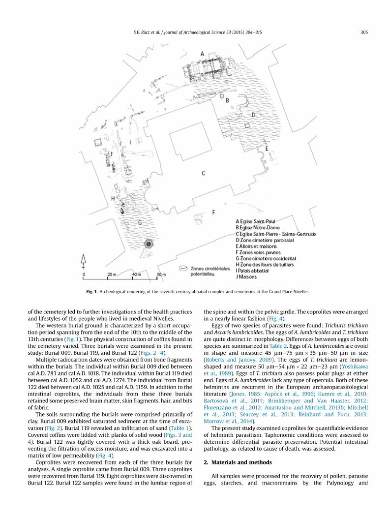
Fig. 1. Archeological rendering of the seventh century abbatial complex and cemeteries at the Grand Place Nivelles.
S.E. R�acz et al. / Journal of Archaeological Science 53 (2015) 304e315 305
of the cemetery led to further investigations of the health practicesand lifestyles of the people who lived in medieval Nivelles.
The western burial ground is characterized by a short occupa-tion period spanning from the end of the 10th to the middle of the13th centuries (Fig. 1). The physical construction of coffins found inthe cemetery varied. Three burials were examined in the presentstudy: Burial 009, Burial 119, and Burial 122 (Figs. 2e4).
Multiple radiocarbon dates were obtained from bone fragmentswithin the burials. The individual within Burial 009 died betweencal A.D. 783 and cal A.D. 1018. The individual within Burial 119 diedbetween cal A.D. 1052 and cal A.D. 1274. The individual from Burial122 died between cal A.D. 1025 and cal A.D. 1159. In addition to theintestinal coprolites, the individuals from these three burialsretained some preserved brainmatter, skin fragments, hair, and bitsof fabric.
The soils surrounding the burials were comprised primarily ofclay. Burial 009 exhibited saturated sediment at the time of exca-vation (Fig. 2). Burial 119 revealed an infiltration of sand (Table 1).Covered coffins were lidded with planks of solid wood (Figs. 3 and4). Burial 122 was tightly covered with a thick oak board, pre-venting the filtration of excess moisture, and was excavated into amatrix of low permeability (Fig. 4).
Coprolites were recovered from each of the three burials foranalyses. A single coprolite came from Burial 009. Three coproliteswere recovered from Burial 119. Eight coprolites were discovered inBurial 122. Burial 122 samples were found in the lumbar region of
the spine and within the pelvic girdle. The coprolites were arrangedin a nearly linear fashion (Fig. 4).
Eggs of two species of parasites were found: Trichuris trichiuraand Ascaris lumbricoides. The eggs of A. lumbricoides and T. trichiuraare quite distinct in morphology. Differences between eggs of bothspecies are summarized in Table 2. Eggs of A. lumbricoides are ovoidin shape and measure 45 mme75 mm� 35 mme50 mm in size(Roberts and Janovy, 2009). The eggs of T. trichiura are lemon-shaped and measure 50 mme54 mm� 22 mme23 mm (Yoshikawaet al., 1989). Eggs of T. trichiura also possess polar plugs at eitherend. Eggs of A. lumbricoides lack any type of opercula. Both of thesehelminths are recurrent in the European archaeoparasitologicalliterature (Jones, 1985; Asp€ock et al., 1996; Kumm et al., 2010;Barto�sov�a et al., 2011; Brinkkemper and Van Haaster, 2012;Florenzano et al., 2012; Anastasiou and Mitchell, 2013b; Mitchellet al., 2013; Searcey et al., 2013; Reinhard and Pucu, 2013;Morrow et al., 2014).
The present study examined coprolites for quantifiable evidenceof helminth parasitism. Taphonomic conditions were assessed todetermine differential parasite preservation. Potential intestinalpathology, as related to cause of death, was assessed.
2. Materials and methods
All samples were processed for the recovery of pollen, parasiteeggs, starches, and macroremains by the Palynology and
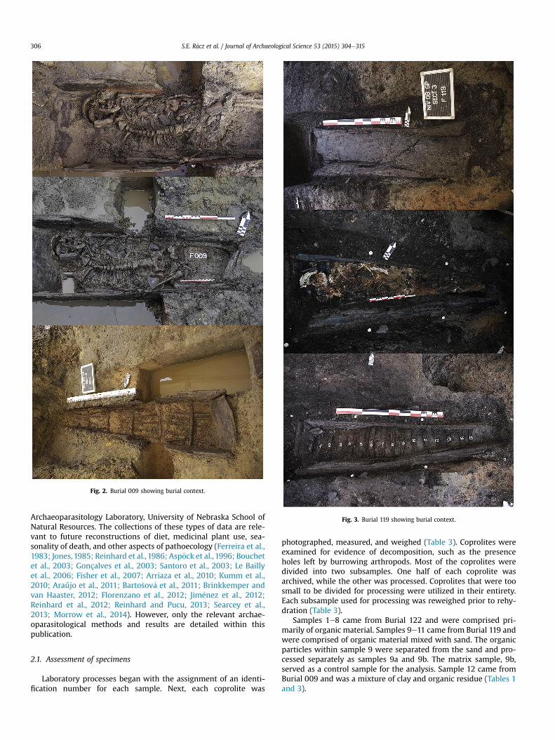
Fig. 2. Burial 009 showing burial context.
Fig. 3. Burial 119 showing burial context.
S.E. R�acz et al. / Journal of Archaeological Science 53 (2015) 304e315306
Archaeoparasitology Laboratory, University of Nebraska School ofNatural Resources. The collections of these types of data are rele-vant to future reconstructions of diet, medicinal plant use, sea-sonality of death, and other aspects of pathoecology (Ferreira et al.,1983; Jones,1985; Reinhard et al., 1986; Asp€ock et al., 1996; Bouchetet al., 2003; Gonçalves et al., 2003; Santoro et al., 2003; Le Baillyet al., 2006; Fisher et al., 2007; Arriaza et al., 2010; Kumm et al.,2010; Araújo et al., 2011; Barto�sov�a et al., 2011; Brinkkemper andvan Haaster, 2012; Florenzano et al., 2012; Jim�enez et al., 2012;Reinhard et al., 2012; Reinhard and Pucu, 2013; Searcey et al.,2013; Morrow et al., 2014). However, only the relevant archae-oparasitological methods and results are detailed within thispublication.
2.1. Assessment of specimens
Laboratory processes began with the assignment of an identi-fication number for each sample. Next, each coprolite was
photographed, measured, and weighed (Table 3). Coprolites wereexamined for evidence of decomposition, such as the presenceholes left by burrowing arthropods. Most of the coprolites weredivided into two subsamples. One half of each coprolite wasarchived, while the other was processed. Coprolites that were toosmall to be divided for processing were utilized in their entirety.Each subsample used for processing was reweighed prior to rehy-dration (Table 3).
Samples 1e8 came from Burial 122 and were comprised pri-marily of organic material. Samples 9e11 came from Burial 119 andwere comprised of organic material mixed with sand. The organicparticles within sample 9 were separated from the sand and pro-cessed separately as samples 9a and 9b. The matrix sample, 9b,served as a control sample for the analysis. Sample 12 came fromBurial 009 and was a mixture of clay and organic residue (Tables 1and 3).
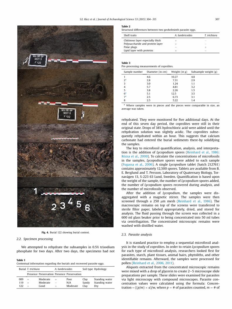
Fig. 4. Burial 122 showing burial context.
Table 2Structural differences between two geohelminth parasite eggs.
Shell traits A. lumbricoides T. trichiura
Chitinous layer especially thick þ �Polysaccharide and protein layer þ �Polar plugs � þLipid layer with proteins þ �
Table 3Pre-processing measurements of coprolites.
Sample number Diameter (in cm) Weight (in g) Subsample weight (g)
1 4.6 10.27 4.82a 2.8 7.31 2.93 3.0 1.24 1.14 5.7 4.81 3.25 3.8 2.26 1.56a 5.1 12.5 3.57a 2.5 6.73 3.18 2.5 5.22 1.4
a Where samples were in pieces and the pieces were comparable in size, anaverage was taken.
S.E. R�acz et al. / Journal of Archaeological Science 53 (2015) 304e315 307
2.2. Specimen processing
We attempted to rehydrate the subsamples in 0.5% trisodiumphosphate for two days. After two days, the specimens had not
Table 1Contextual information regarding the burials and recovered parasite eggs.
Burial T. trichiura A. lumbricoides Soil type Hydrology
Presence Preservation Presence Preservation
009 þ Moderate þ Poor Clay Standing water119 þ Moderate � N/A Sandy Standing water122 þ Good þ Moderate Clay Dry
rehydrated. They were monitored for five additional days. At theend of this seven day period, the coprolites were still in theiroriginal state. Drops of 38% hydrochloric acid were added until therehydration solution was slightly acidic. The coprolites subse-quently rehydrated within an hour. This suggests that calciumcarbonate had entered the burial sediments there-by solidifyingthe samples.
The key to microfossil quantification, analysis, and interpreta-tion is the addition of Lycopodium spores (Reinhard et al., 1986;R�ozsa et al., 2000). To calculate the concentrations of microfossilsin the samples, Lycopodium spores were added to each sample(Fugassa et al., 2006). A single Lycopodium tablet (batch 212761)contains approximately 12,500 spores. Tablets are available from B.E. Berglund and T. Persson, Laboratory of Quaternary Biology, Tor-nav€agen 13, S-223 63 Lund, Sweden. Quantification is based uponthe weight of the sample, the number of Lycopodium spores added,the number of Lycopodium spores recovered during analysis, andthe number of microfossils observed.
After the addition of Lycopodium, the samples were dis-aggregated with a magnetic stirrer. The samples were thenscreened through a 250 mm mesh (Reinhard et al., 1986). Themacroscopic remains on top of the screens were transferred tosterile filter paper, labeled appropriately, dried, and stored foranalysis. The fluid passing through the screen was collected in a600 ml glass beaker prior to being concentrated into 50 ml tubesvia centrifugation. The concentrated microscopic remains werewashed with distilled water.
2.3. Parasite analysis
It is standard practice to employ a sequential microfossil anal-ysis in the study of coprolites. In order to retain Lycopodium sporesfor each type of microfossil analysis, researchers looked first forparasites, starch, plant tissues, animal hairs, phytoliths, and otheridentifiable remains. Afterward, the samples were processed forpollen (Reinhard et al., 2006, 2011).
Aliquots extracted from the concentrated microscopic remainswere mixed with a drop of glycerin to create 2e5 microscope slidepreparations per sample. These slides were examined for parasitesvia light microscopy with compound microscopes. Parasite con-centration values were calculated using the formula: Concen-tration¼ ((p/m) � a)/w, where p ¼ # of parasites counted, m ¼ # of
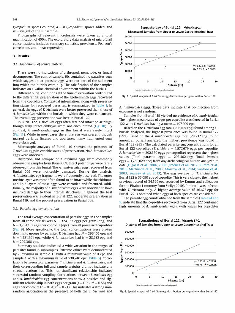
Fig. 5. Spatial analysis of T. trichiura egg distribution per gram within Burial 122.
Fig. 6. Spatial analysis of T. trichiura egg distribution per coprolite within Burial 122.
S.E. R�acz et al. / Journal of Archaeological Science 53 (2015) 304e315308
Lycopodium spores counted, a ¼ # Lycopodium spores added, andw ¼ weight of the subsample.
Photographs of relevant microfossils were taken at a totalmagnification of 400�. The exploratory data analysis of microfossilconcentrations includes summary statistics, prevalence, Pearson'scorrelation, and linear regression.
3. Results
3.1. Taphonomy of source material
There were no indications of arthropod, nematode, or fungaldecomposers. The control sample, 9b, contained no parasites eggswhich suggests that parasite eggs were not part of the sedimentinto which the burials were dug. The calcification of the samplesindicates an alkaline chemical environment within the burials.
Different burial conditions at the time of excavation contributedto the differential preservation of the geohelminth eggs recoveredfrom the coprolites. Contextual information, along with preserva-tion status for recovered parasites, is summarized in Table 1. Ingeneral, the eggs of T. trichiurawere better preserved than those ofA. lumbricoides within the burials in which they were concurrent.The overall egg preservation was best in Burial 122.
In Burial 122, T. trichiura eggs often retained intact polar plugs,though fully intact embryos were not encountered (Fig. 10). Bycontrast, A. lumbricoides eggs in this burial were rarely intact(Fig. 11). While in most cases the entire egg was present, thoughmarred by large fissures and apertures, many fragmented eggswere observed.
Microscopic analyses of Burial 119 showed the presence ofT. trichiura eggs in variable states of preservation. No A. lumbricoideseggs were observed.
Distortion and collapse of T. trichiura eggs were commonlyobserved in samples from Burial 009. Intact polar plugs were rarelyobserved from this burial. The A. lumbricoides eggs recovered fromBurial 009 were noticeably damaged. During the analysis,A. lumbricoides egg fragments were frequently observed. The outeruterine layer was most often found to be intact while the chitinousand lipid layers of these eggs were corroded and fractured. Addi-tionally, the majority of A. lumbricoides eggs were observed to haveincurred damage to their internal structures. In general, the bestpreservation was evident in Burial 122, moderate preservation inBurial 119, and the poorest preservation in Burial 009.
3.2. Parasite egg concentrations
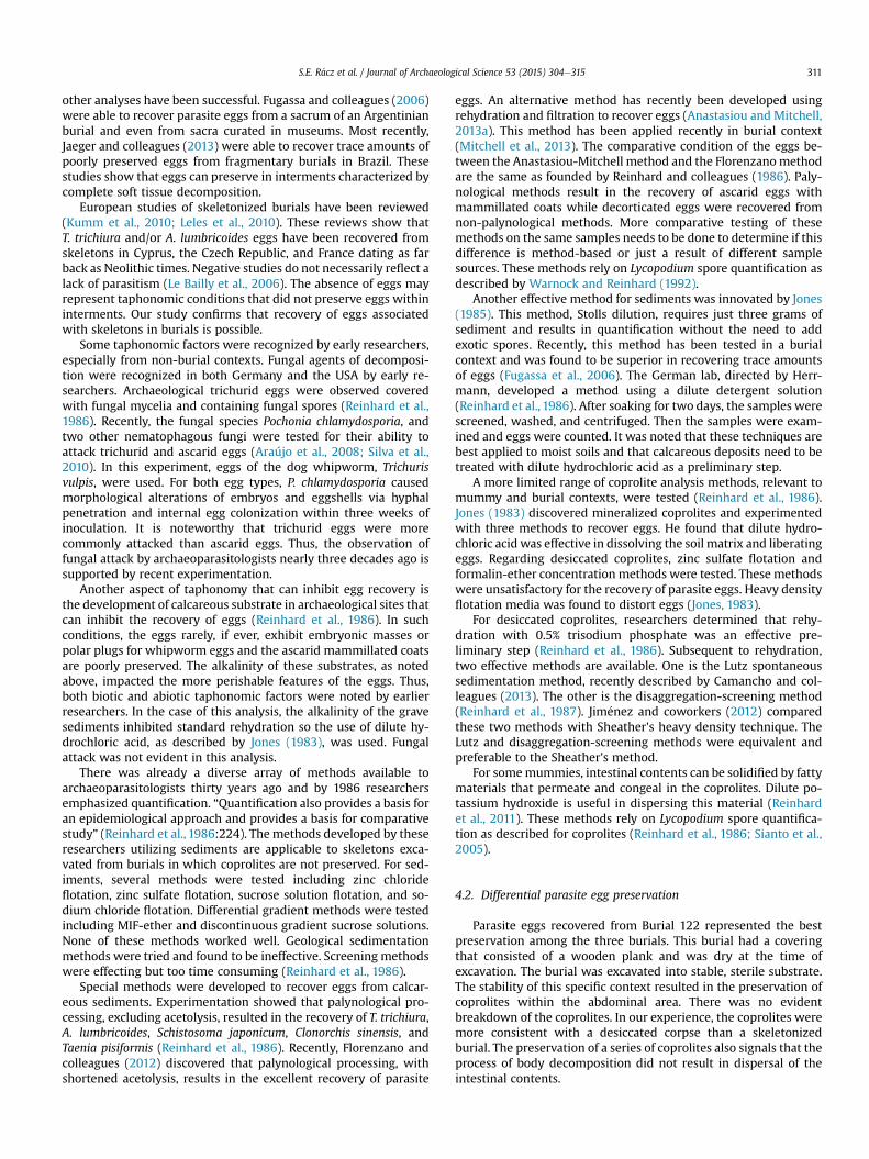
The total average concentration of parasite eggs in the samplesfrom all three burials was N ¼ 324,837 eggs per gram (epg) andN ¼ 1,784,157 eggs per coprolite (epc) from all processed coprolites(Fig. 9). More specifically, the total concentrations were brokendown into groups by parasite; T. trichiura had N ¼ 296,105 epg andN ¼ 1,581,791 epc, while A. lumbricoides had N ¼ 28,732 epg andN ¼ 202,366 epc.
Summary statistics indicated a wide variation in the ranges ofparasites found in subsamples. Extreme values were demonstratedby T. trichiura in sample 11 with a minimum value of 0 epc andsample 1 with a maximum value of 530,240 epc (Table 5). Corre-lations between total parasites, T. trichiura and A. lumbricoides, andtheir corresponding full and sample weights did not indicate anystrong relationships. This non-significant relationship indicatessuccessful random sampling. Correlations between T. trichiura eggand A. lumbricoides egg concentrations show a positive and sig-nificant relationship in both eggs per gram (r ¼ 0.76; r2 ¼ 0.58) andeggs per coprolite (r ¼ 0.84; r2 ¼ 0.71). This indicates a strong non-random association in the presence of both the T. trichiura and
A. lumbricoides eggs. These data indicate that co-infection fromexposure is not random.
Samples from Burial 119 yielded no evidence of A. lumbricoides.The highest mean value of eggs per coprolite was detected in Burial122 with T. trichiura having a mean ¼ 197,209 epc.
Based on the T. trichiura egg total (296,105 epg) found among allburials analyzed, the highest prevalence was found in Burial 122(89%). Based on the A. lumbricoides egg total (28,732 epg) foundamong all burials analyzed, the highest prevalence was found inBurial 122 (99%). The calculated parasite egg concentrations for allBurial 122 coprolites (T. trichiura ¼ 1,577,679 eggs per coprolite,A. lumbricoides ¼ 202,350 eggs per coprolite) represent the highestvalues (Total parasite eggs ¼ 293,402 epg; Total Parasiteeggs ¼ 1,780,029 epc) from any archaeological human analyzed todate (Fugassa et al., 2006, 2008; Jim�enez et al., 2012; Kumm et al.,2010; Martinson et al., 2003; Morrow et al., 2014; Santoro et al.,2003; Searcey et al., 2013). The epg average for T. trichiura forBurial 122 is 33,090 epg of coprolite. This is very close to the highestprevious record of 34,529 epg recorded by Kumm and colleaguesfor the Piraino 1 mummy from Sicily (2010). Piraino 1 was infectedwith T. trichiura only. A higher average value of 36,675 epg forBurial 122 is obtained when eggs of both species are considered.
The parasite egg counts obtained from the samples (Tables 4 and5) indicate that the coprolites recovered from Burial 122 containedhigh amounts of A. lumbricoides eggs, with values for coprolites
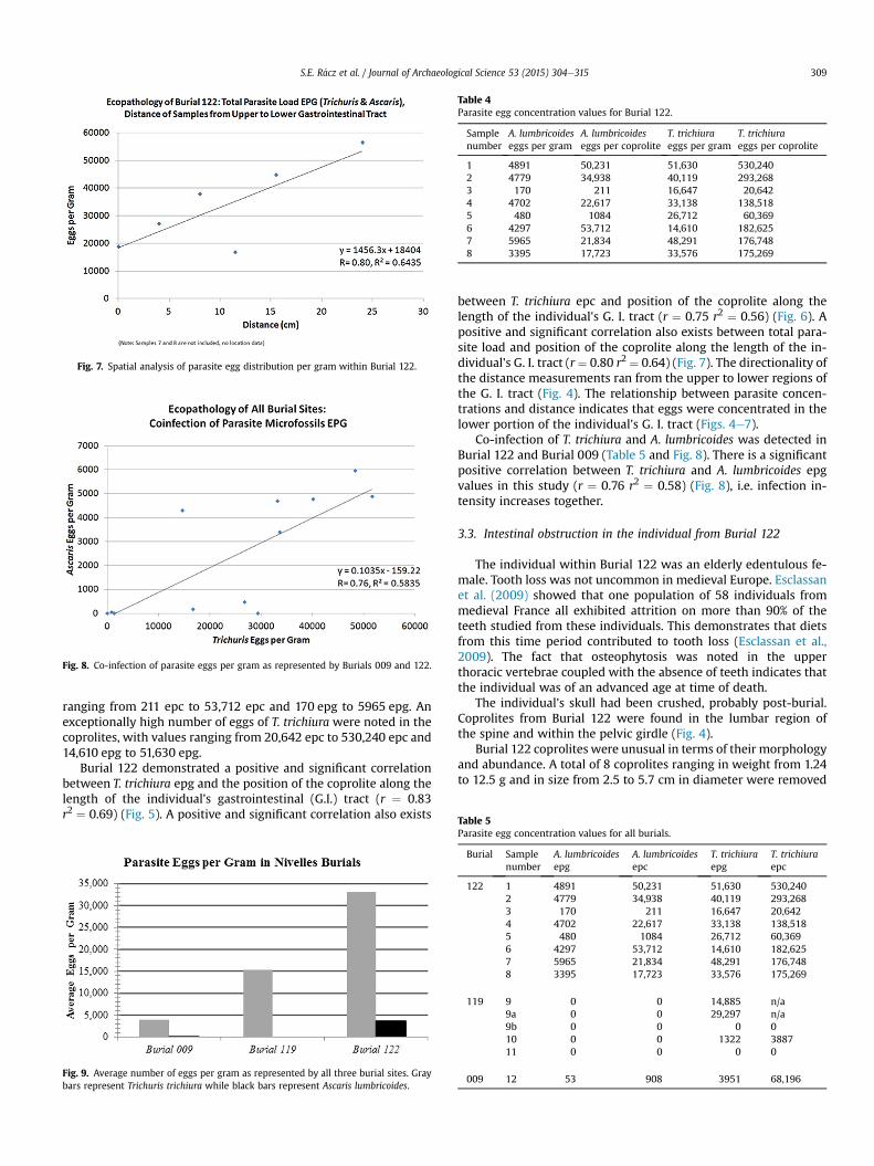
Fig. 7. Spatial analysis of parasite egg distribution per gram within Burial 122.
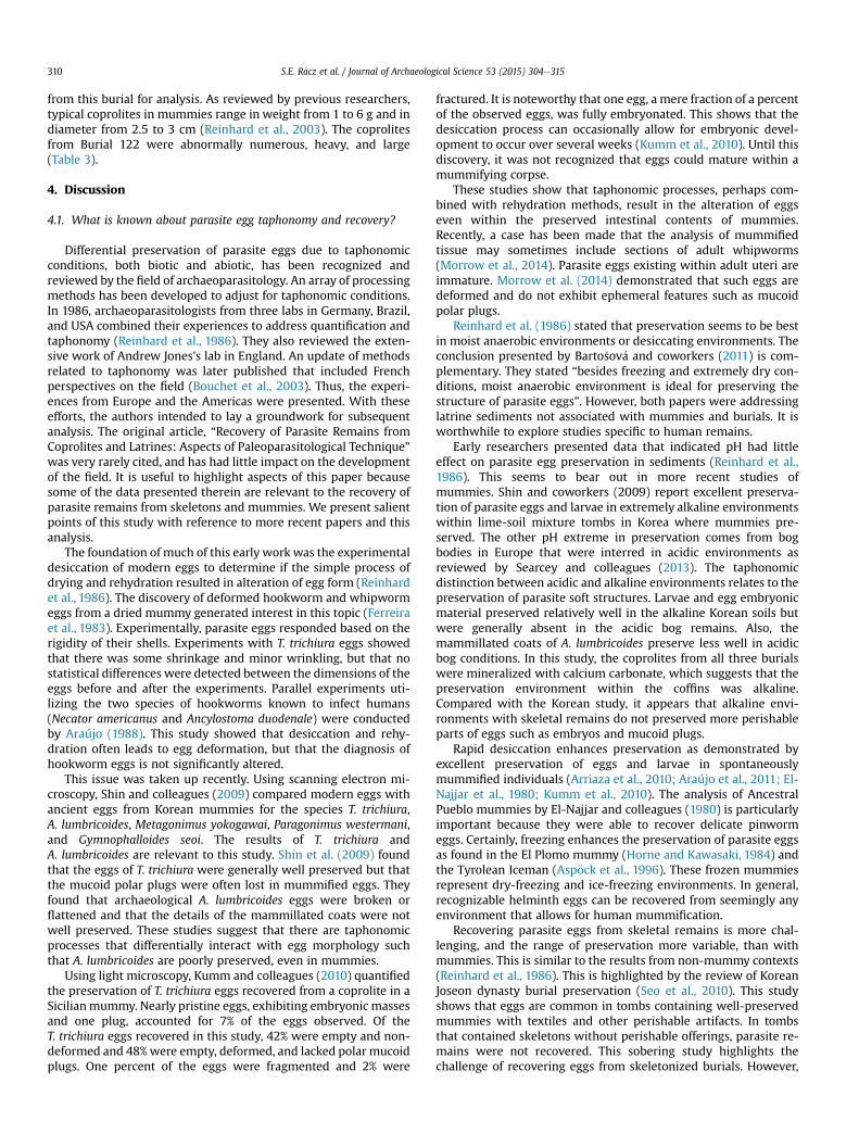
Fig. 8. Co-infection of parasite eggs per gram as represented by Burials 009 and 122.
Table 4Parasite egg concentration values for Burial 122.
Samplenumber
A. lumbricoideseggs per gram
A. lumbricoideseggs per coprolite
T. trichiuraeggs per gram
T. trichiuraeggs per coprolite
1 4891 50,231 51,630 530,2402 4779 34,938 40,119 293,2683 170 211 16,647 20,6424 4702 22,617 33,138 138,5185 480 1084 26,712 60,3696 4297 53,712 14,610 182,6257 5965 21,834 48,291 176,7488 3395 17,723 33,576 175,269
Table 5
S.E. R�acz et al. / Journal of Archaeological Science 53 (2015) 304e315 309
ranging from 211 epc to 53,712 epc and 170 epg to 5965 epg. Anexceptionally high number of eggs of T. trichiura were noted in thecoprolites, with values ranging from 20,642 epc to 530,240 epc and14,610 epg to 51,630 epg.
Burial 122 demonstrated a positive and significant correlationbetween T. trichiura epg and the position of the coprolite along thelength of the individual's gastrointestinal (G.I.) tract (r ¼ 0.83r2 ¼ 0.69) (Fig. 5). A positive and significant correlation also exists
Fig. 9. Average number of eggs per gram as represented by all three burial sites. Graybars represent Trichuris trichiura while black bars represent Ascaris lumbricoides.
between T. trichiura epc and position of the coprolite along thelength of the individual's G. I. tract (r ¼ 0.75 r2 ¼ 0.56) (Fig. 6). Apositive and significant correlation also exists between total para-site load and position of the coprolite along the length of the in-dividual's G. I. tract (r¼ 0.80 r2 ¼ 0.64) (Fig. 7). The directionality ofthe distance measurements ran from the upper to lower regions ofthe G. I. tract (Fig. 4). The relationship between parasite concen-trations and distance indicates that eggs were concentrated in thelower portion of the individual's G. I. tract (Figs. 4e7).
Co-infection of T. trichiura and A. lumbricoides was detected inBurial 122 and Burial 009 (Table 5 and Fig. 8). There is a significantpositive correlation between T. trichiura and A. lumbricoides epgvalues in this study (r ¼ 0.76 r2 ¼ 0.58) (Fig. 8), i.e. infection in-tensity increases together.
3.3. Intestinal obstruction in the individual from Burial 122
The individual within Burial 122 was an elderly edentulous fe-male. Tooth loss was not uncommon in medieval Europe. Esclassanet al. (2009) showed that one population of 58 individuals frommedieval France all exhibited attrition on more than 90% of theteeth studied from these individuals. This demonstrates that dietsfrom this time period contributed to tooth loss (Esclassan et al.,2009). The fact that osteophytosis was noted in the upperthoracic vertebrae coupled with the absence of teeth indicates thatthe individual was of an advanced age at time of death.
The individual's skull had been crushed, probably post-burial.Coprolites from Burial 122 were found in the lumbar region ofthe spine and within the pelvic girdle (Fig. 4).
Burial 122 coprolites were unusual in terms of their morphologyand abundance. A total of 8 coprolites ranging in weight from 1.24to 12.5 g and in size from 2.5 to 5.7 cm in diameter were removed
Parasite egg concentration values for all burials.
Burial Samplenumber
A. lumbricoidesepg
A. lumbricoidesepc
T. trichiuraepg
T. trichiuraepc
122 1 4891 50,231 51,630 530,2402 4779 34,938 40,119 293,2683 170 211 16,647 20,6424 4702 22,617 33,138 138,5185 480 1084 26,712 60,3696 4297 53,712 14,610 182,6257 5965 21,834 48,291 176,7488 3395 17,723 33,576 175,269
119 9 0 0 14,885 n/a9a 0 0 29,297 n/a9b 0 0 0 010 0 0 1322 388711 0 0 0 0
009 12 53 908 3951 68,196
S.E. R�acz et al. / Journal of Archaeological Science 53 (2015) 304e315310
from this burial for analysis. As reviewed by previous researchers,typical coprolites in mummies range in weight from 1 to 6 g and indiameter from 2.5 to 3 cm (Reinhard et al., 2003). The coprolitesfrom Burial 122 were abnormally numerous, heavy, and large(Table 3).
4. Discussion
4.1. What is known about parasite egg taphonomy and recovery?
Differential preservation of parasite eggs due to taphonomicconditions, both biotic and abiotic, has been recognized andreviewed by the field of archaeoparasitology. An array of processingmethods has been developed to adjust for taphonomic conditions.In 1986, archaeoparasitologists from three labs in Germany, Brazil,and USA combined their experiences to address quantification andtaphonomy (Reinhard et al., 1986). They also reviewed the exten-sive work of Andrew Jones's lab in England. An update of methodsrelated to taphonomy was later published that included Frenchperspectives on the field (Bouchet et al., 2003). Thus, the experi-ences from Europe and the Americas were presented. With theseefforts, the authors intended to lay a groundwork for subsequentanalysis. The original article, “Recovery of Parasite Remains fromCoprolites and Latrines: Aspects of Paleoparasitological Technique”was very rarely cited, and has had little impact on the developmentof the field. It is useful to highlight aspects of this paper becausesome of the data presented therein are relevant to the recovery ofparasite remains from skeletons and mummies. We present salientpoints of this study with reference to more recent papers and thisanalysis.
The foundation of much of this early work was the experimentaldesiccation of modern eggs to determine if the simple process ofdrying and rehydration resulted in alteration of egg form (Reinhardet al., 1986). The discovery of deformed hookworm and whipwormeggs from a dried mummy generated interest in this topic (Ferreiraet al., 1983). Experimentally, parasite eggs responded based on therigidity of their shells. Experiments with T. trichiura eggs showedthat there was some shrinkage and minor wrinkling, but that nostatistical differences were detected between the dimensions of theeggs before and after the experiments. Parallel experiments uti-lizing the two species of hookworms known to infect humans(Necator americanus and Ancylostoma duodenale) were conductedby Araújo (1988). This study showed that desiccation and rehy-dration often leads to egg deformation, but that the diagnosis ofhookworm eggs is not significantly altered.
This issue was taken up recently. Using scanning electron mi-croscopy, Shin and colleagues (2009) compared modern eggs withancient eggs from Korean mummies for the species T. trichiura,A. lumbricoides, Metagonimus yokogawai, Paragonimus westermani,and Gymnophalloides seoi. The results of T. trichiura andA. lumbricoides are relevant to this study. Shin et al. (2009) foundthat the eggs of T. trichiura were generally well preserved but thatthe mucoid polar plugs were often lost in mummified eggs. Theyfound that archaeological A. lumbricoides eggs were broken orflattened and that the details of the mammillated coats were notwell preserved. These studies suggest that there are taphonomicprocesses that differentially interact with egg morphology suchthat A. lumbricoides are poorly preserved, even in mummies.
Using light microscopy, Kumm and colleagues (2010) quantifiedthe preservation of T. trichiura eggs recovered from a coprolite in aSicilian mummy. Nearly pristine eggs, exhibiting embryonic massesand one plug, accounted for 7% of the eggs observed. Of theT. trichiura eggs recovered in this study, 42% were empty and non-deformed and 48%were empty, deformed, and lacked polar mucoidplugs. One percent of the eggs were fragmented and 2% were
fractured. It is noteworthy that one egg, a mere fraction of a percentof the observed eggs, was fully embryonated. This shows that thedesiccation process can occasionally allow for embryonic devel-opment to occur over several weeks (Kumm et al., 2010). Until thisdiscovery, it was not recognized that eggs could mature within amummifying corpse.
These studies show that taphonomic processes, perhaps com-bined with rehydration methods, result in the alteration of eggseven within the preserved intestinal contents of mummies.Recently, a case has been made that the analysis of mummifiedtissue may sometimes include sections of adult whipworms(Morrow et al., 2014). Parasite eggs existing within adult uteri areimmature. Morrow et al. (2014) demonstrated that such eggs aredeformed and do not exhibit ephemeral features such as mucoidpolar plugs.
Reinhard et al. (1986) stated that preservation seems to be bestin moist anaerobic environments or desiccating environments. Theconclusion presented by Barto�sov�a and coworkers (2011) is com-plementary. They stated “besides freezing and extremely dry con-ditions, moist anaerobic environment is ideal for preserving thestructure of parasite eggs”. However, both papers were addressinglatrine sediments not associated with mummies and burials. It isworthwhile to explore studies specific to human remains.
Early researchers presented data that indicated pH had littleeffect on parasite egg preservation in sediments (Reinhard et al.,1986). This seems to bear out in more recent studies ofmummies. Shin and coworkers (2009) report excellent preserva-tion of parasite eggs and larvae in extremely alkaline environmentswithin lime-soil mixture tombs in Korea where mummies pre-served. The other pH extreme in preservation comes from bogbodies in Europe that were interred in acidic environments asreviewed by Searcey and colleagues (2013). The taphonomicdistinction between acidic and alkaline environments relates to thepreservation of parasite soft structures. Larvae and egg embryonicmaterial preserved relatively well in the alkaline Korean soils butwere generally absent in the acidic bog remains. Also, themammillated coats of A. lumbricoides preserve less well in acidicbog conditions. In this study, the coprolites from all three burialswere mineralized with calcium carbonate, which suggests that thepreservation environment within the coffins was alkaline.Compared with the Korean study, it appears that alkaline envi-ronments with skeletal remains do not preserved more perishableparts of eggs such as embryos and mucoid plugs.
Rapid desiccation enhances preservation as demonstrated byexcellent preservation of eggs and larvae in spontaneouslymummified individuals (Arriaza et al., 2010; Araújo et al., 2011; El-Najjar et al., 1980; Kumm et al., 2010). The analysis of AncestralPueblo mummies by El-Najjar and colleagues (1980) is particularlyimportant because they were able to recover delicate pinwormeggs. Certainly, freezing enhances the preservation of parasite eggsas found in the El Plomo mummy (Horne and Kawasaki, 1984) andthe Tyrolean Iceman (Asp€ock et al., 1996). These frozen mummiesrepresent dry-freezing and ice-freezing environments. In general,recognizable helminth eggs can be recovered from seemingly anyenvironment that allows for human mummification.
Recovering parasite eggs from skeletal remains is more chal-lenging, and the range of preservation more variable, than withmummies. This is similar to the results from non-mummy contexts(Reinhard et al., 1986). This is highlighted by the review of KoreanJoseon dynasty burial preservation (Seo et al., 2010). This studyshows that eggs are common in tombs containing well-preservedmummies with textiles and other perishable artifacts. In tombsthat contained skeletons without perishable offerings, parasite re-mains were not recovered. This sobering study highlights thechallenge of recovering eggs from skeletonized burials. However,
S.E. R�acz et al. / Journal of Archaeological Science 53 (2015) 304e315 311
other analyses have been successful. Fugassa and colleagues (2006)were able to recover parasite eggs from a sacrum of an Argentinianburial and even from sacra curated in museums. Most recently,Jaeger and colleagues (2013) were able to recover trace amounts ofpoorly preserved eggs from fragmentary burials in Brazil. Thesestudies show that eggs can preserve in interments characterized bycomplete soft tissue decomposition.
European studies of skeletonized burials have been reviewed(Kumm et al., 2010; Leles et al., 2010). These reviews show thatT. trichiura and/or A. lumbricoides eggs have been recovered fromskeletons in Cyprus, the Czech Republic, and France dating as farback as Neolithic times. Negative studies do not necessarily reflect alack of parasitism (Le Bailly et al., 2006). The absence of eggs mayrepresent taphonomic conditions that did not preserve eggs withininterments. Our study confirms that recovery of eggs associatedwith skeletons in burials is possible.
Some taphonomic factors were recognized by early researchers,especially from non-burial contexts. Fungal agents of decomposi-tion were recognized in both Germany and the USA by early re-searchers. Archaeological trichurid eggs were observed coveredwith fungal mycelia and containing fungal spores (Reinhard et al.,1986). Recently, the fungal species Pochonia chlamydosporia, andtwo other nematophagous fungi were tested for their ability toattack trichurid and ascarid eggs (Araújo et al., 2008; Silva et al.,2010). In this experiment, eggs of the dog whipworm, Trichurisvulpis, were used. For both egg types, P. chlamydosporia causedmorphological alterations of embryos and eggshells via hyphalpenetration and internal egg colonization within three weeks ofinoculation. It is noteworthy that trichurid eggs were morecommonly attacked than ascarid eggs. Thus, the observation offungal attack by archaeoparasitologists nearly three decades ago issupported by recent experimentation.
Another aspect of taphonomy that can inhibit egg recovery isthe development of calcareous substrate in archaeological sites thatcan inhibit the recovery of eggs (Reinhard et al., 1986). In suchconditions, the eggs rarely, if ever, exhibit embryonic masses orpolar plugs for whipworm eggs and the ascarid mammillated coatsare poorly preserved. The alkalinity of these substrates, as notedabove, impacted the more perishable features of the eggs. Thus,both biotic and abiotic taphonomic factors were noted by earlierresearchers. In the case of this analysis, the alkalinity of the gravesediments inhibited standard rehydration so the use of dilute hy-drochloric acid, as described by Jones (1983), was used. Fungalattack was not evident in this analysis.
There was already a diverse array of methods available toarchaeoparasitologists thirty years ago and by 1986 researchersemphasized quantification. “Quantification also provides a basis foran epidemiological approach and provides a basis for comparativestudy” (Reinhard et al., 1986:224). The methods developed by theseresearchers utilizing sediments are applicable to skeletons exca-vated from burials in which coprolites are not preserved. For sed-iments, several methods were tested including zinc chlorideflotation, zinc sulfate flotation, sucrose solution flotation, and so-dium chloride flotation. Differential gradient methods were testedincluding MIF-ether and discontinuous gradient sucrose solutions.None of these methods worked well. Geological sedimentationmethods were tried and found to be ineffective. Screening methodswere effecting but too time consuming (Reinhard et al., 1986).
Special methods were developed to recover eggs from calcar-eous sediments. Experimentation showed that palynological pro-cessing, excluding acetolysis, resulted in the recovery of T. trichiura,A. lumbricoides, Schistosoma japonicum, Clonorchis sinensis, andTaenia pisiformis (Reinhard et al., 1986). Recently, Florenzano andcolleagues (2012) discovered that palynological processing, withshortened acetolysis, results in the excellent recovery of parasite
eggs. An alternative method has recently been developed usingrehydration and filtration to recover eggs (Anastasiou and Mitchell,2013a). This method has been applied recently in burial context(Mitchell et al., 2013). The comparative condition of the eggs be-tween the Anastasiou-Mitchell method and the Florenzanomethodare the same as founded by Reinhard and colleagues (1986). Paly-nological methods result in the recovery of ascarid eggs withmammillated coats while decorticated eggs were recovered fromnon-palynological methods. More comparative testing of thesemethods on the same samples needs to be done to determine if thisdifference is method-based or just a result of different samplesources. These methods rely on Lycopodium spore quantification asdescribed by Warnock and Reinhard (1992).
Another effective method for sediments was innovated by Jones(1985). This method, Stolls dilution, requires just three grams ofsediment and results in quantification without the need to addexotic spores. Recently, this method has been tested in a burialcontext and was found to be superior in recovering trace amountsof eggs (Fugassa et al., 2006). The German lab, directed by Herr-mann, developed a method using a dilute detergent solution(Reinhard et al., 1986). After soaking for two days, the samples werescreened, washed, and centrifuged. Then the samples were exam-ined and eggs were counted. It was noted that these techniques arebest applied to moist soils and that calcareous deposits need to betreated with dilute hydrochloric acid as a preliminary step.
A more limited range of coprolite analysis methods, relevant tomummy and burial contexts, were tested (Reinhard et al., 1986).Jones (1983) discovered mineralized coprolites and experimentedwith three methods to recover eggs. He found that dilute hydro-chloric acid was effective in dissolving the soil matrix and liberatingeggs. Regarding desiccated coprolites, zinc sulfate flotation andformalin-ether concentration methods were tested. These methodswere unsatisfactory for the recovery of parasite eggs. Heavy densityflotation media was found to distort eggs (Jones, 1983).
For desiccated coprolites, researchers determined that rehy-dration with 0.5% trisodium phosphate was an effective pre-liminary step (Reinhard et al., 1986). Subsequent to rehydration,two effective methods are available. One is the Lutz spontaneoussedimentation method, recently described by Camancho and col-leagues (2013). The other is the disaggregation-screening method(Reinhard et al., 1987). Jim�enez and coworkers (2012) comparedthese two methods with Sheather's heavy density technique. TheLutz and disaggregation-screening methods were equivalent andpreferable to the Sheather's method.
For somemummies, intestinal contents can be solidified by fattymaterials that permeate and congeal in the coprolites. Dilute po-tassium hydroxide is useful in dispersing this material (Reinhardet al., 2011). These methods rely on Lycopodium spore quantifica-tion as described for coprolites (Reinhard et al., 1986; Sianto et al.,2005).
4.2. Differential parasite egg preservation
Parasite eggs recovered from Burial 122 represented the bestpreservation among the three burials. This burial had a coveringthat consisted of a wooden plank and was dry at the time ofexcavation. The burial was excavated into stable, sterile substrate.The stability of this specific context resulted in the preservation ofcoprolites within the abdominal area. There was no evidentbreakdown of the coprolites. In our experience, the coprolites weremore consistent with a desiccated corpse than a skeletonizedburial. The preservation of a series of coprolites also signals that theprocess of body decomposition did not result in dispersal of theintestinal contents.
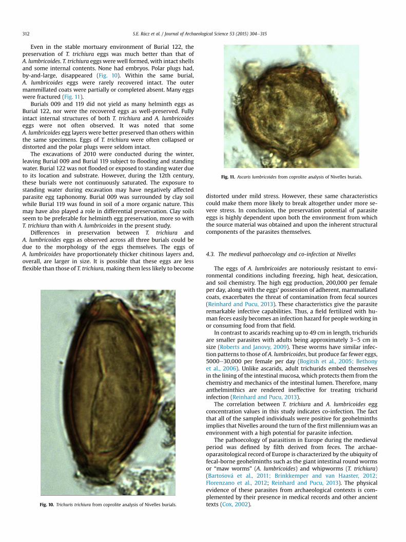
Fig. 11. Ascaris lumbricoides from coprolite analysis of Nivelles burials.
S.E. R�acz et al. / Journal of Archaeological Science 53 (2015) 304e315312
Even in the stable mortuary environment of Burial 122, thepreservation of T. trichiura eggs was much better than that ofA. lumbricoides. T. trichiura eggs werewell formed, with intact shellsand some internal contents. None had embryos. Polar plugs had,by-and-large, disappeared (Fig. 10). Within the same burial,A. lumbricoides eggs were rarely recovered intact. The outermammillated coats were partially or completed absent. Many eggswere fractured (Fig. 11).
Burials 009 and 119 did not yield as many helminth eggs asBurial 122, nor were the recovered eggs as well-preserved. Fullyintact internal structures of both T. trichiura and A. lumbricoideseggs were not often observed. It was noted that someA. lumbricoides egg layers were better preserved than others withinthe same specimens. Eggs of T. trichiura were often collapsed ordistorted and the polar plugs were seldom intact.
The excavations of 2010 were conducted during the winter,leaving Burial 009 and Burial 119 subject to flooding and standingwater. Burial 122 was not flooded or exposed to standing water dueto its location and substrate. However, during the 12th century,these burials were not continuously saturated. The exposure tostanding water during excavation may have negatively affectedparasite egg taphonomy. Burial 009 was surrounded by clay soilwhile Burial 119 was found in soil of a more organic nature. Thismay have also played a role in differential preservation. Clay soilsseem to be preferable for helminth egg preservation, more so withT. trichiura than with A. lumbricoides in the present study.
Differences in preservation between T. trichiura andA. lumbricoides eggs as observed across all three burials could bedue to the morphology of the eggs themselves. The eggs ofA. lumbricoides have proportionately thicker chitinous layers and,overall, are larger in size. It is possible that these eggs are lessflexible than those of T. trichiura, making them less likely to become
Fig. 10. Trichuris trichiura from coprolite analysis of Nivelles burials.
distorted under mild stress. However, these same characteristicscould make them more likely to break altogether under more se-vere stress. In conclusion, the preservation potential of parasiteeggs is highly dependent upon both the environment from whichthe source material was obtained and upon the inherent structuralcomponents of the parasites themselves.
4.3. The medieval pathoecology and co-infection at Nivelles
The eggs of A. lumbricoides are notoriously resistant to envi-ronmental conditions including freezing, high heat, desiccation,and soil chemistry. The high egg production, 200,000 per femaleper day, along with the eggs' possession of adherent, mammallatedcoats, exacerbates the threat of contamination from fecal sources(Reinhard and Pucu, 2013). These characteristics give the parasiteremarkable infective capabilities. Thus, a field fertilized with hu-man feces easily becomes an infection hazard for people working inor consuming food from that field.
In contrast to ascarids reaching up to 49 cm in length, trichuridsare smaller parasites with adults being approximately 3e5 cm insize (Roberts and Janovy, 2009). These worms have similar infec-tion patterns to those of A. lumbricoides, but produce far fewer eggs,5000e30,000 per female per day (Bogitsh et al., 2005; Bethonyet al., 2006). Unlike ascarids, adult trichurids embed themselvesin the lining of the intestinal mucosa, which protects them from thechemistry and mechanics of the intestinal lumen. Therefore, manyanthelminthics are rendered ineffective for treating trichuridinfection (Reinhard and Pucu, 2013).
The correlation between T. trichiura and A. lumbricoides eggconcentration values in this study indicates co-infection. The factthat all of the sampled individuals were positive for geohelminthsimplies that Nivelles around the turn of the first millenniumwas anenvironment with a high potential for parasite infection.
The pathoecology of parasitism in Europe during the medievalperiod was defined by filth derived from feces. The archae-oparasitological record of Europe is characterized by the ubiquity offecal-borne geohelminths such as the giant intestinal round wormsor “maw worms” (A. lumbricoides) and whipworms (T. trichiura)(Barto�sov�a et al., 2011; Brinkkemper and van Haaster, 2012;Florenzano et al., 2012; Reinhard and Pucu, 2013). The physicalevidence of these parasites from archaeological contexts is com-plemented by their presence in medical records and other ancienttexts (Cox, 2002).
S.E. R�acz et al. / Journal of Archaeological Science 53 (2015) 304e315 313
Parasitism has been recognized throughout written history,with the first descriptions coming from Egyptian medical docu-ments dating from 3000 to 400 B.C. Other descriptions involvingparasites were recorded by Greek physicians from 800 to 300 B.C.(Cox, 2002). In fact, both the Greeks and the Romans recognizedA. lumbricoides and other parasitic helminths (Sandison, 1967).More definitive descriptions were recorded in later periods byArabian physicians (Cox, 2002). Medieval physicians recognizedparasitic helminth infections, but did not understand the organ-ismal source (DeMaitre, 2013). All existing materia medicas andpharmacopoeias for Europe and the Mediterranean dating from the5th century B.C. to the 19th century A.D. were summarized by DeVos (2010). This study concludes that certain plant species wereused consistently to treat intestinal helminth infections.
European archaeoparasitological studies have revealed evidenceof A. lumbricoides and T. trichiura infections dating as far back as thePaleolithic (Gonçalves et al., 2003). The eggs of these parasites wereubiquitous in the medieval environment as shown by analysis ofarchaeological sediments. In particular, A. lumbricoides wastremendously abundant, indicating the rampant filth of the period(Leles et al., 2010). Furthermore, A.K.G. Jones (1985) and Herrmann(1985,1986) demonstrated that T. trichiurawas a predictable part ofmedieval background fauna. Herrmann and Schulz (1986) definedthe factors that caused this epidemic, which included commondefecation areas, population structure, and relatively unhygienicconditions. These data are expanded by recent studies document-ing the geographical breadth and temporal depth of the Europeangeohelminth epidemic (Barto�sov�a et al., 2011; Florenzano et al.,2012; Kumm et al., 2010; Mitchell et al., 2013; Morrow et al.,2014; Searcey et al., 2013). In medieval Europe, waste manage-ment was tightly tied to agriculture. The epidemic of geohelminthsparasitism was undoubtedly tied to the recovery of feces from cessdeposits used as fertilizer in near-by agricultural fields (Sterner,2008).
Contemporary sanitization was limited during the medievalperiod. Feces were deposited in refuse pits, yards, in front of houses,or in the streets (Jones,1985). Members of the lower social echelonsoften did not change clothes or bathe due to a lack of runningwater.Regular hand washing and rinsing of fruits and vegetables prior toconsumption were uncommon practices. Water was also widelybelieved to be harmful to the human body (Vondru�ska, 2007). Thislack of sanitation coupled with behavioral avoidance of hygienicpractices created ideal microenvironments for the mechanicaltransmission of geohelminth eggs. Technological and horticulturaladvances after 1300 A.D. gave medieval agriculturalists the abilityto produce more food, which led to a notable increase in Europe'spopulation. Medieval agriculture relied on excrement, allowing forthe continual rotation of geohelminth life cycles, making agricul-tural products reservoirs of infection. Despite most medieval urbanresidences having vegetable gardens, cities depended on ruralagriculture.
Medieval fertilizers were composed almost exclusively oforganic wastes such as biodegradable food scraps, human waste,and offal from urban regions. Waste was dumped in the unpavedcity streets, and was either eaten by animals living in the city, orwas soaked up into urbanmud later used as fertilizer (Reinhard andPucu, 2013; Sterner, 2008). As populations grew, sanitation crisesemerged. Wastes could no longer be absorbed once cities becamepaved, leading to filth accumulation. Uncooked and unwashedvegetables, as well as uncleanwater, were major sources of parasitecontamination. This environment greatly contributed to theemergence of diseases (Sterner, 2008) and inevitably created theconstant risk of parasite contraction and reinfection.
4.4. Burial 122: extreme parasitism and pathology
The especially large coprolites observed in Burial 122 could be asynergistic result of extreme parasitism, diet, and pre-existinghealth conditions associated with aging. This individual waselderly as evinced by her bones and lack of teeth. Dietary analysisyielded evidence of a diet high in fiber, particularly in wheat glumeor “chaff”. There is also a large amount of what appears to be finecharcoal present in Burial 122 coprolites.
The abundance of chaff lends itself to the potential formation ofa bezoar, a rare cause of intestinal blockage (Hall et al., 2011). Abezoar is an obstruction composed of incompletely digested ma-terial that forms within the intestine (DiMarino and Benjamin,2002; Hall et al., 2011). Adhesions are the most common causesof bezoar formation (Hall et al., 2011), especially, in edentulouspatients (DiMarino and Benjamin, 2002; Hall et al., 2011). Incom-plete mastication increases the risk of intestinal obstructionbecause it is much more difficult for the gastrointestinal tract tochemically breakdown foods without an initial mechanical break-down. Because the individual within Burial 122was edentulous andwas shown to have high fiber as part of her diet at the time of death,it is possible that a bezoar could have contributed to her impactionand subsequent death. Additionally, she may have suffered from apre-existing intestinal condition, which would have compromisedher natural ability to pass such a bolus.
Heavy parasite infections also contributed to this individual'sintestinal blockage. Both A. lumbricoides and T. trichiura have beenknown to cause intestinal abnormalities in the event of heavy in-fections (Bethony et al., 2006). This individual's coprolites yieldedegg concentration values of 1,577,679 total eggs for T. trichiura and202,350 total eggs for A. lumbricoides. The numbers ofA. lumbricoides represents the output of one female worm. There-fore, it is not likely that this parasite contributed to pathology.However, the numbers of T. trichiura are suggestive of heavyinfection. Considering that whipworm females lay between5000e30,000 eggs per day, it is probable that the individual inburial 122 was carrying between 53 and 315 female worms.
Despite laying far fewer eggs per day than A. lumbricoides,whipworms often cause more damage to the intestines by bur-rowing further into the intestinal walls. Colonic obstruction is a rarecomplication of extremely high infection. In addition, high burdensof whipworms may also result in pathology such as prolapsedrectum and intussusception (Fishman and Perrone, 1984; Bahonet al., 1997; Palmer and Reeder, 2001).
This individual's exceedingly high T. trichiura egg concentrationsare unprecedented in the paleopathological and clinical literature.Heavy burdens of T. trichiura often leave hosts with decreased in-testinal plasticity leading to problems with absorption andincreasing the potential for intestinal blockages. The extremeparasitism found in this individual leaves little doubt that whip-worms contributed to her death.
In conclusion, the death of the individual in Burial 122 was likelycaused by a combination of dietary and disease factors. Theadvanced age of the edentulous individual, a diet largely comprisedof wheat glumes, and an unprecedented whipworm infection wereall factors that culminated in an intestinal obstruction. Glumes arenot overrepresented due to differential digestion of softer seedparts. Extensive analysis of slides revealed no wheat starch. Thehigh amount of non-masticated ingested fiber coupled with low-ered intestinal plasticity due to extreme parasitismmay have led tothe formation of a bezoar, which caused the fatal intestinalobstruction within this individual buried in the west St. Pierre/St.Gertrude cemetery of the Grand Place Nivelles.
S.E. R�acz et al. / Journal of Archaeological Science 53 (2015) 304e315314
Acknowledgments
The paper was revised and improved based on comments byOtto Brinkkemper (Cultural Heritage Agency, Amersfoort, theNetherlands). Thanks especially to Scott L. Gardner, Curator of theManter Lab, for his energetic support of this research and otherresearch into ancient parasitism. Images were taken in the HaroldW. Manter Laboratory, University of Nebraska State Museum. Wealso thank the University of Nebraska Undergraduate Creative Ac-tivities and Research Experiences Program (UCARE) for lab support.Thanks also to the 2011 University of Nebraska e Lincoln Archae-oparastology class participants for all of their hard work.
References
Anastasiou, E., Mitchell, P.D., 2013a. Simplifying the process for extracting parasiticworm eggs from cesspool and latrine sediments: a trial comparing the efficacyof widely used techniques for disaggregation. Int. J. Paleopathol. 3, 204e207.
Anastasiou, E., Mitchell, P.D., 2013b. Human intestinal parasites from a latrine in the12th century Frankish castle of Saranda Kolones in Cyprus. Int. J. Paleopathol. 3,218e223.
Araújo, A., Ferreira, L.F., Confalonieri, U., Chame, M., 1988. Hookworms and thepeopling of America. Cad. Saúde Pública 4, 226e233.
Araújo, J.V., Braga, F.R., Silva, A.R., Araújo, J.M., Tavela, A.O., 2008. In vitro evaluationof the effect of the nematophagous fungi Duddingtonia flagrans, Mon-acrosporium sinense, and Pochonia chlamydosporia on Ascaris suum eggs. Para-sitol. Res. 102, 787e790.
Araújo, A., Reinhard, K., Leles, D., Sianto, L., I~niguez, A., Fugassa, M., Arriaza, B.,Orellana, N., Ferreira, L.F., 2011. Paleoepidemiology of intestinal parasites andlice in pre-Columbian South Am�erica. Chungar�a Rev. Antropol. Chil. 43,303e313.
Arriaza, B.T., Reinhard, K.J., Araújo, A.G., Orellana, N.C., Standen, V.G., 2010. Possibleinfluence of ENSO phenomenon on the pathoecology of diphyllobothriasis andAnisakiasis in ancient chinchorro populations. Mem. Inst. Oswaldo Cruz 105,66e72.
Asp€ock, H., Auer, H., Picher, O., 1996. Trichuris trichiura eggs in the neolithic glaciermummy from the Alps. Parasitolol. Today 12, 255e256.
Bahon, J., Poirriez, J., Creusy, C., Edriss, A.N., Laget, J.P., Dei Cas, E., 1997. Colonicobstruction and perforation related to heavy T. trichiura infestation. J. Clin.Pathol. 50 (7), 615e616.
Barto�sov�a, L., Ditrich, O., Bene�s, J., Frolík, J., Musil, J., 2011. Paleoparasitologicalfindings in medieval and early modern archaeological deposits from Hradebnístreet, Chrudim, Czech Republic. Interdiscip. Archaeol. Nat. Sci. Archaeol. 2,27e38.
Bethony, J., Brooker, S., Albonico, M., Geiger, S.M., Loukas, A., Diemert, D., Hotez, P.J.,2006. Soil-transmitted helminth infections: ascariasis, trichuriasis, and hook-worm. Lancet 367, 1521e1532.
Bogitsh, B.J., Carter, C.E., Oeltmann, T.N., 2005. Human Parasitology, third ed.Elsevier Incorporated, Oxford, UK.
Bouchet, F., Guidon, N., Dittmar, K., Harter, S., Ferreira, L.F., Miranda, S.C.,Reinhard, K., Araújo, A., 2003. Parasite remains in archaeological sites. Mem.Inst. Oswaldo Cruz 98, 47e52.
Brinkkemper, O., van Haaster, H., 2012. Eggs of intestinal parasites whipworm(Trichuris) and mawworm (Ascaris): non-pollen palynomorphs in archaeolog-ical samples. Rev. Palaeobot. Palynol. 186, 16e21.
Camacho, M., Pessanha, T., Leles, D., Dutra, J.M.F., Silva, R., de Souza, S.M., Araújo, A.,2013. Lutz's spontaneous sedimentation technique and the paleoparasitologicalanalysis of sambaqui (shell mound) sediments. Mem. Inst. Oswaldo Cruz 108,155e159.
Cox, F.E., 2002. History of human parasitology. Clin. Microbiol. Rev. 15 (4), 595e612.DeMaitre, L., 2013. Medieval Medicine: the Art of Healing, from Head to Toe. ABC-
CLIO, LLC, Santa Barbara, CA.De Vos, P., 2010. European materia medica in historical texts: longevity of a tradi-
tion and implications for future use. J. Ethnopharmacol. 132, 28e47.DiMarino, A.J., Benjamin, S.B., 2002. Gastrointestinal Disease: an Endoscopic
Approach, second ed. SLACK Incorporated, Thorofare, N.J.El-Najjar, M.Y., Benitez, J., Fry, G., Lynn, G.E., Ortner, D.J., Reyman, T.A., Small, P.A.,
1980. Autopsies on two native American Mummies. Am. J. Phys. Anthropol. 53,197e202.
Esclassan, R., Grimoud, A.M., Ruas, M.P., Donat, R., Sevin, A., Astie, F., Lucas, S.,Crubezy, E., 2009. Dental caries, tooth wear, and diet in an adult medieval(12th-14th century) population from Mediterranean France. Arch. Oral Biol. 45(3), 287e297.
Ferreira, L.F., Araújo, A., Confalonieri, U., 1983. The finding of helminth eggs in aBrazilian mummy. Trans. R. Soc. Trop. Med. Hyg. 77, 65e67.
Fisher, C.L., Reinhard, K.J., Kirk, M., DiVirgilio, J., 2007. Privies and parasites: thearchaeology of health conditions in Albany, New York. Hist. Archaeol. 41 (4),172e197.
Fishman, J.A., Perrone, T.L., 1984. Colonic obstruction and perforation due toT. trichiura. Am. J. Med. 77 (1), 154e156.
Florenzano, A., Mercuri, A.M., Pederzoli, A., Torri, P., Bosi, G., Olmi, L., Rinaldi, R.,Mazzanti, M.B., 2012. The significance of intestinal parasite remains in pollensamples from medieval pits in the Piazza Garibaldi of Parma, Emilia Romagna,Northern Italy. Geoarchaeol. Int. J. 27, 34e47.
Fugassa, M.H., Araújo, A., Guich�on, R.A., 2006. Quantitative paleoparasitologyapplied to archaeological sediments. Mem. Inst. Oswaldo Cruz 101, 29e33.
Fugassa, M.H., Sardella, N.H., Guich�on, R.A., Denegri, G.M., Araújo, A., 2008. Paleo-parasitological analysis applied to museum-curated sacra from meridionalPatagonian collections. J. Archaeol. Sci. 35, 1408e1411.
Gonçalves, M.L.C., Araújo, A., Ferreira, L.F., 2003. Human intestinal parasites in thepast: new findings and a review. Mem. Inst. Oswaldo Cruz 98 (Suppl. 1),103e118.
Hall, B.M., Shapiro, M.J., Vosswinkel, J.A., Meisel, S., Curci, N., 2011. Phytobezoar as acause of intestinal obstruction. J. Gastrointest. Surg. 15, 2293e2295.
Herrmann, B., 1985. Parasitologisch-epidemiologische Auswertungen mitte-lalterliche Kloaken. Z. Arch€aol. Mittelalt. 13, 131e161.
Herrmann, B., 1986. Parasitologische Untersuchung mittelalterlicher Kloaken. In:Herrmann, B. (Ed.), Mensch und Umwelt im Mittelalter. Deutsche Verlags-Anstalt, Stuttgart, pp. 161e169.
Herrmann, B., Schulz, U., 1986. Parasitologische Untersuchungen einesSp€atmittelalterlich-Frühneuzeitlichen Kloakeninhaltes aus der Fronerei aufdem Schrangen in Lübeck. Lüb. Schriften Arch€aol. Kult.gesch. 12, 167e172.
Horne, P.D., Kawasaki, S.Q., 1984. The Prince of El Plomo: a paleopathological study.Bull. N. Y. Acad. Med. 60, 925e931.
Jaeger, L.H., Taglioretti, V., Fugassa, M.H., Dias, O., Neto, J., I~niguez, A.M., 2013.Paleoparasitological results from XVIII century human remains from Rio deJaneiro, Brazil. Acta Trop. 125, 282e286.
Jim�enez, F.A., Gardner, S.L., Araújo, A., Fugassa, M., Brooks, R.H., R�acz, E.,Reinhard, K.J., 2012. Zoonotic and Human Parasites of Inhabitants of Cueva deLos Muertos Chiquitos, Rio Zape Valley, Durango, M�exico. J. Parasitol. 98,304e309.
Jones, A.K.G., 1983. A coprolite from 6-8 pavement. In: The Archaeology of York: thePast Environment of York, Environment and Living Conditions at Two Anglo-Scandinavian Sites. Council for British Archaeology, pp. 225e229.
Jones, A.K., 1985. Trichurid ova in archaeological deposits: their value as indicatorsof ancient faeces. In: Palaeobiological Investigations. Research Design, Methodsand Data Analysis, Symposia of the Association for Environmental ArchaeologyB. 5, pp. 105e115.
Kumm, K., Reinhard, K.J., Araújo, A., Piombino-Mascali, D., 2010. The parasitology ofthe Piraino 1 Mummy, Sicily, Italy. Anthropol. Int. J. Sci. Man 48, 177e184.
Le Bailly, M., Gonçalves, M.L.C., Lef�evre, C., Roper, D.C., Pye, J.W., Araújo, A.,Bouchet, F., 2006. Parasitism in Kansas in the 1800s e a glimpse to the pastthrough the analysis of grave sediments fromMeadowlark cemetery. Mem. Inst.Oswaldo Cruz 101, 53e56.
Leles, D., Reinhard, K.J., Fugassa, M., Ferreira, L.F., Iniguez, A.M., Araújo, A., 2010.A parasitological paradox: why is ascarid infection so rare in the prehistoricAmericas? J. Archaeol. Sci. 37, 1510e1520.
Martinson, E., Reinhard, K.J., Buikstra, J.E., Dittmar, K., 2003. Pathoecology of Chir-ibaya parasitism. Mem. Inst. Oswaldo Cruz 98, 195e205.
Mitchell, P.D., Yeh, H.-Y., Appleby, J., Buckley, R., 2013. The intestinal parasites ofKing Richard III. Lancet 382, 888.
Morrow, J.J., Larsen, A.S., Jankauskas, R., Kozakait _e, J., Piombino-Mascali, D.,Araújo, A., Reinhard, K.J., 2014. Taphonomic considerations of a whipworminfection in a mummy from the Dominican church of the Holy Spirit, Vilnius,Lithuania. Int. J. Paleopathol. 7, 83e87.
Palmer, P.E.S., Reeder, M.M., 2001. The Imaging of Tropical Diseases with Epide-miological, Pathological, and Clinical Correlation, vol. 1. Springer-Verlag Berlin,Heidelberg. New York.
Reinhard, K.J., Anderson, G.A., Hevly, R.H., 1987. Helminth remains from prehistoriccoprolites on the Colorado Plateau. J. Parasitol. 73, 630e639.
Reinhard, K.J., Confalonieri, U.E., Hermann, B., Ferreira, L.F., Araújo, A.J., 1986. Re-covery of parasite remains from coprolites and latrines: aspects of paleopar-asitological technique. Homo 37, 217e239.
Reinhard, K., Fink, T.M., Skiles, J., 2003. A case of Megacolon in Rio Grande Valley asa possible case of Chagas disease. Mem. Inst. Oswaldo Cruz 89, 165e172.
Reinhard, K.J., Edwards, S.K., Damon, T.R., Meier, D.K., 2006. Pollen concentrationanalysis of ancestral pueblo dietary variation. J. Palaeogeogr. Palaeoclimatol.Palaeoecol. 237, 92e109.
Reinhard, K.J., LeRoy-Toren, S., Arriaza, B., 2011. Where have All the Plant Foodsgone? The Search for Refined Dietary Reconstruction from ChinchorroMummies. In: Yearbook of Mummy Studies, vol. 1, pp. 139e151.
Reinhard, K.J., Johnson, K.L., LeRoy-Toren, S., Wieseman, K., Teixeira-Santos, I.,Vieira, M., 2012. Understanding the pathoecological relationship betweenancient diet and modern diabetes through coprolite analysis: a case examplefrom Antelope Cave, Mojave County, Arizona. Curr. Anthropol. 53, 506e512.
Reinhard, K.J., Pucu, E., 2013. Comparative parasitological perspectives on paleo-epidemiological transitions: Americas and Europe. In: Zuckerman, M.K. (Ed.),Moving the Middle to the Foreground: Interdisciplinary Approaches to Exam-ining the Second Epidemiological Transition. Wiley-Blackwell Publishing,Hoboken, New Jersey.
Roberts, L.S., Janovy Jr., J., 2009. Nematodes: Ascaridida, intestinal large round-worms. In: Foundations of Parasitology, eighth ed. McGraw-Hill, New York, NY,pp. 431e435.
R�ozsa, L., Reiczigel, J., Majoros, G., 2000. Quantifying parasites in samples of hosts.J. Parasitol. 86, 228e232.

S.E. R�acz et al. / Journal of Archaeological Science 53 (2015) 304e315 315
Santoro, C., Vinton, S.D., Reinhard, K., 2003. Inca expansion and parasitism in theLluta Valley: preliminary data. Mem. Inst. Oswaldo Cruz 98, 161e163.
Sandison, A.T., 1967. Sir Marc Armand Ruffer (1859-1917) pioneer of palae-opathology. Med. Hist. 11 (2), 150.
Searcey, N., Reinhard, K.J., Gardner, S.L., Egarter-Vigl, E., Maixner, F., Piombino-Mascali, D., Zink, A., Van Der Sanden, W., Bianucci, R., 2013. Parasitism of theZweeloo Woman bog body. Int. J. Paleopathol. 3, 224e228.
Seo, M., Oh, C.S., Chai, J.Y., Lee, S.J., Park, J.B., Lee, B.H., Park, J.H., Cho, G.H.,Hong, D.W., Park, H.U., Shin, D.H., 2010. The influence of differential burialpreservation on the recovery of parasite eggs in soil samples from Koreanmedieval tombs. J. Parasitol. 96, 366e370.
Shin, D.H., Lim, D.S., Choi, K.J., Oh, C.S., Kim, M.J., Lee, I.S., Kim, S.B., Shin, J.E.,Bok, G.D., Chai, J.Y., Seo, M., 2009. Scanning electron microscope study ofancient parasite eggs recovered from Korean mummies of the Joseon Dynasty.J. Parasitolol. 95, 137e145.
Sianto, L., Reinhard, K.J., Gonçalves, M.L.C., Araújo, A., 2005. The finding of Echi-nostoma (Trematoda: Digenea) and hookworm eggs in coprolites collected froma Brazilian mummified body dated of 600-1,200 years before present.J. Parasitol. 91, 972e975.
Silva, A.R., Araújo, J.V., Braga, F.R., Alves, C.D.F., Frassy, L.N., 2010. Invitroovicidal activityofthe nematophagous fungi Duddingtonia flagrans, Monacrosporium thaumasium, andPochonia chlamydosporia on Trichuris vulpis eggs. Veterinary Parasitol. 172, 76e79.
Sterner, C., 2008. Waste and city form: reconsidering the medieval strategy. J. GreenBuild. 3 (3), 69e78.
Vondru�ska, V., 2007. Intimní Historie od Antiky po Baroko. MOBA, Brno, ISBN 978-80-243-2672-6.
Warnock, P.J., Reinhard, K.J., 1992. Methods for extracting pollen and parasite eggsfrom latrine soils. J. Archaeological Sci. 19, 261e264.
Yoshikawa, H., Yamada, M., Motsumoto, Y., Yoshida, Y., 1989. Variations in egg sizeof T. trichiura. Parasitol. Res. 75 (8), 649e654.