Embed Size (px)
Citation preview


Veterinary Parasitology

Veterinary Parasitology
M.A. Taylor BVMS, PhD, MRCVS, DipEVPC, Dip ECRSHM, CBiol, MRSB
R.L. Coop BSc, PhD
R.L. Wall BSc, MBA, PhD, FRES
Fourth Edition
This edition first published 2016 © 2016 by M.A. Taylor, R.L. Coop and R.L. WallThird edition published in 2007 © 2007 by M.A. Taylor, R.L. Coop and R.L. WallSecond edition published in 1996 © 1996 by Blackwell Scientific Ltd.First edition published in 1987 © 1987 by Longman Scientific & Technical
Registered office: John Wiley & Sons, Ltd, The Atrium, Southern Gate, Chichester, West Sussex, PO19 8SQ, UK
Editorial offices: 9600 Garsington Road, Oxford, OX4 2DQ, UK The Atrium, Southern Gate, Chichester, West Sussex, PO19 8SQ, UK 1606 Golden Aspen Drive, Suites 103 and 104, Ames, Iowa 50010, USA
For details of our global editorial offices, for customer services and for information about how to apply for permission to reuse the copyright material in this book please see our website at www.wiley.com/wiley-blackwell
The right of the author to be identified as the author of this work has been asserted in accordance with the UK Copyright, Designs and Patents Act 1988.
All rights reserved. No part of this publication may be reproduced, stored in a retrieval system, or transmitted, in any form or by any means, electronic, mechanical, photocopying, recording or otherwise, except as permitted by the UK Copyright, Designs and Patents Act 1988, without the prior permission of the publisher.
Designations used by companies to distinguish their products are often claimed as trademarks. All brand names and product names used in this book are trade names, service marks, trademarks or registered trademarks of their respective owners. The publisher is not associated with any product or vendor mentioned in this book. It is sold on the understanding that the publisher is not engaged in rendering professional services. If professional advice or other expert assistance is required, the services of a competent professional should be sought.
The contents of this work are intended to further general scientific research, understanding, and discussion only and are not intended and should not be relied upon as recommending or promoting a specific method, diagnosis, or treatment by health science practitioners for any particular patient. The publisher and the author make no representations or warranties with respect to the accuracy or completeness of the contents of this work and specifically disclaim all warranties, including without limitation any implied warranties of fitness for a particular purpose. In view of ongoing research, equipment modifications, changes in governmental regulations, and the constant flow of information relating to the use of medicines, equipment, and devices, the reader is urged to review and evaluate the information provided in the package insert or instructions for each medicine, equipment, or device for, among other things, any changes in the instructions or indication of usage and for added warnings and precautions. Readers should consult with a specialist where appropriate. The fact that an organization or Website is referred to in this work as a citation and/or a potential source of further information does not mean that the author or the publisher endorses the information the organization or Website may provide or recommendations it may make. Further, readers should be aware that Internet Websites listed in this work may have changed or disappeared between when this work was written and when it is read. No warranty may be created or extended by any promotional statements for this work. Neither the publisher nor the author shall be liable for any damages arising herefrom.
Library of Congress Cataloging-in-Publication DataTaylor, M. A. (Mike A.), author. Veterinary parasitology / M.A. Taylor, R.L. Coop, R.L. Wall.—4th edition. p. ; cm Includes bibliographical references and index. ISBN 978-0-470-67162-7 (cloth) 1. Veterinary parasitology. I. Coop, R. L., author. II. Wall, Richard (Richard L.), author. III. Title. [DNLM: 1. Animals, Domestic—parasitology. 2. Parasitic Diseases, Animal. SF 810.A3] SF810.A3V425 2015 636.089'696—dc23 2015013298
A catalogue record for this book is available from the British Library.
Wiley also publishes its books in a variety of electronic formats. Some content that appears in print may not be available in electronic books.
Set in 9/11pt Minion Pro by Aptara Inc., New Delhi, India
1 2016
v
Contents
Preface to the first edition, xxiAcknowledgements to the first edition, xxiiPreface and acknowledgements to the second edition, xxiiiPreface and acknowledgements to the third edition, xxivPreface and acknowledgements to the fourth edition, xxv
(Contents list continues on pages vii–xx)
vi
Introduction
Based on previous editions of Veterinary Parasitology, this fourth edi-tion has been further developed into a two-part reference text with enhanced and updated parasite taxonomic classification systems.
Part 1 contains expanded individual parasite descriptions and taxonomic status within three new chapters on Veterinary hel-minthology (Chapter 1), Veterinary protozoology (Chapter 2) and Veterinary entomology (Chapter 3). As with previous editions of Veterinary Parasitology, further updated chapters include Labora-tory diagnosis of parasitism (Chapter 4), Antiparasitics (Chapter 5), The epidemiology of parasitic diseases (Chapter 6) and Host resis-tance to parasitic diseases (Chapter 7). Host species chapters have been retained and updated and are now in Part 2 of the edition. Additional information on non-obligate ectoparasites that may be found on several hosts are grouped in a separate chapter, Faculta-tive parasites and arthropod vectors (Chapter 17). In keeping with the third edition of Veterinary Parasitology, extensive indexing and cross-referencing are provided throughout this edition.
There are several slightly different definitions of parasitism and parasites. In the context of this book parasites are considered as organisms which live for a considerable portion of their lives in (endoparasites) or on (ectoparasites) another different kind of organism, the host. Ectoparasites are also frequently vectors of pathogens that may cause disease. Parasites are dependent on that host and benefit from the association, at the host’s expense. They derive nutrition from the host and may also gain other benefits such as a protected habitat in which to grow and reproduce. Para-sites are generally harmful to their host, although this is not always easy to demonstrate as an individual parasite may cause no recog-nisable damage. Disease is frequently a population phenomenon, with small numbers of parasites having no discernible impact but large numbers causing effects ranging from subclinical damage to
debilitating fatal disease. Damage to the host may also results from the host’s defence mechanisms reacting to the presence of parasites.
Many parasites are entirely dependent upon a specific host or hosts in order to complete their life cycle and survive and these organisms are known as an obligate parasite of that particular host. Where an organism can either survive or complete its life cycle indepen-dently from a host, or can have a parasitic existence, then it is called a facultative parasite. There are instances where for various reasons parasites become established in hosts other than their definitive host and in which they cannot complete their normal life cycle. These hosts are termed accidental hosts and in the case of some zoonotic parasites of veterinary importance, humans are an example.
Every parasite must have at least one host in its life cycle and many species have several hosts. The host in which the parasite is best adapted and in which it develops to an adult or sexually mature stage and reproduces is known as the definitive, primary or final host. If there is only a single host in the life cycle, then transmission is said to be direct and the parasite to have a direct life cycle. Many parasites have more complex life cycles with addi-tional hosts in which essential development to a new parasite stage occurs. These stages either do not reproduce or if they do then it is by asexual multiplication. These hosts are known as intermediate or transitional hosts. Some of these hosts can also be vectors (such as ticks, mosquitoes), which carry and spread disease-causing par-asites from one host to another. These life cycles with more than one host are known as indirect or complex life cycles. There are some parasites that use additional hosts to overcome adverse envi-ronmental conditions or to increase the likelihood that the parasite will be transmitted to the definitive host. There is no further devel-opment of the parasite in these hosts. They are known as paratenic or transport hosts.
Part 1: General parasitology including taxonomy, diagnosis, antiparasitics
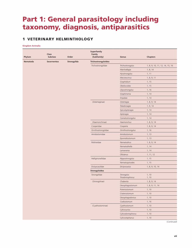
1 VETERINARY HELMINTHOLOGY
Kingdom Animalia
Phylum
Class Subclass
Order
Superfamily Family (Subfamily)
Genus
Chapters
Nematoda Secernentea Strongylida Trichostrongyloidea
Trichostrongylidae Trichostrongylus 1, 8, 9, 10, 11, 12, 14, 15, 16
Marshallagia 1, 8, 14
Hyostrongylus 1, 11
Mecistocirrus 1, 8, 9, 11
Graphidium 1, 15
Obeliscoides 1, 15
Libyostrongylus 1, 16
Graphinema 1, 14
Impalaia 1, 14
(Ostertaginae) Ostertagia 1, 8, 9, 14
Teladorsagia 1, 9, 14
Spiculopteragia 1, 14
Apteragia 1, 14
Camelostrongylus 1, 14
(Haemonchinae) Haemonchus 1, 8, 9, 14
Cooperidae Cooperia 1, 8, 9, 14
Ornithostrongylidae Ornithostrongylus 1, 16
Amidostomidae Amidostomum 1, 13
Epomidiostomum 1, 13
Molineidae Nematodirus 1, 8, 9, 14
Nematodirella 1, 14
Lamanema 1, 14
Ollulanus 1, 11, 12
Helligmonellidae Nippostrongylus 1, 15
Nematospiroides 1, 15
Dictyocaulidae Dictyocaulus 1, 8, 9, 10, 14
Strongyloidea
Strongylidae Strongylus Triodontophorus
1, 101, 10
(Strongylinae) Chabertia 1, 8, 9, 14
Oesophagostomum 1, 8, 9, 11, 14
Poteriostomum 1, 10
Craterostomum 1, 10
Oesophagodontus 1, 10
Codiostomum 1, 16
(Cyathostominae) Cyathostomum 1, 10
Cylicocyclus 1, 10
Cylicodontophorus 1, 10
Cylicostephanus 1, 10
(Continued)
vii
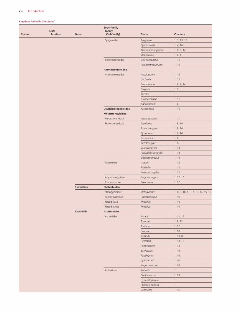
viii Introduction
Syngamidae Syngamus 1, 3, 13, 16
Cyathostoma 1, 4, 16
Mammomonogamus 1, 8, 9, 12
Stephanurus 1, 8, 11
Deletrocephalidae Deletrocephalus 1, 16
Paradeletrocephalus 1, 16
Ancylostomatoidea
Ancylostomatidae Ancylostoma 1, 12
Uncinaria 1, 12
Bunostomum 1, 8, 9, 14
Gaigeria 1, 9
Necator 1
Globocephalus 1, 11
Agriostomum 1, 8
Diaphanocephaloidea Kalicephalus 1, 16
Metastrongyloidea
Metastrongylidae Metastrongylus 1, 11
Protostrongylidae Muellerius 1, 8, 14
Protostrongylus 1, 8, 14
Cystocaulus 1, 8, 14
Spiculocaulus 1, 8
Neostrongylus 1, 8
Varestrongylus 1, 14
Parelaphostrongylus 1, 14
Elaphostrongylus 1, 14
Filaroididae Oslerus 1, 12
Filaroides 1, 12
Aelurostrongylus 1, 12
Angiostrongylidae Angiostrongylus 1, 12, 15
Crenosomidae Crenosoma 1, 12
Rhabditida Rhabditoidea
Strongyloididae Strongyloides 1, 8, 9, 10, 11, 12, 13, 14, 15, 16
Panagrolaimidae Halicephalobus 1, 10
Rhabditidae Rhabditis 1, 10
Rhabdiasidae Rhabdias 1, 16
Ascaridida Ascaridoidea
Ascarididae Ascaris 1, 11, 16
Toxocara 1, 8, 12
Toxascaris 1, 12
Parascaris 1, 10
Ascaridia 1, 13,16
Heterakis 1, 13, 16
Porrocaecum 1, 13
Bayliascaris 1, 16
Polydelphus 1, 16
Ophidascaris 1, 16
Angusticaecum 1, 16
Anisakidae Anisakis 1
Contracaecum 1, 13
Hysterothylacium 1
Pseudoterranova 1
Sulcascaris 1, 16
Phylum
Class Subclass
Order
Superfamily Family (Subfamily)
Genus
Chapters
Kingdom Animalia Continued
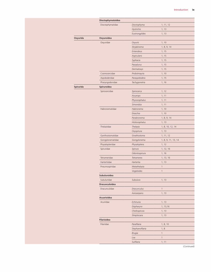
Introduction ix
Dioctophymatoidea
Dioctophymatidae Dioctophyma 1, 11, 12
Hystrichis 1, 13
Eustrongylides 1, 13
Oxyurida Oxyuroidea
Oxyuridae Oxyuris 1, 10
Skrjabinema 1, 8, 9, 14
Enterobius 1, 15
Aspicularis 1, 15
Syphacia 1, 15
Passalurus 1, 15
Dermatoxys 1, 15
Cosmocercidae Probstmayria 1, 10
Aspidoderidae Paraspidodera 1, 15
Pharyngodonidae Tachygonetria 1, 16
Spirurida Spiruroidea
Spirocercidae Spirocerca 1, 12
Ascarops 1, 11
Physocephalus 1, 11
Simondsia 1, 11
Habronematidae Habronema 1, 10
Draschia 1, 10
Parabronema 1, 8, 9, 14
Histiocephalus 1, 13
Thelaziidae Thelazia 1, 8, 10, 12, 14
Oxyspirura 1, 13
Ganthostomatidae Gnathostoma 1, 11, 12
Gongylonematidae Gongylonema 1, 8, 9, 11, 13, 14
Physalopteridae Physaloptera 1, 12
Spiruridae Spirura 1, 12, 16
Odontospirura 1, 16
Tetrameridae Tetrameres 1, 13, 16
Hartertiidae Hartertia 1, 13
Pneumospiridae Metathelazia 1
Vogeloides 1
Subuluroidea
Subuluridae Subulura 1, 13
Dracunculoidea
Dracunculidae Dracunculus 1
Avioserpens 1, 13
Acuarioidea
Acuriidae Echinuria 1, 13
Dispharynx 1, 13,16
Cheilospirura 1, 13
Streptocara 1, 13
Filarioidea
Filariidae Parafilaria 1, 8, 10
Stephanofilaria 1, 8
Brugia 1
Loa 1
Suifilaria 1, 11
(Continued)
x Introduction
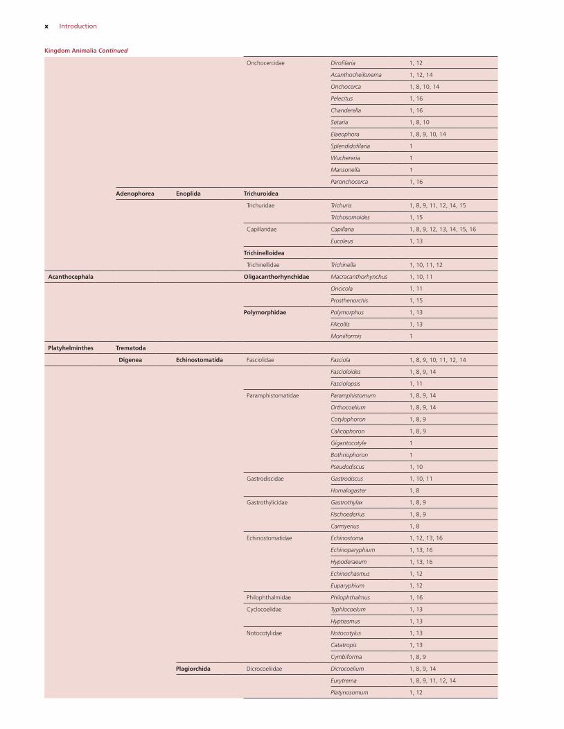
Onchocercidae Dirofilaria 1, 12
Acanthocheilonema 1, 12, 14
Onchocerca 1, 8, 10, 14
Pelecitus 1, 16
Chanderella 1, 16
Setaria 1, 8, 10
Elaeophora 1, 8, 9, 10, 14
Splendidofilaria 1
Wuchereria 1
Mansonella 1
Paronchocerca 1, 16
Adenophorea Enoplida Trichuroidea
Trichuridae Trichuris 1, 8, 9, 11, 12, 14, 15
Trichosomoides 1, 15
Capillaridae Capillaria 1, 8, 9, 12, 13, 14, 15, 16
Eucoleus 1, 13
Trichinelloidea
Trichinellidae Trichinella 1, 10, 11, 12
Acanthocephala Oligacanthorhynchidae Macracanthorhynchus 1, 10, 11
Oncicola 1, 11
Prosthenorchis 1, 15
Polymorphidae Polymorphus 1, 13
Filicollis 1, 13
Moniiformis 1
Platyhelminthes Trematoda
Digenea Echinostomatida Fasciolidae Fasciola 1, 8, 9, 10, 11, 12, 14
Fascioloides 1, 8, 9, 14
Fasciolopsis 1, 11
Paramphistomatidae Paramphistomum 1, 8, 9, 14
Orthocoelium 1, 8, 9, 14
Cotylophoron 1, 8, 9
Calicophoron 1, 8, 9
Gigantocotyle 1
Bothriophoron 1
Pseudodiscus 1, 10
Gastrodiscidae Gastrodiscus 1, 10, 11
Homalogaster 1, 8
Gastrothylicidae Gastrothylax 1, 8, 9
Fischoederius 1, 8, 9
Carmyerius 1, 8
Echinostomatidae Echinostoma 1, 12, 13, 16
Echinoparyphium 1, 13, 16
Hypoderaeum 1, 13, 16
Echinochasmus 1, 12
Euparyphium 1, 12
Philophthalmidae Philophthalmus 1, 16
Cyclocoelidae Typhlocoelum 1, 13
Hyptiasmus 1, 13
Notocotylidae Notocotylus 1, 13
Catatropis 1, 13
Cymbiforma 1, 8, 9
Plagiorchida Dicrocoeliidae Dicrocoelium 1, 8, 9, 14
Eurytrema 1, 8, 9, 11, 12, 14
Platynosomum 1, 12
Kingdom Animalia Continued
Introduction xi
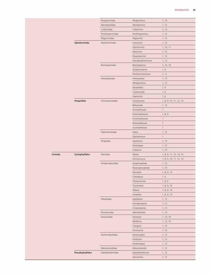
Paragonimidae Paragonimus 1, 14
Nanophyetidae Nanophyetus 1, 12
Collyriclidae Collyriclum 1, 13
Prosthogonimidae Prosthogonimus 1, 13
Plagiorchiidae Plagiorchis 1, 13
Opisthorchida Opisthorchiidae Clonorchis 1, 12
Opisthorchis 1, 10, 11
Metorchis 1, 12
Parametorchis 1, 12
Pseudamphistomum 1, 12
Brachylaemidae Brachylaemus 1, 13, 16
Skrjabinotrema 1, 9
Postharmostomum 1, 11
Heterophyidae Heterophyes 1, 12
Metagonimus 1, 12
Apophallus 1, 6
Cryptocotyle 1, 6
Haplorchis 1, 6
Strigeidida Schistosomatidae Schistosoma 1, 8, 9, 10, 11, 12, 14
Bilharziella 1, 13
Trichobilharzia 1
Orientobilharzia 1, 8, 9
Ornithobilharzia 1
Heterobilharzia 1
Austrobilharzia 1
Diplostomatidae Alaria 1, 12
Diplostomum 1
Strigeidae Apatemon 1, 13
Parastrigea 1, 13
Cotylurus 1, 13
Cestoda Cyclophyllidea Taeniidae Taenia 1, 8, 9, 11, 12, 14, 15
Echinococcus 1, 8, 9, 10, 11, 12, 14
Anoplocephalidae Anoplocephala 1, 10
Paranoplocephala 1, 10
Moniezia 1, 8, 9, 14
Cittotaenia 1, 9
Thysanosoma 1, 8, 9
Thysaniezia 1, 8, 9, 14
Stilesia 1, 8, 9, 14
Avitellina 1, 8, 9, 14
Dilepididae Dipylidium 1, 12
Amoebotaenia 1, 13
Choanotaenia 1, 13
Paruterinidae Metroliasthes 1, 13
Davaineidae Davainea 1, 13, 16
Raillietina 1, 13, 16
Cotugnia 1, 13
Houttuynia 1, 16
Hymenolepididae Hymenolepis 1, 13
Fimbriaria 1, 13
Rodentolepis 1, 15
Mesocestoididae Mesocestoides 1, 12
Pseudophyllidea Diphyllobothriidae Diphyllobothrium 1, 12
Spirometra 1, 12
xii Introduction
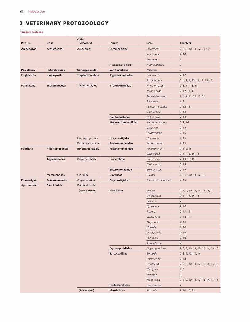
2 VETERINARY PROTOZOOLOGY
Kingdom Protozoa
Phylum
Class
Order (Suborder)
Family
Genus
Chapters
Amoebozoa Archamoeba Amoebida Entamoebidae Entamoeba 2, 8, 9, 10, 11, 12, 13, 16
Iodamoeba 2, 10
Endolimax 2
Acantamoebidae Acanthamoeba 2
Percolozoa Heterolobosea Schizopyrenida Vahlkampfidae Naegleria 2
Euglenozoa Kinetoplasta Trypanosomatida Trypanosomatidae Leishmania 2, 12
Trypanosoma 2, 4, 8, 9, 10, 12, 13, 14, 16
Parabasalia Trichomonadea Trichomonadida Trichomonadidae Tritrichomonas 2, 8, 11, 13, 15
Trichomonas 2, 12, 13, 16
Tetratrichomonas 2, 8, 9, 11, 12, 13, 15
Trichomitus 2, 11
Pentatrichomonas 2, 12, 16
Cochlosoma 2, 13
Dientamoebidae Histomonas 2, 13
Monocercomonadidae Monocercomonas 2, 8, 16
Chilomitus 2, 15
Dientamoeba 2, 15
Honigbergiellida Hexamastigidae Hexamastix 2, 15
Proteromonadida Proteromonadidae Proteromonas 2, 15
Fornicata Retortamonadea Retortamonadida Retortamonadidae Retortamonas 2, 8, 9, 15
Chilomastix 2, 11, 13, 15, 16
Trepamonadea Diplomonadida Hexamitidae Spironucleus 2, 13, 15, 16
Caviomonas 2, 15
Enteromonadidae Enteromonas 2, 15
Metamonadea Giardiida Giardiidae Giardia 2, 8, 9, 10, 11, 12, 15
Preaxostyla Anaeromonadea Oxymonadida Polymastigidae Monocercomonoides 2, 15
Apicomplexa Conoidasida Eucoccidiorida
(Eimeriorina) Eimeriidae Eimeria 2, 8, 9, 10, 11, 13, 14, 15, 16
Cystisospora 2, 11, 12, 14, 16
Isospora 2
Cyclospora 2, 16
Tyzzeria 2, 13, 16
Wenyonella 2, 13, 16
Caryospora 2, 16
Hoarella 2, 16
Octosporella 2, 16
Pythonella 2, 16
Atoxoplasma 2
Cryptosporidiidae Cryptosporidium 2, 8, 9, 10, 11, 12, 13, 14, 15, 16
Sarcocystiidae Besnoitia 2, 8, 9, 12, 14, 16
Hammondia 2, 12
Sarcocystis 2, 8, 9, 10, 11, 12, 13, 14, 15, 16
Neospora 2, 8
Frenkelia 2
Toxoplasma 2, 8, 9, 10, 11, 12, 13, 14, 15, 16
Lankesterellidae Lankesterella 2
(Adeleorina) Klossiellidae Klossiella 2, 10, 15, 16
Introduction xiii
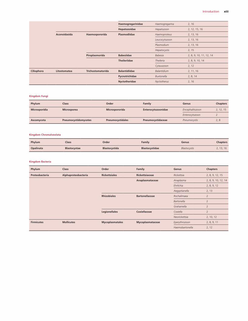
Haemogregarinidae Haemogregarina 2, 16
Hepatozoidae Hepatozoon 2, 12, 15, 16
Aconoidasida Haemospororida Plasmodiidae Haemoproteus 2, 13, 16
Leucocytozoon 2, 13, 16
Plasmodium 2, 13, 16
Hepatocystis 2, 15
Piroplasmorida Babesiidae Babesia 2, 8, 9, 10, 11, 12, 14
Theileriidae Theileria 2, 8, 9, 10, 14
Cytauxzoon 2, 12
Ciliophora Litostomatea Trichostomatorida Balantidiidae Balantidium 2, 11, 16
Pycnotrichidae Buxtonella 2, 8, 14
Nyctotheridae Nyctotherus 2, 16
Kingdom Fungi
Phylum Class Order Family Genus Chapters
Microsporidia Microsporea Microspororida Enterocytozoonidae Encephalitozoon 2, 12, 15
Enterocytozoon 2
Ascomycota Pneumocystidomycetes Pneumocystidales Pneumocystidaceae Pneumocystis 2, 8
Kingdom Chromalveolata
Phylum Class Order Family Genus Chapters
Opalinata Blastocystae Blastocystida Blastocystidae Blastocystis 2, 13, 16
Kingdom Bacteria
Phylum Class Order Family Genus Chapters
Proteobacteria Alphaproteobacteria Rickettsiales Rickettsiaceae Rickettsia 2, 8, 9, 12, 15
Anaplasmataceae Anaplasma 2, 8, 9, 10, 12, 14
Ehrlichia 2, 8, 9, 12
Aegyptianella 2, 13
Rhizobiales Bartonellaceae Rochalimaea 2
Bartonella 2
Grahamella 2
Legionellales Coxiellaceae Coxiella 2
Neorickettsia 2, 10, 12
Firmicutes Mollicutes Mycoplasmatales Mycoplasmataceae Eperythrozoon 2, 8, 9, 11
Haemobartonella 2, 12
xiv Introduction
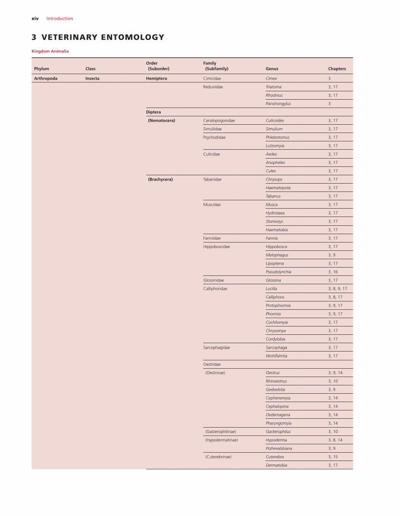
3 VETERINARY ENTOMOLOGY
Kingdom Animalia
Phylum
Class
Order (Suborder)
Family (Subfamily)
Genus
Chapters
Arthropoda Insecta Hemiptera Cimicidae Cimex 3
Reduviidae Triatoma 3, 17
Rhodnius 3, 17
Panstrongylus 3
Diptera
(Nematocera) Ceratopogonidae Culicoides 3, 17
Simuliidae Simulium 3, 17
Psychodidae Phlebotomus 3, 17
Lutzomyia 3, 17
Culicidae Aedes 3, 17
Anopheles 3, 17
Culex 3, 17
(Brachycera) Tabanidae Chrysops 3, 17
Haematopota 3, 17
Tabanus 3, 17
Muscidae Musca 3, 17
Hydrotaea 3, 17
Stomoxys 3, 17
Haematobia 3, 17
Fanniidae Fannia 3, 17
Hippoboscidae Hippobosca 3, 17
Melophagus 3, 9
Lipoptena 3, 17
Pseudolynchia 3, 16
Glossinidae Glossina 3, 17
Calliphoridae Lucilia 3, 8, 9, 17
Calliphora 3, 8, 17
Protophormia 3, 9, 17
Phormia 3, 9, 17
Cochliomyia 3, 17
Chrysomya 3, 17
Cordylobia 3, 17
Sarcophagidae Sarcophaga 3, 17
Wohlfahrtia 3, 17
Oestridae
(Oestrinae) Oestrus 3, 9, 14
Rhinoestrus 3, 10
Gedoelstia 3, 9
Cephenemyia 3, 14
Cephalopina 3, 14
Oedemagena 3, 14
Pharyngomyia 3, 14
(Gasterophilinae) Gasterophilus 3, 10
(Hypodermatinae) Hypoderma 3, 8, 14
Przhevalskiana 3, 9
(Cuterebrinae) Cuterebra 3, 15
Dermatobia 3, 17
Introduction xv
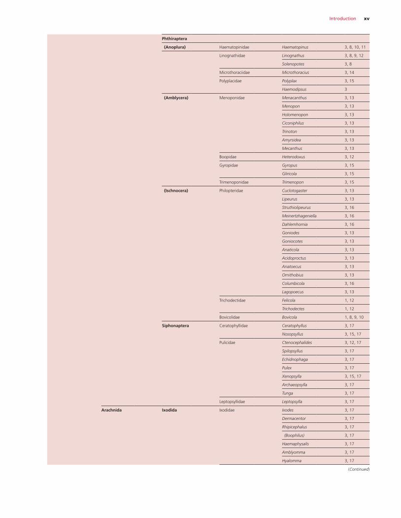
Phthiraptera
(Anoplura) Haematopinidae Haematopinus 3, 8, 10, 11
Linognathidae Linognathus 3, 8, 9, 12
Solenopotes 3, 8
Microthoraciidae Microthoracius 3, 14
Polyplacidae Polyplax 3, 15
Haemodipsus 3
(Amblycera) Menoponidae Menacanthus 3, 13
Menopon 3, 13
Holomenopon 3, 13
Ciconiphilus 3, 13
Trinoton 3, 13
Amyrsidea 3, 13
Mecanthus 3, 13
Boopidae Heterodoxus 3, 12
Gyropidae Gyropus 3, 15
Gliricola 3, 15
Trimenoponidae Trimenopon 3, 15
(Ischnocera) Philopteridae Cuclotogaster 3, 13
Lipeurus 3, 13
Struthiolipeurus 3, 16
Meinertzhageniella 3, 16
Dahlemhornia 3, 16
Goniodes 3, 13
Goniocotes 3, 13
Anaticola 3, 13
Acidoproctus 3, 13
Anatoecus 3, 13
Ornithobius 3, 13
Columbicola 3, 16
Lagopoecus 3, 13
Trichodectidae Felicola 1, 12
Trichodectes 1, 12
Bovicolidae Bovicola 1, 8, 9, 10
Siphonaptera Ceratophyllidae Ceratophyllus 3, 17
Nosopsyllus 3, 15, 17
Pulicidae Ctenocephalides 3, 12, 17
Spilopsyllus 3, 17
Echidnophaga 3, 17
Pulex 3, 17
Xenopsylla 3, 15, 17
Archaeopsylla 3, 17
Tunga 3, 17
Leptopsyllidae Leptopsylla 3, 17
Arachnida Ixodida Ixodidae Ixodes 3, 17
Dermacentor 3, 17
Rhipicephalus 3, 17
(Boophilus) 3, 17
Haemaphysalis 3, 17
Amblyomma 3, 17
Hyalomma 3, 17
(Continued)
xvi Introduction
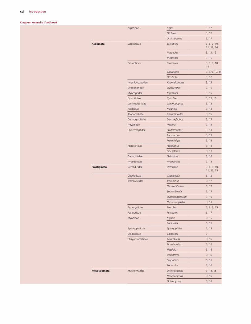
Argasidae Argas 3, 17
Otobius 3, 17
Ornithodoros 3, 17
Astigmata Sarcoptidae Sarcoptes 3, 8, 9, 10, 11, 12, 14
Notoedres 3, 12, 15
Trixacarus 3, 15
Psoroptidae Psoroptes 3, 8, 9, 10, 14
Chorioptes 3, 8, 9, 10, 14
Otodectes 3, 12
Knemidocoptidae Knemidocoptes 3, 13
Listrophoridae Leporacarus 3, 15
Myocoptidae Mycoptes 3, 15
Cytoditidae Cytodites 3, 13, 16
Laminosioptidae Laminosioptes 3, 13
Analgidae Megninia 3, 13
Atopomelidae Chirodiscoides 3, 15
Dermoglyphidae Dermoglyphus 3, 13
Freyanidae Freyana 3, 13
Epidermoptidae Epidermoptes 3, 13
Microlichus 3, 13
Promyialges 3, 13
Pterolichidae Pterolichus 3, 13
Sideroferus 3, 13
Gabuciniidae Gabucinia 3, 16
Hypoderidae Hypodectes 3, 13
Prostigmata Demodicidae Demodex 3, 8, 9, 10, 11, 12, 15
Cheyletidae Cheyletiella 3, 12
Trombiculidae Trombicula 3, 17
Neotrombicula 3, 17
Eutrombicula 3, 17
Leptotrombidium 3, 15
Neoschongastia 3, 13
Psorergatidae Psorobia 3, 8, 9, 15
Pyemotidae Pyemotes 3, 17
Myobidae Myobia 3, 15
Radfordia 3, 15
Syringophilidae Syringophilus 3, 13
Cloacaridae Cloacarus 3
Pterygosomatidae Geckobiella 3, 16
Pimeliaphilus 3, 16
Hirstiella 3, 16
Ixodiderma 3, 16
Scapothrix 3, 16
Zonurobia 3, 16
Mesostigmata Macronyssidae Ornithonyssus 3, 13, 15
Neoliponyssus 3, 16
Ophionyssus 3, 16
Kingdom Animalia Continued
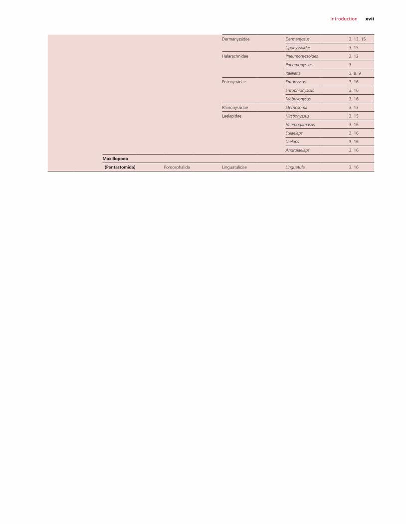
Introduction xvii
Dermanyssidae Dermanyssus 3, 13, 15
Liponyssoides 3, 15
Halarachnidae Pneumonyssoides 3, 12
Pneumonyssus 3
Raillietia 3, 8, 9
Entonyssidae Entonyssus 3, 16
Entophionyssus 3, 16
Mabuyonysus 3, 16
Rhinonyssidae Sternosoma 3, 13
Laelapidae Hirstionyssus 3, 15
Haemogamasus 3, 16
Eulaelaps 3, 16
Laelaps 3, 16
Androlaelaps 3, 16
Maxillopoda
(Pentastomida) Porocephalida Linguatulidae Linguatula 3, 16
xviii Contents
4 Laboratory diagnosis of parasitism, 259Helminth infections, 259
Faecal examination, 259Collection of faeces, 259Methods of examination of faeces, 259Identification of nematode eggs, 261Identification of trematode eggs, 261Larval recovery, 268Recovery of alimentary nematodes, 269Key to the identification of gastrointestinal nematodes of ruminants, 271Recovery of adult lungworms, 274Recovery of trematode and cestode parasites, 275Other aids to diagnosis of ruminant nematodes, 275DNA‐based methods, 276
Protozoal infections, 283Examination of faecal samples for coccidia, 283Examination of faecal samples for other protozoa, 283Examination of blood and lymph, 289Examination of skin, 290Xenodiagnosis, 290
Ectoparasites, 290Insects, 290Ticks and mites, 301Mounting and preservation, 309
Chemicals and solutions, 311Flotation solutions, 311Worm count solutions, 311Stains, 311
5 Antiparasitics, 313Anthelmintics, 313
Anthelmintics and their mode of action, 313Properties of anthelmintic compounds, 317Use of anthelmintics, 317Methods of administration, 318
Antiprotozoals, 318Antiprotozoals and their mode of action, 319Use of antiprotozoals, 323Methods of administration, 323
Ectoparasiticides (insecticides/acaricides), 323Ectoparasiticides and their mode of action, 324Methods of pesticide application and uses, 327
Parasiticide resistance, 328Anthelmintic resistance, 328Antiprotozoal resistance, 333Pesticide resistance, 334
6 The epidemiology of parasitic diseases, 336An increase in the numbers of infective stages, 336
Contamination of the environment, 336Development and survival of infective stages, 337
An alteration in host susceptibility, 338Altered effects of an existing infection, 338Altered susceptibility to the acquisition of new infections, 340
The introduction of susceptible stock, 340Absence of acquired immunity, 340Absence of age immunity, 341Longevity of infective stages, 341Influence of genetic factors, 341Strain of parasite, 341
Introduction of infection into a clean environment, 341Introduction of new stock, 341Role of effluent, 341Role of infected vectors, 342
7 Host resistance to parasitic diseases, 343Species resistance, 343Age resistance, 343Breed resistance, 343Acquired immunity to helminth infections, 344
Effect of the immune response, 344Evasion of the host’s immune response, 345Cost of the immune response, 346
Acquired immunity to protozoal infections, 346Acquired immunity to arthropod infections, 347The future of parasite vaccines, 347
Helminths, 347Protozoa, 349Ectoparasites, 351
Part 2: Host–parasite diseases
8 Parasites of cattle, 352Endoparasites, 352
Parasites of the digestive system, 352Parasites of the respiratory system, 379Parasites of the liver, 383Parasites of the pancreas, 388Parasites of the circulatory system, 389Parasites of the nervous system, 409Parasites of the reproductive/urogenital system, 410Parasites of the locomotory system, 413Parasites of the connective tissue, 417Parasites of the integument, 420
Ectoparasites, 424Flies, 424Lice, 424Mites, 427
Cattle parasite checklist, 431
9 Parasites of sheep and goats, 436Endoparasites, 436
Parasites of the digestive system, 436Parasites of the respiratory system, 474
Parasites of the liver, 480Parasites of the pancreas, 489Parasites of the circulatory system, 489Parasites of the nervous system, 497Parasites of the reproductive/urogenital system, 498Parasites of the locomotory system, 500Parasites of the integument, 502
Ectoparasites, 503Lice, 503Mites, 506Fly strike (myiasis), 511
Sheep parasite checklist, 516Goat parasite checklist, 520
10 Parasites of horses, 524Endoparasites, 524
Parasites of the digestive system, 524Parasites of the respiratory system, 544Parasites of the liver, 545Parasites of the circulatory system, 545Parasites of the nervous system, 552Parasites of the reproductive/urogenital system, 554Parasites of the locomotory system, 555Parasites of the integument, 556
Ectoparasites, 558Lice, 558Mites, 559
Horse parasite checklist, 561
11 Parasites of pigs, 565Endoparasites, 565
Parasites of the digestive system, 565Parasites of the respiratory system, 583Parasites of the liver, 584Parasites of the pancreas, 585Parasites of the circulatory system, 586Parasites of the nervous system, 588Parasites of the reproductive/urogenital system, 588Parasites of the locomotory system, 590Parasites of the integument, 592
Ectoparasites, 592Lice, 592Mites, 593
Pig parasite checklist, 596
12 Parasites of dogs and cats, 599Endoparasites, 599
Parasites of the digestive system, 599Parasites of the respiratory system, 629Parasites of the liver, 635Parasites of the circulatory system, 640Parasites of the nervous system, 654
Parasites of the reproductive/urogenital system, 656Parasites of the locomotory system, 657Parasites of the integument, 659
Ectoparasites, 662Lice, 662Mites, 663
Dog parasite checklist, 670Cat parasite checklist, 674
13 Parasites of poultry and gamebirds, 678Endoparasites, 678
Parasites of the digestive system, 678Parasites of the respiratory system, 720Parasites of the liver, 723Parasites of the circulatory system, 723Parasites of the nervous system, 729Parasites of the reproductive/urogenital system, 729Parasites of the locomotory system, 731Parasites of the integument, 732
Ectoparasites, 733Lice, 733Mites, 737
Chicken parasite checklist, 742Turkey parasite checklist, 746Duck parasite checklist, 749Goose parasite checklist, 752Pheasant parasite checklist, 754Partridge parasite checklist, 756Quail parasite checklist, 758Guinea fowl parasite checklist, 760
14 Parasites of ungulates, 761Deer, 761
Parasites of the digestive system, 761Parasites of the respiratory system, 763Parasites of the liver, 766Parasites of the circulatory system, 768Parasites of the nervous system, 769Parasites of the reproductive/urogenital system, 770Parasites of the locomotory system, 770Parasites of the connective tissue, 771Parasites of the integument, 772Ectoparasites, 773
Camels, 773Parasites of the digestive system, 773Parasites of the respiratory system, 778Parasites of the liver, 779Parasites of the pancreas, 780Parasites of the circulatory system, 780Parasites of the nervous system, 784Parasites of the locomotory system, 784Parasites of the connective tissue, 785Ectoparasites, 785
Contents xix
Llamas, alpacas, guanacos, vicuñas, 787Parasites of the digestive system, 787Parasites of the respiratory system, 790Parasites of the liver, 791Parasites of the nervous system, 792Parasites of the reproductive/urogenital system, 792Parasites of the locomotory system, 792Ectoparasites, 792
Water buffalo, 793Parasites of the digestive system, 793Parasites of the respiratory system, 796Parasites of the liver, 797Parasites of the pancreas, 798Parasites of the circulatory system, 798Parasites of the nervous system, 801Parasites of the reproductive/urogenital system, 801Parasites of the locomotory system, 801Parasites of the connective tissue, 801Parasites of the integument, 802Ectoparasites, 802
Deer parasite checklist, 804Camel parasite checklist, 808Camelid parasite checklist, 811Buffalo parasite checklist, 814
15 Parasites of laboratory animals, 816Rabbits, 816
Parasites of the digestive system, 816Parasites of the respiratory system, 826Parasites of the liver, 826Parasites of the circulatory system, 827Parasites of the nervous system, 829Parasites of the reproductive/urogenital system, 829Parasites of the locomotory system, 829Ectoparasites, 830
Guinea pigs, 833Parasites of the digestive system, 833Parasites of the respiratory system, 836Parasites of the liver, 836Parasites of the circulatory system, 836Parasites of the nervous system, 836Parasites of the reproductive/urogenital system, 836Parasites of the locomotory system, 836Parasites of the integument, 836Ectoparasites, 836
Rats and mice, 839Parasites of the digestive system, 839Parasites of the respiratory system, 847Parasites of the liver, 847Parasites of the circulatory system, 848Parasites of the nervous system, 849Parasites of the reproductive/urogenital system, 849Parasites of the locomotory system, 849Ectoparasites, 849
Primates, 853Parasites of the digestive system, 853Parasites of the respiratory system, 866Parasites of the liver, 868Parasites of the pancreas, 869Parasites of the circulatory system, 869Parasites of the nervous system, 874Parasites of the reproductive/urogenital system, 875Parasites of the locomotory system, 875Parasites of the integument, 876Ectoparasites, 876
Rabbit parasite checklist, 879Guinea pig parasite checklist, 881Rat parasite checklist, 882Mouse parasite checklist, 884Primate parasite checklist, 886
16 Parasites of exotics, 893Pigeons, 893
Parasites of the digestive system, 893Parasites of the respiratory system, 897Parasites of the circulatory system, 898Parasites of the nervous system, 899Parasites of the reproductive/urogenital system, 899Parasites of the locomotory system, 899Parasites of the integument, 899Ectoparasites, 900
Ratites (ostrich, rhea, emu), 903Parasites of the digestive system, 903Parasites of the respiratory system, 906Parasites of the circulatory system, 906Parasites of the nervous system, 907Ectoparasites, 907
Reptiles, 908Parasites of the digestive system, 908Parasites of the respiratory system, 912Parasites of the circulatory system, 912Parasites of the reproductive/urogenital system, 912Ectoparasites, 913
Pigeon parasite checklist, 915Ratite parasite checklist, 918
17 Facultative ectoparasites and arthropod vectors, 921Insects, 921
Biting and nuisance flies, 928Other common hippoboscids, 934Myiasis flies, 935
Ticks, 952Hard ticks, 952Soft ticks (Argasidae), 968
Mites, 972
References and further reading, 975Index, 979
xx Contents
xxi
Preface to the first edition
This book is intended for students of veterinary parasitology, for practising veterinarians and for others requiring information on some aspect of parasitic disease.
Originally intended as a modestly expanded version of the printed notes issued to our students in the third and fourth years of the course, the text, perhaps inevitably, has expanded. This was due to three factors. First, a gradual realization of the deficiencies in our notes: secondly, the necessity of including some of the com-ments normally imparted during the lecture course and thirdly, at the suggestion of the publishers, to the inclusion of certain aspects of parasitic infections not treated in any detail in our course.
We should perhaps repeat that the book is primarily intended for those who are directly involved in the diagnosis, treatment and con-trol of parasitic diseases of domestic animals. The most important of these diseases have therefore been discussed in some detail, the less important dealt with more briefly and the uncommon either omitted or given a brief mention, Also, since details of classifica-tion are of limited value to the veterinarian we have deliberately kept these to the minimum sufficient to indicate the relationships between the various species. For a similar reason, taxonomic detail is only presented at the generic level and, occasionally, for certain parasites, at species level. We have also trod lightly on some other areas such as, for example, the identification of species of tropical ticks and the special significance and epidemiology of some para-sites of regional importance. In these cases, we feel that instruction is best given by an expert aware of the significance of particular species in that region.
Throughout the text we have generally referred to drugs by their chemical, rather than proprietary, names because of the plethora of the latter throughout the world. Also, because formulations are often different, we have avoided stating doses; for these, reference should be made to the data sheets produced by the manufacturer. However, on occasions when a drug is recommended at an unusual dose, we have noted this in the text.
In the chapters at the end of the book we have attempted to review five aspects of veterinary parasitology, epidemiology, immunity, anthelmintics, ectoparasiticides and laboratory diagnosis. We hope that this broader perspective will be of value to students, and par-ticularly to those dismayed by the many complexities of the subject.
There are no references in the text apart from those at the end of the chapter on diagnosis. This was decided with some regret and much relief on the grounds that it would have meant the inclusion, in a book primarily intended for undergraduates, of hundreds of references. We hope that those of our colleagues throughout the world who recognize the results of their work in the text will accept this by way of explanation and apology.
We would, however, like to acknowledge our indebtedness to the authors of several source books on veterinary parasitology whose work we have frequently consulted. These include Medical and Veterinary Protozoology by Adam, Paul & Zaman, Veterinaermed-izinische Parasitologie by Boch & Supperer, Dunn’s Veterinary Hel-minthology, Euzéby’s Les Maladies Vermineuses des Animaux Domes-tiques, Georgi’s Parasitology for Veterinarians, Reinecke’s Veterinary Helminthology, Service’s A Guide to Medical Entomology and Souls-by’s Helminths, Arthropods and Protozoa of Domesticated Animals.
Any student seeking further information on specific topics should consult these or, alternatively, ask his tutor for a suitable review.
The ennui associated with repeated proof reading may occasion-ally (we hope, rarely) have led to some errors in the text. Notifica-tion of these would be welcomed by the authors. Finally we hope that the stresses endured by each of us in this collaborative venture will be more than offset by its value to readers.
G.M. UrquhartJ. Armour
J.L. DuncanA.M. Dunn
F.W. JenningsSeptember 1985
xxii
Acknowledgements to the first edition
We would like to express our gratitude to the following individuals and organizations that assisted us in the preparation of this book.
First, to Drs R. Ashford and W. Beesley of Liverpool; Dr J. Bogan, Glasgow; Dr W. Campbell, Rahway, USA; Dr R. Dalgleish, Brisbane; Dr L. Joyner, Weybridge, England; Dr T. Miller, Florida; Dr M. Murray, Nairobi; Dr R. Purnell, Sandwich, England; Dr S.M. Taylor, Belfast; Professor K. Vickerman, Glasgow. Each of these read and commented on sections of the text in which they are expert. Any errors in these areas are, however, solely the responsibility of the authors.
Secondly, to the following individuals and companies who kindly allowed us to use their photographs or material as illustrations or plates:
Dr E. Allonby, Nairobi (Plate I d, e, f); Dr K. Angus, Edinburgh (Fig. 167); Dr J. Arbuckle, Guildford, England (Fig. 61); Dr E. Batte, North Carolina, USA (Plate IIIf); Dr I. Carmichael, Johannesburg, S. Africa (Fig. 142); Dr L. Cramer, Sao Paulo (Fig. 126b); Crown Copyright, UK (Plate XIVb); Dr J. Dunsmore, Murdoch, W. Aus-tralia (Plate IVd); Professor J. Eckert, Zurich (Fig. 96); Glaxovet, Harefield, England (Plate IIf); Dr I. Herbert, Bangor, Wales (Fig. 172); Dr A. Heydorn, W. Berlin (Figs 170, 171); Professor F. Hörning, Berne (Fig. 82; Plate Ve); Dr B. Iovanitti, Balcarce, Argentina (Figs 22, 23); Dr D. Jacobs, London (Fig. 38); Drs D. Kelly and A. Longstaffe, Bristol (Figs 156, 157); The late Dr I. Lauder, Glasgow (Fig. 65, Plate XIc, e, XIIb); Drs B. Lindemann and J. McCall, Georgia, USA (Fig. 67); Dr N. McEwan, Glasgow (Plate XId, XIIe); Dr G. Mitchell, Ayr, Scotland (Plate VIe); Professor M. Murray, Glasgow (Figs 68, 84, 152); Dr A. Nash, Glasgow (Fig. 138b, Plate XIIc); Dr Julia Nicholls, Adelaide, Australia (Figs 6, 14c, d); Dr R. Purnell, Sandwich, England (Fig. 173, Plate VIIId, e, f); Professor H. Pirie, Glasgow (Fig. 40); Dr J. Reid, Brussels (Plate XIIa); Dr Elaine
Rose, Houghton Poultry Research Station, Huntingdon, England (Figs 160, 163b, 164a, b); Professor I. Selman, Glasgow (Plate XIf); Dr D. Taylor, Glasgow (Plate XIVc); Dr M. Taylor, London (Fig. 85); Dr S. Taylor, Belfast (Plate IIa); Dr H. Thompson, Glasgow (Fig. 92, Plate IVb, c, VId); Dr R. Titchener, Ayr, Scotland (Fig. 113b, Plate VIIIa); Dr A. Waddell, Brisbane, Australia (Fig. 66, Plate IVe); Well-come Research Laboratories, Berkhamsted, England (Plate VIIIc); Dr A. Wright, Bristol (Plate VIb, XIb, XIId, f). In this context we are also extremely grateful to Miss E. Urquhart, Wrexham, Wales who prepared many of the line drawings.
Thirdly, to the pharmaceutical companies of Crown Chemical, Kent, England; Hoechst UK, Bucks; Merck Sharp & Dohme, Herts; Pfizer, Kent; Schering, New Jersey; Syntex Agribusiness, California. Their generosity enabled us to present many of the photographs in colour, thus enhancing their value.
Finally, to those members of the Faculty of Veterinary Medicine, Glasgow, whose cooperation was essential in the production of this book. We would especially like to thank Kenneth Bairden, our chief technician, who prepared much of the material for photography, often at inordinately short notice; Archie Finnie and Allan May, of the Photographic Unit, who, almost uncomplainingly, undertook the extra work of photographing many specimens; our two depart-mental secretaries, Elizabeth Millar and Julie Nybo, without whose skill and attention to detail this book would certainly not have been written.
G.M. UrquhartJ. Armour
J. L. DuncanA. M. Dunn
F. W. JenningsSeptember 1985
xxiii
Preface and acknowledgements to the second edition
The first edition of this book was published in 1987 and the authors considered that a second edition is now necessary for sev-eral reasons.
First, the widespread use of drugs such as avermectins and milbemycins, which have had a significant effect on anthelmintic prophylaxis and control. At the time of the first edition only one, ivermectin, was marketed whereas at the present time there are now several such products, supplemented by a number of new, long- acting chemoprophylactic devices.
Secondly, in many countries the production of a number of older anthelmintics and insecticides has largely ceased or many are dif-ficult to find.
Thirdly, several parasitic diseases have now been described, about which little was known at the time of the first edition. Notably these are neosporosis and Lyme disease. Also included is a short descrip-tion of the nasal mite of dogs, Pneumonyssus caninum, kindly pro-vided by Professor Arvid Uggla of the National Veterinary Institute and Swedish University of Agricultural Sciences, Uppsala, Sweden.
Fourthly, we have taken the opportunity of rewriting some parts of the text, which on reflection, were less clear than we had hoped. In many cases, this has been supplemented by new diagrams or photographs.
Another change in this edition is the adoption of the standard-ized nomenclature of animal parasitic diseases (SNOAPAD) pro-posed by an expert committee appointed by the World Association for the Advancement of Veterinary Parasitology (WAAVP) pub-lished in Veterinary Parasitology (1988) 29, 299–326. Although this may have a discomforting effect on those who have used certain familiar terms for animal parasitic diseases for many years, it is designed to improve the clarity of scientific communication by the
general use of uniform terminology and should, in the long term, prove particularly beneficial in facilitating the retrieval of comput-erized data related to veterinary parasitology.
At the end of the book we have given a list of books and journals, which should be useful to anyone who wishes to pursue a specific sub-ject in greater detail. This is confined to publications which are readily available in most libraries of universities and research institutes.
We wish to thank Drs Ken Bairden, Quintin McKellar and Jac-queline McKeand for helpful comments on the text, also Mr Stuart Brown who assisted in the preparation of some of the new illus-trations and Una B. Shanks RSW who prepared all of the new drawings.
We should mention, with great regret, the death of our co-author Dr Angus M. Dunn, who died in 1991 before this review was started, but we are reasonably certain that he would have approved of all the alterations we have made.
At the start of this revision we had intended to include new sections on parasitic disease of both fish and laboratory animals. However, a subsequent review of the literature currently available on these two subjects indicated that both were adequately covered in existing publications and it seemed more sensible to include the titles of these in the list of suggested reading.
Finally we wish to express our appreciation of the reception accorded to the first edition by reviewers, colleagues and students; we hope this second edition will be equally well received.
G. M. UrquhartJ. Armour
J. L. DuncanA. M. Dunn
F. W. Jennings
xxiv
Preface and acknowledgements to the third edition
The third edition has been written to accommodate a wider read-ership which includes teachers and students in veterinary schools, research groups in universities and institutes, veterinarians in practice and in government service and others who are involved in aspects of parasitic disease. In producing the new edition of Veterinary Parasitology the authors had several aims.
The first was to preserve the spirit of the first and second editions, which had been compiled by eminent and respected veterinary par-asitologists in their field and which provided a solid background on which to consolidate.
The second aim was to expand the sections on protozoa and ecto-parasites and to incorporate a larger selection of parasites, which are of veterinary significance in other parts of the world. The book focuses mainly on core information relating to parasites of livestock and companion animals but new sections on parasites of poultry and gamebirds, laboratory animals, exotic pets, and ‘farmed’ spe-cies have been included. The majority of parasitic diseases are now covered in detail using a standardised format for each parasite to allow easy referencing and for comparison between species within a genus. Where appropriate, reference is made to human infections where there is natural transmission of parasitic disease between vertebrate animals and man (zoonoses).
The third aim was to present the information in a format which is compatible with the current parasitology teaching modules used within many university veterinary schools. This inevitably has had to be a compromise, as approaches to teaching veterinary parasitology differ throughout the world, but, by arranging the parasites under host species and their predilection site within the host and providing a comprehensive check list for each section and extensive cross-referencing, it is hoped that information on particular parasites can be easily located. Taxonomy of the main parasitic phyla and classes are provided within an introductory chapter along with generic descriptions and anatomical features of the parasite orders and families. Additional detailed sections are provided at the back of the book on veterinary antiparasit-ics, with a section on laboratory diagnosis, including numerous tables and identification charts. In keeping with previous editions a series of brief overviews of topics relevant to veterinary parasi-tology have been included to provide the non-expert with basic background information and to also highlight addition sources of reading.
The classification of parasites has been updated to reflect many of the systematic changes introduced, particularly where molecu-lar genetics-based taxonomic re-organisation has been introduced. Throughout, synonyms have been provided reflecting older taxo-nomic nomenclature or where controversy remains. As with the previous edition, parasitic infections are described according to the Standardized Nomenclature of Animal Parasitic Diseases guide-lines (SNOAPAD, 1988; Veterinary Parasitology 29, 299–326). In considering treatment of parasitic infections we have used the generic names of drugs to avoid listing the wide range of products which are currently marketed in different countries. Dose rates of drugs are not always stated as many vary from country to country, being influenced by the relevant regulatory authorities. In all cases, readers are advised to consult the manufacturer’s data sheets for current information and local regulations.
The authors are extremely grateful to Professor Sir James Armour and Professor James Duncan for their interest and support and for reading through the drafts of the text and their constructive comments. Any errors in the book are solely the responsibility of the authors. In order to assist the reader and for clarification we took the decision to produce much of the book and illustrations in colour and we are most grateful for the generous financial sup-port of the following pharmaceutical companies which made this possible: Fort Dodge Animal Health; Pfizer Animal Health; Merial Animal Health; Novartis Animal Health; Intervet UK Ltd.
Finally, the help and support of the following list of people is acknowledged in producing this textbook. Professor Quintin McKellar (previous scientific director) and Professor Julie Fitzpat-rick (current scientific director) of the Moredun Research Institute provided support to Dr Coop allowing him full access to the library facilities following his retirement. Dr Frank Jackson for comments on the manuscript. Michelle Moore, Matthew Carroll and Caroline Chaffer provided invaluable assistance with setting up much of the initial file documentation required to develop the re-organised structure of the book. Ralph Marshall at the Veterinary Laborato-ries Agency provided information on coccidial species of camelids and gamebirds. The technical support of Shelagh Wall is gratefully acknowledged.
Professor Mike TaylorDr Bob Coop
Professor Richard Wall
xxv
Preface and acknowledgements to the fourth edition
This fourth edition has been written to provide detailed parasite descriptions and reference texts for teachers, research groups in universities and institutes, veterinarians in practice and in govern-ment service, and others involved in aspects of parasitic disease. In producing the new edition of Veterinary Parasitology the authors had the following aims and objectives.
The first was to preserve the spirit of the first two editions of the textbook Veterinary Parasitology, which had been compiled by eminent and respected veterinary parasitologists in their field and which provided a solid background on which to further con-solidate.
The second aim was to greatly expand and revise the systematic sections on helminthology, protozoology and entomology and the descriptions of animal parasites which are of veterinary sig-nificance in many parts of the world. This edition focuses mainly on core information relating to parasites of livestock and com-panion animals but sections on parasites of poultry and game-birds, laboratory animals, exotic pets and ‘farmed’ species are included. The majority of parasitic diseases are now covered in detail using standardised formats for each parasite to allow easy referencing and for comparison between species within a genus. Where appropriate, reference is made to human infections where there is natural transmission of parasitic disease between verte-brate animals and humans (zoonoses).
The third aim was to present the information in two formats which are considered compatible with the diversity of current parasitology teaching modules used within many universities and veterinary schools, both for teaching parasite systematics and for teaching of diseases on a host–organ basis. The latter is achieved by arranging the parasites under host species and their predilec-tion sites. By providing both approaches, and because teaching of veterinary parasitology differs throughout the world, it is hoped that information on particular parasites can be easily located. Tax-onomy of the main parasitic kingdoms, phyla, classes, orders and genera are now provided in much greater detail within the intro-ductory chapters and subsections, along with detailed descrip-tions and anatomical features of the parasite orders, families and species. Additional detailed and updated sections are provided on veterinary antiparasitics, with an expanded section on labora-tory diagnosis, including recent molecular developments and an increased number of tables and identification charts. In keeping with previous editions of the textbook, a series of brief overviews of topics relevant to veterinary parasitology have been included to provide the non-expert with basic background information and also to highlight additional sources of reading. An additional fea-ture is the inclusion of a number of additional figures, diagrams and images of parasites.
The classification of parasites reflects many of the systematic changes, particularly where molecular genetics-based taxonomic reclassification has been introduced. Throughout, synonyms have
been provided reflecting older taxonomic nomenclature, or where controversy remains. As with the earlier textbook editions, parasitic infections are described according to the Standardised Nomencla-ture of Animal Parasitic Diseases guidelines (SNOAPAD, 1988; Veterinary Parasitology 29, 299–326). In considering treatment of parasitic infections we have used the generic names of drugs to avoid listing the wide range of products that are currently marketed in different countries. Dose rates of drugs are not always stated as many vary from country to country, being influenced by the rel-evant regulatory authorities. In all cases, readers are advised to consult the manufacturer’s data sheets for current information and local regulations.
The new edition retains the expertise of the three authors of the previous third edition.
Professor Mike Taylor is a veterinary graduate of Glasgow Univer-sity Veterinary College, having studied under the authors of the first and second editions of the textbook Veterinary Parasitology, whose enthusiasm for their subject greatly influenced his interest in veterinary parasitology. After 6 years in general veterinary prac-tice, a large part of his career was spent at the Central Veterinary Laboratory, Weybridge, later to become the Veterinary Laboratories Agency (VLA), where he worked on the epidemiology and control of parasitic helminths, protozoa and ectoparasites of domestic ani-mals, and in particular parasite chemotherapy, parasite control and anthelmintic resistance. During this time he studied for a PhD at the Royal Veterinary College, London, under the expert guidance of Professor Dennis Jacobs. He has spent over 30 years in veterinary parasitology, published 20 book chapters and has over 250 scien-tific publications, as well as contributing extensively to the third and current editions of Veterinary Parasitology. He retired as Head of Veterinary Surveillance at the Central Science Laboratory York (later to become FERA) in 2011, but remains a visiting Professor of Parasitology at the Royal Veterinary College, London, an Emeri-tus Fellow of FERA, and now runs his own veterinary parasitology consultancy company. He is a Diplomate of the European College of Veterinary Parasitology, and a Diplomate of the European College of Small Ruminant Health and Management. He has been a Fellow of Edinburgh University, a former Editor-in-Chief of the Journal Veterinary Parasitology, and past president of the British Associ-ation for Veterinary Parasitology.
Dr Bob Coop graduated in biochemistry from the University of Liverpool and then undertook a PhD in large animal parasitology at the University of Wales, Bangor. He has spent over 35 years in veterinary parasitology research, initially working with lungworm infection in pigs and then on the epidemiology and pathogenesis of gastrointestinal nematode infection in small and large ruminants, and in particular the nutrition–parasite interaction and sustainable control strategies. He has been a Fellow of Edinburgh University
xxvi Preface and acknowledgements to the fourth edition
and a past president of the British Association for Veterinary Para-sitology. Formerly as Head of the Division of Parasitology at the Moredun Research Institute, Edinburgh and now as a Fellow of the Moredun Foundation, he has considerable experience of knowledge transfer to end-user groups and veterinarians in practice. Dr Coop acknowledges full access to the Information Technology Services at the Moredun Research Institute.
Professor Richard Wall graduated in zoology from the Univer-sity of Durham followed by a PhD in insect population ecology at the University of Liverpool. He is now Professor of Zoology at the University of Bristol, where he teaches and heads a research group studying a diverse range of arthropods, focusing particularly on ectoparasites of veterinary importance and insect colonisers of dung and carrion. His research ranges widely from fundamental studies of arthropod taxonomy and physiology, through to field population ecology and farm-level investigations of the application of sustainable control technologies.
The help and support of the following people is acknowledged in producing this edition. Dr Philip Skuce of the Moredun Research
Institute (MRI) wrote the section in Chapter 4 on molecular diag-nostics for which the authors are extremely grateful. Professor James Duncan provided his collection of photographic slides from the Glasgow University Veterinary School, many of which appear as illustrations or figures, both in this and previous editions. Additional photographic material has been used from a collection of digital images gathered under the auspices of the British Associ-ation of Veterinary Parasitology over a period of 20 years. Several of the newer illustrations and figures included in this edition were reproduced from material by former colleagues from the Parasi-tology Department at the Central Veterinary Laboratory (CVL), Weybridge, which sadly now no longer exists. Acknowledgement is therefore made to the following: Dr Martin Gregory, Dr Len Joiner, Janet Catchpole, Chris Norton, Ralph and Jackie Marshall, Dr Mike Peirce, Dr Richard Cawthorne, Keith Hunt, Colin Hong, Barry Lan-caster, Dr Charles Ollerenshaw, Gordon Graham, Dr Joe Donnelly, Paul Phipps, and Drs Alan Kirkwood, David Tarry and Peter Bates whose work and dedication inspired a generation of parasitologists in Britain and around the world. Finally, the authors are grateful to Merial for providing financial sponsorship for several of the figures appearing in this edition.
1
Veterinary Parasitology, Fourth Edition. M.A. Taylor, R.L. Coop and R.L. Wall. © 2016 M.A. Taylor, R.L. Coop and R.L. Wall. Published 2016 by John Wiley & Sons, Ltd.
PrinciPles of classification
When examined, living organisms can be seen to form natural groups with features in common. These similarities may be mor-phological, but increasingly may be based on DNA analysis. Groups of organisms are combined into biologically meaningful groups, usually attempting to represent evolutionary pathways. A group of this sort is called a taxon, and the study of this aspect of biology is called taxonomy. The study of the complex systems of interrela-tionship between living organisms is called systematics.
The taxa into which organisms may be placed are recognised by international agreement; the primary ones are kingdom, phylum, class, order, family, genus and species. The intervals between these are large, and some organisms cannot be allocated to them precisely, so intermediate taxa, prefixed appropriately, have been formed; examples of these are the suborder and the superfamily. As an example, the taxonomic status of one of the common aboma-sal parasites of ruminants may be expressed as shown below.
KingdomPhylumClassOrderSuborderSuperfamilyFamilySubfamilyGenusSpecies
AnimaliaNematodaSecernenteaStrongylidaStrongylinaTrichostrongyloideaTrichostrongylidaeHaemonchinaeHaemonchuscontortus
The names of taxa must follow a set of internationally agreed rules, but it is permissible to anglicise the endings, so that members of the superfamily Trichostrongyloidea in the example above may also be termed trichostrongylids.
The names of the genus and species are expressed in Latin form, the generic name having a capital letter, and they must be in gram-matical agreement. It is customary to print Latin names in italics. Accents are not permitted. If an organism is named after a person, amendment may be necessary; the name of Müller, for example, has been altered in the genus Muellerius.
HELMINTHOLOGY
Parasitic helminths can affect humans, animals and plants, with estimated numbers of between 75,000 and 300,000 species. The higher taxa containing helminths of veterinary importance are:
Major• Nematoda (roundworms)• Platyhelminthes (flatworms)
° Trematoda (Flukes) ° Cestoda (Tapeworms)
Minor• Acanthocephala (thorny-headed worms)
PHYLUM NEMATODA
The nematodes (Nematoda) are commonly called roundworms from their appearance in cross-section, and are parasitic or free-living. In the majority of nematodes the sexes are separate.
CLASS SECERNENTEA
The system of classification of nematodes of veterinary impor-tance, which is based on current taxonomic literature, is given in Table 1.1. In this system, nematode genera and species in the class Secernentea are grouped into 16 superfamilies in which nematodes of veterinary importance occur. The superfamilies can be conveni-ently divided into bursate and non-bursate groups, the most typi-cal features of which are summarised in Table 1.2.
structure and function
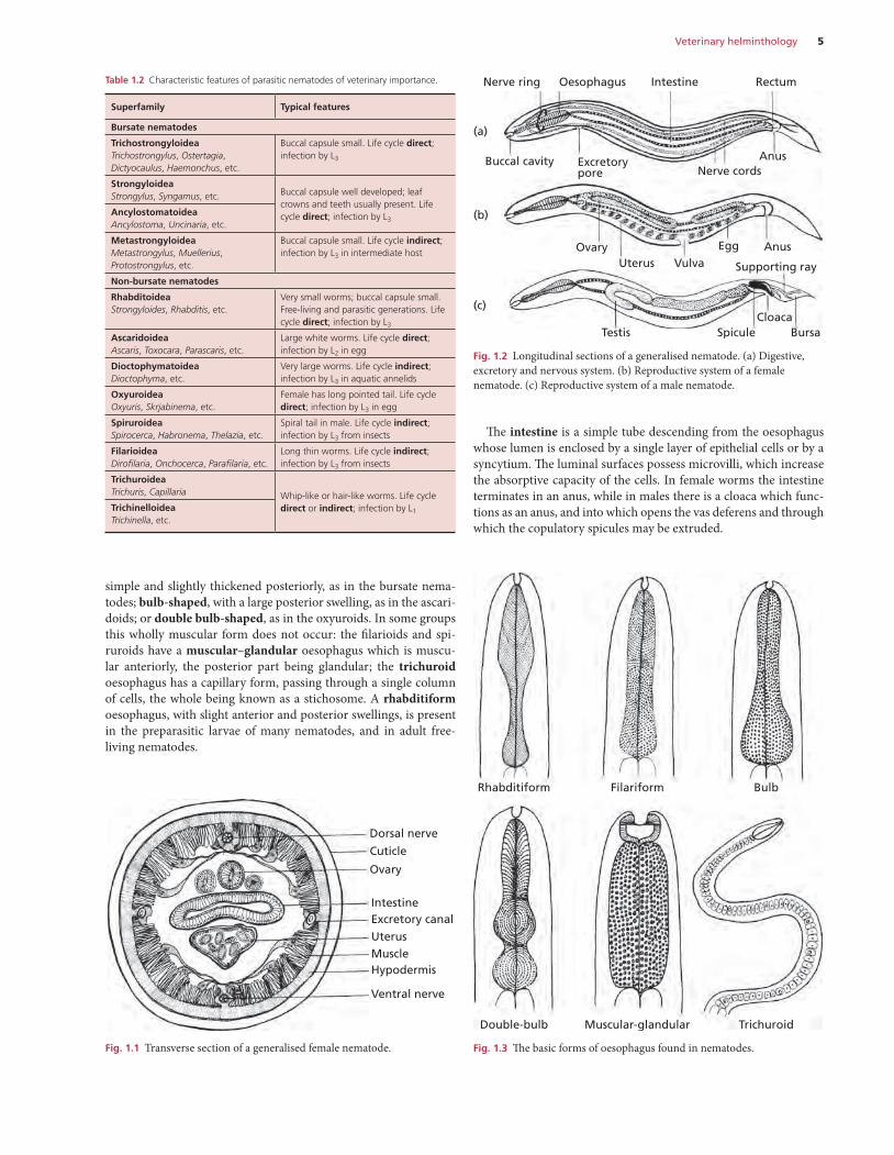
Most nematodes have a cylindrical unsegmented form, tapering at either end, and the body is covered by a colourless, somewhat trans-lucent, layer: the cuticle.
The tough cuticle is secreted by the underlying hypodermis, which projects into the body cavity forming two lateral cords, which carry the excretory canals, and a dorsal and ventral cord carrying the nerves (Fig. 1.1). The muscle cells, arranged longitudinally, lie between the hypodermis and the body cavity. The latter contains fluid at a high pressure, which maintains the turgidity and shape of the body (pseudocoelom). Locomotion is effected by undulating waves of muscle contraction and relaxation that alternate on the dorsal and ventral aspects of the worm. A circular muscle layer is absent in nematodes. Most of the internal organs are filamentous and suspended in the fluid-filled body cavity (Fig. 1.1).
The digestive system is tubular (Fig. 1.2a). The mouth, or stoma, of many nematodes is a simple opening, which may be surrounded by two or three lips, and leads directly into the oesophagus. Where
CHAPTER 1
Veterinary helminthology
PART 1 General parasitology including taxonomy, diagnosis, antiparasitics
2 Part 1: General parasitology including taxonomy, diagnosis, antiparasitics
Table 1.1 Classification of parasitic nematodes of veterinary importance.
Kingdom Phylum Class Order Superfamily Family (Subfamily) Genus
Animalia Nematoda Secernentea Strongylida Trichostrongyloidea TrichostrongylidaeTrichostrongylusMarshallagiaHyostrongylusMecistocirrusGraphidiumObeliscoidesLibyostrongylusGraphinemaImpalaia (Ostertaginae)OstertagiaTeladorsagiaSpiculopteragiaApteragiaCamelostrongylus (Haemonchinae)Haemonchus
CooperidaeCooperia
OrnithostrongylidaeOrnithostrongylus
AmidostomidaeAmidostomumEpomidiostomum
MolineidaeNematodirusNematodirellaLamanemaMolineusOllulanusTupaiostrongylus
HeligmonellidaeNippostrongylusNematospiroides
DictyocaulidaeDictyocaulus
Strongyloidea Strongylidae (Strongylinae)StrongylusTriodontophorusChabertiaOesophagostomumPoteriostomumCraterostomumOesophagodontusCodiostomum (Cyathostominae)CyathostomumCyclicocyclusCylicodonthophorusCylicostephanusSyngamidaeSyngamusCyathostomaMammomonogamusStephanurus
DeletrocephalidaeDeletrocephalusParadeletrocephalus
Ancylostomatoidea AncylostomatidaeAncylostomaUncinariaBunostomumGaigeriaNecatorGlobocephalusAgriostomum
Diaphanocephaloidea DiaphanocephalidaeKalicephalus
Veterinary helminthology 3
Kingdom Phylum Class Order Superfamily Family (Subfamily) Genus
Animalia Nematoda Secernentea Metastrongyloidea MetastrongylidaeMetastrongylusProtostrongylidaeMuelleriusProtostrongylusCystocaulusSpiculocaulusNeostrongylusVarestrongylusParelaphostrongylusElaphostrongylusFilaroididaeOslerusFilaroidesAngiostrongylidaeAngiostrongylusCrenosomidaeCrenosoma
Rhabditida Rhabditoidea StrongyloididaeStrongyloidesPanagrolaimidaeHalicephalobusRhabditidaeRhabditisRhabdiasidaeRhabdias
Ascaridida Ascaridoidea AscarididaeAscarisToxocaraToxascarisParascarisAscaridiaHeterakisPorrocaecumBayliascarisPolydelphusOphidascarisAngusticaecumAnisakidaeAnisakisContracaecumHysterothylaciumPseudoterranovaSulcascaris
Dioctophymatoidea DioctophymatidaeDioctophymaHystrichisEustrongylides
Oxyurida Oxyuroidea OxyuridaeOxyurisSkrjabinemaAspicularisSyphaciaPassalurusDermatoxysEnterobiusCosmocercidaeProbstmayriaAspidoderidaeParaspidoderaPharyngodonidaeTachygonetria
Spirurida Spiruroidea SpirocercidaeSpirocercaAscaropsPhysocephalusSimondsiaStreptoparagusHabronematidaeHabronemaDraschiaParabronemaHistiocephalus
Continued
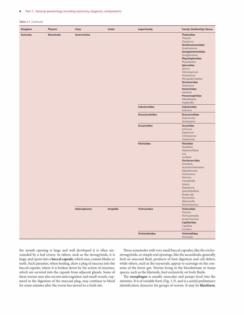
4 Part 1: General parasitology including taxonomy, diagnosis, antiparasitics
Kingdom Phylum Class Order Superfamily Family (Subfamily) Genus
Animalia Nematoda Secernentea ThelazidaeThelaziaOxyspiruraGnathostomatidaeGnathostomaGongylonematidaeGongylonemaPhysalopteridaePhysalopteraSpiruridaeSpiruraOdontospiruraProtospiruraPterygodermatitiesTetrameridaeTetrameresHartertiidaeHartertiaPneumospiridaeMetathelaziaVogeloides
Subuluroidea SubuluridaeSubulura
Dracunculoidea DracunculidaeDracunculusAvioserpens
Acuarioidea AcuaridaeEchinuriaDispharynxCheilospiruraStreptocara
Filarioidea FilariidaeParafilariaStephanofilariaLoaSuifilariaOnchocercidaeDirofilariaAcanthocheilonemaDipetalonemaOnchocercaPelecitusChanderellaSetariaElaeophoraSplendidofilariaBrugia spp.WuchereriaMansonellaParonchocerca
Adenophorea Enoplida Trichuroidea TrichuridaeTrichurisTrichosomoidesAnatrichosomaCapillaridaeCapillariaEucoleus
Trichinelloidea TrichinellidaeTrichinella
the mouth opening is large and well developed it is often sur-rounded by a leaf crown. In others, such as the strongyloids, it is large, and opens into a buccal capsule, which may contain blades or teeth. Such parasites, when feeding, draw a plug of mucosa into the buccal capsule, where it is broken down by the action of enzymes, which are secreted into the capsule from adjacent glands. Some of these worms may also secrete anticoagulant, and small vessels, rup-tured in the digestion of the mucosal plug, may continue to bleed for some minutes after the worm has moved to a fresh site.
Those nematodes with very small buccal capsules, like the tricho-strongyloids, or simple oral openings, like the ascaridoids, generally feed on mucosal fluid, products of host digestion and cell debris, while others, such as the oxyuroids, appear to scavenge on the con-tents of the lower gut. Worms living in the bloodstream or tissue spaces, such as the filarioids, feed exclusively on body fluids.
The oesophagus is usually muscular and pumps food into the intestine. It is of variable form (Fig. 1.3), and is a useful preliminary identification character for groups of worms. It may be filariform,
Table 1.1 Continued
Veterinary helminthology 5
The intestine is a simple tube descending from the oesophagus whose lumen is enclosed by a single layer of epithelial cells or by a syncytium. The luminal surfaces possess microvilli, which increase the absorptive capacity of the cells. In female worms the intestine terminates in an anus, while in males there is a cloaca which func-tions as an anus, and into which opens the vas deferens and through which the copulatory spicules may be extruded.
simple and slightly thickened posteriorly, as in the bursate nema-todes; bulb-shaped, with a large posterior swelling, as in the ascari-doids; or double bulb-shaped, as in the oxyuroids. In some groups this wholly muscular form does not occur: the filarioids and spi-ruroids have a muscular–glandular oesophagus which is muscu-lar anteriorly, the posterior part being glandular; the trichuroid oesophagus has a capillary form, passing through a single column of cells, the whole being known as a stichosome. A rhabditiform oesophagus, with slight anterior and posterior swellings, is present in the preparasitic larvae of many nematodes, and in adult free-living nematodes.
Table 1.2 Characteristic features of parasitic nematodes of veterinary importance.
Superfamily Typical features
Bursate nematodes
TrichostrongyloideaTrichostrongylus, Ostertagia, Dictyocaulus, Haemonchus, etc.
Buccal capsule small. Life cycle direct; infection by L3
StrongyloideaStrongylus, Syngamus, etc. Buccal capsule well developed; leaf
crowns and teeth usually present. Life cycle direct; infection by L3
AncylostomatoideaAncylostoma, Uncinaria, etc.
MetastrongyloideaMetastrongylus, Muellerius, Protostrongylus, etc.
Buccal capsule small. Life cycle indirect; infection by L3 in intermediate host
Non-bursate nematodes
RhabditoideaStrongyloides, Rhabditis, etc.
Very small worms; buccal capsule small. Free-living and parasitic generations. Life cycle direct; infection by L3
AscaridoideaAscaris, Toxocara, Parascaris, etc.
Large white worms. Life cycle direct; infection by L2 in egg
DioctophymatoideaDioctophyma, etc.
Very large worms. Life cycle indirect; infection by L3 in aquatic annelids
OxyuroideaOxyuris, Skrjabinema, etc.
Female has long pointed tail. Life cycle direct; infection by L3 in egg
SpiruroideaSpirocerca, Habronema, Thelazia, etc.
Spiral tail in male. Life cycle indirect; infection by L3 from insects
FilarioideaDirofilaria, Onchocerca, Parafilaria, etc.
Long thin worms. Life cycle indirect; infection by L3 from insects
TrichuroideaTrichuris, Capillaria Whip-like or hair-like worms. Life cycle
direct or indirect; infection by L1TrichinelloideaTrichinella, etc.
Fig. 1.1 Transverse section of a generalised female nematode.
Dorsal nerve
Cuticle
Ovary
IntestineExcretory canal
UterusMuscleHypodermis
Ventral nerve
Fig. 1.2 Longitudinal sections of a generalised nematode. (a) Digestive, excretory and nervous system. (b) Reproductive system of a female nematode. (c) Reproductive system of a male nematode.
Buccal cavity
Nerve ring Oesophagus RectumIntestine
(a)
(b)
(c)
Excretorypore Nerve cords
Anus
AnusEgg
Supporting rayVulvaUterusOvary
Testis Spicule BursaCloaca
Fig. 1.3 The basic forms of oesophagus found in nematodes.
Rhabditiform Filariform Bulb
Double-bulb Muscular-glandular Trichuroid
6 Part 1: General parasitology including taxonomy, diagnosis, antiparasitics
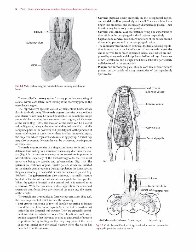
• Cervical papillae occur anteriorly in the oesophageal region, and caudal papillae posteriorly at the tail. They are spine-like or finger-like processes, and are usually diametrically placed. Their function may be sensory or supportive.
• Cervical and caudal alae are flattened wing-like expansions of the cuticle in the oesophageal and tail regions respectively.
• Cephalic and cervical vesicles are inflations of the cuticle around the mouth opening and in the oesophageal region.
• The copulatory bursa, which embraces the female during copula-tion, is important in the identification of certain male nematodes and is derived from much expanded caudal alae, which are sup-ported by elongated caudal papillae called bursal rays. It consists of two lateral lobes and a single small dorsal lobe. It is particularly well developed in the strongylids.
• Plaques and cordons are plate-like and cord-like ornamentations present on the cuticle of many nematodes of the superfamily Spiruroidea.
The so-called ‘excretory system’ is very primitive, consisting of a canal within each lateral cord joining at the excretory pore in the oesophageal region.
The reproductive systems consist of filamentous tubes, which float in the body cavity. The female organs comprise ovary, oviduct and uterus, which may be paired (didelphic) or sometimes single (monodelphic), ending in a common short vagina, which opens at the vulva (Fig. 1.2b). The location of the vulva can be a useful aid in diagnosis, being at the anterior end (opisthodelphic), middle (amphidelphic) or the posterior end (prodelphic). At the junction of uterus and vagina in some species there is a short muscular organ, the ovejector, which regulates and assists in egg laying. A vulval flap may also be present. Nematodes can be oviparous, ovoviviparous or viviparous.
The male organs consist of a single continuous testis and a vas deferens terminating in a muscular ejaculatory duct into the clo-aca (Fig. 1.2c). Accessory male organs are sometimes important in identification, especially of the trichostrongyloids, the two most important being the spicules and gubernaculum (Fig. 1.4). The spicules are chitinous organs, usually paired, which are inserted in the female genital opening during copulation. In some species they are absent (e.g. Trichinella) or only one spicule is present (e.g. Trichuris). The gubernaculum, also chitinous, is a small structure located in the dorsal wall, which acts as a guide for the spicules. When the guide is located in the ventral wall it is referred to as a telamon. With the two sexes in close apposition the amoeboid sperm are transferred from the cloaca of the male into the uterus of the female.
The cuticle may be modified to form various structures (Fig. 1.5), the more important of which include the following.• Leaf crowns consisting of rows of papillae occurring as fringes
round the rim of the buccal capsule (external leaf crowns) or just inside the rim (internal leaf crowns). They are especially promi-nent in certain nematodes of horses. Their function is not known, but it is suggested that they may be used to pin a patch of mucosa in position during feeding, or that they may prevent the entry of foreign matter into the buccal capsule when the worm has detached from the mucosa.
Fig. 1.4 Male trichostrongylid nematode bursa showing spicules and bursa.
Spicules
Gubernaculum
Bursa
Leaf crownsCephalic vesicle
Cervical vesicle
Cervical ala
Cervical papilla
(a)
Spicule
Gubernaculum
Bursal lobe Ventral rays
Lateral raysDorsal rays(b) Externo-dorsal rays
Fig. 1.5 Cuticular modifications of a generalised nematode: (a) anterior region; (b) posterior region of a male.
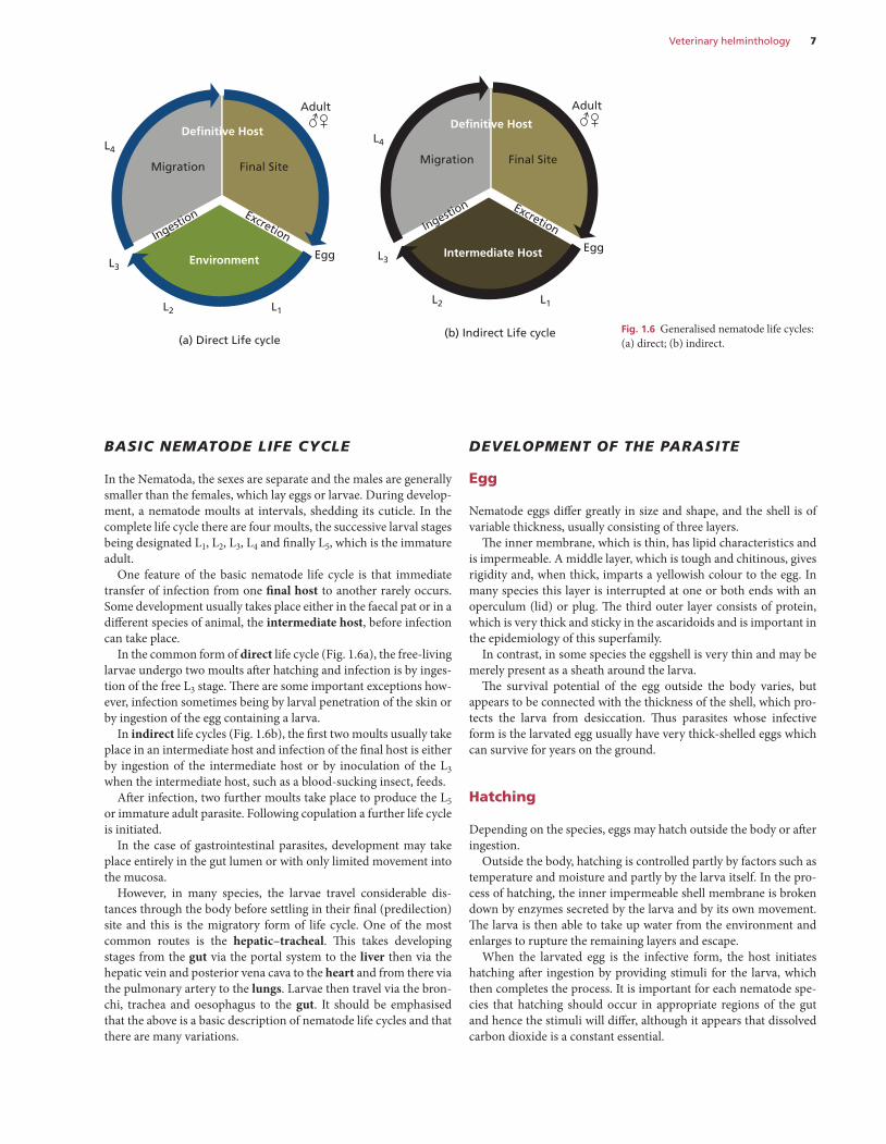
Veterinary helminthology 7
develoPment of the Parasite
Egg
Nematode eggs differ greatly in size and shape, and the shell is of variable thickness, usually consisting of three layers.
The inner membrane, which is thin, has lipid characteristics and is impermeable. A middle layer, which is tough and chitinous, gives rigidity and, when thick, imparts a yellowish colour to the egg. In many species this layer is interrupted at one or both ends with an operculum (lid) or plug. The third outer layer consists of protein, which is very thick and sticky in the ascaridoids and is important in the epidemiology of this superfamily.
In contrast, in some species the eggshell is very thin and may be merely present as a sheath around the larva.
The survival potential of the egg outside the body varies, but appears to be connected with the thickness of the shell, which pro-tects the larva from desiccation. Thus parasites whose infective form is the larvated egg usually have very thick-shelled eggs which can survive for years on the ground.
Hatching
Depending on the species, eggs may hatch outside the body or after ingestion.
Outside the body, hatching is controlled partly by factors such as temperature and moisture and partly by the larva itself. In the pro-cess of hatching, the inner impermeable shell membrane is broken down by enzymes secreted by the larva and by its own movement. The larva is then able to take up water from the environment and enlarges to rupture the remaining layers and escape.
When the larvated egg is the infective form, the host initiates hatching after ingestion by providing stimuli for the larva, which then completes the process. It is important for each nematode spe-cies that hatching should occur in appropriate regions of the gut and hence the stimuli will differ, although it appears that dissolved carbon dioxide is a constant essential.
Basic nematode life cycle
In the Nematoda, the sexes are separate and the males are generally smaller than the females, which lay eggs or larvae. During develop-ment, a nematode moults at intervals, shedding its cuticle. In the complete life cycle there are four moults, the successive larval stages being designated L1, L2, L3, L4 and finally L5, which is the immature adult.
One feature of the basic nematode life cycle is that immediate transfer of infection from one final host to another rarely occurs. Some development usually takes place either in the faecal pat or in a different species of animal, the intermediate host, before infection can take place.
In the common form of direct life cycle (Fig. 1.6a), the free-living larvae undergo two moults after hatching and infection is by inges-tion of the free L3 stage. There are some important exceptions how-ever, infection sometimes being by larval penetration of the skin or by ingestion of the egg containing a larva.
In indirect life cycles (Fig. 1.6b), the first two moults usually take place in an intermediate host and infection of the final host is either by ingestion of the intermediate host or by inoculation of the L3 when the intermediate host, such as a blood-sucking insect, feeds.
After infection, two further moults take place to produce the L5 or immature adult parasite. Following copulation a further life cycle is initiated.
In the case of gastrointestinal parasites, development may take place entirely in the gut lumen or with only limited movement into the mucosa.
However, in many species, the larvae travel considerable dis-tances through the body before settling in their final (predilection) site and this is the migratory form of life cycle. One of the most common routes is the hepatic–tracheal. This takes developing stages from the gut via the portal system to the liver then via the hepatic vein and posterior vena cava to the heart and from there via the pulmonary artery to the lungs. Larvae then travel via the bron-chi, trachea and oesophagus to the gut. It should be emphasised that the above is a basic description of nematode life cycles and that there are many variations.
Fig. 1.6 Generalised nematode life cycles: (a) direct; (b) indirect.
(b) Indirect Life cycle
L1
(a) Direct Life cycle
L1
EggEnvironment
Definitive Host
Final SiteMigration
ExcretionIngestion
L2
L3
L4
Adult
EggIntermediate Host
Definitive Host
Final SiteMigration
ExcretionIngestion
L2
L3
L4
Adult
8 Part 1: General parasitology including taxonomy, diagnosis, antiparasitics
metaBolism
The main food reserve of preparasitic nematode larvae, whether inside the eggshell or free-living, is lipid, which may be seen as droplets in the lumen of the intestine. The infectivity of these stages is often related to the amount of lipid present; larvae which have depleted their reserves are not as infective as those which still retain quantities of lipid.
Apart from these reserves, the free-living first- and second-stage larvae of most nematodes feed on bacteria. However, once they reach the infective third stage, they are sealed in the retained cuticle of the second stage, cannot feed and are completely dependent on their stored reserves.
In contrast, the adult parasite stores its energy as glycogen, mainly in the lateral cords and muscles, and this may constitute 20% of the dry weight of the worm.
Free-living and developing stages of nematodes usually have an aerobic metabolism, whereas adult nematodes can metabolise car-bohydrate by both glycolysis (anaerobic) and oxidative decarboxyl-ation (aerobic). However, in the latter, pathways may operate which are not present in the host and it is at this level that some antipara-sitic drugs operate.
The oxidation of carbohydrates requires the presence of an elec-tron transport system, which in most nematodes can operate aer-obically down to oxygen tensions of 5.0 mmHg or less. Since the oxygen tension at the mucosal surface of the intestine is around 20 mmHg, nematodes in close proximity to the mucosa normally have sufficient oxygen for aerobic metabolism. Otherwise, if the nematode is temporarily or permanently some distance from the mucosal surface, energy metabolism is probably largely anaerobic.
As well as the conventional cytochrome and flavoprotein elec-tron transport system, many nematodes have ‘haemoglobin’ in their body fluids which gives them a red pigmentation. This nematode haemoglobin is chemically similar to myoglobin and has the high-est affinity for oxygen of any known animal haemoglobin. The main function of nematode haemoglobin is thought to be to transport oxygen, acquired by diffusion through the cuticle or gut, into the tissues; blood-sucking worms presumably ingest a considerable amount of oxygenated nutrients in their diet.
The end products of the metabolism of carbohydrates, fats or proteins are excreted through the anus or cloaca, or by diffusion through the body wall. Ammonia, the terminal product of protein metabolism, must be excreted rapidly and diluted to non-toxic levels in the surrounding fluids. During periods of anaerobic carbohydrate metabolism, the worms may also excrete pyruvic acid rather than retaining it for future oxidation when aerobic metabolism is possible.
The ‘excretory system’ terminating in the excretory pore is almost certainly not concerned with excretion, but rather with osmoregu-lation and salt balance.
Two phenomena which affect the normal parasitic life cycle of nematodes and which are of considerable biological and epidemio-logical importance are arrested larval development and the peri-parturient rise in faecal egg counts.
arrested larval develoPment
(Synonyms: inhibited larval development, hypobiosis)This phenomenon may be defined as the temporary cessation in development of a nematode at a precise point in its parasitic devel-opment. It is usually a facultative characteristic and affects only a
Larval development and survival
Three of the important superfamilies, the trichostrongyloids, the strongyloids and the rhabditoids, have a completely free-living pre-parasitic phase. The first two larval stages usually feed on bacteria, but the L3, sealed off from the environment by the retained cuti-cle of the L2, cannot feed and must survive on the stored nutrients acquired in the early stages. Growth of the larva is interrupted dur-ing moulting by periods of lethargus in which it neither feeds nor moves.
The cuticle of the L2 is retained as a sheath around the L3; this is important in larval survival with a protective role analogous to that of the eggshell in egg-infective groups.
The two most important components of the external environ-ment are temperature and humidity. The optimal temperature for the development of the maximum number of larvae in the short-est feasible time is generally in the range 18–26°C. At higher tem-peratures, development is faster and the larvae are hyperactive, thus depleting their lipid reserves. The mortality rate then rises, so that few will survive to L3. As the temperature falls the process slows, and below 10°C the development from egg to L3 usually cannot take place. Below 5°C movement and metabolism of L3 is minimal, which in many species favours survival.
The optimal humidity is 100%, although some development can occur down to 80% relative humidity. It should be noted that even in dry weather where the ambient humidity is low, the microclimate in faeces or at the soil surface may be sufficiently humid to permit continuing larval development.
In the trichostrongyloids and strongyloids, the embryonated egg and the ensheathed L3 are best equipped to survive in adverse con-ditions such as freezing or desiccation; in contrast, the L1 and L2 are particularly vulnerable. Although desiccation is generally con-sidered to be the most lethal influence in larval survival, there is increasing evidence that by entering a state of anhydrobiosis, cer-tain larvae can survive severe desiccation.
On the ground most larvae are active, although they require a film of water for movement and are stimulated by light and tem-perature. It is now thought that larval movement is mostly random and encounter with grass blades accidental.
Infection
As noted previously, infection may be by ingestion of the free-living L3, and this occurs in the majority of trichostrongyloid and stron-gyloid nematodes. In these, the L3 sheds the retained sheath of the L2 within the alimentary tract of the host, the stimulus for exsheath-ment being provided by the host in a manner similar to the hatching stimulus required by egg-infective nematodes. In response to this stimulus the larva releases its own exsheathing fluid, containing an enzyme, leucine aminopeptidase, which dissolves the sheath from within, either at a narrow collar anteriorly so that a cap detaches, or by splitting the sheath longitudinally. The larva can then wriggle free of the sheath.
As in the preparasitic stage, growth of the larva during parasitic development is interrupted by two moults, each of these occurring during a short period of lethargus.
The time taken for development from infection until mature adult parasites are producing eggs or larvae is known as the pre-patent period and this is of known duration for each nematode species.
Veterinary helminthology 9
protein. This relaxation of immunity can be largely restored by sup-plementation with rumen-undegradable protein and is also influ-enced by the body protein status of the ewe.
The source of the PPR is threefold: 1 Maturation of larvae arrested due to host immunity. 2 An increased establishment of infections acquired from the pas-
tures and a reduced turnover of existing adult infections. 3 An increased fecundity of existing adult worm populations.
Contemporaneously, but not associated with the relaxation of host immunity, the PPR may be augmented by the maturation of hypobiotic larvae.
The importance of the PPR is that it occurs at a time when the numbers of new susceptible hosts are increasing and so ensures the survival and propagation of the worm species. Depending on the magnitude of infection, it may also cause a loss of production in lactating animals and, by contamination of the environment, lead to clinical disease in susceptible young stock.
NEMATODE SUPERFAMILIES
suPerfamily trichostronGyloidea
The trichostrongyloids are small, often hair-like, worms in the bursate group, which, with the exception of the lungworm Dictyo-caulus, parasitise the alimentary tract of animals and birds. Struc-turally they have few cuticular appendages and the buccal capsule is vestigial and possesses no leaf crowns. Teeth are usually absent. The males have a well developed bursa with large lateral lobes and two spicules, the configuration of which is used for species differ-entiation. The life cycle is direct and usually non-migratory and the ensheathed L3 is the infective stage.
The trichostrongylids are responsible for considerable mortal-ity and widespread morbidity, especially in ruminants. The most important alimentary genera are Ostertagia (and other members of the subfamily Ostertaginae, Teladorsagia, Spiculopteragia, Aptera-gia, Camelostrongylus), Haemonchus, Trichostrongylus, Cooperia, Nematodirus, Hyostrongylus, Marshallagia and Mecistocirrus. Dic-tyocaulus is an important genus affecting the respiratory tract of ruminants and horses.
Other genera of lesser importance are Graphidium, Obeliscoides, Ollulanus, Libyostrongylus, Graphinema, Impalaia, Ornithostrongy-lus, Amidostomum, Epomidiostomum, Nematodirella, Lamanema, Nippostrongylus and Nematospiroides.
family trichostronGylidae
trichostrongylus
Adult worms are small, slightly reddish/brown in colour, slender and hair-like, usually less than 7.0 mm long (Fig 1.7) and difficult to see with the naked eye. The worms have no obvious buccal capsule and cephalic inflations are absent. A most useful generic character is the distinct excretory notch in the oesophageal region (Fig. 1.8). The male bursa has long lateral lobes, while the dorsal lobe is not well defined with a slender dorsal ray, which is cleft near its tip into two branches. The ventro-ventral ray is well separated from the other rays. The spicules are thick and unbranched and a gubernacu-lum is present. Species identification is based on the shape and size of the spicules (Table 1.3). In the female, the tail is bluntly tapered
proportion of the worm population. Some strains of nematodes have a high propensity for arrested development while in others this is low. The stage at which larvae become arrested varies between species of nematodes; for example, L3 stage in Trichostrongylus, Cyathostominae, and Ancylostoma, L4 stage in Ostertagia, Tela-dorsagia, Haemonchus, and Obeliscoides, or immature adults as in Dictyocaulus.
Conclusive evidence for the occurrence of arrested larval devel-opment can only be obtained by examination of the worm popula-tion in the host. It is usually recognised by the presence of large numbers of larvae at the same stage of development in animals withheld from infection for a period longer than that required to reach that particular larval stage.
The nature of the stimulus for arrested development and for the subsequent maturation of the larvae is still a matter of debate. Although there are apparently different circumstances which initi-ate arrested larval development, most commonly the stimulus is an environmental one received by the free-living infective stages prior to ingestion by the host. It may be seen as a ruse by the parasite to avoid adverse climatic conditions for its progeny by remaining sex-ually immature in the host until more favourable conditions return. The name commonly applied to this seasonal arrestment is hypo-biosis. Thus the accumulation of arrested larvae often coincides with the onset of cold autumn/winter conditions in the northern hemisphere, or very dry conditions in the subtropics or tropics. In contrast, the maturation of these larvae coincides with the return of environmental conditions suitable to their free-living development, although it is not clear what triggers the signal to mature and how it is transmitted.
The degree of adaptation to these seasonal stimuli and therefore the proportion of larvae which do become arrested seem to be a heritable trait and is affected by various factors, including grazing systems and the degree of adversity in the environment. For exam-ple, in Canada where the winters are severe, most trichostrongyloid larvae ingested in late autumn or winter become arrested, whereas in southern Britain with moderate winters, about 50–60% are arrested. In the humid tropics where free-living larval development is possible all the year round, relatively few larvae become arrested.
However, arrested development may also occur as a result of both acquired and age immunity in the host and, although the propor-tions of larvae arrested are not usually so high as in hypobiosis, they can play an important part in the epidemiology of nematode infec-tions. Maturation of these arrested larvae seems to be linked with the breeding cycle of the host and occurs at or around parturition.
The epidemiological importance of arrested larval development from whatever cause is that, first, it ensures the survival of the nematode during periods of adversity and, second, the subsequent maturation of arrested larvae increases the contamination of the environment and can sometimes result in clinical disease.
PeriParturient rise in faecal eGG counts
(Synonyms: postparturient rise, spring rise)Periparturient rise (PPR) refers to an increase in the numbers of nematode eggs in the faeces of animals around parturition. This phenomenon is most marked in ewes, goats and sows and recent data support the hypothesis that there is competition for nutrients between the immune system, the rapidly growing fetus in late preg-nancy and the udder during lactation, particularly metabolisable
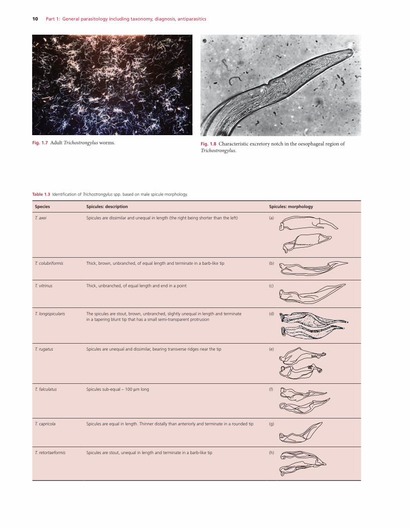
10 Part 1: General parasitology including taxonomy, diagnosis, antiparasitics
Fig. 1.7 Adult Trichostrongylus worms. Fig. 1.8 Characteristic excretory notch in the oesophageal region of Trichostrongylus.
Table 1.3 Identification of Trichostrongylus spp. based on male spicule morphology.
Species Spicules: description Spicules: morphology
T. axei Spicules are dissimilar and unequal in length (the right being shorter than the left) (a)
T. colubriformis Thick, brown, unbranched, of equal length and terminate in a barb-like tip (b)
T. vitrinus Thick, unbranched, of equal length and end in a point (c)
T. longispicularis The spicules are stout, brown, unbranched, slightly unequal in length and terminate in a tapering blunt tip that has a small semi-transparent protrusion
(d)
T. rugatus Spicules are unequal and dissimilar, bearing transverse ridges near the tip (e)
T. falculatus Spicules sub-equal – 100 μm long (f)
T. capricola Spicules are equal in length. Thinner distally than anteriorly and terminate in a rounded tip (g)
T. retortaeformis Spicules are stout, unequal in length and terminate in a barb-like tip (h)
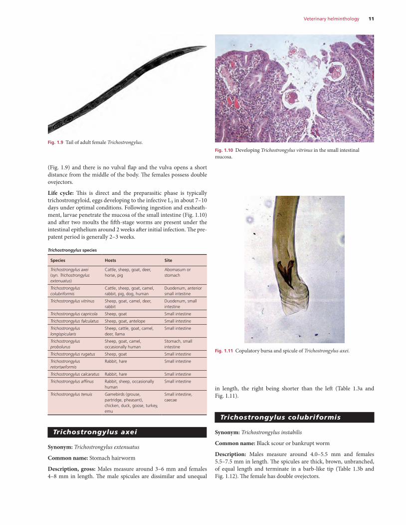
Veterinary helminthology 11
in length, the right being shorter than the left (Table 1.3a and Fig. 1.11).
trichostrongylus colubriformis
Synonym: Trichostrongylus instabilis
Common name: Black scour or bankrupt worm
Description: Males measure around 4.0–5.5 mm and females 5.5–7.5 mm in length. The spicules are thick, brown, unbranched, of equal length and terminate in a barb-like tip (Table 1.3b and Fig. 1.12). The female has double ovejectors.
(Fig. 1.9) and there is no vulval flap and the vulva opens a short distance from the middle of the body. The females possess double ovejectors.
Life cycle: This is direct and the preparasitic phase is typically trichostrongyloid, eggs developing to the infective L3 in about 7–10 days under optimal conditions. Following ingestion and exsheath-ment, larvae penetrate the mucosa of the small intestine (Fig. 1.10) and after two moults the fifth-stage worms are present under the intestinal epithelium around 2 weeks after initial infection. The pre-patent period is generally 2–3 weeks.
Trichostrongylus species
Species Hosts Site
Trichostrongylus axei(syn. Trichostrongylus extenuatus)
Cattle, sheep, goat, deer, horse, pig
Abomasum or stomach
Trichostrongylus colubriformis
Cattle, sheep, goat, camel, rabbit, pig, dog, human
Duodenum, anterior small intestine
Trichostrongylus vitrinus Sheep, goat, camel, deer, rabbit
Duodenum, small intestine
Trichostrongylus capricola Sheep, goat Small intestine
Trichostrongylus falculatus Sheep, goat, antelope Small intestine
Trichostrongylus longispicularis
Sheep, cattle, goat, camel, deer, llama
Small intestine
Trichostrongylus probolurus
Sheep, goat, camel, occasionally human
Stomach, small intestine
Trichostrongylus rugatus Sheep, goat Small intestine
Trichostrongylus retortaeformis
Rabbit, hare Small intestine
Trichostrongylus calcaratus Rabbit, hare Small intestine
Trichostrongylus affinus Rabbit, sheep, occasionally human
Small intestine
Trichostrongylus tenuis Gamebirds (grouse, partridge, pheasant), chicken, duck, goose, turkey, emu
Small intestine, caecae
trichostrongylus axei
Synonym: Trichostrongylus extenuatus
Common name: Stomach hairworm
Description, gross: Males measure around 3–6 mm and females 4–8 mm in length. The male spicules are dissimilar and unequal
Fig. 1.9 Tail of adult female Trichostrongylus.Fig. 1.10 Developing Trichostrongylus vitrinus in the small intestinal mucosa.
Fig. 1.11 Copulatory bursa and spicule of Trichostrongylus axei.
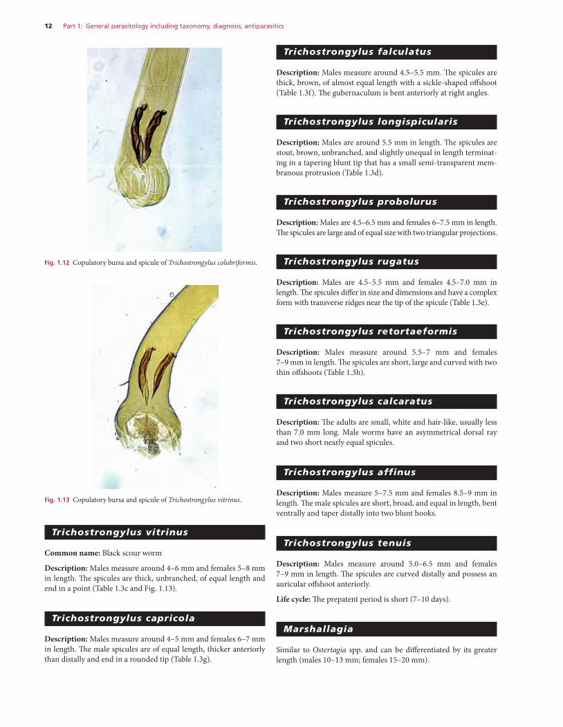
12 Part 1: General parasitology including taxonomy, diagnosis, antiparasitics
trichostrongylus falculatus
Description: Males measure around 4.5–5.5 mm. The spicules are thick, brown, of almost equal length with a sickle-shaped offshoot (Table 1.3f). The gubernaculum is bent anteriorly at right angles.
trichostrongylus longispicularis
Description: Males are around 5.5 mm in length. The spicules are stout, brown, unbranched, and slightly unequal in length terminat-ing in a tapering blunt tip that has a small semi-transparent mem-branous protrusion (Table 1.3d).
trichostrongylus probolurus
Description: Males are 4.5–6.5 mm and females 6–7.5 mm in length. The spicules are large and of equal size with two triangular projections.
trichostrongylus rugatus
Description: Males are 4.5–5.5 mm and females 4.5–7.0 mm in length. The spicules differ in size and dimensions and have a complex form with transverse ridges near the tip of the spicule (Table 1.3e).
trichostrongylus retortaeformis
Description: Males measure around 5.5–7 mm and females 7–9 mm in length. The spicules are short, large and curved with two thin offshoots (Table 1.3h).
trichostrongylus calcaratus
Description: The adults are small, white and hair-like, usually less than 7.0 mm long. Male worms have an asymmetrical dorsal ray and two short nearly equal spicules.
trichostrongylus affinus
Description: Males measure 5–7.5 mm and females 8.5–9 mm in length. The male spicules are short, broad, and equal in length, bent ventrally and taper distally into two blunt hooks.
trichostrongylus tenuis
Description: Males measure around 5.0–6.5 mm and females 7–9 mm in length. The spicules are curved distally and possess an auricular offshoot anteriorly.
Life cycle: The prepatent period is short (7–10 days).
marshallagia
Similar to Ostertagia spp. and can be differentiated by its greater length (males 10–13 mm; females 15–20 mm).
trichostrongylus vitrinus
Common name: Black scour worm
Description: Males measure around 4–6 mm and females 5–8 mm in length. The spicules are thick, unbranched, of equal length and end in a point (Table 1.3c and Fig. 1.13).
trichostrongylus capricola
Description: Males measure around 4–5 mm and females 6–7 mm in length. The male spicules are of equal length, thicker anteriorly than distally and end in a rounded tip (Table 1.3g).
Fig. 1.12 Copulatory bursa and spicule of Trichostrongylus colubriformis.
Fig. 1.13 Copulatory bursa and spicule of Trichostrongylus vitrinus.


