Embed Size (px)
Citation preview

Clinical 131 (2007) 28–35www.elsevier.com/locate/autneu
Autonomic Neuroscience: Basic and
Parasympathetic dysfunction is associated with baroreflex andchemoreflex impairment in streptozotocin-induced diabetes in rats
Pedro Dall'Ago a, Beatriz D'Agord Schaan b,⁎, Viviane Oliveira Kenne da Silva a,Juliana Werner a, Pedro Paulo da Silva Soares d, Kátia de Angelis c,e, Maria Cláudia Irigoyen c
a Department of Physiological Sciences, Federal School Foundation of Medical Sciences of Porto Alegre and Unilasalle, RS, Brazilb Institute of Cardiology of Rio Grande do Sul/University Foundation of Cardiology, RS, Brazil
c Heart Institute (Incor), University of Sao Paulo, SP, Brazild Department of Physiology and Pharmacology, Fluminense Federal University, RJ, Brazil
e Sao Judas Tadeu University, Sao Paulo, SP, Brazil
Received 9 February 2006; received in revised form 19 May 2006; accepted 8 June 2006
Abstract
This study explored physiological mechanisms of diabetic dysfunction in baroreceptors and chemoreceptors-mediated hemodynamicresponses, and cholinergic neurotransmission in 30-day diabetic rats (n=14) and controls (n=14). Basal hemodynamic data and vagalresponse to electrical stimulation and methacholine injection were also evaluated. Muscarinic receptors were characterized using aradioligand receptor binding assay ([3H]N methylscopolamine). Experimental diabetes (50mg/kg of STZ, i.v.) decreased systolic, diastolic,and mean arterial pressure and basal heart rate. Heart rate (HR) responses to vagal electrical stimulation (16, 32, and 64Hz) were 15%,11%, and 14% higher in diabetics vs non-diabetics, as were HR responses to methacholine injection (−130±24, −172±18, −206±15bpmvs. −48±15, −116±12, −151±18bpm, P<0.05). Muscarinic receptor density was higher (267.4±11 vs 193.5±22fmol/mg/prot, P<0.05)in the atria of diabetic rats than in those of controls; the affinity was similar between groups. Diabetes-induced reduction of reflex responsesto baro- (reflex bradycardia: −3.4±0.3 and −2.7±0.2bpm/mm Hg; reflex tachycardia: −1.6±0.1 and −1.4±0.07bpm/mm Hg, in controland diabetics, P<0.05) and chemoreceptor stimulation, enhancement of HR responsiveness to cardiac vagal electrical stimulation andmethacholine stimulation, plus an increase in the number of atrial muscarinic receptors indicates reduced parasympathetic activity, which isprobably derived from central nervous system derangement.© 2006 Elsevier B.V. All rights reserved.
Keywords: Diabetes mellitus; Blood pressure; Baroreflex; Chemoreceptors; Autonomic nervous system; Muscarinic receptors
1. Introduction
It is well known that diabetes mellitus produces changesin blood vessel structure and function, involving sensory,motor, and autonomic nervous systems. Significantincreased morbidity and mortality, represented by orthostatic
⁎ Corresponding author. Unidade de Pesquisa do IC/FUC - Av. PrincesaIsabel, 370 90.620-001 Porto Alegre-RS, Brazil. Tel./fax: +55 5132192812x22,23,24.
E-mail addresses: [email protected],[email protected] (B. D'Agord Schaan).
1566-0702/$ - see front matter © 2006 Elsevier B.V. All rights reserved.doi:10.1016/j.autneu.2006.06.005
hypotension, painless myocardial infarction, and suddendeath, is observed in patients diagnosed with cardiovascularautonomic neuropathy (Ewing et al., 1980; Hilsted, 1982;Sampson et al., 1990). Extensive studies had been performedto clarify the mechanisms underlying such changes.Pathologic conditions, such as cardiomyopathy (Makino etal., 1987; Tahiliani and McNeill, 1986), increased lipidperoxidation (Tada et al., 1992), metabolic disturbances(Rodrigues and McNeill, 1992), and sympathetic nervoussystem structural and functional changes (Monckton andPehowich, 1980; Takiguchi et al., 1988) are all contributorsto this cardiovascular dysfunction.

29P. Dall'Ago et al. / Autonomic Neuroscience: Basic and Clinical 131 (2007) 28–35
Streptozotocin (STZ)-induced diabetes in rats causeshypotension and bradycardia, probably related to pacemakercell dysfunction (Maeda et al., 1995) associated with adepression in cardiac function (Jackson and Carrier, 1983;Shimabukuro et al., 1996). Indeed, baroreflex-mediatedbradycardia and tachycardia in response to arterial pressurechanges was found to be attenuated in diabetic rats after STZ(Dall'Ago et al., 2002) or alloxan (McDowell et al., 1994)administration. This impairment has been attributed, at leastin part, to parasympathetic dysfunction, because earlydepressed vagal tonus has been consistently demonstratedin diabetic rats (Yagihashi, 1995). Moreover, evidencesuggests that cardiovascular responses evoked by chemore-flex activation induced by potassium cyanide (KCN) inconscious rats mostly depends on cardiac vagal activationwith some contribution of the cardiac-sympathetic activationin determining the final pressor responses (Franchini andKrieger, 1993). It was also showed before that STZ-diabeticrats have impaired cardiovascular responses evoked by KCN(Dall'Ago et al., 1997).
Chronotropic responses to cholinergic agonists wereevaluated in isolated, spontaneously beating atria, but notin the conscious diabetic rat (Aronstam and Carrier, 1989;Carrier et al., 1984). Diminished muscarinic receptorpopulation in cardiac tissue was observed in experimentaldiabetes (Aronstam and Carrier, 1989; Carrier et al., 1984;Kofo-Abayomi and Lucas, 1987), a change that is reversibleby insulin (Aronstam and Carrier, 1989) and aldosereductase inhibitor (Kofo-Abayomi and Lucas, 1987)treatment, suggesting a metabolic cause for this abnormality.The relationship between hyperglycemia and cardiovasculardysfunction has already been demonstrated (Schaan et al.,2004). Although depressed vagal function can be associatedwith changes in peripheral or central nervous systems, nosystematic functional studies have been undertaken todeterminate the role of the parasympathetic efferent pathwayon heart function in diabetic rats.
The aim of the present study was to evaluate the role of30-day STZ-induced diabetes on the baro- and chemoreflexcontrol of circulation by evaluating the heart rate responsesto: (1) vagal electrical stimulation and (2) intravenousmethacholine injection, a muscarinic receptor agonist, inrats. Atrial muscarinic receptor density and affinity were alsoquantified.
2. Methods
2.1. General procedures
Experiments were performed on 28 age-matched maleWistar rats, housed in individual cages, with free access tofood and water. They were kept in a temperature-controlledroom (22°C) with a 12–12-h dark–light cycle. The rats wererandomly assigned to 1 of 2 groups: control (C, n=14, bodyweight 224.2±5g) and diabetic (D, n=14, body weight226.8±6g). Rats were injected with a single intravenous (i.v.)
injection of STZ (50mg/kg, Sigma Chemical Company, St.Louis MO, USA) dissolved in citrate buffer (0.01M, pH 4.5,D) or only citrate buffer (C) into the lateral tail vein. Toprevent severe hypoglycemia in the first few hours afterinjection, both STZ- and citrate buffer-treated rats were givena 5% (wt/vol) dextrose solution to drink overnight. Experi-ments were performed in conscious rats whenever theprocedures could be performed in this way (basal hemody-namic records, baro- and chemoreflex sensitivity evaluation).
2.2. Basal hemodynamic status
Thirty days after STZ administration, 2 catheters (PE10)filled with heparin in normal saline were installed in theanimal, under general anesthesia (IP ketamine, 9mg/kg,Parker, Davis, Brazil and xylasine, 1mg/kg, Bayer, Brazil),these catheters being placed into the abdominal aorta andinferior vena cava through the left femoral artery and vein,respectively, tunneled subcutaneously and exteriorized at theback of the neck. The catheters were used for directmeasurement of mean arterial pressure (MAP) and drugadministration, respectively. One day after catheter place-ment, cardiovascular records were obtained. The arterialcatheter was attached to a 20-cm (PE90) polyvinyl tubeconnected to a strain-gauge pressure transducer (P23 Db,Gould Statham, Oxnard, CA, USA). Blood pressure signalswere recorded for 40min with a microcomputer equippedwith an analog-to-digital converter board (CODAS, 1kHz,Dataq Instruments, and Akron, OH, USA). The recordeddata were analyzed on a beat-to-beat basis to quantify MAPand heart rate (HR) at rest.
2.3. Baroreflex and chemoreflex sensitivity
After basal MAP recording, baroreflex-mediated changeswere measured during peak increases or decreases in MAPafter phenylephrine (0.02–0.32μg) or sodium nitroprusside(0.05–0.16μg) injections, and the corresponding peak reflexchanges in HR was recorded after each dose of the drugs.The changes of MAP were within the range of 10–30mmHg. The maximum changes in MAP and HR were measured,and baroreflex sensitivity was determined as the slope of theMAP/HR ratio (bpm/mm Hg). The slopes obtained for eachgroup of rats evaluated were then statistically compared.
Chemoreflex sensitivity was tested 30min after the end ofthe baroreflex test session, by the administration ofincreasing intravenous doses of KCN (60, 100, 140, and180μg/kg) (Dall'Ago et al., 1997; Franchini and Krieger,1993). Mean AP and HR were measured continuously for10s before and for 15s after the injections of KCN.
2.4. Bradycardic responses to vagal nerve stimulation andmethacholine injection
The vagal electrical stimulation and muscarinic responsesto methacholine were tested 24h after baroreflex and

Table 1Characterization of diabetic and control groups
Control (n=14) STZ (n=14)
Body wt (g) 265±5 197±6⁎
Blood glucose level (mg/dL) 122±3 439±22⁎
Diastolic AP (mm Hg) 95±3 80±2⁎
Systolic AP (mm Hg) 139±3 116±3⁎
Mean AP (mm Hg) 114±3 98±2⁎
Heart rate (bpm) 331±9 299±11⁎
Baseline metabolic and cardiovascular characteristics of the animals studied.Values are means±S.E.M.⁎ Significant differences between groups (P<0.05).
30 P. Dall'Ago et al. / Autonomic Neuroscience: Basic and Clinical 131 (2007) 28–35
chemoreflex evaluation. The decrease in HR produced by theelectrical stimulation (5V, 2ms, 2–64Hz, for 10s) of the cutright vagus nerve distal portion was studied in anesthetizedrats (thiopental, 35mg/kg, i.v.). The interval between stimuliwas determined by the time required for HR to return toprestimulation levels. After vagal stimulation, the sensitivityof the heart muscarinic receptors was tested by evaluatingHR responses to intravenous injections of increasing dosesof methacholine (5, 7.5, and 10μg/kg) in the same rats. Bodytemperature was maintained at 37°C by external heating.
2.5. Muscarinic receptor binding assay
Saturation binding experiments were performed inmembrane preparations of diabetic and control rats' atria.The procedures for membrane protein preparation havebeen described previously (Carrier et al., 1984). Briefly, therats were killed by cervical dislocation, and the atria wereremoved from the heart, weighed, and washed with ice-cold phosphate-buffered saline (PBS) buffer. The tissueswere then homogenized with a Polytron Homogenizer(GlennMills, Clinton, NJ, USA) in 10-ml of ice-cold lysisbuffer containing 5mM Tris–HCl, 2mM EDTA, pH 7.4,and a protease inhibitor cocktail consisting of 5mg/mlphenylmethylsulfonyl fluoride, 10mg/ml benzamidine, and5mg/ml soybean trypsin inhibitor. The homogenate wascentrifuged at 500×g for 15min at 4°C. The pellets werethen homogenized as before, spun again, and the super-natants pooled. The supernatants were centrifuged at45,000×g for 15min, and the pellets washed twice in thesame buffer. The membrane fractions were resuspended ina buffer containing 50mM Tris–HCl, pH 7.4, 2mMMgCl2, and 5mM EDTA. The protein content wasdetermined with a Bio-Rad Protein Assay kit (Bio-Rad,Mississauga, ON, Canada), using bovine serum albumin asthe standard. A muscarinic acetylcholine receptor non-selective antagonist [N-methyl-3H]scopolamine methylchloride ([3H]NMS, 82Ci/mmol) ranging from 10−11 to10−8M was used in the absence (total binding) andpresence (nonspecific binding) of atropine sulphate (SigmaChemical Company, St. Louis, MO, EUA) for 90min.Nonspecific binding was defined as that measured in thepresence of 1μM atropine. Specific binding was deter-mined by subtracting nonspecific from total binding.Binding measurements were obtained in duplicate foreach experiment. Incubations at a volume of 1ml (90min atroom temperature) were terminated by rapid filtration withWhatman GF/C filters (Xymotech, Montreal, PQ, Canada),and radioactivity was counted with an LS6000 ScintillationCounter (Beckman, Fullerton, CA).
2.6. Blood analysis
Arterial blood samples were obtained from the catheterplaced in the femoral artery to determine blood glucose (teststrips, Advantage, Roche, Indianapolis, IN, USA).
All experimental procedures described above wereapproved by the Ethical Committee for Animal Researchof the Federal School Foundation of Medical Sciences ofPorto Alegre.
2.7. Statistical analysis
Data are reported as means±S.E.M., and the unpairedStudent's t-test was used for comparison between groups.Baroreflex sensitivity was evaluated by regression analysisof different groups, and the differences between the slopes ofthe relation between AP and HR response to pressor anddepressor agents were evaluated for statistical significanceusing the Student's t-test for unpaired data. Differences inMAP and HR responses to KCN and the HR changesobtained by vagal stimulation and methacholine injectionsbetween groups were compared by analysis of variance forrepeated measures, post hoc Student–Newmann–Keuls test.Binding data were analyzed using curve-fitting functions inGraphPad Prism software (GraphPad Software, San Diego,CA). Linear regression was performed on the percentage ofbound versus the ratio of bound over free ligand, and onlydata with a regression coefficient of ≥0.9 were used foranalysis. One- and two-site models were tested for all datasets, and the model yielding the least residual sum of squareswas taken to describe the data. Probability levels <0.05 wereconsidered significant for all tests.
3. Results
3.1. Body weight and blood glucose
These data are represented in Table 1. Thirty days afterSTZ administration, body weights were lower in diabeticthan in control rats (P<0.0001). All 14 rats given citratebuffer remained normoglycemic throughout the 4-weekstudy. Blood glucose levels were significantly higher in thediabetic compared with the control rats (P<0.0001).
3.2. Baseline hemodynamic status
Table 1 also shows the systolic, diastolic, and meanarterial pressure at rest, which were markedly reduced in

Fig. 1. Regression lines showing the effects of STZ-induced diabetes on bradycardic (lower right) and tachycardic (upper left) responses to pressor changesinduced by increasing doses of phenylephrine and sodium nitroprusside, respectively. STZ-diabetic rats presented an impairment of tachycardic responses, whilereflex bradycardia was unaltered. The slope of the lines obtained by linear regression was −3.40 vs. −2.70bpm/mm Hg (P<0.05, unpaired Student's t-test) forcontrol (CON) and diabetic (STZ) rats, respectively after a decrease in MAP.
31P. Dall'Ago et al. / Autonomic Neuroscience: Basic and Clinical 131 (2007) 28–35
STZ-diabetic rats, P<0.0001. Also, resting heart rate wassignificantly lower in the diabetic vs control group,P<0.0001.
3.3. Baroreflex and chemoreflex sensitivity
The reflex tachycardia elicited by sodium nitroprusside-induced hypotension was significantly reduced by diabetes,as indicated by the slope of the regression line plottingchanges in HR with changes in MAP (b: −3.4±0.3 and−2.7±0.2bpm/mmHg, in control and diabetics, respectively,P<0.05). The reflex bradycardia elicited by phenylephrine-
Fig. 2. Line graphs showing the effects of streptozotocin-induced diabetes on heartcontrol (filled triangles, n=14) and diabetic (filled squares, n=14) rats to increasingcontrols (ANOVA, post hoc Student Newmann–Keuls).
induced hypertensive stimulus was similar between groups(b: −1.6±0.1 and −1.4±0.07bpm/mm Hg, in control anddiabetics, respectively, P=0.177). These data are shown inFig. 1.
Fig. 2 shows the dose-dependent bradycardic and pressorresponses to intravenous injection of KCN produced in bothcontrol and diabetic rats. Bradycardia evoked by thechemoreflex activation was reduced in diabetic rats at allKCN doses employed (Fig. 2A). The pressor responsesinduced by chemoreflex stimulation (Fig. 2B) were alsomarkedly decreased in the diabetic group at all KCN dosesemployed.
rate (HR) responses (A) and mean arterial pressure (MAP) responses (B) ofdoses of KCN. Data are reported as means±S.E.M. ⁎P<0.05 compared with
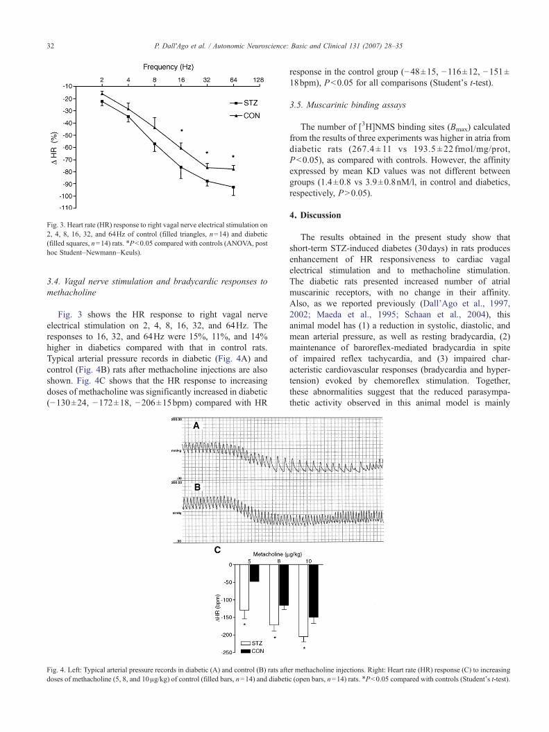
Fig. 3. Heart rate (HR) response to right vagal nerve electrical stimulation on2, 4, 8, 16, 32, and 64Hz of control (filled triangles, n=14) and diabetic(filled squares, n=14) rats. ⁎P<0.05 compared with controls (ANOVA, posthoc Student–Newmann–Keuls).
32 P. Dall'Ago et al. / Autonomic Neuroscience: Basic and Clinical 131 (2007) 28–35
3.4. Vagal nerve stimulation and bradycardic responses tomethacholine
Fig. 3 shows the HR response to right vagal nerveelectrical stimulation on 2, 4, 8, 16, 32, and 64Hz. Theresponses to 16, 32, and 64Hz were 15%, 11%, and 14%higher in diabetics compared with that in control rats.Typical arterial pressure records in diabetic (Fig. 4A) andcontrol (Fig. 4B) rats after methacholine injections are alsoshown. Fig. 4C shows that the HR response to increasingdoses of methacholine was significantly increased in diabetic(−130±24, −172±18, −206±15bpm) compared with HR
Fig. 4. Left: Typical arterial pressure records in diabetic (A) and control (B) rats aftdoses of methacholine (5, 8, and 10μg/kg) of control (filled bars, n=14) and diabeti
response in the control group (−48±15, −116±12, −151±18bpm), P<0.05 for all comparisons (Student's t-test).
3.5. Muscarinic binding assays
The number of [3H]NMS binding sites (Bmax) calculatedfrom the results of three experiments was higher in atria fromdiabetic rats (267.4 ± 11 vs 193.5 ± 22 fmol/mg/prot,P<0.05), as compared with controls. However, the affinityexpressed by mean KD values was not different betweengroups (1.4±0.8 vs 3.9±0.8nM/l, in control and diabetics,respectively, P>0.05).
4. Discussion
The results obtained in the present study show thatshort-term STZ-induced diabetes (30days) in rats producesenhancement of HR responsiveness to cardiac vagalelectrical stimulation and to methacholine stimulation.The diabetic rats presented increased number of atrialmuscarinic receptors, with no change in their affinity.Also, as we reported previously (Dall'Ago et al., 1997,2002; Maeda et al., 1995; Schaan et al., 2004), thisanimal model has (1) a reduction in systolic, diastolic, andmean arterial pressure, as well as resting bradycardia, (2)maintenance of baroreflex-mediated bradycardia in spiteof impaired reflex tachycardia, and (3) impaired char-acteristic cardiovascular responses (bradycardia and hyper-tension) evoked by chemoreflex stimulation. Together,these abnormalities suggest that the reduced parasympa-thetic activity observed in this animal model is mainly
er methacholine injections. Right: Heart rate (HR) response (C) to increasingc (open bars, n=14) rats. ⁎P<0.05 compared with controls (Student's t-test).

33P. Dall'Ago et al. / Autonomic Neuroscience: Basic and Clinical 131 (2007) 28–35
derived from a central nervous system derangement. Wecould not exclude the possibility that defects in reflexresponses could lie on the afferent side, or on eitherefferent or afferent side to neuropathy.
The hypotension observed in the present study in diabeticrats confirms our previously published results (Dall'Ago etal., 1997, 2002; De Angelis et al., 2000a; Maeda et al., 1995;Schaan et al., 1997) and is in accordance with resultsreported by others (De Angelis et al., 2000b; Jackson andCarrier, 1983; Tomlinson et al., 1982). Some authors havedescribed a so-called streptozotocin hypertension, whichprobably reflects discrepancies between the direct andindirect blood pressure measurements (Bunag et al., 1982;Kusaka et al., 1987). Resting bradycardia observed after STZtreatment has also been previously demonstrated (Bunag etal., 1982; Dall'Ago et al., 2002; Jackson and Carrier, 1983;Maeda et al., 1995). Using pharmacological blockade, DeAngelis et al. (2002) demonstrated resting HR fell 5daysafter STZ administration. This change was maintained for3months and seems to be related to a reduced intrinsic heartrate, suggesting that bradycardia in STZ-induced diabeticrats is associated with changes in the electrical activity of thesinoatrial node.
Baseline changes in MAP and HR could be related tochanges in reflex mechanisms that control circulation. In fact,the reflex tachycardia response elicited by decreasing MAPwas attenuated in the 30-day diabetic group. Previous reportsfrom our laboratory have shown similar baroreflex control ofHR impairment after 5 and 15days of the STZ injection,suggesting that early alterations in reflex control weremaintained during longer periods (1month), as shown bythe present study, or even later (De Angelis et al., 2002).Although bradycardic responses to increasing MAP weremaintained in this experiment, reports show impairment inHR-reducing mechanisms. These differences seem to berelated to methodological differences: undernutrition of thediabetic rats and prolonged diabetes duration (Chang andLund, 1986) and experiments in anesthetized preparations(Van Buren et al., 1998).
The impairment of central parasympathetic pathwayfunction with preservation of sympathetic control havebeen suggested as causal mechanisms involved in theattenuation of bradycardic responses in diabetic rabbits(McDowell et al., 1994). In this context, we observed in thepresent study a reduction in cardiac-vagal activation evokedby chemoreceptors in the diabetic group, as shown by thereduced bradycardia produced by KCN injection. Thebradycardic responses evoked by KCN represent thecardiac-vagal activation and were not modulated by respira-tory hyperventilation induced by chemoreflex activation.Because baroreflex- and chemoreflex-mediated responses areintegrated by a common neural network in the central nervoussystem, we were not surprised that similar vagal efferentresponses could be evidenced during both receptor stimuli. Inthe rat, the respiratory-dependent effects exert little influencein the final cardiovascular responses evoked by chemoreflex,
since bradycardia and hypertension, simultaneously tohyperventilation are observed in response to carotid bodychemoreceptors activation (Franchini and Krieger, 1993).However, the magnitude of bradycardia and hypertension, aswell as the specific response (tachycardia or bradycardia)could be changed if the secondary respiratory and beha-vioural responses are controlled by anesthetic agents. The useof anesthetized rats and the type and level of anesthesia canalter the final cardiovascular responses to chemoreflexstimulation, since defense areas activation during hypoxiamay occur (Marshall, 1998).
The pressor responses produced by chemoreflex activa-tion of vascular sympathetic pathway were also reduced indiabetic rats. In the present study and in previous ones fromour group (Dall'Ago et al., 2002) and other authors (Jacksonand Carrier, 1983), it has been demonstrated that the diabeticstate induced not only an impairment in heart responses, butalso a depressed vascular reactivity in this animal model.Jackson and Carrier (1983) induced blood pressureresponses with norepinephrine and angiotensin II, and weused phenylephrine. These responses were depressed in theshort-term diabetic rat in both studies; however, thebaroreceptor reflexes in these rats were more sensitive toincreases in blood pressure. Therefore, it appears that sometype of nonspecific alteration occurs in the responsiveness ofthe cardiovascular system to the vasopressor agonists in theshort-term diabetes in the rat.
Although the reduced reflex bradycardia elicited by baro-and chemoreceptor stimulation in diabetic rats has beenattributed to the impairment in efferent parasympatheticfunction to the heart, there is no data to distinguish whetherthis dysfunction originates from central or peripheral nervoussystem derangements. The greater bradycardic responsesobtained by vagal electrical stimulation or by methacholineinjection in diabetic rats in our study suggest that the efferentpathway is actually more effective in diabetic than in normalrats.We (Dall'Ago et al., 2002;Maeda et al., 1995) and others(McDowell et al., 1994) have previously reported on theenhancement of efferent parasympathetic function. Theresponses to vagal nerve stimulation and methacholineinjection are necessarily performed under general anesthesia(thiopental, 35mg/kg, i.v.); the evaluation of a control groupallowed us to exclude anesthesia-induced changes as thedeterminant of the responses obtained.
These findings, associated with the increased density ofmuscarinic receptors in the heart, that we also observed,suggest an up-regulation of these receptors to compensate fora reduction in central parasympathetic activity. This is thefirst study showing a complete analysis of the parasympa-thetic efferent to the heart in experimental diabetes. Anincrease in the density of cardiac muscarinic receptorsmatches the observed cholinergic supersensitivity in thediabetic atria. Muscarinic receptor populations are regulatedby the degree of effective neurotransmission. These receptorpopulations are decreased by chronic exposure to agonists orby inhibition of acetylcholinesterase (Gazit et al., 1979),

34 P. Dall'Ago et al. / Autonomic Neuroscience: Basic and Clinical 131 (2007) 28–35
whereas they are increased by exposure to antagonists (Wiseet al., 1980). However, atrial muscarinic receptor density indiabetes has been variably reported to decrease (Carrier et al.,1984; Kofo-Abayomi and Lucas, 1987), not change (Wegneret al., 1987) or to increase in humans (Richardson et al., 2004)and in rats (Liu et al., 2005). These 2 last studies specificallyevaluated the muscarinic receptor protein and mRNAdensities in the heart, techniques that add to our results. Liuet al also showed that the up-regulation of these receptorscould be normalized by inhibiting the sorbitol pathway withan aldose reductase inhibitor (Liu et al., 2005). Also,muscarinic receptor density was increased in the urinarybladder (Tong et al., 2002) and ileum (Coulson et al., 2004) ofdiabetic rats. It is well accepted that in the atria of mammalsthe most important muscarinic acetylcholine receptor is of theM2 subtype, however it cannot be excluded the possiblepresence of the M1 or M3 subtypes (Krejci and Tucek, 2002;Wang et al., 2001, 2004). In the present study a nonselectiveantagonist was used in the binding assays and this matter wasnot addressed. Future studies are necessary to investigate thepossible expression of muscarinic acetylcholine receptorsothers than the M2 subtype in diabetic rats' atria.
5. Conclusion
STZ-induced experimental diabetes promotes hypoten-sion accompanied by resting bradycardia, impaired baro- andchemoreflex control of circulation, enhancement of HRresponsiveness to cardiac vagal stimulation, and an increaseddensity of muscarinic receptors in the heart. These latterfindings allowed us to hypothesize that the mechanismsinvolved in the impaired HR reflex changes depend on areduced central parasympathetic tonus generation. Thisvagal impairment is associated with an up-regulation ofmuscarinic heart function, although sympathetic activitychanges should also be considered. Further studies arenecessary to test these latter hypotheses.
References
Aronstam, R.S., Carrier, G.O., 1989. Insulin prevention of alteredmuscarinic receptor-G protein coupling in diabetic rat atria. Diabetes38 (12), 1611–1616.
Bunag, R.D., Tomita, T., Sasaki, S., 1982. Streptozotocin diabetic rats arehypertensive despite reduced hypothalamic responsiveness. Hyperten-sion 4 (4), 556–565.
Carrier, G.O., Edwards, A.D., Aronstam, R.S., 1984. Cholinergic super-sensitivity and decreased number of muscarinic receptors in atria fromshort-term diabetic rats. J. Mol. Cell. Cardiol. 16 (10), 963–965.
Chang, K.S., Lund, D.D., 1986. Alterations in the baroreceptor reflexcontrol of heart rate in streptozotocin diabetic rats. J. Mol. Cell. Cardiol.18 (6), 617–624.
Coulson, F.R., Jacoby, D.B., Fryer, A.D., 2004. Insulin regulatesneuronal M2 muscarinic receptor function in the ileum of diabeticrats. J. Pharmacol. Exp. Ther. 308 (2), 760–766.
Dall'Ago, P., Fernandes, T.G., Machado, U.F., Bello, A.A., Irigoyen, M.C.,1997. Baroreflex and chemoreflex dysfunction in streptozotocin-diabeticrats. Braz. J. Med. Biol. Res. 30 (1), 119–124.
Dall'Ago, P., Silva, V.O., De Angelis, K.L., Irigoyen, M.C., Fazan Jr., R.,Salgado, H.C., 2002. Reflex control of arterial pressure and heart rate inshort-term streptozotocin diabetic rats. Braz. J. Med. Biol. Res. 35 (7),843–849.
De Angelis, K.L., Cestari, I.A., Barp, J., Dall'Ago, P., Fernandes, T.G., deBittencourt, P.I., Bello-Klein, A., Bello, A.A., Llesuy, S., Irigoyen, M.C.,2000a. Oxidative stress in the latissimus dorsi muscle of diabetic rats.Braz. J. Med. Biol. Res. 33 (11), 1363–1368.
De Angelis, K.L., Oliveira, A.R., Dall'Ago, P., Peixoto, L.R., Gadonski, G.,Lacchini, S., Fernandes, T.G., Irigoyen, M.C., 2000b. Effects of exercisetraining on autonomic and myocardial dysfunction in streptozotocin-diabetic rats. Braz. J. Med. Biol. Res. 33 (6), 635–641.
De Angelis, K., Schaan, B.D., Maeda, C.Y., Dall'Ago, P., Wichi, R.B.,Irigoyen, M.C., 2002. Cardiovascular control in experimental diabetes.Braz. J. Med. Biol. Res. 35 (9), 1091–1100.
Ewing, D.J., Campbell, I.W., Clarke, B.F., 1980. The natural history ofdiabetic autonomic neuropathy. Q. J. Med. 49 (193), 95–108.
Franchini, K.G., Krieger, E.M., 1993. Cardiovascular responses ofconscious rats to carotid body chemoreceptor stimulation by intravenousKCN. J. Auton. Nerv. Syst. 42 (1), 63–69.
Gazit, H., Silman, I., Dudai, Y., 1979. Administration of an organophosphatecauses a decrease in muscarinic receptor levels in rat brain. Brain Res.174 (2), 351–356.
Hilsted, J., 1982. Pathophysiology in diabetic autonomic neuropathy:cardiovascular, hormonal, and metabolic studies. Diabetes 31 (8 Pt 1),730–737.
Jackson, C.V., Carrier, G.O., 1983. Influence of short-term experimentaldiabetes on blood pressure and heart rate in response to norepinephrineand angiotensin II in the conscious rat. J. Cardiovasc. Pharmacol. 5 (2),260–265.
Kofo-Abayomi, A., Lucas, P.D., 1987. Muscarinic receptor density isreduced in diabetic rat atria, an effect prevented by the aldose reductaseinhibitor, Statil. J. Pharm. Pharmacol. 39 (12), 1019–1021.
Krejci, A., Tucek, S., 2002. Quantitation of mRNAs for M(1) to M(5)subtypes of muscarinic receptors in rat heart and brain cortex. Mol.Pharmacol. 61 (6), 1267–1272.
Kusaka, M., Kishi, K., Sokabe, H., 1987. Does so-called streptozocinhypertension exist in rats? Hypertension 10 (5), 517–521.
Liu, T.P., Juang, S.W., Cheng, J.T., Tong, Y.C., 2005. The role ofsorbitol pathway and treatment effect of aldose reductase inhibitorONO2235 in the up-regulation of cardiac M2-muscarinic receptorsin streptozotocin-induced diabetic rats. Neurosci. Lett. 383 (1–2),131–135.
Maeda, C.Y., Fernandes, T.G., Timm, H.B., Irigoyen, M.C., 1995.Autonomic dysfunction in short-term experimental diabetes. Hyperten-sion 26 (6 Pt 2), 1100–1104.
Makino, N., Dhalla, K.S., Elimban, V., Dhalla, N.S., 1987. SarcolemmalCa2+ transport in streptozotocin-induced diabetic cardiomyopathy inrats. Am. J. Physiol. 253 (2 Pt 1), E202–E207.
Marshall, J.M., 1998. Chemoreceptors and cardiovascular control in acuteand chronic systemic hypoxia. Braz. J. Med. Biol. Res. 31 (7), 863–888.
McDowell, T.S., Chapleau, M.W., Hajduczok, G., Abboud, F.M., 1994.Baroreflex dysfunction in diabetes mellitus: I. Selective impairment ofparasympathetic control of heart rate. Am. J. Physiol. 266 (1 Pt 2),H235–H243.
Monckton, G., Pehowich, E., 1980. Autonomic neuropathy in thestreptozotocin diabetic rat. Can. J. Neurol. Sci. 7 (2), 135–142.
Richardson, M.D., Kilts, J.D., Kwatra, M.M., 2004. Increased expression ofGi-coupled muscarinic acetylcholine receptor and Gi in atrium of elderlydiabetic subjects. Diabetes 53 (9), 2392–2396.
Rodrigues, B., McNeill, J.H., 1992. The diabetic heart: metabolic causes forthe development of a cardiomyopathy. Cardiovasc. Res. 26 (10),913–922.
Sampson, M.J., Wilson, S., Karagiannis, P., Edmonds, M., Watkins, P.J.,1990. Progression of diabetic autonomic neuropathy over a decade ininsulin-dependent diabetics. Q. J. Med. 75 (278), 635–646.

35P. Dall'Ago et al. / Autonomic Neuroscience: Basic and Clinical 131 (2007) 28–35
Schaan, B.D., Maeda, C.Y., Timm, H.B., Medeiros, S., Moraes, R.S., Ferlin,E., Fernandes, T.G., Ribeiro, J.P., Schmid, H., Irigoyen, M.C., 1997.Time course of changes in heart rate and blood pressure variability instreptozotocin-induced diabetic rats treated with insulin. Braz. J. Med.Biol. Res. 30 (9), 1081–1086.
Schaan BDDA, P., Maeda, C., Ferlin, E., Fernandes, T.G., Schmid, H.,Irigoyen, M.C., 2004. Relationship between cardiovascular dysfunctionand hyperglycemia in streptozotocin-induced diabetes. Braz. J. Med.Biol. Res. 37 (8), 889–893.
Shimabukuro, M., Higa, S., Shinzato, T., Nagamine, F., Komiya, I., Takasu,N., 1996. Cardioprotective effects of troglitazone in streptozotocin-induced diabetic rats. Metabolism 45 (9), 1168–1173.
Tada, H., Oida, K., Kutsumi, Y., Shimada, Y., Nakai, T., Miyabo, S., 1992.Effects of probucol on impaired cardiac performance and lipidmetabolism in streptozotocin-induced diabetic rats. J. Cardiovasc.Pharmacol. 20 (2), 179–186.
Tahiliani, A.G., McNeill, J.H., 1986. Diabetes-induced abnormalities in themyocardium. Life Sci. 38 (11), 959–974.
Takiguchi, Y., Satoh, N., Hashimoto, H., Nakashima, M., 1988. Changes invascular reactivity in experimental diabetic rats: comparison withhypothyroid rats. Blood Vessels 25 (5), 250–260.
Tomlinson, D.R., Holmes, P.R., Mayer, J.H., 1982. Reversal, by treatmentwith an aldose reductase inhibitor, of impaired axonal transport andmotor nerve conduction velocity in experimental diabetes mellitus.Neurosci. Lett. 31 (2), 189–193.
Tong, Y.C., Cheng, J.T., Wan, W.C., 2002. Effects of Ba-Wei-Die-Huang-Wan on the cholinergic function and protein expression of M2muscarinic receptor of the urinary bladder in diabetic rats. Neurosci.Lett. 330 (1), 21–24.
Van Buren, T., Kasbergen, C.M., Gispen, W.H., De Wildt, D.J., 1998. Invivo cardiovascular reactivity and baroreflex activity in diabetic rats.Cardiovasc. Res. 38 (3), 763–771.
Wang, H., Han, H., Zhang, L., Shi, H., Schram, G., Nattel, S., Wang, Z.,2001. Expression of multiple subtypes of muscarinic receptors andcellular distribution in the human heart. Mol. Pharmacol. 59 (5),1029–1036.
Wang, Z., Shi, H., Wang, H., 2004. Functional M3 muscarinic acetylcho-line receptors in mammalian hearts. Br. J. Pharmacol. 142 (3),395–408.
Wegner, J.A., Lund, D.D., Overton, J.M., Edwards, J.G., Oda, R.P., Tipton,C.M., 1987. Select cardiovascular and metabolic responses of diabeticrats to moderate exercise training. Med. Sci. Sports Exerc. 19 (5),497–503.
Wise, B.C., Shoji, M., Kuo, J.F., 1980. Decrease or increase in cardiacmuscarinic cholinergic receptor number in rats treated with methacho-line or atropine. Biochem. Biophys. Res. Commun. 92 (4),1136–1142.
Yagihashi, S., 1995. Pathology and pathogenetic mechanisms of diabeticneuropathy. Diabetes Metab. Rev. 11 (3), 193–225.


