Embed Size (px)
Citation preview

The left ventricle is richly innervated with non-myelinatedvagal afferents, stimulation of which leads to profounddepressor responses (see Hainsworth, 1991). Dischargefrom these nerve endings may be sparse and irregular(Coleridge et al. 1964) or show cardiac rhythm relating toboth ventricular systolic and diastolic pressures (Thorén,1977). The physiological or pathophysiological role ofreflexes originating in the left ventricle, however, is stillnot established. We recently reported that discretestimulation of mechanosensitive ventricular afferents,which required end-diastolic distension, resulted in relativelysmall reflex responses whereas much larger responsesoccurred following moderate pressure changes in thecoronary arteries (Wright et al. 2000; Drinkhill et al. 2001).We therefore suggested that the large responses frommechanical stimulation of the ventricle seen in earlierstudies were likely to have been due to stimulation ofcoronary baroreceptors. The question then remains as towhat exactly is the role of left ventricular mechano-receptors.
One widely held view is that left ventricular receptors arelikely to be involved in conditions associated with abnormaldistension (Mark, 1983; Abboud, 1989; Persson, 1991;Thames & Dibner-Dunlap, 1991; Thames et al. 1993). It is
suggested, in particular, that during myocardial ischaemia orin heart failure, when the left ventricle is likely to beabnormally distended, one of the effects of this may be tocause depression of the baroreceptor reflex. Evidence for suchreflex interactions has been sought in a variety of ways. Theseinclude examining the effects on responses to carotidbaroreceptor stimulation of interruption of cardiac vagalafferents by vagal cooling (Öberg & White, 1970; Mancia etal. 1973, 1975, 1976) or vagotomy (Guazzi et al. 1962; Manciaet al. 1973). Clearly, such procedures do not provide stimulithat are localised to ventricular receptors. Others havestimulated ventricular chemosensitive afferents with chemicalagents and reported a depression of baroreflex responses(Chen, 1979; Holmberg et al. 1983). These results, however,are not related to mechanical events and interpretation is alsocomplicated by tachyphylaxis, which is known to occur inresponse to the repeated application of chemical stimulants.
There have been no adequate studies that have applied adiscrete stimulus to ventricular mechanoreceptors andexamined the effects on other reflex responses. It was thepurpose of the present study to do this, and in particular toexamine effects of ventricular distension on responses tostimulation of carotid and coronary baroreceptors and leftventricular chemosensitive endings. In carrying out this study,
Responses to stimulation of coronary and carotid baroreceptors andthe coronary chemoreflex at different ventricular distending
pressures in anaesthetised dogs
C. I. Wright, M. J. Drinkhill* and R. Hainsworth
Institute of Cardiovascular Research, University of Leeds, Leeds LS2 9JT, UK
(Received 23 January 2001; accepted 21 March 2001)
Stimulation of left ventricular mechanoreceptors was believed not only to exert important effects on the circulation, butalso to influence the responses to baroreceptor reflexes. However, most previous work is flawed due to inadequatelocalisation of stimuli to specific reflexogenic areas. In this study, we applied a discrete stimulus to left ventricularmechanoreceptors to examine other reflexes known to effect the circulation. Dogs were anaesthetised, artificiallyventilated and a cardiopulmonary bypass established. The pressure distending the left ventricle was controlled throughan apical cannula with the aortic valve obstructed by a balloon. Changes in ventricular systolic and end-diastolic pressurehad only a small effect on vascular resistance, assessed as perfusion pressure in the systemic circulation (flow constant).Responses to changes in carotid or coronary pressure or to stimulation of chemosensitive afferents by injectingveratridine into the coronary circulation were always much larger. Responses to stimulation of these reflexes were littleaffected by the level of stimulus to the ventricular receptors. These experiments confirm that responses to stimulation ofventricular mechanoreceptors are very small and show that they remain small at different levels of input to otherbaroreceptive regions. There was no evidence of interaction between ventricular mechanoreceptor reflexes and carotidor coronary baroreceptors or ventricular chemosensitive reflexes. Experimental Physiology (2001) 86.3, 381–390.
2208
Publication of The Physiological Society * Corresponding author: [email protected]
) by guest on May 24, 2011ep.physoc.orgDownloaded from Exp Physiol (
we paid particular attention to the definition of reflexinteraction by Tutt et al. (1988), which required a change tobe demonstrated in the stimulus–response relationship of areflex that was not due solely to non-linearities or saturationof the efferent limb.
METHODSAnimals and preparationSixteen beagle dogs of both sexes weighing between 15 and 20 kg(mean, 17.2 ± 0.3 kg) were anaesthetised with a-chloralose(100 mg kg_1 I.V. in saline; Vickers Laboratories Ltd, Pudsey,Yorks, UK) infused through a catheter inserted under localanaesthesia (2 % lignocaine hydrochloride) through the rightsaphenous vein, so that its tip lay in the inferior vena cava.Surgical anaesthesia was maintained throughout the duration ofthe experiments by further infusions of a-chloralose(0.5–1.0 mg kg_1 min_1). Prior to major surgical procedures,alfentanil (Janssen-Cilag Ltd, High Wycombe, Bucks, UK) wasgiven intravenously (30 µg kg_1 over a 10 min period), and thenit was infused at 2.5 µg min_1 until 60 min prior to the start of theexperimental protocol. At intervals throughout the experiment,the depth of anaesthesia was assessed from the stability of bloodpressure and observing only small muscular contractions inresponse to toe pinch or to a sharp tap on the surgical table.
A longitudinal midline incision was made in the neck, thetrachea intubated and the lungs artificially ventilated with 40 %oxygen-enriched air using a Starling ‘Ideal’ pump set at17 ml kg_1 and 18 strokes min_1. Arterial PO2
, PCO2and pH were
determined at intervals during the experiments using a pH/bloodgas analyser (Instrumentation Laboratory, model IL 1610) andwere maintained within normal limits (see below) by altering thestroke volume of the respiratory pump, the rate of oxygen inflowand infusions of molar sodium bicarbonate solution, whenrequired.
Both carotid sinus regions were prepared by ligating all branchesarising from the carotid bifurcations, except the external carotidand lingual arteries, which were used for subsequent perfusion.The left side of the chest was exposed by a mid-sternal split andby dividing the fifth intercostal space. Once the pleural cavitywas opened, the expiratory output from the pump was immersedin 3 cm of water (2.3 mmHg, expiratory resistance) to preventlung collapse. The upper six pairs of intercostal arteries were tiedand then divided to mobilise a length (approximately 5 cm) ofthe descending aorta. The left subclavian artery was dissectedfree. Two snares were threaded around the brachiocephalicartery, carefully avoiding the ansae subclaviae. The pericardiumwas opened to allow access to the atria and left ventricles.Finally, a nylon cord was passed around the root of theascending aorta (0.5–1.0 cm from its origin), just distal to thecoronary ostia.
The animal received an intravenous injection of heparin(500 i.u. kg_1 I.V.; Leo Laboratories Ltd, Princes Risborough,UK) prior to cannulation and connection of the perfusion circuit(see Fig. 1). The perfusion circuit was part-filled with 2 l of amixture of mammalian Ringer solution (g l_1: NaCl, 6.9; KCl,0.35; CaCl2, 0.28; MgSO4, 0.14; NaHCO3, 2.09; KH2PO4, 0.16;glucose, 1.0), and Dextran in dextrose solution (50 : 50 mixture,50 g l_1 dextran, molecular weight 181 000). Blood cells from aprevious experiment, which had been centrifuged and washed,were added to the perfusate. The perfusion circuit was thenconnected in the following sequence. A curved stainless-steelcannula was inserted into the root of the aorta to convey blood
to the main arterial reservoir (see A in Fig. 1). The descendingaorta was cannulated and a pump perfused the sub-diaphragmatic circulation at a constant flow, which was initiallyset to give a systemic perfusion pressure of 150 mmHg. Thecentral and peripheral ends of the left subclavian artery werecannulated and the aortic arch and cephalic regions wereperfused from a reservoir maintained at a constant pressure (seeC in Fig. 1). A full heart–lung bypass was achieved by insertingcannulae into the left and right atria and the inferior vena cava(7 and 10 mm I.D.), to drain blood to an open reservoir (see D inFig. 1). The blood from reservoir D was pumped through amembrane gas exchange unit (Sorin Monolyth IntegratedMembrane Lung, Sorin Biomedica Cardio, Saluggia, Italy) andinto reservoir A from which it was distributed to the remainderof the perfusion circuit. A stab incision was made in the apex ofthe left ventricle, and a cannula (8 mm i.d.) was inserted andsecured into position by a purse-string suture. From reservoir A,blood was pumped at constant flow through a damping chamberand into the left ventricle. The outflow from the ventricle passedthrough the same cannula with the pressure controlled by aStarling resistor (Knowlton & Starling, 1912), to drain intoreservoir D.
Left and right common carotid arteries were cannulated andperfused at a constant pressure (see B in Fig. 1) and drained bycannulae inserted into the central ends of both lingual arteries.The snare placed around the root of the ascending aorta was tiedonto the aortic root cannula to create a pouch of the aorta (seeFig. 2). The pressure perfusing the aortic pouch, which alsocontrolled cephalic pressure in most experiments, was controlledby an independent reservoir (C in Fig. 1), which was connectedto cannulae in the central and peripheral ends of the leftsubclavian artery. The pressure applied to the main arterialreservoir (A in Fig. 1) controlled pressure at the aortic root(coronary pressure).
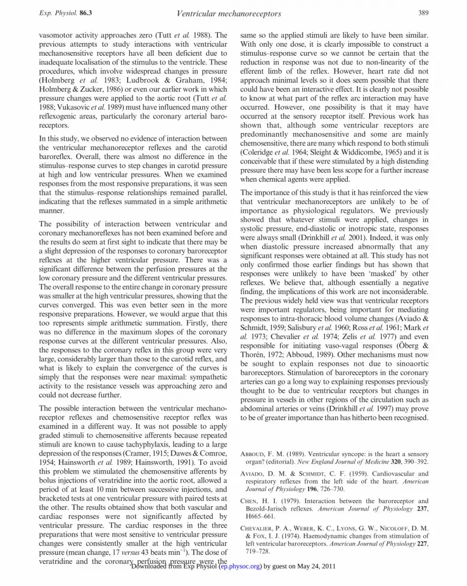
The method by which the left ventricle was isolated from thecoronary circulation has already been described (see Wright etal. 2000). Isolation of pressure stimuli to the left ventricle andcoronary arteries was achieved by passing a balloon catheter(atrioseptostomy catheter, Baxter International Inc., McGawPark, IL, USA) through the aortic root cannula and into the leftventricle. This balloon catheter was inflated with 2–3 ml of salineand then withdrawn sufficiently to occlude the aortic valve (seeFig. 2). Adequate obstruction was inferred from theindependence of coronary and left ventricular pressures. Theposition of this balloon catheter was always inspected and itsposition confirmed post mortem. Finally a catheter (see Fig. 2)was advanced through the lumen of the aortic root cannula andwas positioned adjacent to the coronary ostia to permit thecoronary injections of veratridine.
Nylon catheters attached to strain gauges (Gould-Statham P23ID, Oxnard, CA, USA) were used to record pressures in: theright carotid cannula (carotid sinus pressure); the left subclavianartery cannula (aortic and cephalic perfusion pressures); theaortic root cannula (coronary arterial pressure); the apical leftventricular cannula (left ventricular pressures); the right femoralartery cannula (systemic arterial perfusion pressure); and, insome, the atrial cannula (left atrial pressure). All pressures wererecorded on a direct-writing electrostatic recorder (Model ES1000, Gould Electronics, Ballainvilliers, France) and a magnetictape (Racal V-store, Racal Recorders Ltd, Southampton, UK).Data were analysed on-line using a real-time data acquisitionunit (Fastdaq, Lectromed, Letchworth, UK). The temperatureof the animal was recorded by placing a thermistor probe in theoesophagus and temperature was maintained between 37 and
C. I. Wright, M. J. Drinkhill and R. Hainsworth382 Exp. Physiol. 86.3
) by guest on May 24, 2011ep.physoc.orgDownloaded from Exp Physiol (

Ventricular mechanoreceptorsExp. Physiol. 86.3 383
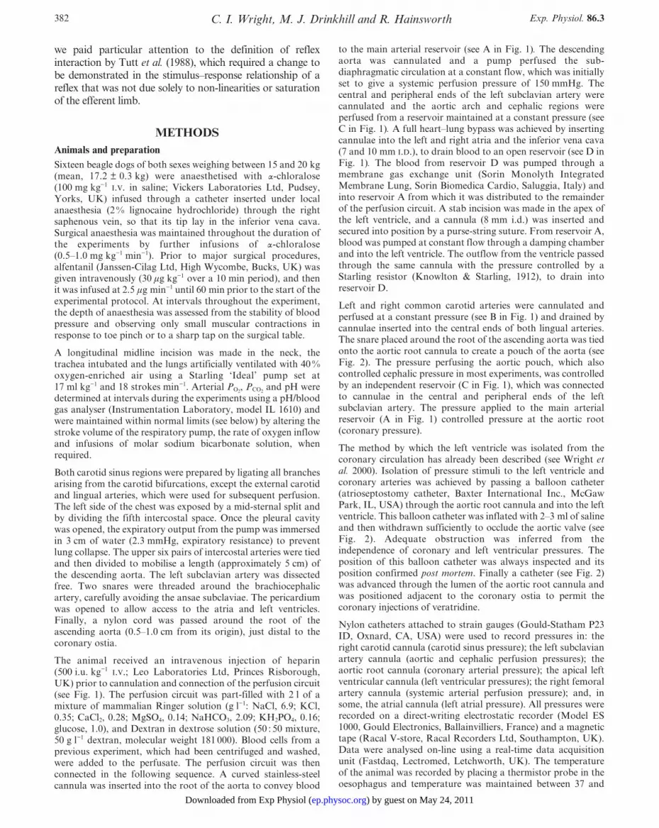
Figure 1
Diagram of experimental preparation. A large curved stainless-steel cannula tied into the aorta, distal toboth the coronary ostia and the left subclavian artery (LscA), created a pouch of the aorta outside thiscannula and conveyed blood to a pressurised main arterial reservoir. A total cardiopulmonary bypass wasachieved by draining blood through cannulae tied in the inferior vena cava and both atrial cannulae andinto reservoir D, from which it was pumped through an integrated heat exchanger/oxygenator and then toreservoir A. From this reservoir, blood was pumped to: (i) reservoir B and at a constant pressure intocannulae tied into both common carotid arteries; (ii) reservoir C and at constant pressure into cannulaetied into the central and peripheral ends of the left subclavian artery; (iii) the descending aorta at constantflow; and (iv) the left ventricle (LV) through a damping chamber at constant flow and out through aStarling resistor to closed reservoir D. Cannulae inserted in both lingual arteries drained blood from thecarotid bifurcation region to reservoir D. The LV was isolated from the coronary circulation by a ballooncatheter inserted into the LV, which was positioned to occlude the aortic valve. The insertion of an aorticroot catheter, positioned to lie adjacent to the coronary arteries, provided a site for the intra-coronaryinjection of veratridine. Abbreviations: AoP, aortic pouch; BcA, brachial cephalic artery; CP, constantpressure; DC, damping chamber; LscA, left subclavian artery; IVC., inferior vena cava; SG, strain gaugetransducer; P, pump; Res, reservoir; SR, Starling resistor.
) by guest on May 24, 2011ep.physoc.orgDownloaded from Exp Physiol (

39 °C by use of a heat exchanger incorporated into the perfusioncircuit and by heating the animal table.
All experiments were conducted in accordance with the currentUK legislation, the Animals (Scientific Procedures) Act, 1986,and all experiments were terminated by exsanguination of theanimal while under deep anaesthesia.
Experimental protocolFollowing the connection of the perfusion circuit, 30 min wasallowed for the animal to reach a stable state. During this time,arterial blood gases were analysed and corrected so that therespective values of pH, PO2
and PCO2were 7.4 ± 0.1, 219.7 ± 51.5
and 38.7 ± 2.5 mmHg (means ± S.D.). The haematocrit ofarterial blood was 19 ± 3 % (mean ± S.D.; range 12–24).
The following procedures were performed.
Carotid and coronary pressure tests. Single large-pressurestimuli were applied at the start of and at intervals during theexperiment to establish the viability of the preparation (see Dataanalysis, below). Coronary pressure was increased in a step from60 to 180 mmHg and carotid pressure in a step from 60 to240 mmHg. Pressures were held at each value for 1–2 min toallow steady-state responses to be obtained.
Coronary pressure tests at different ventricular pressures.Coronary arterial pressure was increased in steps of 30 mmHgfrom 60 to 180 mmHg with left ventricular systolic and end-diastolic pressures held at approximately 70 and 12 mmHg,respectively. Pressures were held at each step for 60 s to allowsteady states to be achieved. Coronary pressure was thendecreased to 60 mmHg and the averages of the values at the twolow coronary pressures were taken as baseline values. Vascularand heart rate responses to each step were calculated from thesebaseline values. Next, ventricular pressures were increased toapproximately 147 and 41 mmHg and the sequence of coronarypressure steps was repeated. Finally, ventricular pressures werereduced to the low values and the coronary pressure test againrepeated. In some experiments, we reversed the order of pressurechanges, going from high to low to high ventricular pressures.
Carotid baroreceptor tests at different ventricular pressures. Asimilar procedure was carried out as described above. Carotidpressure was changed in 30 mmHg steps from 60 to 240 mmHgwith ventricular systolic and end-diastolic pressures held first atthe low, then high, and finally at the low value again. As before,the order of the ventricular pressures was varied betweenexperiments.
Intra-coronary injection of veratridine at low and highventricular pressures. Heart rate and perfusion pressureresponses were assessed to the intra-coronary injections ofveratridine (30–60 µg). This procedure was performed atdifferent ventricular pressures. Steady-state values taken beforeand after the injection of veratridine were averaged to givecontrol values. Responses to veratridine were taken as the peakchange (from 5 s running averages) from the control level. Inbetween tests, at least 15 min was allowed to lapse to avoidtachyphylaxis. The responses obtained at the higher ventricularpressures were compared with those recorded at the low levels,obtained before and after the increase in ventricular pressures. Insome experiments, the order in which different ventricularpressures were applied was changed to high–low–high.
Data analysisVascular responses were accepted for analysis from those animalsin which a step increase in coronary pressure from 60 to 180 mmHgdecreased systemic perfusion pressure by at least 23 mmHg(approximately a 20% decrease in vascular resistance). In tests inwhich the effects of changing ventricular pressure were assessed,animals were divided into two groups. The first included the resultsfrom all the animals studied, and the second group included onlythose in which a step increase in ventricular pressure reducedsystemic perfusion pressure by 6% or more.
Plots were drawn of systemic vascular resistance againstmechanoreceptor distending pressures for both low and highventricular pressures (control values for perfusion pressure at thelow ventricular pressures expressed as 100 %). Stimulus–response curves were further analysed using a commercial curve-fitting computer program (GraphPad v2.0; GraphPad SoftwareInc., San Diego, CA, USA) which derived a sigmoid curve to fitthe points. From each curve, various measurements werederived. Saturation pressure was taken as the mechanoreceptorpressure corresponding to 95 % of the total perfusion pressureresponse. Values of maximum slope were obtained from thepeak differential of the fitted sigmoid function, and the inflexionpressures were those which corresponded to the peak of thedifferential.
Stimulus–response curves performed at the low and highventricular pressures were compared, and levels of significance
C. I. Wright, M. J. Drinkhill and R. Hainsworth384 Exp. Physiol. 86.3
Figure 2
Diagram showing the position of the left ventricularballoon catheter. This balloon catheter was passedretrogradely across the aortic valve, inflated withinthe cavity of the left ventricle and then withdrawnand positioned to occlude the aortic valve, in orderto isolate the left ventricle from the coronarycirculation. The position of the aortic root catheter(site of intra-coronary injection) is also shown andthis was positioned to lie adjacent to the coronaryostia.
) by guest on May 24, 2011ep.physoc.orgDownloaded from Exp Physiol (

were assessed using Student’s paired t test and consideredstatistically significant when P < 0.05. Data presented are ofmeans ± S.E.M.
RESULTSDuring the testing of responses to stimulation of carotid,coronary and ventricular mechanoreceptor and ventricularchemosensitive nerves (see procedures below), pressures
perfusing the regions not being investigated were heldconstant at the following values: carotid, 64.1 ± 0.8 mmHg;coronary, 82.2 ± 3.0 mmHg; aortic pouch (and cephaliccirculation), 124.4 ± 5.1 mmHg; ventricular systolic and end-diastolic pressures, 57.8 ± 4.2 and 11.4 ± 1.3 mmHg. In fiveanimals, the cephalic region was perfused separately from theaortic pouch at 133.4 ± 9.7 mmHg. Also, in 12 animals inwhich the left atrial pressure was recorded, the bypassmaintained it at a mean value of 0.2 ± 0.2 mmHg.
Ventricular mechanoreceptorsExp. Physiol. 86.3 385
Figure 3
Response of systemic perfusion pressure to stepwise changes in coronary perfusion pressure between 60and 180 mmHg at low (A) and high (B) ventricular pressures. Traces are shown of coronary perfusionpressure (CPP), left ventricular pressure (LVP), aortic pouch pressure (AoP), systemic perfusionpressure (SPP) and carotid sinus pressure (CSP). Note the reduction in systemic perfusion pressure atthe higher ventricular pressures, at a coronary pressure of 60 mmHg.
) by guest on May 24, 2011ep.physoc.orgDownloaded from Exp Physiol (

Responses to single-step changes in aortic root (coronaryarterial) or carotid sinus pressureIn 16 dogs, a step increase in coronary arterial pressurebetween 63.3 ± 1.0 and 180.5 ± 2.7 mmHg resulted in adecrease in systemic perfusion pressure from 170.7 ± 9.1to 101.3 ± 6.5 mmHg (_39.2 ± 3.5 %; P < 0.0001), but nosignificant change in heart rate (from 175 ± 8 to 176 ±6 beats min_1; P > 0.05, Student’s paired t test). In thesesame animals, increasing carotid sinus pressure between63.8 ± 0.4 and 194.5 ± 4.3 mmHg decreased systemicperfusion pressure from 139.2 ± 4.1 to 87.2 ± 4.4 mmHg(_36.7 ± 3.2 %; P < 0.0001) and heart rate from 171 ± 6to 146 ± 7 beats min_1 (_25 ± 6; P < 0.001, Student’spaired t test).
Responses to changes in coronary pressure at differentventricular pressuresIn seven animals coronary pressure was increased in steps ofapproximately 30 mmHg between 65.4 ± 0.8 and 183.4 ±0.7 mmHg with ventricular pressures held at the low values of69.7 ± 4.3 and 12.0 ± 2.2 mmHg (peak and end-diastolic) andthe high values of 146.5 ± 12.9 and 40.7 ± 17.6 mmHg. Thisresulted in step decreases in perfusion pressure from179.2 ± 15.3 to 100.2 ± 6.3 mmHg (_40.6 ± 7.4%; P < 0.005)and at the high pressures from 167.5 ± 15.2 to 99.5 ±7.0 mmHg (_34.4 ± 7.5%; P < 0.005). An example of tracesobtained from one of these dogs is shown in Fig. 3 and thisemphasises the large responses to changes in coronarypressure and the smallness of the effect of the ventricularpressure. Mean vascular responses are compared in Fig. 4A.
Overall, vascular responses were significantly (P < 0.05)smaller at the higher ventricular pressures, but values ofmaximum slope, and inflexion and saturation pressures werenot different (P > 0.05; see Table 1).
Figure 4B shows the results from those three animals mostresponsive to changes in ventricular pressure. At the higherventricular pressure, the decreases in vascular resistance weresmaller (decreasing by 37.3% (range, 21.5–60.8%) comparedwith 46.7% (32.5–67.1%) at the low ventricular pressures).However, until the responses approached saturation values,the curves were approximately parallel. Values of maximumslope, and inflexion and saturation pressures were similar (seeTable 1).
Responses to changes in carotid sinus pressure at low andhigh ventricular pressuresIn eight animals, carotid sinus pressure was increased in30 mmHg steps between 64.2 ± 0.4 and 243.2 ± 1.0 mmHgat the high and low ventricular pressures. Overall, at thelower ventricular pressures, perfusion pressure and heartrate decreased from 154.4 ± 10.9 to 109.2 ± 9.4 mmHg(_29.4 ± 2.9%; P < 0.0001) and from 187 ± 17 to 166 ±19 beats min_1 (_21 ± 5 beats min_1; P < 0.005) . At thehigher ventricular pressures, perfusion pressure and heartrate decreased from 151.4 ± 10.8 to 105.6 ± 8.5 mmHg(_28.2 ± 3.7%; P = 0.0001) and from 182 ± 15 to 161 ±15 beats min_1 (_22 ± 5 beats min_1; P < 0.005, Student’spaired t test). The responses were not significantly differentfrom those obtained at the lower distension pressures
C. I. Wright, M. J. Drinkhill and R. Hainsworth386 Exp. Physiol. 86.3
Figure 4Reflex responses of systemic perfusion pressure to stepincreases in coronary pressure at low (1) and high (0)ventricular pressures. A shows the results from all sevenof the animals studied, whereas B shows the results fromthose three animals most responsive to changes inventricular pressure. Resting levels of perfusion pressurewere: A, 179.2 ± 15.3 and 167.5 ± 15.2 mmHg;B, 174.6 mmHg (range, 141.2–217.8 mmHg) and156.6 mmHg (122.9–201.8 mmHg) (low and high,respectively). ‘+’ indicates P < 0.05, when correspondingvalues at the low and high ventricular pressures werecompared (Student’s paired t test). Overall, there was areduction in the vascular response at the higherventricular pressures, but there was no difference inmaximal slopes (see Table 1). Perfusion pressures arerelated to the values recorded at the low ventricularpressures that were expressed as 100 % and values arepresented as means ± S.E.M. (A) or means (B).
) by guest on May 24, 2011ep.physoc.orgDownloaded from Exp Physiol (

(P > 0.05, Student’s paired t test). The responses ofperfusion pressure at each step in carotid pressure and atboth ventricular pressures are plotted in Fig. 5. Figure 5Ashows data from all experiments, and clearly the stimulus–response curves overlapped. In Fig. 5B, obtained from themore responsive animals, the stimulus–response curvesappeared parallel and, unlike the responses to changes incoronary pressure shown in Fig. 4, there was no evidence ofconvergence. No difference was found between maximalslopes, and inflexion and saturation pressures (see Table 1).
Responses to veratridine at low and high ventricularpressuresFigure 6A shows the maximal responses from six animals tothe coronary injection of veratridine at the two ventricularpressures. At the low and high levels of ventricular pressure,perfusion pressure decreased from 138 ± 6.5 to 104 ±6.5 mmHg (_24.2 ± 2.0%; P < 0.0001) and from 126 ± 5.2 to99 ± 7.4 mmHg (_22 ± 3.4%; P < 0.005). These responseswere not significantly different (P > 0.05, Student’s paired ttest). Heart rate decreased from 190 ± 16 to 158 ±
Ventricular mechanoreceptorsExp. Physiol. 86.3 387
Table 1. Values of maximum slopes, inflexion pressures and saturation pressures for coronary andcarotid baroreflexes, at low and high ventricular pressures (LVP)
Slope (mmHg mmHg_1) Inflexion pressure (mmHg) Saturation pressure (mmHg)No. of ———————————— —————————————— ——————————————
Baroreceptor dogs Low LVP High LVP Low LVP High LVP Low LVP High LVP
Coronary 7 9.5 ± 2.2 10.0 ± 2.6 103.0 ± 4.5 102.8 ± 7.7 157.1 ± 5.5 157.2 ± 7.7
3 10.0 9.4 98.7 90.1 157.5 158.7(7.4–14.6) (4.3–16.3) (91.1–110.8) (85.3–93.0) (142.1–170.2) (147.5–167.0)
Carotid 8 4.2 ± 0.7 5.4 ± 0.8 151.6 ± 10.0 146.9 ± 13.7 198.1 ± 8.1 201.3 ± 7.9
3 3.5 5.4 174.2 164.6 214.6 208.5(1.9–5.5) (3.0–6.7) (152.9–187.9) (134.5–203.9) (200.0–236.8) (183.0–240.1)
No significant difference was found when corresponding values at low and high ventricular pressureswere compared (P > 0.05, Student’s paired t test). Numbers in parentheses are ranges and all othervalues are presented as means ± S.E.M.
Figure 5Responses of systemic perfusion pressure to stepwisechanges in carotid sinus pressure at low (1) and high (0)left ventricular pressures. A shows the results from alleight animals studied and B shows the means from threeanimals most responsive to changes in ventricularpressures. Resting levels of perfusion pressure were: A,154.4 ± 10.9 and 151.4 ± 10.8 mmHg; B, 151.0 mmHg(range, 133.2–161.8 mmHg) and 139.3 mmHg(121.7–148.7 mmHg) (low and high, respectively). Theresults in B indicate that at the high ventricular pressuresthere was a downwards and parallel shift in perfusionpressure, which was not evident in A.
) by guest on May 24, 2011ep.physoc.orgDownloaded from Exp Physiol (

12 beats min_1 (_32 ± 10 beats min_1; P < 0.05) and from184 ± 11 to 160 ± 17 beats min_1 (_24 ± 7 beats min_1;P < 0.05) at low and high ventricular pressures, respectively.Although the response at the high ventricular pressure wassmaller, it was not significant different (P > 0.05, Student’spaired t test).
Figure 6B shows the maximal responses from the threeanimals most responsive to changes in ventricular pressure.Vascular responses were little different at the different levelsof ventricular pressure, perfusion pressure decreased by22.9% (range, 17.5 to 28.5%) and 18.4% (12.7 to 26.4%) atthe low and high levels, respectively. The heart rate responsewas, however, smaller at the higher ventricular pressures,decreasing by 17 beats min_1 (range, 15–21 beats min_1)compared with 43 beats min_1 (range, 16–71 beats min_1) atthe lower level.
DISCUSSIONAlthough there have been several previous investigationswhich attempted to examine interactions between ventricularreceptors and baroreceptor reflexes, none has succeeded in
applying localised mechanical stimuli to two or more specificregions. This study is the first in which the pressuresdistending ventricular mechanoreceptors and other reflexo-genic areas have been controlled in a discrete manner. Wepreviously showed that the responses even to gross distensionof the left ventricle were small and the present study confirmsthat the ventricle is not of major importance on its own incardiovascular regulation. Furthermore, the absence of largeresponses to changes in ventricular pressure could not havebeen due to the preparation being unresponsive because ofnear-maximal vasoconstriction or vasodilatation, since thebaroreceptor stimulus–response curves virtually overlapped.
Previous work by others has suggested that the stimulation ofventricular receptors decreases the sensitivity of the carotidbaroreceptor reflex. However, in those studies in which aninteractive effect was claimed to have been shown, only thoseof ventricular receptors with chemosensitive endings werestimulated (Chen, 1979; Holmberg et al. 1983). In fact, eventhe apparent interactions seen when stimulating thechemosensitive endings may not actually be a true interactionbecause the reduction of responses may well have been due tolimitation of the efferent limb of the reflexes as neurogenic
C. I. Wright, M. J. Drinkhill and R. Hainsworth388 Exp. Physiol. 86.3
Figure 6
Changes in systemic perfusion pressure (4) and heart rate (5) to the coronary injection of veratridine(30–60 µg) at low (L) and high (H) ventricular pressures. A shows the results from all six animals studiedand B shows the means from those three animals most responsive to changes in ventricular pressures.Resting levels of perfusion pressure and heart rate were: A, 138.0 ± 6.5 and 126.0 ± 5.2 mmHg and190 ± 16 and 184 ± 11 beats min_1; B, 147.4 mmHg (range, 135.1–159.2 mmHg) and 125.8 mmHg(range, 110.9–135.6 mmHg) and 220 beats min_1 (range, 190–248 beats min_1) and 206 beats min_1
(range, 196–220 beats min_1) (low and high, respectively). In all the six animals studied, no significantdifference was observed between the maximal responses of vascular resistance or heart rate. In the threemore responsive animals, the heart rate response was reduced at the higher ventricular pressures.Absolute changes in heart rate are shown. Responses of perfusion pressure are presented as a changefrom control conditions (taken as 0 %), with values presented as either mean ± S.E.M. (A) or means (B).
) by guest on May 24, 2011ep.physoc.orgDownloaded from Exp Physiol (
vasomotor activity approaches zero (Tutt et al. 1988). Theprevious attempts to study interactions with ventricularmechanosensitive receptors have all been deficient due toinadequate localisation of the stimulus to the ventricle. Theseprocedures, which involve widespread changes in pressure(Holmberg et al. 1983; Ludbrook & Graham, 1984;Holmberg & Zucker, 1986) or even our earlier work in whichpressure changes were applied to the aortic root (Tutt et al.1988; Vukasovic et al. 1989) must have influenced many otherreflexogenic areas, particularly the coronary arterial baro-receptors.
In this study, we observed no evidence of interaction betweenthe ventricular mechanoreceptor reflexes and the carotidbaroreflex. Overall, there was almost no difference in thestimulus–response curves to step changes in carotid pressureat high and low ventricular pressures. When we examinedresponses from the most responsive preparations, it was seenthat the stimulus–response relationships remained parallel,indicating that the reflexes summated in a simple arithmeticmanner.
The possibility of interaction between ventricular andcoronary mechanoreflexes has not been examined before andthe results do seem at first sight to indicate that there may bea slight depression of the responses to coronary baroreceptorreflexes at the higher ventricular pressure. There was asignificant difference between the perfusion pressures at thelow coronary pressure and the different ventricular pressures.The overall response to the entire change in coronary pressurewas smaller at the high ventricular pressures, showing that thecurves converged. This was even better seen in the moreresponsive preparations. However, we would argue that thistoo represents simple arithmetic summation. Firstly, therewas no difference in the maximum slopes of the coronaryresponse curves at the different ventricular pressures. Also,the responses to the coronary reflex in this group were verylarge, considerably larger than those to the carotid reflex, andwhat is likely to explain the convergence of the curves issimply that the responses were near maximal: sympatheticactivity to the resistance vessels was approaching zero andcould not decrease further.
The possible interaction between the ventricular mechano-receptor reflexes and chemosensitive receptor reflex wasexamined in a different way. It was not possible to applygraded stimuli to chemosensitive afferents because repeatedstimuli are known to cause tachyphylaxis, leading to a largedepression of the responses (Cramer, 1915; Dawes & Comroe,1954; Hainsworth et al. 1989; Hainsworth, 1991). To avoidthis problem we stimulated the chemosensitive afferents bybolus injections of veratridine into the aortic root, allowed aperiod of at least 10 min between successive injections, andbracketed tests at one ventricular pressure with paired tests atthe other. The results obtained show that both vascular andcardiac responses were not significantly affected byventricular pressure. The cardiac responses in the threepreparations that were most sensitive to ventricular pressurechanges were consistently smaller at the high ventricularpressure (mean change, 17 versus 43 beats min_1). The dose ofveratridine and the coronary perfusion pressure were the
same so the applied stimuli are likely to have been similar.With only one dose, it is clearly impossible to construct astimulus–response curve so we cannot be certain that thereduction in response was not due to non-linearity of theefferent limb of the reflex. However, heart rate did notapproach minimal levels so it does seem possible that therecould have been an interactive effect. It is clearly not possibleto know at what part of the reflex arc interaction may haveoccurred. However, one possibility is that it may haveoccurred at the sensory receptor itself. Previous work hasshown that, although some ventricular receptors arepredominantly mechanosensitive and some are mainlychemosensitive, there are many which respond to both stimuli(Coleridge et al. 1964; Sleight & Widdicombe, 1965) and it isconceivable that if these were stimulated by a high distendingpressure there may have been less scope for a further increasewhen chemical agents were applied.
The importance of this study is that it has reinforced the viewthat ventricular mechanoreceptors are unlikely to be ofimportance as physiological regulators. We previouslyshowed that whatever stimuli were applied, changes insystolic pressure, end-diastolic or inotropic state, responseswere always small (Drinkhill et al. 2001). Indeed, it was onlywhen diastolic pressure increased abnormally that anysignificant responses were obtained at all. This study has notonly confirmed those earlier findings but has shown thatresponses were unlikely to have been ‘masked’ by otherreflexes. We believe that, although essentially a negativefinding, the implications of this work are not inconsiderable.The previous widely held view was that ventricular receptorswere important regulators, being important for mediatingresponses to intra-thoracic blood volume changes (Aviado &Schmidt, 1959; Salisbury et al. 1960; Ross et al. 1961; Mark etal. 1973; Chevalier et al. 1974; Zelis et al. 1977) and evenresponsible for initiating vaso-vagal responses (Öberg &Thorén, 1972; Abboud, 1989). Other mechanisms must nowbe sought to explain responses not due to sinoaorticbaroreceptors. Stimulation of baroreceptors in the coronaryarteries can go a long way to explaining responses previouslythought to be due to ventricular receptors but changes inpressure in vessels in other regions of the circulation such asabdominal arteries or veins (Drinkhill et al. 1997) may proveto be of greater importance than has hitherto been recognised.
ABBOUD, F. M. (1989). Ventricular syncope: is the heart a sensoryorgan? (editorial). New England Journal of Medicine 320, 390–392.
AVIADO, D. M. & SCHMIDT, C. F. (1959). Cardiovascular andrespiratory reflexes from the left side of the heart. AmericanJournal of Physiology 196, 726–730.
CHEN, H. I. (1979). Interaction between the baroreceptor andBezold-Jarisch reflexes. American Journal of Physiology 237,H665–661.
CHEVALIER, P. A., WEBER, K. C., LYONS, G. W., NICOLOFF, D. M.& FOX, I. J. (1974). Haemodynamic changes from stimulation ofleft ventricular baroreceptors. American Journal of Physiology 227,719–728.
Ventricular mechanoreceptorsExp. Physiol. 86.3 389
) by guest on May 24, 2011ep.physoc.orgDownloaded from Exp Physiol (

COLERIDGE, H. M., COLERIDGE, J. C. & KIDD, C. (1964). Cardiacreceptors in the dog, with particular reference to two types ofafferent ending in the ventricular wall. Journal of Physiology 174,323–339.
CRAMER, W. J. (1915). On the action of veratrum viride. Journal ofPharmacology and Experimental Therapeutics 7, 63–82.
DAWES, G. S. & COMROE, J. H. (1954). Chemoreflexes from theheart and lungs. Physiological Reviews 34, 167–201.
DRINKHILL, M. J., DOE, C. P., MYERS, D. S., SELF, D. A. &HAINSWORTH, R. (1997). Reflex vascular responses to alterationsin abdominal arterial pressure and flow in anaesthetised dogs.Experimental Physiology 82, 995–1005.
DRINKHILL, M. J., WRIGHT, C. & HAINSWORTH, R. (2001). Reflexvascular responses to independent changes in left ventricular end-diastolic and peak systolic pressures and inotropic state inanaesthetised dogs. Journal of Physiology (in the Press).
GUAZZI, M., LIBRETTI, A. & ZANCHETTI, A. (1962). Tonic reflexregulation of the cat’s blood pressure through vagal afferents fromthe cardiopulmonary region. Circulation Research 11, 7–16.
HAINSWORTH, R. (1991). Reflexes from the heart. PhysiologicalReviews 71, 617–658.
HAINSWORTH, R., CARE, F. M. S., COULSHED, D. & MCWILLIAM, P.N. (1989). Response of cardiac receptors to chemical andmechanical stimulation. Society of Neuroscience Abstracts 15,1184.
HOLMBERG, M. J., GORMAN, A. J., CORNISH, K. G. & ZUCKER, I. H.(1983). Attenuation of arterial baroreflex control of heart rate byleft ventricular receptor stimulation in the conscious dog.Circulation Research 52, 597–607.
HOLMBERG, M. J. & ZUCKER, I. H. (1986). Increased left ventricularpressure attenuates the baroreflex in unanaesthetised dogs.American Journal of Physiology 251, R23–31.
KNOWLTON, F. P. & STARLING, E. H. (1912). The influence ofvariations in temperature and blood pressure on the performanceof the isolated mammalian heart. Journal of Physiology 44,206–219.
LUDBROOK, J. & GRAHAM, W. F. (1984). The role of cardiacreceptor and arterial baroreceptor reflexes in control of thecirculation during acute change of blood volume in the consciousrabbit. Circulation Research 54, 424–435.
MANCIA, G., DONALD, D. E. & SHEPHERD, J. T. (1973). Inhibitionof adrenergic outflow to peripheral blood vessels by vagalafferents from the cardiopulmonary region. Circulation Research33, 713–721.
MANCIA, G., DONALD, D. E. & SHEPHERD, J. T. (1975). Role ofcardiac, pulmonary and carotid mechanoreceptors in the controlof hind limb and renal circulation in dogs. Circulation Research 37,200–208.
MANCIA, G., DONALD, D. E. & SHEPHERD, J. T. (1976). Interplayamong carotid sinus, cardiopulmonary, and carotid body reflexesin dogs. American Journal of Physiology 230, 19–24.
MARK, A. L. (1983). The Bezold–Jarisch reflex revisited: clinicalimplications of inhibitory reflexes originating in the heart. Journalof the American College of Cardiology 1, 90–102.
MARK, A. L., ABBOUD, F. M., SCHMID, P. G., HEISTAD, D. D. &JOHANNSEN, U. J. (1973). Reflex vascular responses to leftventricular outflow obstruction and activation of ventricularbaroreceptors in dogs. Journal of Clinical Investigation 52,1147–1153.
ÖBERG, B. & THORÉN, P. N. (1972). Increased activity in leftventricular receptors during haemorrhage or occlusion of cavalveins in the cat. A possible cause of the vaso-vagal reaction. ActaPhysiologica Scandinavica 85, 164–173.
ÖBERG, B. & WHITE, S. (1970). Circulatory effects of interruptionand stimulation of cardiac vagal afferents. Acta PhysiologicaScandinavica 80, 383–394.
PERSSON, P. B. (1991). Interaction of arterial and cardiopulmonaryreflexes. In Baroreceptor Reflexes: Integrative Functions andClinical Aspects, ed. PERSSON, P. B. & KIRCHEIM, H. R., pp. 126–153.Springer-Verlag, Berlin.
ROSS, J., FRAHM, C. J. & BRAUNWALD, E. (1961). The influence ofintra-thoracic baroreceptors on venous return, systemic vascularvolume and peripheral resistance. Journal of Clinical Investigation40, 563–572.
SALISBURY, P. F., CROSS, C. E. & RIEBEN, P. A. (1960). Reflexeffects of left ventricular distension. Circulation Research 8,530–534.
SLEIGHT, P. & WIDDICOMBE, J. G. (1965). Action potentials in fibersfrom receptors in the epicardium and myocardium of the dog’s leftventricle. Journal of Physiology 181, 235–258.
THAMES, M. D. & DIBNER-DUNLAP, M. E. (1991). Baroreflexes incongestive heart failure. In Baroreceptor Reflexes: IntegrativeFunctions and Clinical Aspects, ed. PERSSON, P. B. & KIRCHEIM,H. R., pp. 257–270. Springer-Verlag, Berlin.
THAMES, M. D., DIBNER-DUNLAP, M. E. & MINISI, A. J. (1993).Cardiovascular reflexes during myocardial ischaemia andinfarction. In Cardiovascular Reflex Control in Health and Disease,ed. HAINSWORTH, R. & MARK, A. L., pp. 235–255. CambridgeUniversity Press, Cambridge.
THORÉN, P. N. (1977). Characteristics of left ventricular receptorswith nonmedullated vagal afferents in cats. Circulation Research40, 415–421.
TUTT, S. M., MCGREGOR, K. H. & HAINSWORTH, R. (1988). Reflexvascular responses to changes in left ventricular pressure inanaesthetised dogs. Quarterly Journal of Experimental Physiology73, 425–437.
VUKASOVIC, J. L., TUTT, S. M., CRISP, A. J. & HAINSWORTH, R.(1989). The influence of left ventricular pressure on the vascularresponses to changes in carotid sinus pressure in anaesthetiseddogs. Quarterly Journal of Experimental Physiology 74, 735–746.
WRIGHT, C., DRINKHILL, M. J. & HAINSWORTH, R. (2000). Reflexeffects of independent stimulation of coronary and left ventricularmechanoreceptors in anaesthetised dogs. Journal of Physiology528, 349–358.
ZELIS, R., LOTYSH, M., BRAIS, M., PENG, C. L., HURLEY, E. &MASON, D. J. (1977). Effects of isolated right and left ventricularstretch on regional arteriolar resistance. Cardiovascular Research11, 419–426.
AcknowledgementsThis research was funded by a studentship (FS/97075) from theBritish Heart Foundation and by the Medical Research Council(G9809405). The technical assistance of Mr D. Myers is alsogratefully acknowledged.
C. I. Wright, M. J. Drinkhill and R. Hainsworth390 Exp. Physiol. 86.3
) by guest on May 24, 2011ep.physoc.orgDownloaded from Exp Physiol (


