Embed Size (px)
Citation preview
203.865.6784 • ct-ortho.com
Moving Forward Together
Pediatric Forearm and Distal Radius Fractures
Richard A. Bernstein, M.D.
Forearm fractures in children are common and are managed differently than similar injuries in
adults. Historically, the results of nonoperative treatment of adult forearm fractures have been
poor, with reports of nonunion, malalignment, and stiffness due to the lengthy immobilization
required for union. Currently, most adults with both-bone forearm fractures are treated by open
reduction and internal fixation. In pediatric patients, treatment is primarily nonoperative because
of uniformly rapid healing and the potential for remodeling of residual deformity.
Although the outcomes in children are usually good, treatment of individual patients and
education of families can be challenging. Beyond the sometimes-difficult mechanics of fracture
reduction and maintenance, the clinician is faced with controversies regarding techniques of
reduction, position of immobilization, and definition of an acceptable reduction.
The purpose of this article is to critically summarize available information and present treatment
recommendations based on a literature review and the previous experience of the senior author
(C.T.P.). The scope of this discussion will be limited to the more common entities, such as
pediatric forearm and distal radius fractures, and will not include articular fractures, plastic
deformation, and fracture-dislocations, such as Monteggia lesions.
Functional Anatomy
The ulna is a relatively straight bone around which the curved radius rotates during pronation
and supination. The axis of rotation passes obliquely from the distal ulnar head to the proximal
radial head. The two bones are stabilized distally and proximally by the triangular fibrocartilage
complex and the annular ligament, respectively. Further stabilization is provided by the
interosseous membrane, with oblique fibers passing distally from the radius to the ulna; these
fibers are somewhat relaxed in supination and tighter in pronation.
The pronator quadratus (distally) and pronator teres (inserting on the middle portion of the
radius) actively pronate the forearm, while the biceps and supinator (proximal insertions)
provide supination. The insertions of these four muscles can partially account for fragment
position in complete fractures. In distal-third fractures, the proximal fragment will be in neutral to
slight supination, and the weight of the hand combined with the pronator quadratus tends to
pronate the distal fragment. In proximal-third fractures, the distal fragment is pronated, and the
proximal fragment is supinated. Mid-shaft fractures tend to leave both fragments in a neutral
position with the distal fragment slightly pronated and the proximal fragment slightly supinated.
203.865.6784 • ct-ortho.com
Moving Forward Together
Several anatomic differences distinguish pediatric forearms from those of adults. The pediatric
radial and ulnar shafts are proportionately smaller, with narrow medullary canals, and the
metaphysis contains more trabecular bone. In addition, the periosteum in children is much
thicker than that in adults; this feature can both hinder and help in the management of pediatric
fractures.
Normal Growth and Implications for Remodeling
The proximal and distal physes provide longitudinal growth, which contributes to remodeling
after fracture healing. The distal radial and ulnar growth plates are responsible for 75% and 81%
of the longitudinal growth of each bone, respectively. 1 This is consistent with the oft-made
observation that distal forearm fractures have greater potential for remodeling than do more
proximal fractures. 2-4 Additional remodeling can also be attributed to elevation of the thick
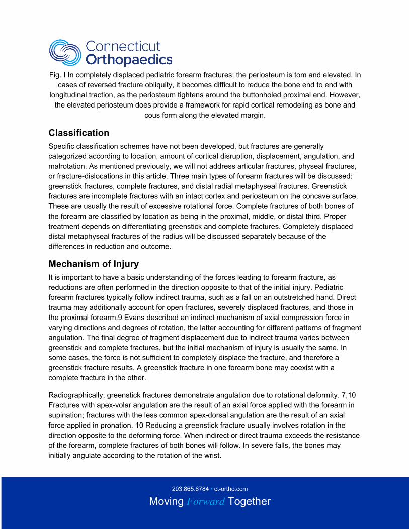
osteogenic periosteum after fracture (Fig. 1). Intramembranous ossification by the periosteum
will assist in rapid healing and subsequent remodeling of residual diaphyseal deformity. Normal
Function and Treatment Objectives. The goal of treatment of forearm and distal radius injuries is
to facilitate union of the fracture in a position that restores functional range of motion to the
elbow and forearm. The predominant motions affected by malunion are pronation and
supination, which are a function of skeletal length and axial and rotational alignment. Normal
supination from neutral is 80 to 120 degrees; normal pronation from neutral is 50 to 80 degrees.
5 It is important to realize that “normal" motion may not be what is needed for normal function
Biomechanical testing has revealed that common activities of daily living require 100 degrees of
forearm rotation, equally split between pronation and supination. 6 Limited pronation is more
easily compensated for by shoulder abduction. Secondary concerns include cosmetic
alignment; however, acceptable reduction usually precludes gross malalignment. Ulnar
alignment is the most important cosmetic determinant.
203.865.6784 • ct-ortho.com
Moving Forward Together
Fig. I In completely displaced pediatric forearm fractures; the periosteum is tom and elevated. In
cases of reversed fracture obliquity, it becomes difficult to reduce the bone end to end with
longitudinal traction, as the periosteum tightens around the buttonholed proximal end. However,
the elevated periosteum does provide a framework for rapid cortical remodeling as bone and
cous form along the elevated margin.
Classification
Specific classification schemes have not been developed, but fractures are generally
categorized according to location, amount of cortical disruption, displacement, angulation, and
malrotation. As mentioned previously, we will not address articular fractures, physeal fractures,
or fracture-dislocations in this article. Three main types of forearm fractures will be discussed:
greenstick fractures, complete fractures, and distal radial metaphyseal fractures. Greenstick
fractures are incomplete fractures with an intact cortex and periosteum on the concave surface.
These are usually the result of excessive rotational force. Complete fractures of both bones of
the forearm are classified by location as being in the proximal, middle, or distal third. Proper
treatment depends on differentiating greenstick and complete fractures. Completely displaced
distal metaphyseal fractures of the radius will be discussed separately because of the
differences in reduction and outcome.
Mechanism of Injury
It is important to have a basic understanding of the forces leading to forearm fracture, as
reductions are often performed in the direction opposite to that of the initial injury. Pediatric
forearm fractures typically follow indirect trauma, such as a fall on an outstretched hand. Direct
trauma may additionally account for open fractures, severely displaced fractures, and those in
the proximal forearm.9 Evans described an indirect mechanism of axial compression force in
varying directions and degrees of rotation, the latter accounting for different patterns of fragment
angulation. The final degree of fragment displacement due to indirect trauma varies between
greenstick and complete fractures, but the initial mechanism of injury is usually the same. In
some cases, the force is not sufficient to completely displace the fracture, and therefore a
greenstick fracture results. A greenstick fracture in one forearm bone may coexist with a
complete fracture in the other.
Radiographically, greenstick fractures demonstrate angulation due to rotational deformity. 7,10
Fractures with apex-volar angulation are the result of an axial force applied with the forearm in
supination; fractures with the less common apex-dorsal angulation are the result of an axial
force applied in pronation. 10 Reducing a greenstick fracture usually involves rotation in the
direction opposite to the deforming force. When indirect or direct trauma exceeds the resistance
of the forearm, complete fractures of both bones will follow. In severe falls, the bones may
initially angulate according to the rotation of the wrist.
203.865.6784 • ct-ortho.com
Moving Forward Together
However, when completely broken by either indirect or direct forces, the bones shorten,
angulate, and rotate within the confines of the surrounding periosteum, interosseous membrane,
and muscle attachments. Because the final positioning in complete fractures depends to some
degree on the relationship of fracture location and the insertions of the pronating and supinating
muscles, reduction is more complex than for simple greensick fractures.
Distal radius fractures usually follow a fall on an outstretched hand. The resultant angulation
may also be accompanied by rotational deformity. Apex-volar angulation (the most common
deformity) is accompanied by supination and apex-dorsal angulation with pronation. In our
experience, solely ulnar fractures are less common, and probably result from direct trauma.
Patient Assessment and Radiographic Evaluation
The diagnosis of forearm fractures is usually self-evident from the history and the obvious
deformity. Child abuse must always be considered in patients less than 3 years of age.
Inspection and palpation should be carefully performed; occasionally, soft tissue swelling will
obscure gross malalignment. The wrist and elbow should be examined for swelling, tenderness,
and unusual prominences that may signify a Monteggia or Galeazzi fracture. Cursory
examination of the humerus and clavicle may detect fractures that have also resulted from a fall
on an outstretched hand. Detailed neurovascular examination is necessary before and after
reduction; median, ulnar, and posterior interosseous neurapraxias have been documented.
Such deficits usually resolve with observation in 2 to 3 weeks.
Radiographic evaluation should include anteroposterior (AP) and lateral views of the forearm. If
the elbow and wrist are not adequately visualized, corresponding views should be obtained to
eliminate radial head dislocation, supracondylar fracture, and distal radioulnar joint injury.
Forearm radiographs are examined to determine fracture pattern (complete or greenstick),
location (proximal, middle, or distal third), displacement, angulation, and rotation.
Displacement and angulation are fairly easy to document on AP and lateral views. Although
deformities can often be quantified and described on these standard views, it is important to
remember that fracture angulation and displacement are always in a single plane, between
those obtained on orthogonal radiographs. The magnitude of the deformity is at least as great
as or greater than that seen on each view. Malrotation in complete fractures can be difficult to
detect and assess, but can be suspected when the cortical, medullary, or bone diameters of
both fragments are not equal. Malrotation can be gauged from deviations of normal orientation
of proximal and distal bony prominences.
On a standard AP view, the radial tuberosity is seen in profile on the medial side, while the
radial styloid and thumb are seen 180 degrees opposite on the lateral side. On this same view,
ulnar styloid and coronoid process are not seen. Lateral views reveal the ulnar styloid pointing
posterior and the coronoid process pointing directly anterior; the aforementioned radial

203.865.6784 • ct-ortho.com
Moving Forward Together
prominences will not be seen. Another useful method for determining rotation of the proximal
fragment utilizes the tuberosity view described by Evans. This technique allows a quantitative
assessment of proximal fragment rotation. The distal fragment can then be manipulated and
rotated into a corresponding position.
Anesthesia (edited)
In many centers, a large proportion of forearm and distal radius fractures are treated outside the
surgical suite, requiring the treating surgeon to consider and administer appropriate anesthesia.
Strict guidelines for conscious sedation have been established by the American Academy of
Pediatries.14 A survey of orthopaedic surgeons completed in 1993 indicated that as many as
one third of orthopaedic surgeons were not in compliance with these guidelines during fracture
reduction. 15
The chosen method should be as safe as possible, induce the least trauma, including fracture
reduction. As no one method completely meets these criteria, several different choices exist,
each with its own advantages and disadvantages.
Options include quick reduction without anesthesia, hematoma block which involves an injection
of the anesthetic in the area of the fracture or going to the hospital for either a block type or a
general anesthetic. Intravenous sedation entails the potential for overdosage and
cardiopulmonary depression.
Regional intravenous blocks have the advantages of rapid onset of effect, simple administration,
and good muscle relaxation. Disadvantages include pain when the injured limb is exsanguinated
by wrapping or elevation. Premature cuff deflation may lead to major neurologic and cardiac
complications when high doses are used.
Use of general anesthesia relieves the surgeon of the burden of providing safe and effective
anesthesia. This allows the surgeon to concentrate on reduction and stabilization
unencumbered by the proximity of anxious parents. In addition, if several reduction attempts are
required, general anesthesia provides total relaxation with minimal constraints. Furthermore, if
reduction is inadequate or unstable, it easy to convert to operative stabilization.
Adequacy of Reduction and Results of Closed Treatment
Anatomic reduction is usually not required for pediatric forearm fractures due to the potential for
growth and remodeling. However, the treating physician must be able to define reasonable
residual malalignment by answering several important questions: What are the acceptable limits
of displacement at healing, and to what degree do the deformities remodel over time? How is
remodeling potential affected by variables such as age and location of the fracture? Does
malalignment at healing and follow-up correlate with loss of motion? What degree of
documented motion loss is associated with poor function and patient dissatisfaction?
203.865.6784 • ct-ortho.com
Moving Forward Together
It is uniformly agreed that post-traumatic angular deformities in children have variable
remodeling potential; however, it has not been consistently proved that deformities
characterized by rotational malalignment will also remodel. Many studies have documented
better radiographic remodeling of distal fracture and fractures in patients less than 9 or 10 years
of age. It is important to realize that fracture location and age may not be independent variables.
Creasman et al 22 documented better results in distal fractures; however, their patients were on
average 3 years younger than patients with proximal fractures. Whether anatomic alignment
correlates with final range of motion is controversial. Fuller and McCullough4 demonstrated a
positive relationship with residual angulation and eventual range of motion. However, there are
certainly examples of excessive malunion with good motion.
Conversely, cases of "anatomic" healing with documented motion loss have been reported.
Carey et al 24 reported the follow-up data on 33 patients with both bone forearm fractures and
demonstrated average angulation of 12 degrees in patients aged 6 to 10 years and 9 degrees in
patients aged 11 to 15 years. While almost all patients in the former group had full motion, those
in the latter group had a small loss of rotation averaging 20 to 30 degrees. This disparity
suggests that factors other than alignment may affect range of motion. Perhaps motion loss in
such cases is due to contracture of the interosseous membrane from the injury and/or
immobilization.
However, it is clear from in vitro studies that fracture malrotation proportionally decreases
forearm rotation.27 Published discrepancies between residual angular deformity and final
forearm rotation may be due to inability to accurately document and record radiographic
malrotation. Finally, what is the subjective outcome in pediatric patients with fractures of both
forearm bones, and does residual deformity or motion loss correlate with decreased function?
Although several authors have demonstrated decreased remodeling potential in proximal
fractures, Holdsworth and Sloan found that only 3 of 51 proximal forearm malunions showed
marked loss of function, with a mean attendant loss of 65 degrees of forearm rotation. Studies
of documented malunions demonstrate that good function can be obtained in all patients with
motion loss up to 50 degrees, and that more symptomatic losses of 90 degrees can be partially
compensated for with shoulder abduction. Other authors have demonstrated little functional loss
with decreases in forearm rotation of 35 to 40 degrees. Higgstrom et al 3 found that some
patients with a limitation of 60 degrees or less in the range of pronation and supination
appeared to be unaware of their incapacity. In addition, it is conceivable that patients with
initially unsatisfactory motion may have improvement with time. Although differing definitions of
acceptable alignment have been delineated in the literature, many patients with residual
deformity have good functional results.
Our recommendations are based on previous studies of malunion in children with relatively
good function. In fractures at any level in children less than 9 years of age, we accept complete
displacement, 15 degrees of angulation, and 45 degrees of malrotation. In children 9 years of
203.865.6784 • ct-ortho.com
Moving Forward Together
age and older, we continue to accept bayonet apposition but only 30 degrees of malrotation;
acceptable angulation is 10 degrees in proximal fractures and 15 degrees in more distal
fractures. In distal radial metaphyseal fractures, we accept complete displacement and up to 20
degrees of angulation. In cases of completely displaced and slightly angulated distal radius
fractures, it is important to inform the family that cosmetic deformity may be noted initially after
fracture healing; however, remodeling can be expected to improve the appearance as long as 2
years of growth remains.
Reduction and Casting Greenstick Fractures
Historically, incomplete fractures were treated by completing the fracture and then manipulating
the bones into an acceptable position. This approach has the theoretical advantage of
increasing the size of the fracture callus and decreasing the risk of refracture. Currently, it is
recognized that residual angulation is a result of malrotation and that the fracture should be
reduced by rotating in the direction opposite to the deforming force. Traction and manipulation
of the apex while rotating will often assist in the reduction. Most greenstick fractures are
supination injuries with apex-volar angulation, which can be reduced with varying degrees of
pronation. It can be difficult to remember whether to pronate or supinate the hand. Most
fractures can be reduced by rotating the palm toward the deformity. Fractures with apex-volar
angulation are a result of axial load in supination; there- fore, the palm should be rotated volarly
(pronation). Fractures with apexdorsal angulation are a result of pronation force; therefore, the
palm should be rotated dorsally (supination). It is not uncommon to see a greenstick fracture of
one bone and a complete fracture of the other. in these cases, we use the same principles of
reduction by rotation.
After reduction, the forearm should be immobilized in the same position that reduced the
fracture. Studies have documented 10% to 16% rates of redisplacement when greenstick
fractures were not adequately rotated in the cast. Complete Fractures Complete both-bone
forearm fractures are reduced with a combination of sustained traction and manipulation. The
fingers are taped to prevent sores and placed in finger traps with the elbow at 90 degrees of
flexion. Countertraction is provided by 10 to 15 lb. of weight suspended from a sling over the
distal humerus. The fracture and soft tissues are slowly brought out to length for 10 to 15
minutes, and the arm is allowed to find its own rotation.12 end-to-end apposition is then
attempted with deformity exaggeration and direct manipulation. If attempts to achieve bone
apposition are unsuccessful, complete overriding of fracture fragments is accepted as long as
rotation and angulation are reduced (Fig. 2). Fracture alignment in traction is assessed with
fluoroscopy or plain radiography. If alignment is adequate, the distal part of the long arm cast is
applied and molded while the arm is still in traction. Residual malrotation is addressed before
cast application by rotating the forearm. It was traditionally taught that the hand should be
casted in a position dictated by the relationship of fracture location with the insertions of the
pronators and supinators. This principle is used to direct distal forearm positioning when
203.865.6784 • ct-ortho.com
Moving Forward Together
residual malrotation is present. Because most displaced both-bone fractures are in the middle
region, the hand is placed in a neutral or slightly supinated position, which usually
accommodates rotation and angulation. Pronation is rarely employed for complete fractures and
may result in a functional loss of supination due to soft-tissue contracture.
Figure 2A
Figure 2B
Figure 2C
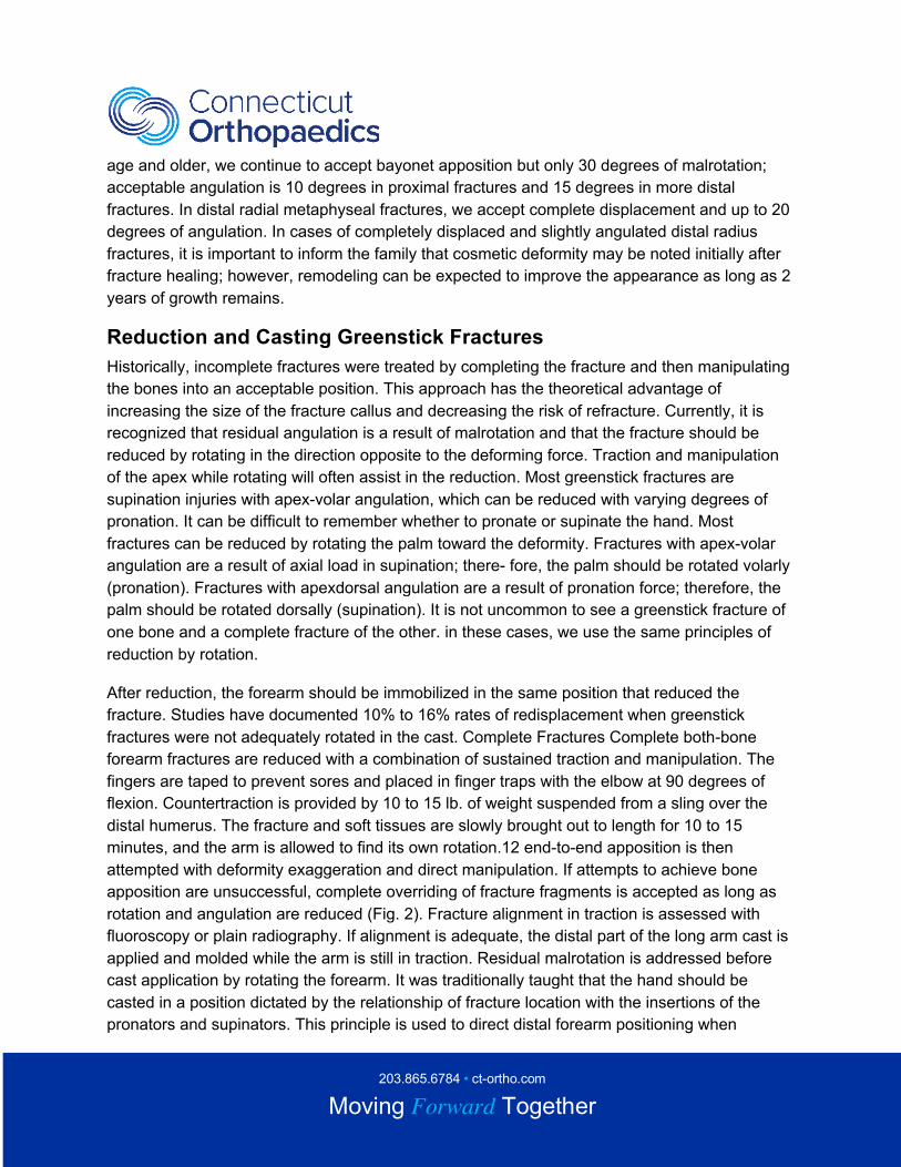
Fig. 2A, Displaced midshaft fracture of the radius and ulna in a girl aged 9 years I month. 2B,
The fracture was reduced in neutral position. Bayonet apposition with minimal angulation and no
203.865.6784 • ct-ortho.com
Moving Forward Together
rotational malalignment was accepted. The fracture united in this position. 2C, Radiographs
obtained 6 years later demonstrate complete remodeling. Clinical examination demonstrated full
range of motion in pronation and supination.
Distal Radius Fractures
Distal radius fractures are reduced with a combination of traction, angulation, and rotation of the
palm in the direction of the angulation. In the case of completely displaced and bayoneted
fractures, sustained longitudinal traction is used with finger traps, as previously described. After
the fracture has been brought out to length, deformity exaggeration and rotation may produce
end-to-end contact. It may be difficult to obtain apposition, as torn periosteum tightens around
the buttonholed proximal fragments (Fig. 1). In these cases, it is acceptable to leave the
fragments overlapped as long as rotation and angulation are reduced (Fig. 3). Typically, these
fractures are immobilized in casts. Sugar-tong splinting is another form of immobilization
commonly used immediately after reduction. If this method is selected, it is important to tighten
the splint or convert to a cast when the initial swelling resolves in 2 or 3 days; high rates of
reangulation in distal radius fractures have been reported. Distal radius fractures without ulnar
fracture are immobilized in a lesser degree of pronation or supination depending on the apex
direction. As these fractures are the result of an angulatory force as well as rotation, the position
of the wrist is less critical. There is some suggestion that distal radius fractures are more stable
in supination because of the action of the brachioradialis.
203.865.6784 • ct-ortho.com
Moving Forward Together
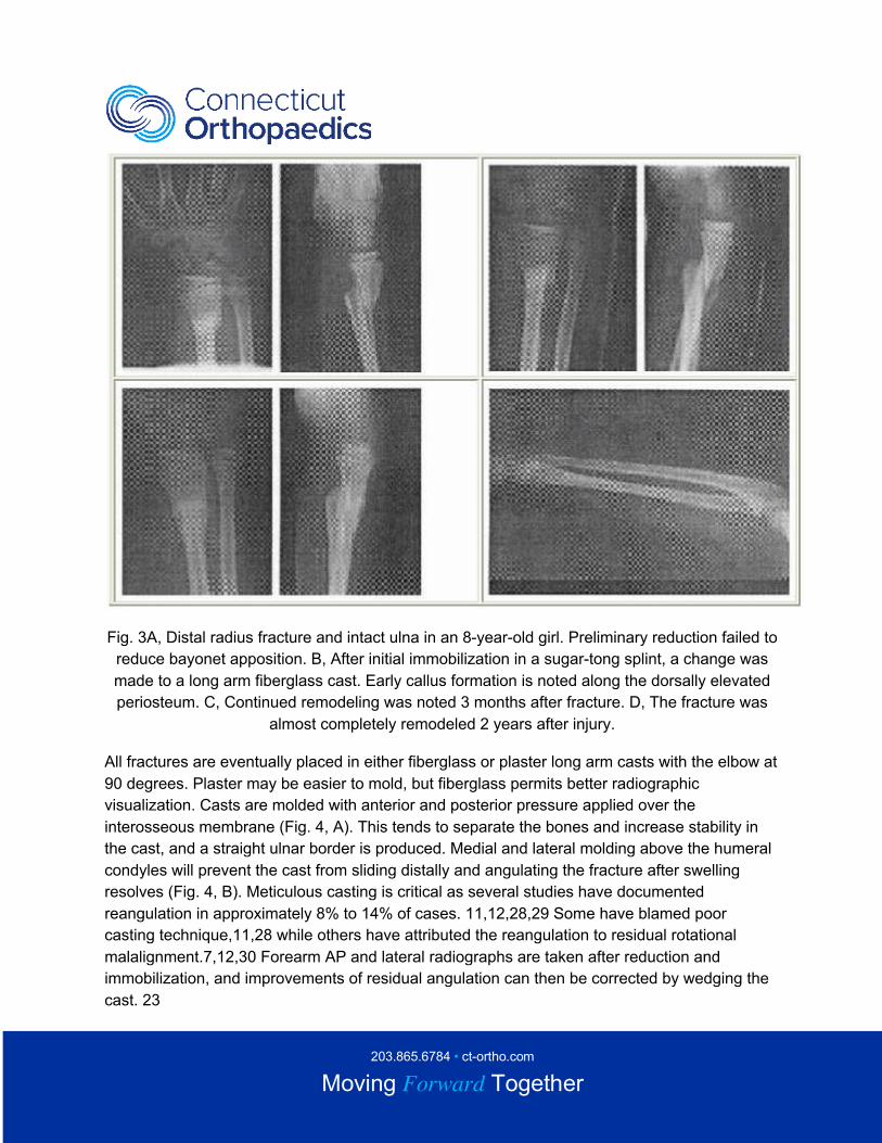
Fig. 3A, Distal radius fracture and intact ulna in an 8-year-old girl. Preliminary reduction failed to
reduce bayonet apposition. B, After initial immobilization in a sugar-tong splint, a change was
made to a long arm fiberglass cast. Early callus formation is noted along the dorsally elevated
periosteum. C, Continued remodeling was noted 3 months after fracture. D, The fracture was
almost completely remodeled 2 years after injury.
All fractures are eventually placed in either fiberglass or plaster long arm casts with the elbow at
90 degrees. Plaster may be easier to mold, but fiberglass permits better radiographic
visualization. Casts are molded with anterior and posterior pressure applied over the
interosseous membrane (Fig. 4, A). This tends to separate the bones and increase stability in
the cast, and a straight ulnar border is produced. Medial and lateral molding above the humeral
condyles will prevent the cast from sliding distally and angulating the fracture after swelling
resolves (Fig. 4, B). Meticulous casting is critical as several studies have documented
reangulation in approximately 8% to 14% of cases. 11,12,28,29 Some have blamed poor
casting technique,11,28 while others have attributed the reangulation to residual rotational
malalignment.7,12,30 Forearm AP and lateral radiographs are taken after reduction and
immobilization, and improvements of residual angulation can then be corrected by wedging the
cast. 23
203.865.6784 • ct-ortho.com
Moving Forward Together
After adequate reduction and immobilization, patients typically return for a follow-up radiograph
1 to 2 weeks after injury. Several studies have documented reangulation during the first 2
weeks. If reangulation is documented, cast removal and re-reduction under general anesthesia
are recommended. Good results of re-reduction have been documented if performed within a
few weeks of the initial fracture. If no reangulation is appreciated, the cast is continued for 6 to 8
weeks or until there is radiographic evidence of healing. Patients cannot participate in contact
sports for 4 to 6 months, but all other activities are permitted. Refractures are uncommon; when
they do occur, it is usually within several months of cast removal.
Copyright © 2020, Connecticut Orthopaedics All rights reserved.


