Embed Size (px)
Citation preview
Cancer Cell
Article
Persistently Activated Stat3Maintains Constitutive NF-kB Activity in TumorsHeehyoung Lee,1 Andreas Herrmann,1 Jie-Hui Deng,1 Maciej Kujawski,1 Guilian Niu,2 Zhiwei Li,2 Steve Forman,1
Richard Jove,1 Drew M. Pardoll,3,* and Hua Yu1,*1Beckman Research Institute, City of Hope National Medical Center, Duarte, CA 91010, USA2H. Lee Moffitt Cancer Center & Research Institute, Tampa, FL 33610, USA3Sidney Kimmel Comprehensive Cancer Center, Johns Hopkins University School of Medicine, Baltimore, MD 20892, USA
*Correspondence: [email protected] (D.M.P.), [email protected] (H.Y.)
DOI 10.1016/j.ccr.2009.02.015
SUMMARY
NF-kB (RelA) is constitutively active in many cancers, where it upregulates antiapoptotic and other oncogenicgenes. While proinflammatory stimulus-induced NF-kB activation involves IKK-dependent nuclear transloca-tion, mechanisms for maintaining constitutive NF-kB activity in tumors have not been elucidated. We showhere that maintenance of NF-kB activity in tumors requires Stat3, which is also frequently constitutively acti-vated in cancer. Stat3 prolongs NF-kB nuclear retention through acetyltransferase p300-mediated RelA acet-ylation, thereby interfering with NF-kB nuclear export. Stat3-mediated maintenance of NF-kB activity occursin both cancer cells and tumor-associated hematopoietic cells. Both murine and human cancers displayhighly acetylated RelA, which is associated with Stat3 activity. This Stat3/NF-kB interaction is thus centralto both the transformed and nontransformed elements in tumors.
INTRODUCTION
NF-kB is a central transcription factor in both innate and adap-
tive immunity. There has been increasing interest in NF-kB’s role
in both cancer initiation (in particular, inflammation-induced
carcinogenesis) and maintenance of established cancer, where
it is frequently constitutively activated and plays a major role in
the transcriptional activation of multiple antiapoptotic and other
oncogenic genes. NF-kB consists of five Rel-related proteins,
and the prototypical NF-kB complex is a RelA/p50 heterodimer,
which is important for NF-kB-mediated antiapoptotic effects
(Karin et al., 2002). In the absence of appropriate stimuli, NF-kB
is sequestered in the cytoplasm by IkBa protein. IkB kinases
(IKKs) are activated upon stimulation of Toll-like receptors and
intracellular sensors such as RIG-I, MDA-5, and NOD1/2 by
various pathogen-associated molecular patterns (PAMPs) or
proinflammatory cytokines such as TNF, leading to serine phos-
phorylation of IkBa and its subsequent proteasome-mediated
degradation, which is critical for NF-kB nuclear translocation
(Ghosh et al., 1998). An important role of IKKb-dependent
NF-kB activation involving RelA/p50 has been documented
both during pathogen infection and in cancers caused by
chronic inflammation and other stimuli (Naugler et al., 2007;
Stancovski and Baltimore, 1997). However, with only a few
exceptions, such as lymphoid tumors, where activating muta-
tions of upstream IKK activators such as CARD11 have been
identified (Lenz et al., 2008; Pomerantz et al., 2002), IKK is not
often continuously activated in cultured cancer cells but rather
is inducible by proinflammatory stimuli. Several studies have
provided evidence that secretion of cytokines and growth
factors, many of which are encoded by NF-kB target genes, is
critical for constitutive activation of NF-kB in cancer cells (Lu
et al., 2004; Lu and Stark, 2004). Since IKK is not often consti-
tutively activated in tumors, the question remains whether one
or more additional mechanisms involving signaling pathways
or molecules downstream of some of these cytokines and
growth factors might directly contribute to constitutive activa-
tion of NF-kB.
SIGNIFICANCE
Development of innate and adaptive immunity in response to proinflammatory stimuli requires induction of NF-kB, whichinvolves its nuclear translocation followed by expression of proinflammation/immunity-related genes. In contrast, NF-kBcan be constitutively activated without continuous proinflammatory stimuli in cancer cells, where it serves a very differentrole: upregulating genes necessary for tumor progression. How NF-kB stays constitutively active in cancer remains to befully defined. The current work reveals a mechanism whereby constitutively activated Stat3 maintains constitutive NF-kBactivity in cancers by inhibiting its export from the nucleus. This Stat3/NF-kB interaction observed in cancer providesinsights into carcinogenesis and strategies for developing cancer therapeutics.
Cancer Cell 15, 283–293, April 7, 2009 ª2009 Elsevier Inc. 283
Cancer Cell
Stat3 and NF-kB Interactions in Tumors
It has been suggested that NF-kB/IkBa can shuttle in and out of
the nucleus in the absence of stimuli, although the rate of nuclear
export is greater than the rate of nuclear import (Huang et al.,
2000). Recent studies have demonstrated that the amplitude
and half-life of nuclear NF-kB can be influenced by acetylation
of RelA (Chen and Greene, 2004), which requires prior RelA phos-
phorylation (Chen et al., 2005). In particular, endogenous RelA is
acetylated in a signal-coupled manner following stimulation
(Chen et al., 2001, 2002; Chen and Greene, 2004). The p300/
CBP cofactors are acetyltransferases that mediate RelA acetyla-
tion, which is subject to deacetylation by histone deacetylases
(HDACs) (Chen et al., 2001). Reversible acetylation of RelA is
essential for the duration of NF-kB activity, due to its role in regu-
lating the assembly of RelA/IkBa complexes necessary for RelA
nuclear export and its presence in the cytoplasm. Acetylated
RelA interacts only weakly with IkBa, while deacetylation of RelA
by HDACs markedly increases the binding of RelA to IkBa (Chen
et al., 2001; Chen and Greene, 2004). Although RelA acetylation
has been studied in the context of inflammatory stimuli, whether
it has a role in constitutive NF-kB activation in cancer is unknown.
Recent research has documented the importance of cytokines
and growth factors secreted by tumor cells, which often depend
on IKK-mediated NF-kB activation for their production, as a caus-
ative factor in constitutive NF-kB activation in cancer cells and
tumors (Greten et al., 2004; Lu et al., 2004; Lu and Stark, 2004).
Some of these cytokines and growth factors, such as inter-
leukin-6 (IL-6) and fibroblast growth factor (FGF), are activators
of signal transducer and activator of transcription 3 (Stat3) (Deo
et al., 2002; Zhong et al., 1994). Stat3 is a transcription factor
that can promote oncogenesis (Bromberg et al., 1999), and it
is commonly activated in cancer (Darnell, 2002; Yu and Jove,
2004) as well as in tumor-associated myeloid cells (Kortylewski
et al., 2005; Kujawski et al., 2008). Stat3 and NF-kB stimulate
a highly overlapping repertoire of prosurvival, proliferative, and
proangiogenic genes (Catlett-Falcone et al., 1999; Darnell, 2002;
Lo et al., 2005; Yu and Jove, 2004). A recent study further demon-
strated that Stat3 interaction with RelA leads to upregulation of
the immunosuppressive IL-23/p19 gene (Kortylewski et al.,
2009). Although Stat3 has been implicated in inhibiting IKK activity
in normal immune cells (Welte et al., 2003), whether constitutively
activated Stat3 and RelA directly interact in both cancer cells
and immune cells within the tumor microenvironment remains
unknown. In the current study, we explored the possibility that
constitutively activated Stat3 maintains NF-kB activity in tumors.
RESULTS
Stat3 Is Required to Maintain Tumor NF-kB ActivityIt has been shown that phospho-IkBa (p-IkBa) levels are
increased in Stat3�/� dendritic cells (DCs), suggesting that
Stat3 signaling inhibits IKK activity in the context of normal
immune responses (Welte et al., 2003). We confirmed that
Stat3 negatively regulates IKK activity in normal immune cells,
by determining the ratio of p-IkBa to IkBa in splenic cells with
or without Stat3. Generation of mice containing a functional dele-
tion of Stat3 alleles in the myeloid compartment has been
described previously (Lee et al., 2002). Data from these experi-
ments showed that the p-IkBa/IkBa ratio was higher in Stat3-
deficient splenocytes relative to their wild-type (WT) counter-
284 Cancer Cell 15, 283–293, April 7, 2009 ª2009 Elsevier Inc.
parts (see Figure S1A available online). Using in vitro IKK kinase
assays, we further demonstrated that in tumor cells with both
constitutive Stat3 and NF-kB activity, such as human A2058
melanoma and DU145 prostate cancer cell lines, blocking
Stat3 by a small-molecule Stat3 inhibitor (Turkson et al., 2004)
or siRNA also increased IkBa phosphorylation by IKK, which
was further upregulated by TNFa (Figure S1B).
Our data and that of others (Welte et al., 2003) suggest that
Stat3 signaling has inhibitory effects on stimulus-induced IKK in
both immune and tumor cells. However, these findings raise the
question of how constitutively activated Stat3 and NF-kB can
coexist in cancer cells. When we examined growing B16 mela-
noma tumors for NF-kB activity, we found that tumor RelA was
constitutively bound to its consensus DNA sequence as deter-
mined by EMSA (Figure 1A, left); we also observed increased
RelA (Ser536) and Stat3 (Tyr705) phosphorylation. B16 tumors
growing in mice with Stat3�/� myeloid cells displayed reduced
Stat3 activity (Figure 1A, left), likely due to the interruption of
Stat3-mediated crosstalk between tumor cells and tumor stromal
myeloid cells (Kortylewski et al., 2005; Kujawski et al., 2008). To
our surprise, there was little constitutive RelA activity in B16
tumors when Stat3 activity was abrogated (Figure 1A, left). These
observations suggested a possible requirement of Stat3 for
constitutive RelA activity in tumors. The human A2058 melanoma
cell line represents a typical example of a tumor with both consti-
tutive Stat3 (Niu et al., 2002) and NF-kB activity (Figure 1A, right).
Similar to in B16 tumors, when Stat3 is silenced in A2058 tumor
cells, NF-kB (RelA) activity is greatly diminished in the tumor cells.
These results obtained from both murine tumors and human
tumor cells are the opposite of what would be predicted if the
primary effect of Stat3 on the NF-kB pathway in tumors were inhi-
bition of IKK, and if IKK were mainly responsible for maintaining
constitutive NF-kB activity in tumor cells. In contrast to Stat3
siRNA treatment, silencing IKKb alone did not affect nuclear
NF-kB activity over a 48 hr period (Figure 1A). Similarly, knocking
down IKKa alone had no effect on NF-kB in tumor cells (data not
shown). However, silencing both Stat3 and IKKb with siRNA
greatly diminished NF-kB activity in cancer cells (Figure 1A).
While these results indicated that maintenance of existing consti-
tutive NF-kB activity in tumors depends more on Stat3 than IKK
activity, they did not rule out the need for IKK to initiate NF-kB
activation by facilitating its nuclear translocation. They nonethe-
less reveal an important IKK-independent downstream mecha-
nism for enhanced NF-kB activity in tumors.
Given our data suggesting that Stat3 also inhibits IKK (Figures
S1A and S1B), we investigated whether the effect of Stat3 on
NF-kB activity depended on NF-kB activity being driven by
a proinflammatory stimulus (and was thus dependent on IKK
activity) or whether it was constitutively activated, independent
of immune stimuli. A2058 tumor cells display relatively high
RelA activity, which is difficult to further increase by additional
proinflammatory stimuli. However, DU145 prostate cancer cells
have lower RelA activity (Figure 1B) and were thus used to confirm
our findings that Stat3 facilitates maintenance of constitutive NF-
kB activity in tumor cells, and to further test the role of Stat3 in in-
hibiting inflammatory signal-induced NF-kB activity. As in A2058
tumor cells, siRNA knockdown of Stat3, but not siRNA knock-
down of IKKb, resulted in the reduction of endogenous RelA
activity in DU145 tumor cells (Figure 1B). The immunostimulatory
Cancer Cell
Stat3 and NF-kB Interactions in Tumors
C
α++
−−
κ α
β
β
α
β
κ
β
β
α
β β β
− α
κ
A
++
−−
++
α
β
B
β
β
β β β β
− α
Cancer Cell 15, 283
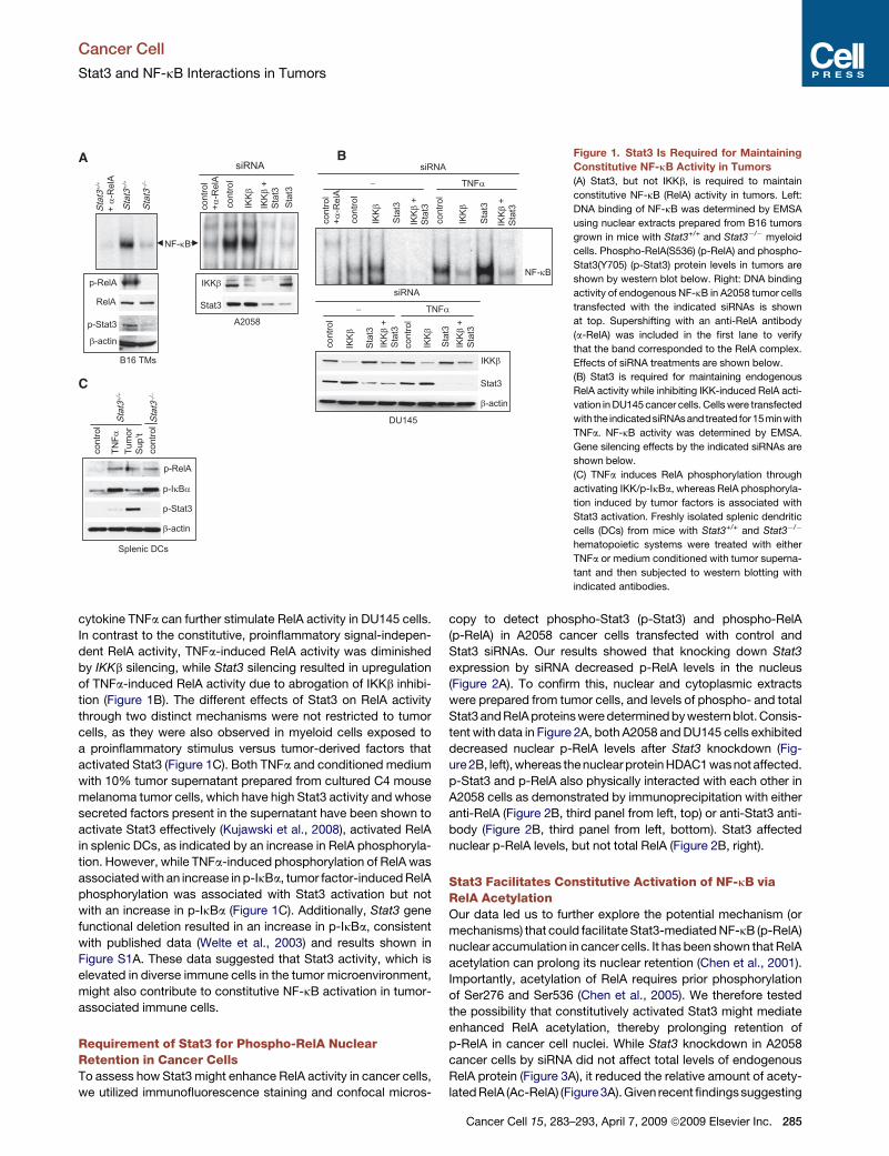
Figure 1. Stat3 Is Required for Maintaining
Constitutive NF-kB Activity in Tumors
(A) Stat3, but not IKKb, is required to maintain
constitutive NF-kB (RelA) activity in tumors. Left:
DNA binding of NF-kB was determined by EMSA
using nuclear extracts prepared from B16 tumors
grown in mice with Stat3+/+ and Stat3�/� myeloid
cells. Phospho-RelA(S536) (p-RelA) and phospho-
Stat3(Y705) (p-Stat3) protein levels in tumors are
shown by western blot below. Right: DNA binding
activity of endogenous NF-kB in A2058 tumor cells
transfected with the indicated siRNAs is shown
at top. Supershifting with an anti-RelA antibody
(a-RelA) was included in the first lane to verify
that the band corresponded to the RelA complex.
Effects of siRNA treatments are shown below.
(B) Stat3 is required for maintaining endogenous
RelA activity while inhibiting IKK-induced RelA acti-
vation in DU145 cancer cells. Cells were transfected
with the indicatedsiRNAs and treated for15minwith
TNFa. NF-kB activity was determined by EMSA.
Gene silencing effects by the indicated siRNAs are
shown below.
(C) TNFa induces RelA phosphorylation through
activating IKK/p-IkBa, whereas RelA phosphoryla-
tion induced by tumor factors is associated with
Stat3 activation. Freshly isolated splenic dendritic
cells (DCs) from mice with Stat3+/+ and Stat3�/�
hematopoietic systems were treated with either
TNFa or medium conditioned with tumor superna-
tant and then subjected to western blotting with
indicated antibodies.
cytokine TNFa can further stimulate RelA activity in DU145 cells.
In contrast to the constitutive, proinflammatory signal-indepen-
dent RelA activity, TNFa-induced RelA activity was diminished
by IKKb silencing, while Stat3 silencing resulted in upregulation
of TNFa-induced RelA activity due to abrogation of IKKb inhibi-
tion (Figure 1B). The different effects of Stat3 on RelA activity
through two distinct mechanisms were not restricted to tumor
cells, as they were also observed in myeloid cells exposed to
a proinflammatory stimulus versus tumor-derived factors that
activated Stat3 (Figure 1C). Both TNFa and conditioned medium
with 10% tumor supernatant prepared from cultured C4 mouse
melanoma tumor cells, which have high Stat3 activity and whose
secreted factors present in the supernatant have been shown to
activate Stat3 effectively (Kujawski et al., 2008), activated RelA
in splenic DCs, as indicated by an increase in RelA phosphoryla-
tion. However, while TNFa-induced phosphorylation of RelA was
associated with an increase in p-IkBa, tumor factor-induced RelA
phosphorylation was associated with Stat3 activation but not
with an increase in p-IkBa (Figure 1C). Additionally, Stat3 gene
functional deletion resulted in an increase in p-IkBa, consistent
with published data (Welte et al., 2003) and results shown in
Figure S1A. These data suggested that Stat3 activity, which is
elevated in diverse immune cells in the tumor microenvironment,
might also contribute to constitutive NF-kB activation in tumor-
associated immune cells.
Requirement of Stat3 for Phospho-RelA NuclearRetention in Cancer CellsTo assess how Stat3 might enhance RelA activity in cancer cells,
we utilized immunofluorescence staining and confocal micros-
copy to detect phospho-Stat3 (p-Stat3) and phospho-RelA
(p-RelA) in A2058 cancer cells transfected with control and
Stat3 siRNAs. Our results showed that knocking down Stat3
expression by siRNA decreased p-RelA levels in the nucleus
(Figure 2A). To confirm this, nuclear and cytoplasmic extracts
were prepared from tumor cells, and levels of phospho- and total
Stat3 and RelA proteins were determined by western blot. Consis-
tent with data in Figure 2A, both A2058 and DU145 cells exhibited
decreased nuclear p-RelA levels after Stat3 knockdown (Fig-
ure2B, left), whereas the nuclear protein HDAC1 wasnot affected.
p-Stat3 and p-RelA also physically interacted with each other in
A2058 cells as demonstrated by immunoprecipitation with either
anti-RelA (Figure 2B, third panel from left, top) or anti-Stat3 anti-
body (Figure 2B, third panel from left, bottom). Stat3 affected
nuclear p-RelA levels, but not total RelA (Figure 2B, right).
Stat3 Facilitates Constitutive Activation of NF-kB viaRelA AcetylationOur data led us to further explore the potential mechanism (or
mechanisms) that could facilitate Stat3-mediated NF-kB (p-RelA)
nuclear accumulation in cancer cells. It has been shown that RelA
acetylation can prolong its nuclear retention (Chen et al., 2001).
Importantly, acetylation of RelA requires prior phosphorylation
of Ser276 and Ser536 (Chen et al., 2005). We therefore tested
the possibility that constitutively activated Stat3 might mediate
enhanced RelA acetylation, thereby prolonging retention of
p-RelA in cancer cell nuclei. While Stat3 knockdown in A2058
cancer cells by siRNA did not affect total levels of endogenous
RelA protein (Figure 3A), it reduced the relative amount of acety-
lated RelA (Ac-RelA) (Figure 3A). Given recent findings suggesting
–293, April 7, 2009 ª2009 Elsevier Inc. 285
Cancer Cell
Stat3 and NF-kB Interactions in Tumors
that NF-kB p50 is an acetylated protein (Chen and Greene, 2003;
Deng and Wu, 2003), we tested whether p50 acetylation was also
regulated by Stat3. Compared to RelA, the p50 acetylation level
was relatively low in A2058 tumor cells and was not affected by
Stat3 knockdown (Figure S2).
These findings prompted us to further investigate a potential
role of Stat3 in regulating RelA acetylation. Trichostatin A (TSA),
a selective inhibitor of multiple HDACs, has been shown to
increase acetylation of RelA and inhibit its interaction with IkBa
(Chen et al., 2001). Treatment of A2058 melanoma cells with
TSA increased the proportion of acetylated RelA (Figure 3B).
Moreover, the presence of Stat3C, a constitutively activated
Stat3 mutant (Bromberg et al., 1999), led to further enhancement
of RelA acetylation accompanied by the loss of RelA/IkBa
complexes, as shown by the absence of IkBa protein in RelA
complexes assessed by immunoprecipitation (Figure 3B). This
A
B
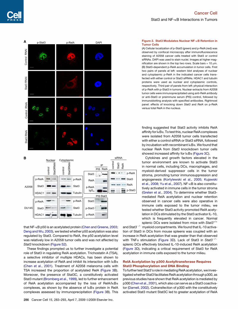
Figure 2. Stat3 Modulates Nuclear NF-kB Retention in
Tumor Cells
(A) Cellular localization of p-Stat3 (green) and p-RelA (red) was
observed by confocal microscopy after immunofluorescence
staining of A2058 cancer cells treated with Stat3 or control
siRNAs. DAPI was used to stain nuclei. Images at higher mag-
nification are shown in the top two rows. Scale bars = 10 mm.
(B) Stat3-dependent p-RelA accumulation in tumor cells. First
two pairs of panels at left: western blot analyses of nuclear
and cytoplasmic p-RelA in the indicated cancer cells trans-
fected with either control or Stat3 siRNAs. HDAC1 and tubulin
proteins were used as nuclear and cytoplasmic controls,
respectively. Third pair of panels from left: physical interaction
of p-RelA with p-Stat3 in tumors. Nuclear extracts from A2058
tumor cells were immunoprecipitated using anti-RelA antibody
or anti-Stat3 or preimmune serum (PIS) control, followed by
immunoblotting analysis with specified antibodies. Rightmost
panel: effects of knocking down Stat3 and RelA on p-RelA
versus total RelA in the nucleus.
finding suggested that Stat3 activity inhibits RelA
affinity for IkBa. To test this, nuclear RelA complexes
were isolated from A2058 tumor cells transfected
with either a control siRNA or Stat3 siRNA, followed
by incubation with recombinant IkBa. We found that
nuclear RelA from Stat3 knockdown tumor cells
showed increased affinity for IkBa (Figure 3C).
Cytokines and growth factors elevated in the
tumor environment are known to activate Stat3
in normal cells, including DCs, macrophages, and
myeloid-derived suppressor cells in the tumor
stroma, promoting tumor immunosuppression and
angiogenesis (Kortylewski et al., 2005; Kujawski
et al., 2008; Yu et al., 2007). NF-kB is also constitu-
tively activated in immune cells in the tumor stroma
(Greten et al., 2004). To determine whether Stat3-
mediated RelA acetylation and nuclear retention
observed in cancer cells were also operative in
immune cells exposed to the tumor milieu, we
tested whether Stat3 activity promoted RelA acety-
lation in DCs stimulated by the Stat3 activator IL-10,
which is frequently elevated in cancer. Normal
splenic DCs were isolated from mice with Stat3+/+
and Stat3�/�myeloid compartments. We found that IL-10 activa-
tion of Stat3 in DCs from mouse spleens was coupled with an
increase in RelA acetylation that was greater than that observed
with TNFa stimulation (Figure 3D). Lack of Stat3 in Stat3�/�
splenic DCs effectively blocked IL-10-induced RelA acetylation
(Figure 3D), indicating a critical requirement of Stat3 for RelA
acetylation in immune cells exposed to the tumor milieu.
RelA Acetylation by p300 Acetyltransferase RequiresStat3 Phosphorylation and DNA BindingTo further test Stat3’s role in mediating RelA acetylation, we inves-
tigated whetherStat3 facilitates RelA acetylation through p300, as
previous studies have shown that RelA acetylation is mediated by
p300 (Chen et al., 2001), which also can serve as a Stat3 coactiva-
tor (Darnell, 2002). Cotransfection of p300 with the constitutively
activated Stat3 mutant Stat3C led to greater acetylation of RelA
286 Cancer Cell 15, 283–293, April 7, 2009 ª2009 Elsevier Inc.
Cancer Cell
Stat3 and NF-kB Interactions in Tumors
than transfection of either one alone (Figure 4A, left). Coexpres-
sion of Stat3C and p300 in 3T3 fibroblasts also resulted in
increased nuclear RelA levels (data not shown). Furthermore,
levels of p300 and acetylated RelA in the complex were Stat3
dependent in both B16 melanoma (Figure 4A, right) and A2058
cancer cells (data not shown). To demonstrate that RelA activity
in tumors was dependent upon the interaction with Stat3 and
p300, we performed NF-kB DNA binding assays using an NF-kB
DNA-binding oligonucleotide and nuclear extracts prepared from
growing B16 melanoma tumors. Constitutive NF-kB activity in
tumors was blocked by preincubation with either Stat3 or p300
antibody (Figure 4B), but not by anti-c-Rel antibody, suggesting
that constitutive activation of the NF-kB complex in tumors
involves both Stat3 and p300 (Figure 4B, upper left). To further
confirm that the NF-kB DNA-binding complex contained Stat3
and p300, an oligo binding assay using biotin-labeled NF-kB
DNA-binding sequences was performed. After incubation with
nuclear extract, NF-kB complexes were pulled down by strepta-
vidin-conjugated magnetic beads, followed by western blot anal-
C
B
−−−
−−+
+−+
+++
κ α
κ α
A
β
D
α α
+ + − −
β
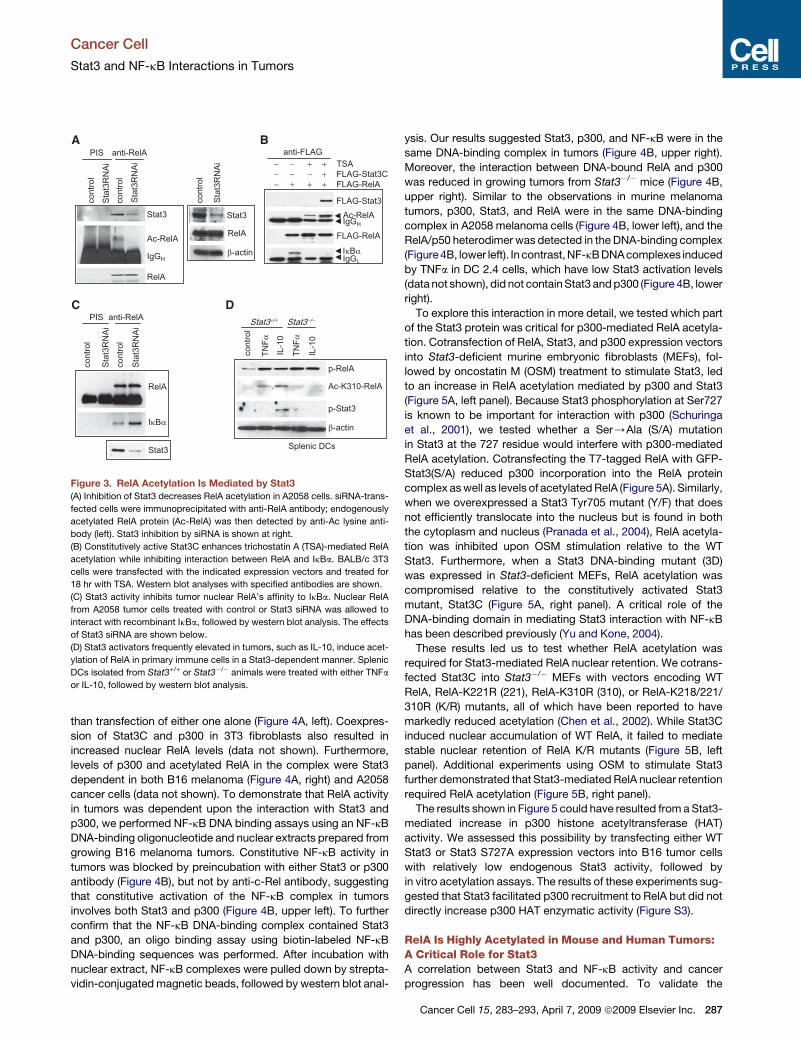
Figure 3. RelA Acetylation Is Mediated by Stat3
(A) Inhibition of Stat3 decreases RelA acetylation in A2058 cells. siRNA-trans-
fected cells were immunoprecipitated with anti-RelA antibody; endogenously
acetylated RelA protein (Ac-RelA) was then detected by anti-Ac lysine anti-
body (left). Stat3 inhibition by siRNA is shown at right.
(B) Constitutively active Stat3C enhances trichostatin A (TSA)-mediated RelA
acetylation while inhibiting interaction between RelA and IkBa. BALB/c 3T3
cells were transfected with the indicated expression vectors and treated for
18 hr with TSA. Western blot analyses with specified antibodies are shown.
(C) Stat3 activity inhibits tumor nuclear RelA’s affinity to IkBa. Nuclear RelA
from A2058 tumor cells treated with control or Stat3 siRNA was allowed to
interact with recombinant IkBa, followed by western blot analysis. The effects
of Stat3 siRNA are shown below.
(D) Stat3 activators frequently elevated in tumors, such as IL-10, induce acet-
ylation of RelA in primary immune cells in a Stat3-dependent manner. Splenic
DCs isolated from Stat3+/+ or Stat3�/� animals were treated with either TNFa
or IL-10, followed by western blot analysis.
ysis. Our results suggested Stat3, p300, and NF-kB were in the
same DNA-binding complex in tumors (Figure 4B, upper right).
Moreover, the interaction between DNA-bound RelA and p300
was reduced in growing tumors from Stat3�/� mice (Figure 4B,
upper right). Similar to the observations in murine melanoma
tumors, p300, Stat3, and RelA were in the same DNA-binding
complex in A2058 melanoma cells (Figure 4B, lower left), and the
RelA/p50 heterodimer was detected in the DNA-binding complex
(Figure 4B, lower left). In contrast, NF-kB DNA complexes induced
by TNFa in DC 2.4 cells, which have low Stat3 activation levels
(data not shown), did not contain Stat3 and p300 (Figure 4B, lower
right).
To explore this interaction in more detail, we tested which part
of the Stat3 protein was critical for p300-mediated RelA acetyla-
tion. Cotransfection of RelA, Stat3, and p300 expression vectors
into Stat3-deficient murine embryonic fibroblasts (MEFs), fol-
lowed by oncostatin M (OSM) treatment to stimulate Stat3, led
to an increase in RelA acetylation mediated by p300 and Stat3
(Figure 5A, left panel). Because Stat3 phosphorylation at Ser727
is known to be important for interaction with p300 (Schuringa
et al., 2001), we tested whether a Ser/Ala (S/A) mutation
in Stat3 at the 727 residue would interfere with p300-mediated
RelA acetylation. Cotransfecting the T7-tagged RelA with GFP-
Stat3(S/A) reduced p300 incorporation into the RelA protein
complex as well as levels of acetylated RelA (Figure 5A). Similarly,
when we overexpressed a Stat3 Tyr705 mutant (Y/F) that does
not efficiently translocate into the nucleus but is found in both
the cytoplasm and nucleus (Pranada et al., 2004), RelA acetyla-
tion was inhibited upon OSM stimulation relative to the WT
Stat3. Furthermore, when a Stat3 DNA-binding mutant (3D)
was expressed in Stat3-deficient MEFs, RelA acetylation was
compromised relative to the constitutively activated Stat3
mutant, Stat3C (Figure 5A, right panel). A critical role of the
DNA-binding domain in mediating Stat3 interaction with NF-kB
has been described previously (Yu and Kone, 2004).
These results led us to test whether RelA acetylation was
required for Stat3-mediated RelA nuclear retention. We cotrans-
fected Stat3C into Stat3�/� MEFs with vectors encoding WT
RelA, RelA-K221R (221), RelA-K310R (310), or RelA-K218/221/
310R (K/R) mutants, all of which have been reported to have
markedly reduced acetylation (Chen et al., 2002). While Stat3C
induced nuclear accumulation of WT RelA, it failed to mediate
stable nuclear retention of RelA K/R mutants (Figure 5B, left
panel). Additional experiments using OSM to stimulate Stat3
further demonstrated that Stat3-mediated RelA nuclear retention
required RelA acetylation (Figure 5B, right panel).
The results shown in Figure 5 could have resulted from a Stat3-
mediated increase in p300 histone acetyltransferase (HAT)
activity. We assessed this possibility by transfecting either WT
Stat3 or Stat3 S727A expression vectors into B16 tumor cells
with relatively low endogenous Stat3 activity, followed by
in vitro acetylation assays. The results of these experiments sug-
gested that Stat3 facilitated p300 recruitment to RelA but did not
directly increase p300 HAT enzymatic activity (Figure S3).
RelA Is Highly Acetylated in Mouse and Human Tumors:A Critical Role for Stat3A correlation between Stat3 and NF-kB activity and cancer
progression has been well documented. To validate the
Cancer Cell 15, 283–293, April 7, 2009 ª2009 Elsevier Inc. 287
Cancer Cell
Stat3 and NF-kB Interactions in Tumors
importance of Stat3-mediated RelA acetylation in cancer, we
utilized tumor-associated myeloid cells to examine whether
Stat3 was required for RelA acetylation in vivo. While CD11b+
myeloid cells isolated from B16 tumors contained both phos-
phorylated and acetylated RelA, in vivo targeted functional
deletion of Stat3 in the myeloid compartment diminished levels
of both phospho-RelA and acetylated RelA (Figure 6A). Further-
more, immunohistochemical analyses confirmed the heavy pres-
ence of acetylated RelA in B16 tumors, mainly in the nuclear
compartment (Figure 6B, left panels). In contrast, the level of
acetylated RelA was greatly diminished in whole B16 tumors
with minimal Stat3 activity (Figure 6B, right panels), due to
Stat3 ablation in the tumor-infiltrating myeloid cells (Figure 1A,
left panel). Of the eight slides examined, only one section of
B16 tumor tissue from a mouse with a Stat3�/�myeloid compart-
ment exhibited detectable (but low) levels of acetylated RelA
(data not shown). Other sections did not exhibit detectable acet-
ylated RelA (Figure 6B, right panels). These results emphasize
that the Stat3-dependent NF-kB acetylation/nuclear retention
described here is not totally cell-autonomous, but rather that
crosstalk between tumor cells and nontransformed hematopoi-
etic cells in the tumor microenvironment is particularly important
to amplify this interaction in multiple cellular components within
the tumor in vivo.
In order to determine whether our findings regarding Stat3-
mediated RelA acetylation are important for human cancers, we
next analyzed human tumors and normal tissues for phospho-
RelA, acetylated RelA, and phospho-Stat3. Malignant human
tissues of different origins were subject to immunohistochemical
staining and confocal microscopic analyses. We used sections
+−+
−++
+++
− +−
A
αα α α
κ
B
++
−−
κ
αα α α
α
α
αααα α
κ
β
+
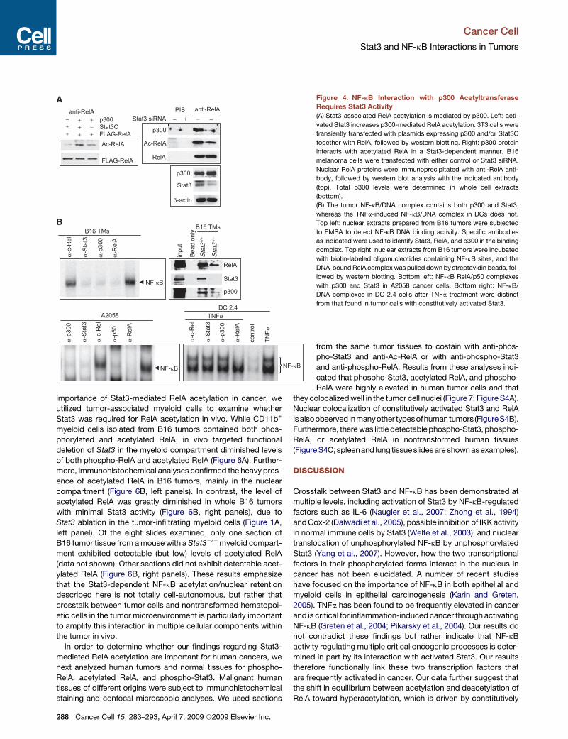
Figure 4. NF-kB Interaction with p300 Acetyltransferase
Requires Stat3 Activity
(A) Stat3-associated RelA acetylation is mediated by p300. Left: acti-
vated Stat3 increases p300-mediated RelA acetylation. 3T3 cells were
transiently transfected with plasmids expressing p300 and/or Stat3C
together with RelA, followed by western blotting. Right: p300 protein
interacts with acetylated RelA in a Stat3-dependent manner. B16
melanoma cells were transfected with either control or Stat3 siRNA.
Nuclear RelA proteins were immunoprecipitated with anti-RelA anti-
body, followed by western blot analysis with the indicated antibody
(top). Total p300 levels were determined in whole cell extracts
(bottom).
(B) The tumor NF-kB/DNA complex contains both p300 and Stat3,
whereas the TNFa-induced NF-kB/DNA complex in DCs does not.
Top left: nuclear extracts prepared from B16 tumors were subjected
to EMSA to detect NF-kB DNA binding activity. Specific antibodies
as indicated were used to identify Stat3, RelA, and p300 in the binding
complex. Top right: nuclear extracts from B16 tumors were incubated
with biotin-labeled oligonucleotides containing NF-kB sites, and the
DNA-bound RelA complex was pulled down by streptavidin beads, fol-
lowed by western blotting. Bottom left: NF-kB RelA/p50 complexes
with p300 and Stat3 in A2058 cancer cells. Bottom right: NF-kB/
DNA complexes in DC 2.4 cells after TNFa treatment were distinct
from that found in tumor cells with constitutively activated Stat3.
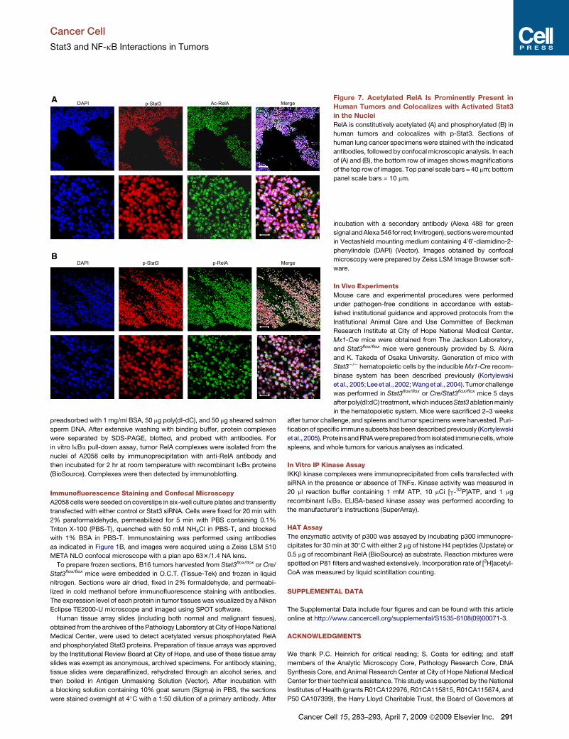
from the same tumor tissues to costain with anti-phos-
pho-Stat3 and anti-Ac-RelA or with anti-phospho-Stat3
and anti-phospho-RelA. Results from these analyses indi-
cated that phospho-Stat3, acetylated RelA, and phospho-
RelA were highly elevated in human tumor cells and that
they colocalized well in the tumor cell nuclei (Figure 7; Figure S4A).
Nuclear colocalization of constitutively activated Stat3 and RelA
isalsoobserved inmanyother typesofhumantumors (FigureS4B).
Furthermore, there was little detectable phospho-Stat3, phospho-
RelA, or acetylated RelA in nontransformed human tissues
(FigureS4C;spleenand lungtissueslidesare shown asexamples).
DISCUSSION
Crosstalk between Stat3 and NF-kB has been demonstrated at
multiple levels, including activation of Stat3 by NF-kB-regulated
factors such as IL-6 (Naugler et al., 2007; Zhong et al., 1994)
and Cox-2 (Dalwadi et al., 2005), possible inhibition of IKK activity
in normal immune cells by Stat3 (Welte et al., 2003), and nuclear
translocation of unphosphorylated NF-kB by unphosphorylated
Stat3 (Yang et al., 2007). However, how the two transcriptional
factors in their phosphorylated forms interact in the nucleus in
cancer has not been elucidated. A number of recent studies
have focused on the importance of NF-kB in both epithelial and
myeloid cells in epithelial carcinogenesis (Karin and Greten,
2005). TNFa has been found to be frequently elevated in cancer
and is critical for inflammation-induced cancer through activating
NF-kB (Greten et al., 2004; Pikarsky et al., 2004). Our results do
not contradict these findings but rather indicate that NF-kB
activity regulating multiple critical oncogenic processes is deter-
mined in part by its interaction with activated Stat3. Our results
therefore functionally link these two transcription factors that
are frequently activated in cancer. Our data further suggest that
the shift in equilibrium between acetylation and deacetylation of
RelA toward hyperacetylation, which is driven by constitutively
288 Cancer Cell 15, 283–293, April 7, 2009 ª2009 Elsevier Inc.
Cancer Cell
Stat3 and NF-kB Interactions in Tumors
activated Stat3, contributes to NF-kB activation in both tumor
cells and the tumor microenvironment. Our studies also reconcile
the roles of NF-kB and Stat3 in mediating the complex interac-
tions between the tumor and its immune microenvironment.
The data generated by expressing various Stat3 mutants in
MEFs lacking intact Stat3 alleles suggest that Stat3-mediated
RelA acetylation requires serine (727) and tyrosine (705) phos-
phorylation, as well as the DNA-binding domain of Stat3 protein.
The reason that serine phosphorylation is important for RelA
acetylation is likely that it is the critical site for Stat3 interaction
with p300 (Schuringa et al., 2001). It has been documented
that both interaction of RelA with p300 and acetylation of RelA
by p300 require phosphorylation of RelA (Chen et al., 2005).
Because unphosphorylated Stat3 (Y705F) preferentially inter-
acts with unphosphorylated RelA (Yang et al., 2007), it is plau-
sible that only phosphorylated Stat3 is able to interact with
p300/phosphorylated RelA efficiently, leading to RelA acetyla-
tion. As for why mutation of the DNA-binding domain of Stat3
inhibits its ability to activate NF-kB, it has been reported that
the Stat3 DNA-binding domain is critical for mediating interac-
tion with RelA (Yu and Kone, 2004). Our data are consistent
with these reports in that S/A and Y/F Stat3 proteins were able
to interact with RelA but Stat3D was not (Figure 5A). At the
same time, these Stat3 mutants, which do not efficiently interact
with p300, RelA, or phosphorylated RelA, were not able to facil-
itate RelA acetylation. Although our results suggest an important
role of p300 in facilitating acetylation of phosphorylated RelA, it is
possible that other acetyltransferases, such as Tip60 or NcoA/
SRC1, which interact with Stat3 (Giraud et al., 2002; Xiao et al.,
2003), could also contribute to RelA acetylation.
A
+−+
+−+
+++
+++
+−+
+++
+−+−
+−−
++
++
++−
++
++−−
++−
+++
+++
+++
+++
+−
++
++
++
++
B
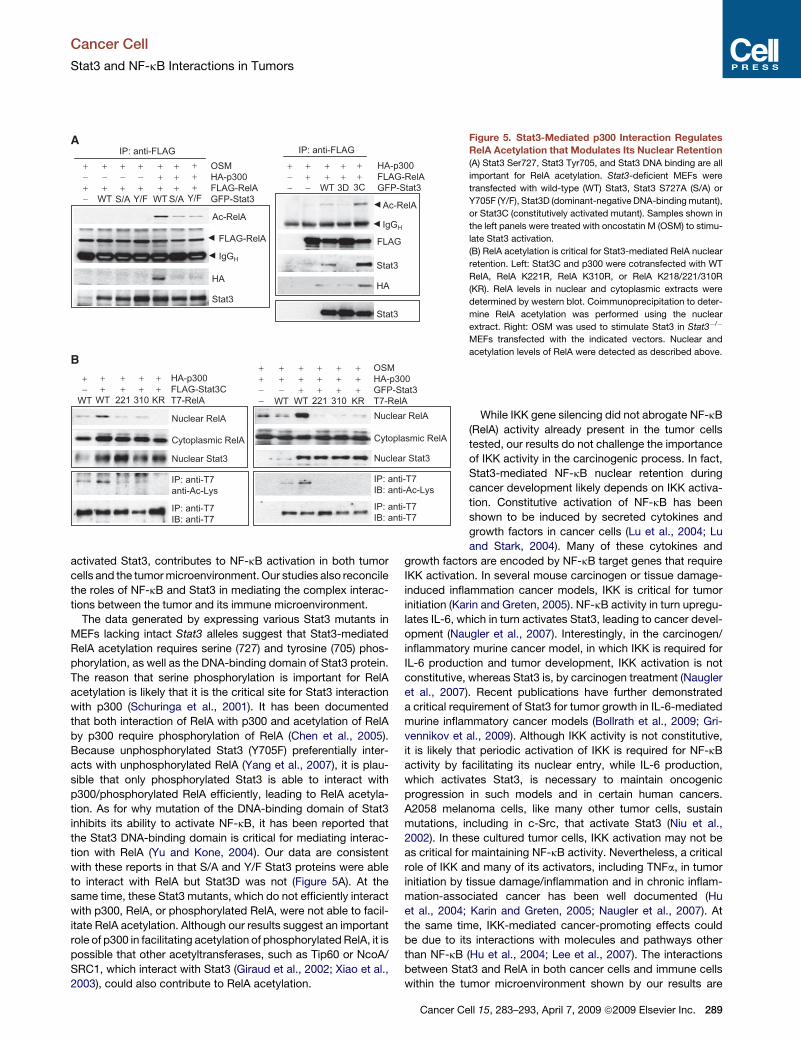
Figure 5. Stat3-Mediated p300 Interaction Regulates
RelA Acetylation that Modulates Its Nuclear Retention
(A) Stat3 Ser727, Stat3 Tyr705, and Stat3 DNA binding are all
important for RelA acetylation. Stat3-deficient MEFs were
transfected with wild-type (WT) Stat3, Stat3 S727A (S/A) or
Y705F (Y/F), Stat3D (dominant-negative DNA-binding mutant),
or Stat3C (constitutively activated mutant). Samples shown in
the left panels were treated with oncostatin M (OSM) to stimu-
late Stat3 activation.
(B) RelA acetylation is critical for Stat3-mediated RelA nuclear
retention. Left: Stat3C and p300 were cotransfected with WT
RelA, RelA K221R, RelA K310R, or RelA K218/221/310R
(KR). RelA levels in nuclear and cytoplasmic extracts were
determined by western blot. Coimmunoprecipitation to deter-
mine RelA acetylation was performed using the nuclear
extract. Right: OSM was used to stimulate Stat3 in Stat3�/�
MEFs transfected with the indicated vectors. Nuclear and
acetylation levels of RelA were detected as described above.
While IKK gene silencing did not abrogate NF-kB
(RelA) activity already present in the tumor cells
tested, our results do not challenge the importance
of IKK activity in the carcinogenic process. In fact,
Stat3-mediated NF-kB nuclear retention during
cancer development likely depends on IKK activa-
tion. Constitutive activation of NF-kB has been
shown to be induced by secreted cytokines and
growth factors in cancer cells (Lu et al., 2004; Lu
and Stark, 2004). Many of these cytokines and
growth factors are encoded by NF-kB target genes that require
IKK activation. In several mouse carcinogen or tissue damage-
induced inflammation cancer models, IKK is critical for tumor
initiation (Karin and Greten, 2005). NF-kB activity in turn upregu-
lates IL-6, which in turn activates Stat3, leading to cancer devel-
opment (Naugler et al., 2007). Interestingly, in the carcinogen/
inflammatory murine cancer model, in which IKK is required for
IL-6 production and tumor development, IKK activation is not
constitutive, whereas Stat3 is, by carcinogen treatment (Naugler
et al., 2007). Recent publications have further demonstrated
a critical requirement of Stat3 for tumor growth in IL-6-mediated
murine inflammatory cancer models (Bollrath et al., 2009; Gri-
vennikov et al., 2009). Although IKK activity is not constitutive,
it is likely that periodic activation of IKK is required for NF-kB
activity by facilitating its nuclear entry, while IL-6 production,
which activates Stat3, is necessary to maintain oncogenic
progression in such models and in certain human cancers.
A2058 melanoma cells, like many other tumor cells, sustain
mutations, including in c-Src, that activate Stat3 (Niu et al.,
2002). In these cultured tumor cells, IKK activation may not be
as critical for maintaining NF-kB activity. Nevertheless, a critical
role of IKK and many of its activators, including TNFa, in tumor
initiation by tissue damage/inflammation and in chronic inflam-
mation-associated cancer has been well documented (Hu
et al., 2004; Karin and Greten, 2005; Naugler et al., 2007). At
the same time, IKK-mediated cancer-promoting effects could
be due to its interactions with molecules and pathways other
than NF-kB (Hu et al., 2004; Lee et al., 2007). The interactions
between Stat3 and RelA in both cancer cells and immune cells
within the tumor microenvironment shown by our results are
Cancer Cell 15, 283–293, April 7, 2009 ª2009 Elsevier Inc. 289
Cancer Cell
Stat3 and NF-kB Interactions in Tumors
distinct, having opposite consequences for overall NF-kB
activity in tumor versus normal immune cells responding to im-
munostimulatory signals. These findings define a cooperativity
between Stat3 and NF-kB in cancer and help explain why both
transcription factors appear to stimulate a highly overlapping
repertoire of prosurvival, proliferative, and proangiogenic genes
(Catlett-Falcone et al., 1999; Darnell, 2002; Lo et al., 2005; Yu
and Jove, 2004). Our finding that this Stat3/NF-kB interaction
extends to tumor-associated hematopoietic cells emphasizes
++
−−
κ α
β
A
+ +
B− −
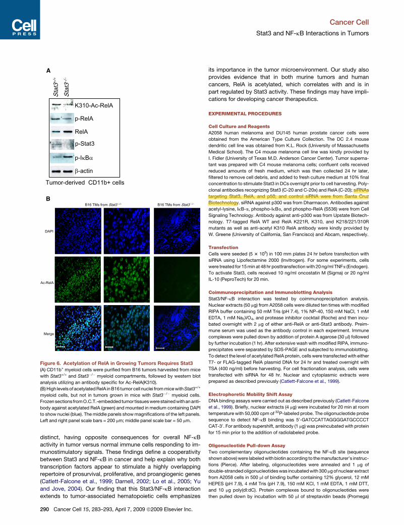
Figure 6. Acetylation of RelA in Growing Tumors Requires Stat3
(A) CD11b+ myeloid cells were purified from B16 tumors harvested from mice
with Stat3+/+ and Stat3�/� myeloid compartments, followed by western blot
analysis utilizing an antibody specific for Ac-RelA(K310).
(B) High levels of acetylated RelA in B16 tumor cell nuclei from mice with Stat3+/+
myeloid cells, but not in tumors grown in mice with Stat3�/� myeloid cells.
Frozen sections fromO.C.T.-embedded tumor tissueswere stained withan anti-
body against acetylated RelA (green) and mounted in medium containing DAPI
to show nuclei (blue). The middle panels show magnifications of the left panels.
Left and right panel scale bars = 200 mm; middle panel scale bar = 50 mm.
290 Cancer Cell 15, 283–293, April 7, 2009 ª2009 Elsevier Inc.
its importance in the tumor microenvironment. Our study also
provides evidence that in both murine tumors and human
cancers, RelA is acetylated, which correlates with and is in
part regulated by Stat3 activity. These findings may have impli-
cations for developing cancer therapeutics.
EXPERIMENTAL PROCEDURES
Cell Culture and Reagents
A2058 human melanoma and DU145 human prostate cancer cells were
obtained from the American Type Culture Collection. The DC 2.4 mouse
dendritic cell line was obtained from K.L. Rock (University of Massachusetts
Medical School). The C4 mouse melanoma cell line was kindly provided by
I. Fidler (University of Texas M.D. Anderson Cancer Center). Tumor superna-
tant was prepared with C4 mouse melanoma cells; confluent cells received
reduced amounts of fresh medium, which was then collected 24 hr later,
filtered to remove cell debris, and added to fresh culture medium at 10% final
concentration to stimulate Stat3 in DCs overnight prior to cell harvesting. Poly-
clonal antibodies recognizing Stat3 (C-20 and C-20x) and RelA (C-20); siRNAs
targeting Stat3, RelA, and p50; and control siRNA were from Santa Cruz
Biotechnology. siRNA against p300 was from Dharmacon. Antibodies against
acetyl-lysine, IkB-a, phospho-IkBa, and phospho-RelA (S536) were from Cell
Signaling Technology. Antibody against anti-p300 was from Upstate Biotech-
nology. T7-tagged RelA WT and RelA K221R, K310, and K218/221/310R
mutants as well as anti-acetyl K310 RelA antibody were kindly provided by
W. Greene (University of California, San Francisco) and Abcam, respectively.
Transfection
Cells were seeded (5 3 105) in 100 mm plates 24 hr before transfection with
siRNA using Lipofectamine 2000 (Invitrogen). For some experiments, cells
were treated for 15 min at 48 hr posttransfection with 20 ng/ml TNFa (Endogen).
To activate Stat3, cells received 10 ng/ml oncostatin M (Sigma) or 20 ng/ml
IL-10 (PeproTech) for 20 min.
Coimmunoprecipitation and Immunoblotting Analysis
Stat3/NF-kB interaction was tested by coimmunoprecipitation analysis.
Nuclear extracts (50 mg) from A2058 cells were diluted ten times with modified
RIPA buffer containing 50 mM Tris (pH 7.4), 1% NP-40, 150 mM NaCl, 1 mM
EDTA, 1 mM Na3VO4, and protease inhibitor cocktail (Roche) and then incu-
bated overnight with 2 mg of either anti-RelA or anti-Stat3 antibody. Preim-
mune serum was used as the antibody control in each experiment. Immune
complexes were pulled down by addition of protein A agarose (30 ml) followed
by further incubation (1 hr). After extensive wash with modified RIPA, immuno-
precipitates were separated by SDS-PAGE and subjected to immunoblotting.
To detect the level of acetylated RelA protein, cells were transfected with either
T7- or FLAG-tagged RelA plasmid DNA for 24 hr and treated overnight with
TSA (400 ng/ml) before harvesting. For cell fractionation analysis, cells were
transfected with siRNA for 48 hr. Nuclear and cytoplasmic extracts were
prepared as described previously (Catlett-Falcone et al., 1999).
Electrophoretic Mobility Shift Assay
DNA binding assays were carried out as described previously (Catlett-Falcone
et al., 1999). Briefly, nuclear extracts (4 mg) were incubated for 20 min at room
temperature with 50,000 cpm of 32P-labeled probe. The oligonucleotide probe
sequence to detect NF-kB binding was 50-GATCCATTAGGGGATGCCCCT
CAT-30. For antibody supershift, antibody (1 mg) was preincubated with protein
for 15 min prior to the addition of radiolabeled probe.
Oligonucleotide Pull-down Assay
Two complementary oligonucleotides containing the NF-kB site (sequence
shown above) were labeled with biotin according to the manufacturer’s instruc-
tions (Pierce). After labeling, oligonucleotides were annealed and 1 mg of
double-stranded oligonucleotides was incubated with 300 mg of nuclear extract
from A2058 cells in 500 ml of binding buffer containing 12% glycerol, 12 mM
HEPES (pH 7.9), 4 mM Tris (pH 7.9), 150 mM KCl, 1 mM EDTA, 1 mM DTT,
and 10 mg poly(dI:dC). Protein complexes bound to oligonucleotides were
then pulled down by incubation with 50 ml of streptavidin beads (Promega)
Cancer Cell
Stat3 and NF-kB Interactions in Tumors
preadsorbed with 1 mg/ml BSA, 50 mg poly(dI-dC), and 50 mg sheared salmon
sperm DNA. After extensive washing with binding buffer, protein complexes
were separated by SDS-PAGE, blotted, and probed with antibodies. For
in vitro IkBa pull-down assay, tumor RelA complexes were isolated from the
nuclei of A2058 cells by immunoprecipitation with anti-RelA antibody and
then incubated for 2 hr at room temperature with recombinant IkBa proteins
(BioSource). Complexes were then detected by immunoblotting.
Immunofluorescence Staining and Confocal Microscopy
A2058 cells were seeded on coverslips in six-well culture plates and transiently
transfected with either control or Stat3 siRNA. Cells were fixed for 20 min with
2% paraformaldehyde, permeabilized for 5 min with PBS containing 0.1%
Triton X-100 (PBS-T), quenched with 50 mM NH4Cl in PBS-T, and blocked
with 1% BSA in PBS-T. Immunostaining was performed using antibodies
as indicated in Figure 1B, and images were acquired using a Zeiss LSM 510
META NLO confocal microscope with a plan apo 633/1.4 NA lens.
To prepare frozen sections, B16 tumors harvested from Stat3flox/flox or Cre/
Stat3flox/flox mice were embedded in O.C.T. (Tissue-Tek) and frozen in liquid
nitrogen. Sections were air dried, fixed in 2% formaldehyde, and permeabi-
lized in cold methanol before immunofluorescence staining with antibodies.
The expression level of each protein in tumor tissues was visualized by a Nikon
Eclipse TE2000-U microscope and imaged using SPOT software.
Human tissue array slides (including both normal and malignant tissues),
obtained from the archives of the Pathology Laboratory at City of Hope National
Medical Center, were used to detect acetylated versus phosphorylated RelA
and phosphorylated Stat3 proteins. Preparation of tissue arrays was approved
by the Institutional Review Board at City of Hope, and use of these tissue array
slides was exempt as anonymous, archived specimens. For antibody staining,
tissue slides were deparaffinized, rehydrated through an alcohol series, and
then boiled in Antigen Unmasking Solution (Vector). After incubation with
a blocking solution containing 10% goat serum (Sigma) in PBS, the sections
were stained overnight at 4�C with a 1:50 dilution of a primary antibody. After
A
B
Figure 7. Acetylated RelA Is Prominently Present in
Human Tumors and Colocalizes with Activated Stat3
in the Nuclei
RelA is constitutively acetylated (A) and phosphorylated (B) in
human tumors and colocalizes with p-Stat3. Sections of
human lung cancer specimens were stained with the indicated
antibodies, followed by confocal microscopic analysis. In each
of (A) and (B), the bottom row of images shows magnifications
of the top row of images. Top panel scale bars = 40 mm; bottom
panel scale bars = 10 mm.
incubation with a secondary antibody (Alexa 488 for green
signal and Alexa 546 for red; Invitrogen), sections were mounted
in Vectashield mounting medium containing 4060-diamidino-2-
phenylindole (DAPI) (Vector). Images obtained by confocal
microscopy were prepared by Zeiss LSM Image Browser soft-
ware.
In Vivo Experiments
Mouse care and experimental procedures were performed
under pathogen-free conditions in accordance with estab-
lished institutional guidance and approved protocols from the
Institutional Animal Care and Use Committee of Beckman
Research Institute at City of Hope National Medical Center.
Mx1-Cre mice were obtained from The Jackson Laboratory,
and Stat3flox/flox mice were generously provided by S. Akira
and K. Takeda of Osaka University. Generation of mice with
Stat3�/� hematopoietic cells by the inducible Mx1-Cre recom-
binase system has been described previously (Kortylewski
et al., 2005;Lee et al., 2002;Wanget al., 2004). Tumor challenge
was performed in Stat3flox/flox or Cre/Stat3flox/flox mice 5 days
after poly(dI:dC) treatment, which induces Stat3 ablation mainly
in the hematopoietic system. Mice were sacrificed 2–3 weeks
after tumor challenge, and spleens and tumor specimens were harvested. Puri-
fication of specific immune subsets has been described previously (Kortylewski
et al., 2005). Proteinsand RNA were prepared from isolated immunecells, whole
spleens, and whole tumors for various analyses as indicated.
In Vitro IP Kinase Assay
IKKb kinase complexes were immunoprecipitated from cells transfected with
siRNA in the presence or absence of TNFa. Kinase activity was measured in
20 ml reaction buffer containing 1 mM ATP, 10 mCi [g-32P]ATP, and 1 mg
recombinant IkBa. ELISA-based kinase assay was performed according to
the manufacturer’s instructions (SuperArray).
HAT Assay
The enzymatic activity of p300 was assayed by incubating p300 immunopre-
cipitates for 30 min at 30�C with either 2 mg of histone H4 peptides (Upstate) or
0.5 mg of recombinant RelA (BioSource) as substrate. Reaction mixtures were
spotted on P81 filters and washed extensively. Incorporation rate of [3H]acetyl-
CoA was measured by liquid scintillation counting.
SUPPLEMENTAL DATA
The Supplemental Data include four figures and can be found with this article
online at http://www.cancercell.org/supplemental/S1535-6108(09)00071-3.
ACKNOWLEDGMENTS
We thank P.C. Heinrich for critical reading; S. Costa for editing; and staff
members of the Analytic Microscopy Core, Pathology Research Core, DNA
Synthesis Core, and Animal Research Center at City of Hope National Medical
Center for their technical assistance. This study was supported by the National
Institutes of Health (grants R01CA122976, R01CA115815, R01CA115674, and
P50 CA107399), the Harry Lloyd Charitable Trust, the Board of Governors at
Cancer Cell 15, 283–293, April 7, 2009 ª2009 Elsevier Inc. 291
Cancer Cell
Stat3 and NF-kB Interactions in Tumors
City of Hope, gifts from the Topercer family, Mrs. Dorothy Needle, Mr. John
Goldsmith, the Seraph Foundation, and the Janney Fund. Antibodies against
Ac-RelA(K310) and Ac-RelA mutants were from W. Greene (University of
California, San Francisco). FLAG-RelA plasmid DNA was kindly provided by
M.W. Mayo (University of Virginia). Stat3-deficient MEFs were from V. Poli
(University of Turin). R.J., D.P., and H.Y. wish to dedicate this article in memory
of Dr. Tsai-Fan Yu, a female physician-scientist active in medical science from
the 1930s through the early 2000s. Her pioneering and seminal contributions to
elucidating the metabolic basis and defining treatments for gout are a paragon
of translational biomedical research.
Received: August 25, 2008
Revised: December 2, 2008
Accepted: February 12, 2009
Published: April 6, 2009
REFERENCES
Bollrath, J., Phesse, T.J., von Burstin, V.A., Putoczki, T., Bennecke, M.,
Bateman, T., Nebelsiek, T., Lundgren-May, T., Canli, O., Schwitalla, S., et al.
(2009). gp130-mediated Stat3 activation in enterocytes regulates cell survival
and cell-cycle progression during colitis-associated tumorigenesis. Cancer
Cell 15, 91–102.
Bromberg, J.F., Wrzeszczynska, M.H., Devgan, G., Zhao, Y., Pestell, R.G.,
Albanese, C., and Darnell, J.E., Jr. (1999). Stat3 as an oncogene. Cell 98,
295–303.
Catlett-Falcone, R., Landowski, T.H., Oshiro, M.M., Turkson, J., Levitzki, A.,
Savino, R., Ciliberto, G., Moscinski, L., Fernandez-Luna, J.L., Nunez, G.,
et al. (1999). Constitutive activation of Stat3 signaling confers resistance to
apoptosis in human U266 myeloma cells. Immunity 10, 105–115.
Chen, L.F., and Greene, W.C. (2003). Regulation of distinct biological activities
of the NF-kappaB transcription factor complex by acetylation. J. Mol. Med. 81,
549–557.
Chen, L.F., and Greene, W.C. (2004). Shaping the nuclear action of NF-kap-
paB. Nat. Rev. Mol. Cell Biol. 5, 392–401.
Chen, L.F., Fischle, W., Verdin, E., and Greene, W.C. (2001). Duration of
nuclear NF-kappaB action regulated by reversible acetylation. Science 293,
1653–1657.
Chen, L.F., Mu, Y., and Greene, W.C. (2002). Acetylation of RelA at discrete
sites regulates distinct nuclear functions of NF-kappaB. EMBO J. 21,
6539–6548.
Chen, L.F., Williams, S.A., Mu, Y., Nakano, H., Duerr, J.M., Buckbinder, L., and
Greene, W.C. (2005). NF-kappaB RelA phosphorylation regulates RelA acety-
lation. Mol. Cell. Biol. 25, 7966–7975.
Dalwadi, H., Krysan, K., Heuze-Vourc’h, N., Dohadwala, M., Elashoff, D.,
Sharma, S., Cacalano, N., Lichtenstein, A., and Dubinett, S. (2005). Cyclooxy-
genase-2-dependent activation of signal transducer and activator of transcrip-
tion 3 by interleukin-6 in non-small cell lung cancer. Clin. Cancer Res. 11,
7674–7682.
Darnell, J.E., Jr. (2002). Transcription factors as targets for cancer therapy.
Nat. Rev. Cancer 2, 740–749.
Deng, W.G., and Wu, K.K. (2003). Regulation of inducible nitric oxide synthase
expression by p300 and p50 acetylation. J. Immunol. 171, 6581–6588.
Deo, D.D., Axelrad, T.W., Robert, E.G., Marcheselli, V., Bazan, N.G., and
Hunt, J.D. (2002). Phosphorylation of STAT-3 in response to basic fibroblast
growth factor occurs through a mechanism involving platelet-activating factor,
JAK-2, and Src in human umbilical vein endothelial cells. Evidence for a dual
kinase mechanism. J. Biol. Chem. 277, 21237–21245.
Ghosh, S., May, M.J., and Kopp, E.B. (1998). NF-kappa B and Rel proteins:
evolutionarily conserved mediators of immune responses. Annu. Rev. Immu-
nol. 16, 225–260.
Giraud, S., Bienvenu, F., Avril, S., Gascan, H., Heery, D.M., and Coqueret, O.
(2002). Functional interaction of STAT3 transcription factor with the coactivator
NcoA/SRC1a. J. Biol. Chem. 277, 8004–8011.
292 Cancer Cell 15, 283–293, April 7, 2009 ª2009 Elsevier Inc.
Greten, F.R., Eckmann, L., Greten, T.F., Park, J.M., Li, Z.W., Egan, L.J.,
Kagnoff, M.F., and Karin, M. (2004). IKKbeta links inflammation and tumorigen-
esis in a mouse model of colitis-associated cancer. Cell 118, 285–296.
Grivennikov, S., Karin, E., Terzic, J., Mucida, D., Yu, G.Y., Vallabhapurapu, S.,
Scheller, J., Rose-John, S., Cheroutre, H., Eckmann, L., and Karin, M. (2009).
IL-6 and Stat3 are required for survival of intestinal epithelial cells and devel-
opment of colitis-associated cancer. Cancer Cell 15, 103–113.
Hu, M.C., Lee, D.F., Xia, W., Golfman, L.S., Ou-Yang, F., Yang, J.Y., Zou, Y.,
Bao, S., Hanada, N., Saso, H., et al. (2004). IkappaB kinase promotes tumor-
igenesis through inhibition of forkhead FOXO3a. Cell 117, 225–237.
Huang, T.T., Kudo, N., Yoshida, M., and Miyamoto, S. (2000). A nuclear export
signal in the N-terminal regulatory domain of IkappaBalpha controls cyto-
plasmic localization of inactive NF-kappaB/IkappaBalpha complexes. Proc.
Natl. Acad. Sci. USA 97, 1014–1019.
Karin, M., and Greten, F.R. (2005). NF-kappaB: linking inflammation and
immunity to cancer development and progression. Nat. Rev. Immunol. 5,
749–759.
Karin, M., Cao, Y., Greten, F.R., and Li, Z.W. (2002). NF-kappaB in cancer:
from innocent bystander to major culprit. Nat. Rev. Cancer 2, 301–310.
Kortylewski, M., Kujawski, M., Wang, T., Wei, S., Zhang, S., Pilon-Thomas, S.,
Niu, G., Kay, H., Mule, J., Kerr, W.G., et al. (2005). Inhibiting Stat3 signaling in
the hematopoietic system elicits multicomponent antitumor immunity. Nat.
Med. 11, 1314–1321.
Kortylewski, M., Xin, H., Kujawski, M., Lee, H., Liu, Y., Harris, T., Drake, C.,
Pardoll, D., and Yu, H. (2009). Regulation of the IL-23 and IL-12 balance by
Stat3 signaling in the tumor microenvironment. Cancer Cell 15, 114–123.
Kujawski, M., Kortylewski, M., Lee, H., Herrmann, A., Kay, H., and Yu, H.
(2008). Stat3 mediates myeloid cell-dependent tumor angiogenesis in mice.
J. Clin. Invest. 118, 3367–3377.
Lee, C.K., Raz, R., Gimeno, R., Gertner, R., Wistinghausen, B., Takeshita, K.,
DePinho, R.A., and Levy, D.E. (2002). STAT3 is a negative regulator of granu-
lopoiesis but is not required for G-CSF-dependent differentiation. Immunity 17,
63–72.
Lee,D.F.,Kuo, H.P., Chen, C.T., Hsu,J.M., Chou, C.K.,Wei, Y., Sun,H.L., Li, L.Y.,
Ping, B., Huang, W.C., et al. (2007). IKK beta suppression of TSC1 links inflam-
mation and tumor angiogenesis via the mTOR pathway. Cell 130, 440–455.
Lenz,G., Davis,R.E.,Ngo, V.N., Lam, L., George,T.C., Wright,G.W., Dave, S.S.,
Zhao, H., Xu, W., Rosenwald, A., et al. (2008). Oncogenic CARD11 mutations in
human diffuse large B cell lymphoma. Science 319, 1676–1679.
Lo, H.W., Hsu, S.C., Ali-Seyed, M., Gunduz, M., Xia, W., Wei, Y., Bartholo-
meusz, G., Shih, J.Y., and Hung, M.C. (2005). Nuclear interaction of EGFR
and STAT3 in the activation of the iNOS/NO pathway. Cancer Cell 7, 575–589.
Lu, T., and Stark, G.R. (2004). Cytokine overexpression and constitutive
NFkappaB in cancer. Cell Cycle 3, 1114–1117.
Lu, T., Sathe, S.S., Swiatkowski, S.M., Hampole, C.V., and Stark, G.R. (2004).
Secretion of cytokines and growth factors as a general cause of constitutive
NFkappaB activation in cancer. Oncogene 23, 2138–2145.
Naugler, W.E., Sakurai, T., Kim, S., Maeda, S., Kim, K., Elsharkawy, A.M., and
Karin, M. (2007). Gender disparity in liver cancer due to sex differences in
MyD88-dependent IL-6 production. Science 317, 121–124.
Niu, G., Bowman, T., Huang, M., Shivers, S., Reintgen, D., Daud, A., Chang, A.,
Kraker, A., Jove, R., and Yu, H. (2002). Roles of activated Src and Stat3
signaling in melanoma tumor cell growth. Oncogene 21, 7001–7010.
Pikarsky, E., Porat, R.M., Stein, I., Abramovitch, R., Amit, S., Kasem, S.,
Gutkovich-Pyest, E., Urieli-Shoval, S., Galun, E., and Ben-Neriah, Y. (2004).
NF-kappaB functions as a tumour promoter in inflammation-associated
cancer. Nature 431, 461–466.
Pomerantz, J.L., Denny, E.M., and Baltimore, D. (2002). CARD11 mediates
factor-specific activation of NF-kappaB by the T cell receptor complex.
EMBO J. 21, 5184–5194.
Pranada, A.L., Metz, S., Herrmann, A., Heinrich, P.C., and Muller-Newen, G.
(2004). Real time analysis of STAT3 nucleocytoplasmic shuttling. J. Biol.
Chem. 279, 15114–15123.
Cancer Cell
Stat3 and NF-kB Interactions in Tumors
Schuringa, J.J., Schepers, H., Vellenga, E., and Kruijer, W. (2001). Ser727-
dependent transcriptional activation by association of p300 with STAT3
upon IL-6 stimulation. FEBS Lett. 495, 71–76.
Stancovski, I., and Baltimore, D. (1997). NF-kappaB activation: the I kappaB
kinase revealed? Cell 91, 299–302.
Turkson,J., Zhang,S., Palmer, J., Kay,H., Stanko,J., Mora, L.B., Sebti,S., Yu,H.,
and Jove, R. (2004). Inhibition of constitutive signal transducer and activator of
transcription 3 activation by novel platinum complexes with potent antitumor
activity. Mol. Cancer Ther. 3, 1533–1542.
Wang, T., Niu, G., Kortylewski, M., Burdelya, L., Shain, K., Zhang, S., Bhatta-
charya, R., Gabrilovich, D., Heller, R., Coppola, D., et al. (2004). Regulation of
the innate and adaptive immune responses by Stat-3 signaling in tumor cells.
Nat. Med. 10, 48–54.
Welte, T., Zhang, S.S., Wang, T., Zhang, Z., Hesslein, D.G., Yin, Z., Kano, A.,
Iwamoto, Y., Li, E., Craft, J.E., et al. (2003). STAT3 deletion during hematopoi-
esis causes Crohn’s disease-like pathogenesis and lethality: a critical role of
STAT3 in innate immunity. Proc. Natl. Acad. Sci. USA 100, 1879–1884.
Xiao, H., Chung, J., Kao, H.Y., and Yang, Y.C. (2003). Tip60 is a co-repressor
for STAT3. J. Biol. Chem. 278, 11197–11204.
Yang, J., Liao, X., Agarwal, M.K., Barnes, L., Auron, P.E., and Stark, G.R.
(2007). Unphosphorylated STAT3 accumulates in response to IL-6 and acti-
vates transcription by binding to NFkappaB. Genes Dev. 21, 1396–1408.
Yu, H., and Jove, R. (2004). The STATs of cancer–new molecular targets come
of age. Nat. Rev. Cancer 4, 97–105.
Yu, H., Kortylewski, M., and Pardoll, D. (2007). Crosstalk between cancer and
immune cells: role of STAT3 in the tumour microenvironment. Nat. Rev. Immu-
nol. 7, 41–51.
Yu, Z., and Kone, B.C. (2004). The STAT3 DNA-binding domain mediates
interaction with NF-kappaB p65 and iuducible nitric oxide synthase transre-
pression in mesangial cells. J. Am. Soc. Nephrol. 15, 585–591.
Zhong, Z., Wen, Z., and Darnell, J.E., Jr. (1994). Stat3: a STAT family member
activated by tyrosine phosphorylation in response to epidermal growth factor
and interleukin-6. Science 264, 95–98.
Cancer Cell 15, 283–293, April 7, 2009 ª2009 Elsevier Inc. 293


