Embed Size (px)
Citation preview
Microbes and Infection 13 (2011) 691e696www.elsevier.com/locate/micinf
Original article
Phlebotomus sergenti (Parrot, 1917) identified as Leishmania killicki hostin Ghardaıa, south Algeria
S.C. Boubidi a,*, K. Benallal a, A. Boudrissa a, L. Bouiba a, B. Bouchareb b, R. Garni a,A. Bouratbine c, C. Ravel d, V. Dvorak e, J. Votypka e, P. Volf e, Z. Harrat a
a Service d’Eco-Epidemiologie Parasitaire, Institut Pasteur, 1 Rue du petit Staoueli, Delly Ibrahim, Algiers, Algeriab Service de Prevention, Ghardaıa, Algeria
c Service de Parasitologie et de Mycologie Medicale, Institut Pasteur, Tunis, Tunisied Laboratoire de Parasitologie-Mycologie, CHU de Montpellier, France
eDepartment of Parasitology, Charles University in Prague, Faculty of Science, Czech Republic
Received 17 October 2010; accepted 21 February 2011
Available online 5 March 2011
Abstract
Since 2005, an outbreak of human cutaneous leishmaniasis (CL) in Ghardaıa, south Algeria, was studied and one output of these investi-gations was the identification of two Leishmania species, Leishmania major and Leishmania killicki, as the CL causative agents. In the presentstudy, we were curious to focus on sand fly fauna present in this area and detection of Leishmania-positive sand fly females. Sand flies (3717)were collected during two seasons using sticky papers and CDC light traps in urban, rural and sylvatic sites. Twelve Phlebotomus species wereidentified. Phlebotomus papatasi was dominant in the urban site while Phlebotomus sergenti and Phlebotomus riouxi/chabaudi were dominant inthe sylvatic site. Out of 74 P. sergenti females captured by CDC light traps in the sylvatic site populated by Ghardaıas’ Gundi (Massoutieramzabi), three ones were hosting Leishmania promastigotes. PCR-RFLP and sequencing of seven single-copy coding DNA sequences identifiedthe promastigotes as L. killicki. Furthermore, laboratory experiments revealed that L. killicki isolate sampled from a CL patient inhabiting thestudied region develop well in P. sergenti females. Our findings strongly suggest that the human cutaneous leishmaniases caused by L. killicki isa zoonotic disease with P. sergenti sand flies acting as hosts and vectors and gundi rodents as reservoirs.� 2011 Institut Pasteur. Published by Elsevier Masson SAS. All rights reserved.
Keywords: Cutaneous leishmaniasis; Leishmania tropica killicki; Phlebotomus sergenti; Ghardaıa; Algeria
1. Introduction
In Algeria, leishmaniases occur in two clinical forms:visceral leishmaniasis (VL) caused by Leishmania infantumand cutaneous leishmaniasis (CL) caused by three Leishmaniaspecies: L. infantum, Leishmania killicki and Leishmaniamajor which is the most widespread one [1]. During 2005,almost thirty thousands (30,000) CL cases were reported in thecountry, the province of Ghardaıa being particularly affectedwith more than two thousands cases recorded. In this region,
* Corresponding author. Tel./fax: þ 213 2137 6851.
E-mail addresses: [email protected], [email protected] (S.C. Boubidi).
1286-4579/$ - see front matter � 2011 Institut Pasteur. Published by Elsevier Ma
doi:10.1016/j.micinf.2011.02.008
the disease is caused by either L. killicki or L. major, whichcoexist sympatrically [2].
Recently, L. killicki spread was reported from central andsouth-western areas of Tunisia, e.g. Metlaoui and Ain Jloula[3e5], and L. killicki parasites were isolated from patients alsoin Kenya, Yemen [6] and Libya [7]. Apparent increase of thegeographic distribution of L. killicki may correspond withhistorical difficulties in distinguishing between L. major andLeishmania tropica complex which coupled with a low diseaseburden may have masked the presence of the parasite frompast discovery [8].
CL caused by L. major is a zoonosis with rodents serving asreservoir hosts. In Algeria, L. major is transmitted by Phle-botomus papatasi, gerbils Psammomys obesus and Meriones
sson SAS. All rights reserved.
692 S.C. Boubidi et al. / Microbes and Infection 13 (2011) 691e696
shawi were proven as reservoirs [9,10]. On the other hand,transmission cycle of L. killicki is still unknown.
L. killicki (Rioux, Lanotte and Pratlong 1986) was firstdescribed in Tataouine, southeast Tunisia by Rioux et al.(1986) [12] and in the same year included in the L. tropicacomplex. Species denoted as L. tropica are highly heteroge-neous [13], strains are readily distinguishable by antigenic,biochemical and molecular techniques [14], nevertheless, theposition of L. killicki as a separate species is still questionable.Based on the similarity with L. tropica, Rioux et al. (1986c)[15] first mentioned Phlebotomus sergenti as a suspectedvector of L. killicki. However, since its description in Algeriain 1917 by Parrot [16], P. sergenti was never found in thiscountry infected by promastigotes and neither was any othersand fly species belonging to subgenus Paraphlebotomus.
CL caused by L. tropica complex is usually regarded as ananthroponosis occurring in hyperendemic situations whereman-to-man transmission is well ascertained. L. tropicaparasites were, however, isolated from black rats, dogs androck hyraxes (Procavia capensis) [13,17e19] and recentlynew zoonotic foci of L. tropica are emerging in different partsof the Mediterranean countries [19], including Tunisia andAlgeria [2]. The location of sporadic human cases in theprovince of Ghardaıa also suggests zoonotic transmission.
In the present study, we proved P. sergenti as a competentvector of L. killicki. We found natural infection in P. sergenticaptured in the neighborhood of the Mzab Gundi’s (Massou-tiera mzabi) burrows with L. killicki and experimentallyconfirmed susceptibility of P. sergenti to L. killicki.
2. Materials and methods
2.1. Study area
Localization of Ghardaıa (32�300/32�410N; 03�370/03�420E)and description of the region was presented by Harrat et al.(2009) [2]. For the entomological investigations, three sites withdifferent biotopes were prospected in respect to the incidence ofhuman cases, population density and abundance of wildrodents. Urban site was located in Ksar (Castle) of Ghardaıa(32�290N; 03�420E), one of the five cities of Ghardaıa province,where high number of CL cases was reported during theoutbreak in 2005 [20]. This site is characterized by a highconcentration of houses and inhabitants, some of them breedinggoats, chickens and donkeys. Rural site was located in theOases of Ghardaıa named Ghaba (32�290N; 03�390E) whereinhabitants of urban sites move during hot summer season torest in their second homes dispersed in the palm plantations.Sylvatic site consists of natural caves in the rocky hill ofChaabat Telli (32�480N; 03�630E), located in the periphery ofGhardaıa’s valley. These caves provide shelters for the MzabGundi (M. mzabi), a rodent frequently encountered in this area.
2.2. Sand fly collection, dissection and identification
Samplings were performed in 2008 and 2009, twicea month from April to November, using sticky traps and four
CDC light traps (John W. Hock, Gainesville, FL). Sticky trapswere placed in each site, near human dwellings, in front ofrodents’ burrows and inside the caves. Captured flies werestored in 70% alcohol and most of them subsequently treatedby KOH 20% and embedded in Marc Andre medium forspecies identification using keys of Abonnenc (1972) [21],Dedet (1984) [22] and Depaquit (1998) [23].
Female sand flies caught by CDC light traps in sylvatic site inOctober 2009 were washed in 70% ethanol and dissected insterile saline. For species identification, the head and theposterior part of the abdomen of each sand fly were mounted ina drop of sterile saline and the gutwas examined for the presenceof promastigotes under microscope. Upon microscopicaldetection of Leishmania infection, the material from the slidewas divided into two parts. The first part of each gut containingpromastigotes was aseptically inoculated into NNN medium(9& sterile saline; 50 mg/ml streptomycin; 100.000 UI peni-cillin; 1.000 mg/ml Nystatine). The second part of parasitesuspensions was placed in duplicate wells containing a drop ofTris-EDTA buffer for DNA extraction and Leishmania identi-fication as a backup in case of unsuccessful cultivation.
In some specimens belonging to Phlebotomus riouxi/cha-baudi, the thorax was preserved in alcohol and used for DNAextraction and molecular identification based on PCR-RFLPand sequencing of the cytochrome c oxidase I (COI) gene (seebelow).
2.3. DNA extraction, Leishmania typing and P. riouxi/chabaudi identification
Extractions of total DNA from the material preserved inTris-EDTA buffer (parasites from naturally infected sand flies)and in ethanol (sand fly thorax) were performed using a DNAextraction kit (Roche, France) according to the manufacturer’sinstruction.
Samples containing Leishmania parasites from naturallyinfected sand flies were analyzed by the sequencing of sevensingle-copy coding DNA sequences (Multi Locus SequenceTyping) located on six different chromosomes [C. Ravel,unpublished]. Obtained sequences (GenBank accessionnumber Ph3:HM135420eHM135426; LEM4995:HM135427eHM135433; Ph13:HM135434eHM135440) wereconcatenated (4839 bp in all) and compared to 22 L. tropicacomplex strains and one L. major strain as an out-group(Table 1). Analysis was performed using PhyML 3.0 softwareunder GTR substitution model and 1000 bootstraps.
The molecular identification of specimens belonging tosibling species P. riouxi/chabaudi was based on PCR-restric-tion fragment length polymorphism (PCR-RFLP) and ondirect sequencing of a 689 bp variable region of COI gene asdescribed by Boudabous et al. (2009) [24].
2.4. Development of L. killicki in P. sergenti
P. sergenti colony originating from females collected inAmnun, Northern Israel in 2001 was maintained in conditionsdescribed by Benkova and Volf (2007) [25]. Female sand flies
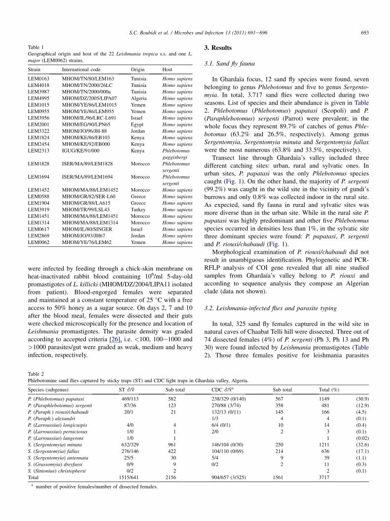
Table 1
Geographical origin and host of the 22 Leishmania tropica s.s. and one L.
major (LEM0062) strains.
Strain International code Origin Host
LEM0163 MHOM/TN/80/LEM163 Tunisia Homo sapiens
LEM4018 MHOM/TN/2000/26LC Tunisia Homo sapiens
LEM3987 MHOM/TN/2000/000a Tunisia Homo sapiens
LEM4995 MHOM/DZ/2005/LIPA07 Algeria Homo sapiens
LEM1015 MHOM/YE/86/LEM1015 Yemen Homo sapiens
LEM0955 MHOM/YE/86/LEM955 Yemen Homo sapiens
LEM3956 MHOM/IL/96/LRC-L691 Israel Homo sapiens
LEM2001 MHOM/EG/90/LPN65 Egypt Homo sapiens
LEM3322 MHOM/JO/96/JH-88 Jordan Homo sapiens
LEM1824 MHOM/KE/86/EB103 Kenya Homo sapiens
LEM2454 MHOM/KE/92/EB000 Kenya Homo sapiens
LEM2313 IGUG/KE/91/000 Kenya Phlebotomus
guggisbergi
LEM1828 ISER/MA/89/LEM1828 Morocco Phlebotomus
sergenti
LEM1694 ISER/MA/89/LEM1694 Morocco Phlebotomus
sergenti
LEM1452 MHOM/MA/88/LEM1452 Morocco Homo sapiens
LEM0588 MHOM/GR/82/SER-L60 Greece Homo sapiens
LEM1904 MHOM/GR/88/LA615 Greece Homo sapiens
LEM3919 MHOM/TR/99/LSL43 Turkey Homo sapiens
LEM1451 MHOM/MA/88/LEM1451 Morocco Homo sapiens
LEM1314 MHOM/MA/88/LEM1314 Morocco Homo sapiens
LEM0617 MHOM/IL/80/SINGER Israel Homo sapiens
LEM2869 MHOM/JO/93/JH67 Jordan Homo sapiens
LEM0062 MHOM/YE/76/LEM62 Yemen Homo sapiens
693S.C. Boubidi et al. / Microbes and Infection 13 (2011) 691e696
were infected by feeding through a chick-skin membrane onheat-inactivated rabbit blood containing 106/ml 5-day-oldpromastigotes of L. killicki (MHOM/DZ/2004/LIPA11 isolatedfrom patient). Blood-engorged females were separatedand maintained at a constant temperature of 25 �C with a freeaccess to 50% honey as a sugar source. On days 2, 7 and 10after the blood meal, females were dissected and their gutswere checked microscopically for the presence and location ofLeishmania promastigotes. The parasite density was gradedaccording to accepted criteria [26], i.e. <100, 100e1000 and>1000 parasites/gut were graded as weak, medium and heavyinfection, respectively.
Table 2
Phlebotomine sand flies captured by sticky traps (ST) and CDC light traps in Gha
Species (subgenus) ST _/\ Sub total
P. (Phlebotomus) papatasi 469/113 582
P. (Paraphlebotomus) sergenti 87/36 123
P. (Paraph.) riouxi/chabaudi 20/1 21
P. (Paraph.) alexandri
P. (Larroussius) longicuspis 4/0 4
P. (Larroussius) perniciosus 1/0 1
P. (Larroussius) langeroni 1/0 1
S. (Sergentomyia) minuta 632/329 961
S. (Sergentomyia) fallax 276/146 422
S. (Sergentomyia) antennata 25/5 30
S. (Grassomyia) dreyfussi 0/9 9
S. (Sintonius) christophersi 0/2 2
Total 1515/641 2156
a number of positive females/number of dissected females.
3. Results
3.1. Sand fly fauna
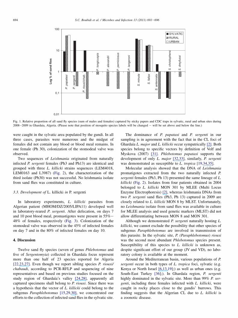
In Ghardaıa focus, 12 sand fly species were found, sevenbelonging to genus Phlebotomus and five to genus Sergento-myia. In total, 3.717 sand flies were collected during twoseasons. List of species and their abundance is given in Table2. Phlebotomus (Phlebotomus) papatasi (Scopoli) and P.(Paraphlebotomus) sergenti (Parrot) were prevalent; in thewhole focus they represent 89.7% of catches of genus Phle-botomus (63.2% and 26.5%, respectively). Among genusSergentomyia, Sergentomyia minuta and Sergentomyia fallaxwere the most numerous (63.8% and 33.5%, respectively).
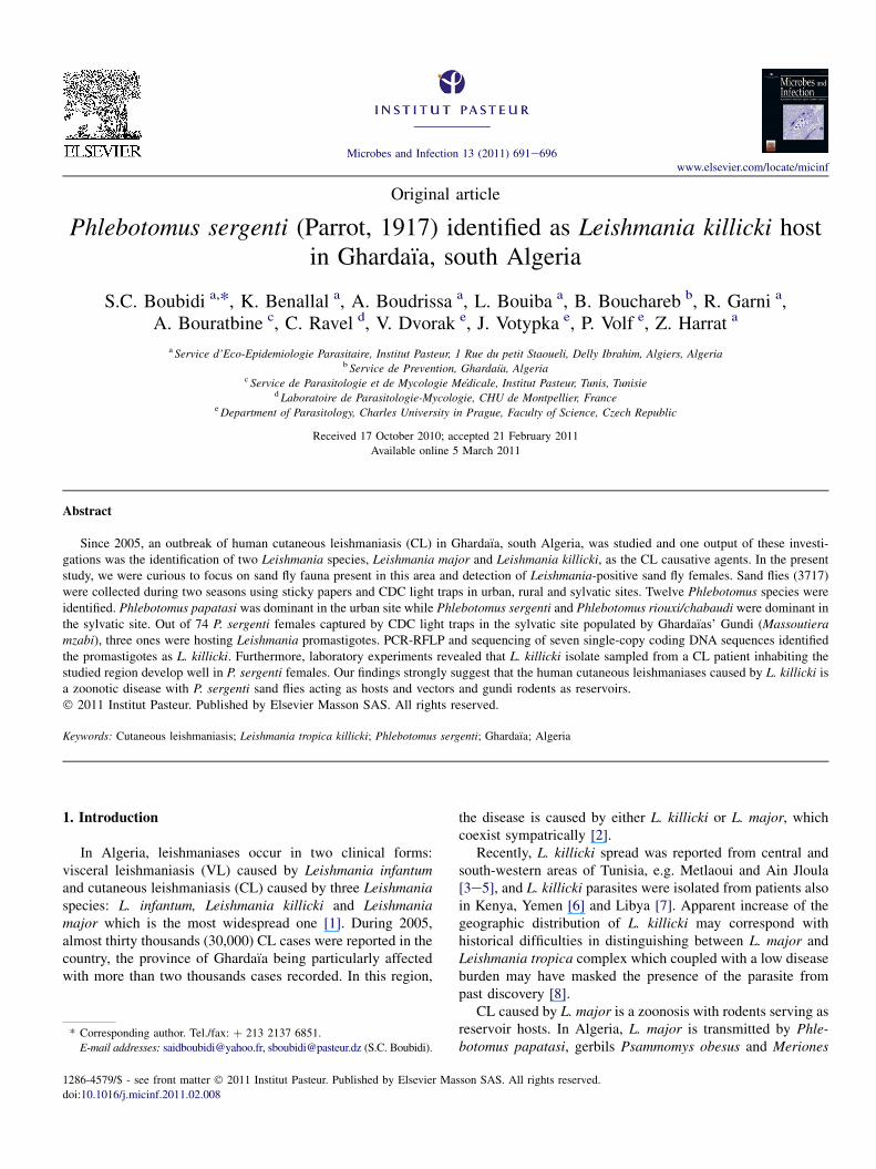
Transect line through Ghardaıa’s valley included threedifferent catching sites: urban, rural and sylvatic ones. Inurban sites, P. papatasi was the only Phlebotomus speciescaught (Fig. 1). On the other hand, the majority of P. sergenti(99.2%) was caught in the wild site in the vicinity of gundi’sburrows and only 0.8% was collected indoor in the rural site.As expected, sand fly fauna in rural and sylvatic sites wasmore diverse than in the urban site. While in the rural site P.papatasi was highly predominant and other five Phlebotomusspecies occurred in densities less than 1%, in the sylvatic sitethree dominant species were found: P. papatasi, P. sergentiand P. riouxi/chabaudi (Fig. 1).
Morphological examination of P. riouxi/chabaudi did notresult in unambiguous identification. Phylogenetic and PCR-RFLP analysis of COI gene revealed that all nine studiedsamples from Ghardaıa’s valley belong to P. riouxi andaccording to sequence analysis they compose an Algerianclade (data not shown).
3.2. Leishmania-infected flies and parasite typing
In total, 325 sand fly females captured in the wild site innatural caves of Chaabat Telli hill were dissected. Three out of74 dissected females (4%) of P. sergenti (Ph 3, Ph 13 and Ph30) were found infected by Leishmania promastigotes (Table2). Those three females positive for leishmania parasites
rdaıa valley, Algeria.
CDC _/\a Sub total Total (%)
238/329 (0/140) 567 1149 (30.9)
270/88 (3/74) 358 481 (12.9)
132/13 (0/11) 145 166 (4.5)
1/3 4 4 (0.1)
6/4 (0/1) 10 14 (0.4)
2/0 2 3 (0.1)
1 (0.02)
146/104 (0/30) 250 1211 (32.6)
104/110 (0/69) 214 636 (17.1)
5/4 9 39 (1.1)
0/2 2 11 (0.3)
2 (0.1)
904/657 (3/325) 1561 3717
S. d
re
yfu
ssi
S. ch
ris
to
ph
ersi
S. a
nte
nn
ata
S. fa
lla
x
S. m
inu
ta
P. la
ng
ero
ni
P. p
ern
icio
su
s
P. a
lexa
nd
ri
P. lo
ng
icu
sp
is
P. rio
uxi/ch
ab
.
P. se
rg
en
ti
P. p
ap
ata
si
0,01
0,1
1
10
100
Lo
g %
o
f s
an
d flie
s
SYLVATICRURALURBAN
Fig. 1. Relative proportion of all sand fly species (sum of males and females) captured by sticky papers and CDC traps in sylvatic, rural and urban sites during
2008e2009 in Ghardaia, Algeria. (Please note that position of mosquito species labels will be changed e will be set above and below the line.)
694 S.C. Boubidi et al. / Microbes and Infection 13 (2011) 691e696
were caught in the sylvatic area populated by the gundi. In allthree cases, parasites were numerous and the midgut offemales did not contain any blood or blood meal remains. Inone female (Ph 30), colonization of the stomodeal valve wasobserved.
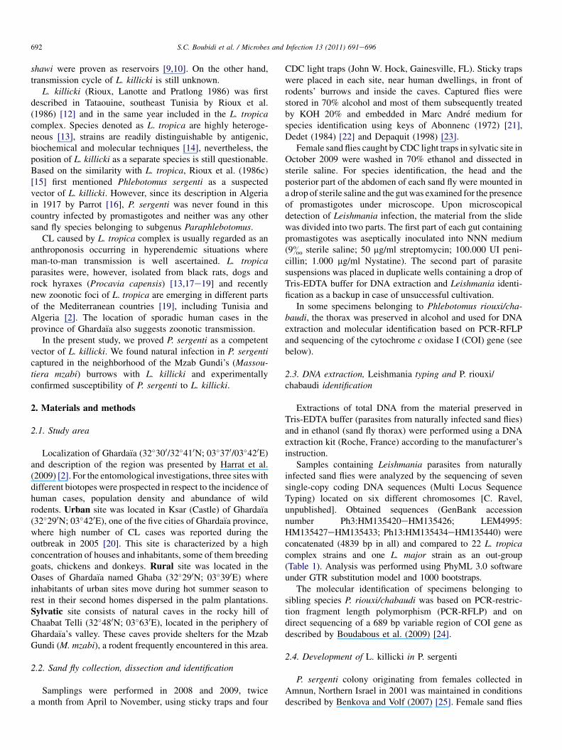
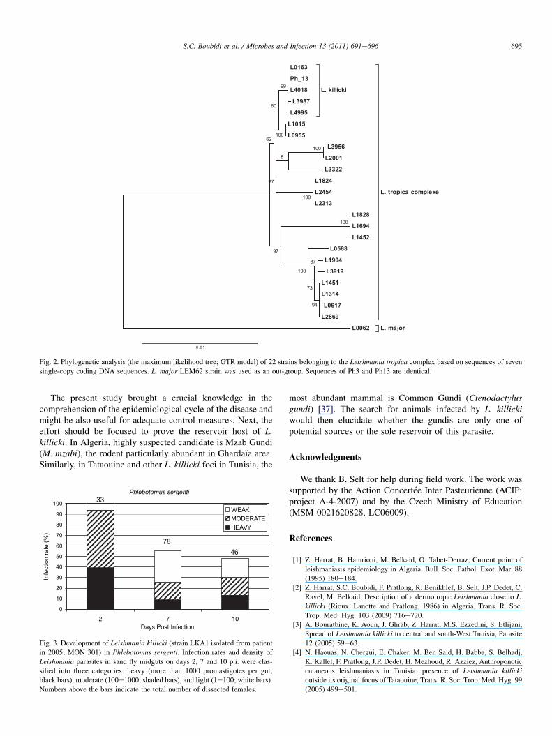
Two sequences of Leishmania originated from naturallyinfected P. sergenti females (Ph3 and Ph13) are identical andgrouped with three L. killicki strains sequences (LEM4018,LEM0163 and L3987) (Fig. 2), the characterization of thethird isolate (Ph30) was not successful. No leishmania isolatefrom sand flies was constituted in culture.
3.3. Development of L. killicki in P. sergenti
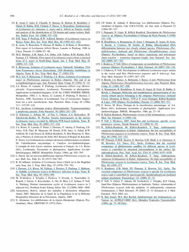
In laboratory experiments, L. killicki parasites fromAlgerian patient (MHOM/DZ/2005/LIPA11) developed wellin laboratory-reared P. sergenti. After defecation, on days 7and 10 post blood meal, promastigotes were present in 55%e48% of females, respectively (Fig. 3). Colonization of thestomodeal valve was observed in the 45% of infected femaleson day 7 and in the 80% of infected females on day 10.
4. Discussion
Twelve sand fly species (seven of genus Phlebotomus andfive of Sergentomyia) collected in Ghardaıa focus representmore than one half of 23 species reported for Algeria[22,23,27]. Even though we report sibling species P. riouxi/chabaudi, according to PCR-RFLP and sequencing of ninerepresentatives and based on previous studies focused on thestudy region of Ghardaıa’s valley [24,28], apparently allcaptured specimens shall belong to P. riouxi. Since there wasa hypothesis that the vector of L. killicki could belong to thesubgenus Paraphlebotomus [15,29,30], we concentrated ourefforts to the collection of infected sand flies in the sylvatic site.
The dominance of P. papatasi and P. sergenti in oursampling is in agreement with the fact that in the CL foci ofGhardaıa L. major and L. killicki occur sympatrically [2]. Bothspecies belong to specific vectors by definition of Volf andMyskova (2007) [31]. Phlebotomus papatasi supports thedevelopment of only L. major [32,33], similarly, P. sergentiwas demonstrated as susceptible to L. tropica [19,34,35].
Molecular analysis showed that the DNA of Leishmaniapromatigotes extracted from the two naturally infected P.sergenti females (Ph3, Ph 13) presented the same lineage of L.killicki (Fig. 2). Isolates from four patients obtained in 2004belonged to L. killicki MON 301 by MLEE (Multi LocusEnzyme Electrophoresis) [2], whereas leishmania DNAs fromtwo P. sergenti sand flies (Ph3, Ph 13) captured in 2009 areclosely related to L. killicki MON 8 by MLST. Unfortunately,no Leishmania isolate from sand flies was available in culturefor MLEE analysis and used genetic markers (MLST) did notallow differentiating between MON 8 and MON 301.
Although we demonstrated P. sergenti naturally hosting L.killicki, we cannot exclude the possibility that other species ofsubgenus Paraphlebotomus are involved in transmission ofthis parasite. In the sylvatic site, P. (Paraphlebotomus) riouxiwas the second most abundant Phlebotomus species present.Susceptibility of this species to L. killicki is unknown as,despite significant effort of our group (JV and VD), no labo-ratory colony is available at the moment.
Around the Mediterranean basin, various populations of P.sergenti occur in both types of L. tropica foci, sylvatic (e.g.Kenya or North Israel [6,13,19];) as well as urban ones (e.g.South-East Turkey [36];). In Ghardaıa region, P. sergentihighly dominated in the sylvatic site. More than 99% P. ser-genti, including three females infected with L. killicki, werecaught in rocky places close to the gundis’ burrows. Thisfinding suggests that the Algerian CL due to L. killicki isa zoonotic disease.
L0163
Ph_13
L4018
L3987
L4995
L. killicki
L1015
L0955
L3956
L2001
L3322
L1824
L2454
L2313
L1828
L1694
L1452
L0588
L1904
L3919
L1451
L1314
L0617
L2869
L. tropica complexe
L. major L0062
99
100
60
100
81
100
37
62
87
94
73
100
97
100
0.01
Fig. 2. Phylogenetic analysis (the maximum likelihood tree; GTR model) of 22 strains belonging to the Leishmania tropica complex based on sequences of seven
single-copy coding DNA sequences. L. major LEM62 strain was used as an out-group. Sequences of Ph3 and Ph13 are identical.
695S.C. Boubidi et al. / Microbes and Infection 13 (2011) 691e696
The present study brought a crucial knowledge in thecomprehension of the epidemiological cycle of the disease andmight be also useful for adequate control measures. Next, theeffort should be focused to prove the reservoir host of L.killicki. In Algeria, highly suspected candidate is Mzab Gundi(M. mzabi), the rodent particularly abundant in Ghardaıa area.Similarly, in Tataouine and other L. killicki foci in Tunisia, the
Phlebotomus sergenti
4678
33
0
10
20
30
40
50
60
70
80
90
100
0172Days Post Infection
Infe
ctio
n ra
te (%
)
WEAKMODERATEHEAVY
Fig. 3. Development of Leishmania killicki (strain LKA1 isolated from patient
in 2005; MON 301) in Phlebotomus sergenti. Infection rates and density of
Leishmania parasites in sand fly midguts on days 2, 7 and 10 p.i. were clas-
sified into three categories: heavy (more than 1000 promastigotes per gut;
black bars), moderate (100e1000; shaded bars), and light (1e100; white bars).
Numbers above the bars indicate the total number of dissected females.
most abundant mammal is Common Gundi (Ctenodactylusgundi) [37]. The search for animals infected by L. killickiwould then elucidate whether the gundis are only one ofpotential sources or the sole reservoir of this parasite.
Acknowledgments
We thank B. Selt for help during field work. The work wassupported by the Action Concertee Inter Pasteurienne (ACIP:project A-4-2007) and by the Czech Ministry of Education(MSM 0021620828, LC06009).
References
[1] Z. Harrat, B. Hamrioui, M. Belkaid, O. Tabet-Derraz, Current point of
leishmaniasis epidemiology in Algeria, Bull. Soc. Pathol. Exot. Mar. 88
(1995) 180e184.
[2] Z. Harrat, S.C. Boubidi, F. Pratlong, R. Benikhlef, B. Selt, J.P. Dedet, C.
Ravel, M. Belkaid, Description of a dermotropic Leishmania close to L.
killicki (Rioux, Lanotte and Pratlong, 1986) in Algeria, Trans. R. Soc.
Trop. Med. Hyg. 103 (2009) 716e720.[3] A. Bouratbine, K. Aoun, J. Ghrab, Z. Harrat, M.S. Ezzedini, S. Etlijani,
Spread of Leishmania killicki to central and south-West Tunisia, Parasite
12 (2005) 59e63.
[4] N. Haouas, N. Chergui, E. Chaker, M. Ben Said, H. Babba, S. Belhadj,
K. Kallel, F. Pratlong, J.P. Dedet, H. Mezhoud, R. Azziez, Anthroponotic
cutaneous leishmaniasis in Tunisia: presence of Leishmania killicki
outside its original focus of Tataouine, Trans. R. Soc. Trop. Med. Hyg. 99
(2005) 499e501.
696 S.C. Boubidi et al. / Microbes and Infection 13 (2011) 691e696
[5] K. Aoun, F. Amri, E. Chouihi, N. Haouas, K. Bedoui, R. Benikhlef, J.
Ghrab, H. Babba, M.K. Chahed, Z. Harrat, A. Bouratbine, Epidemiology
of Leishmania (L.) infantum, L. major and L. killicki in Tunisia: results
and analysis of the identification of 226 human and canine isolates, Bull.
Soc. Pathol. Exot. 101 (2008) 323e328.[6] D.K. Sang, F. Pratlong, R.W. Ashford, Identity of Leishmania tropica in
Kenya, Trans. Roy. Soc. Trop. Med. Hyg. 86 (1992) 621e622.
[7] K. Aoun, N. Bousslimi, N. Haouas, H. Babba, A. El-Buni, A. Bouratbine,
First report of Leishmania killicki Rioux, Lanotte et Pratlong, 1986 in
Libya, Parasite 13 (2006) 87e88.
[8] M.G. Shehata, A.M. Samy, S.A. Doha, A.R. Fahmy, R.M. Kaldas, B.D.
Furman, J.T. Villinski, First report of Leishmania tropica from a classical
focus of L. major in North-Sinai, Egypt, Am. J. Trop. Med. Hyg. 81
(2009) 213e218.
[9] S. Belazzoug, Isolation of Leishmania major Yakimoff, Schokhor, 1914
from Psammomys obesus Gretzschmar, 1828 (Rodentia: Gerbillidae) in
Algeria, Trans. R. Soc. Trop. Med. Hyg. 77 (1983) 876.
[10] M.A. Izri, S. Belazzoug, F. Pratlong, J.A. Rioux, Isolation of Leishmania
major in Phlebotomus papatasi in Biskra (Algeria). The end of an
ecoepidemiological saga, Ann. Parasitol. Hum. Comp. 67 (1992) 31e32.
[12] J.A. Rioux, G. Lanotte, F. Pratlong, Leishmania killicki n.sp (Kineto-
plastida: Trypaosomatidae). Leishmania. Taxonomie et phylogenese.
Applications ecoepidemiologiques. (Coll. Int. CNRS/ INSERM. IMEEE,
Montpellier, 1984, J. A. Rioux, G. Lanotte, F. Pratlong, P. Bastien, J.
Perieres, 1990. Taxonomy of Leishmania. Use of isoenzymes, Sugges-
tions for a new classification, Ann. Parasitol. Hum. Comp. 65 (1986)
111e115139e142.[13] R.L. Jacobson, Leishmania tropica (Kinetoplastida: Trypanosomatidae)
a perplexing parasite, Folia Parasitol. 50 (2003) 241e250.
[14] G. Schonian, L. Schnur, M. . el Fari, L. Oskam, A.A. Kolesnikov, W.
Sokolowska-Kohler, W. Presber, Genetic heterogeneity in the species
Leishmania tropica revealed by different PCR-based methods, Trans. R.
Soc. Trop. Med. Hyg. 95 (2001) 217e224.
[15] J.A. Rioux, G. Lanotte, F. Petter, J. Dereure, O. Akalay, F. Pratlong, I.D.
Velez, N.B. Fikri, R. Maazoun, M. Denial, D.M. Jarry, A. Zahaf, R.W.
Ashford, M. Cadi-Soussi, R. Killick-Kendrick, N. Ben Mansour, G. Mor-
eno, J. Perieres, E.Guilvard,M.Zribi,M.F.Kennou, P. Rispail, R.Knechtli,
E. Serres, Les leishmanioses cutanees du bassin mediterraneen occidental.
De l’identification enzymatique a‘ l’analyse eco-epidemiologique.
L’exemple de trois foyers, tunisien, marocain et francais. in: J.A. Rioux
(Ed.), Leishmania, Taxonomie et phylogenese, Applications eco-epi-
demiologiques. IMEEE, Montpellier, France, 1986c, pp. 365e395.
[16] L. Parrot, Sur un nouveau phlebotome algerien Phlebotomus sergenti, sp.
nov, Bull. Soc. Path. Ex 10 (1917) 564e567.
[17] B. El-Adhami, Isolation of Leishmania from a black rat in the Baghdad
area, Iraq, Am. J. Trop. Med. Hyg. 25 (1976) 759e761.
[18] J. Dereure, J.A. Rioux, M. Gallego, J. Perieres, F. Pratlong, J. Mahjour,
A. Saddiki, Leishmania tropica in Morocco: infection in dogs, Trans. R.
Soc. Trop. Med. Hyg. 85 (1991) 595.
[19] M. Svobodova, J. Votypka, J. Peckova, V. Dvorak, A. Nasereddin, G.
Baneth, J. Sztern, V. Kravchenko, A. Orr, D. Meir, L.F. Schnur, P. Volf,
A. Warburg, Distinct transmission cycles of Leishmania tropica in 2
adjacent foci, Northern Israel, Emerg. Infect. Dis. 12 (2006) 1860e1868.[20] Anonymous, Releve, annuel des maladies a declaration obligatoire,
Annee 2005. Ministere de la Sante de la Population et de la Reforme
Hospitaliere Direction de la Prevention, Algerie, 2005.
[21] E. Abonnenc, Les phlebotomes de la region ethiopienne (Diptcra, Psy-
chodidae), Mem. ORSTOM 55 (1972) Paris.
[22] J.P. Dedet, K. Addadi, S. Belazzoug, Les phlebotomes (Diptera, Psy-
chodidae) d’Algerie, Cah. O.R.S.T.O.M., ser. Ent. med. et Parasitol 22
(1984) 99e127.
[23] J. Depaquit, N. Leger, R. Killick Kendrick, Description de Phlebotomus
riouxi n. sp. (Diptera: Psychodidae) d’Afrique du Nord, Parasite 5 (1998)
151e158.
[24] R. Boudabous, A. Bounamous, D. Jouet, J. Depaquit, D. Augot, H. Ferte,
S. Berchi, A. Couloux, M. Veuille, H. Babba, Mitochondrial DNA
differentiation between two closely related species, Phlebotomus (Par-
aphlebotomus) chabaudi and Phlebotomus (Paraphlebotomus) riouxi
(Diptera: Psychodidae), based on direct sequencing and polymerase
chain reaction e restriction fragment length, Ann. Entomol. Soc. Am.
102 (2009) 347e353.
[25] I. Benkova, P. Volf, Effect of temperature on metabolism of Phlebotomus
papatasi (Diptera: Psychodidae), J. Med. Entomol. 44 (2007) 150e154.
[26] J. Cihakova, P. Volf, Development of different Leishmania major strains
in the vector sand flies Phlebotomus papatasi and P. duboscqi, Ann.
Trop. Med. Parasitol. 91 (1997) 267e279.
[27] S. Berchi, J.A. Rioux, A. Belmonte, J. Russo, A new Phlebotomus from
Algeria, Phlebotomus (Paraphlebotomus) kazeruni, Ann. Parasitol. Hum.
Comp. 61 (1986) 507e508.
[28] A. Bounamous, R. Boudabous, D. Jouet, D. Augot, H. Ferte, H. Babba, S.
Berchi, J. Depaquit, Molecular and morphological characterisation of two
closely related species belonging to the subgenus Phlebotomus chabaudi
Croset, Abonnenc & Rioux, 1970 et P. riouxi Depaquit, Killick-Kendrick
& Leger, 1998 (Diptera: Psychodidae), Parasite 15 (2008) 565e571.
[29] E. Serres, M. Roux, Pratique de la classification automatique. in: J.A.
Rioux (Ed.), leishmania taxonomie et phylogenese. Application eco-
epidemiologique. IMEEE, Montpellier, 1986, pp. 27e39.
[30] R. Killick-Kendrick, Phlebotomine vectors of the leishmaniasis: a review,
Med. Vet. Entomol. 4 (1990) 124.
[31] P. Volf, J. Myskova, 2007, Sand flies and Leishmania: specific versus
permissive vectors, Trends Parasitol. 23 (1990) 91e92.
[32] R. Killick-Kendrick, M. Killick-Kendrick, Y. Tang, Anthroponotic
cutaneous leishmaniasis in Kabul, Afghanistan: the low susceptibility of
Phlebotomus papatasi to Leishmania tropica, Trans. R. Soc. Trop. Med.
Hyg. 88 (1994) 252e253.
[33] P.F. Pimenta, E.M.B. Saraiva, E. Rowton, G.B. Modi, L.A. Garraway, S.
M. Beverley, S.J. Turco, D.L. Sacks, Evidence that the vectorial
competence of phlebotomine sandflies for different species of Leish-
mania is controlled by structural polymorphisms in the surface lip-
ophosphoglycan, Proc. Natl. Acad. Sci. USA 91 (1994) 9155e9159.[34] R. Killick-Kendrick, M. Killick-Kendrick, Y. Tang, Anthroponotic
cutaneous leishmaniasis in Kabul, Afghanistan: the high susceptibility of
Phlebotomus sergenti to Leishmania tropica, Trans. R. Soc. Trop. Med.
Hyg. 89 (1995) 477.
[35] S. Kamhawi, G.B. Modi, P.F. Pimenta, E. Rowton, D.L. Sacks, The
vectorial competence of Phlebotomus sergenti is specific for Leishmania
tropica and is controlled by speciesspecific, lipophosphoglycan-mediated
midgut attachment, Parasitology 121 (2000) 25e33.
[36] P. Volf, Y. Ozbel, F. Akkafa, M. Svobodova, J. Votypka, K.P. Chang,
Sand flies (Diptera: Phlebotominae) in Sanliurfa, Turkey: relationship of
Phlebotomus sergenti with the epidemic of anthroponotic cutaneous
leishmaniasis, J. Med. Entomol. 39 (2002) 12e15 (Erratum in: J. Med.
Entomol. 39(3) 2002; iii).
[37] R. Ben Ismail, M.S. Ben Rachid, Epidemiologie des leishmanioses en
Tunisie. in: AUPELF-UREF (Ed.), Maladies Tropicales Transmissibles
(1989), pp. 73e80 Paris.