Embed Size (px)
Citation preview

Phosphoinositide 3-kinase (PI3K) and nutrient sensing mTOR(mammalian target of rapamycin) pathways control Tlymphocyte trafficking
Linda V Sinclair1, David Finlay1, Carmen Feijoo1, Georgina H Cornish2, Alex Gray3, AnnAger4, Klaus Okkenhaug5, Thijs J Hagenbeek6,7, Hergen Spits6,7, and Doreen A Cantrell1
1Department of Cell Biology and Immunology, University of Dundee, DD1 5EH, UK 2Immune CellBiology, The National Institute for Medical Research, London, NW7 1AA, UK 3Division ofMolecular Physiology, University of Dundee, DD1 5EH, UK 4Department of Medical Biochemistryand Immunology, Cardiff University, CF14 4XN, UK 5Laboratory of Lymphocyte Signalling andDevelopment, Babraham Institute, Cambridge, CB2 4AT, UK 6Department of Immunology, TheNetherlands Cancer Institute, 1066 CX Amsterdam, The Netherlands 7Department of ImmunologyDiscovery, Genentech Inc., South San Francisco, California CA 94080, USA
SummaryPI3K and mTOR are evolutionarily conserved regulators of cell metabolism. Here we show PI3Kand mTOR determine the repertoire of adhesion and chemokine receptors expressed by Tlymphocytes. Key lymph node homing receptors, CD62L (L-selectin) and CCR7, are highlyexpressed on naive T lymphocytes but downregulated following immune activation. CD62Ldownregulation occurs via ectodomain proteolysis and suppression of gene transcription. PI3Kp110δ controls CD62L proteolysis via mitogen-activated protein (MAP) kinases whereas PI3Kp110δ control of CD62L transcription is mediated by the nutrient sensor mTOR via regulation ofthe transcription factor KLF2. PI3K-mTOR nutrient sensing pathways also determined expressionof the chemokine receptor CCR7 and regulate lymphocyte trafficking in vivo. Hence, lymphocytesutilize PI3K and mTOR to match metabolism and trafficking.
KeywordsCD62L; L-selectin; CCR7; PI3K; p110δ; rapamycin; MAP kinases; KLF2
IntroductionThe lipid product of phosphoinositide 3-kinases (PI3Ks), phosphoinositide (3,4,5) tri-phosphate (PI(3,4,5)P3), binds to the pleckstrin homology (PH) domains of proteins andcontrols an array of signaling molecules including serine or threonine kinases such as PKB(also known as AKT), Tec family tyrosine kinases and guanine nucleotide exchange proteinsfor Rac and Rho-family GTPases1. T cell activation induces rapid and sustained production
Correspondence to: Doreen A Cantrell1 email: [email protected] ContributionsL.V.S., most in vitro assays and in vivo adoptive transfer; D.F., analysis of PTEN KO(T) cells; C.F., Real-Time PCR; G.H.C., in vitromigration assay; A.G., PI(3,4,5)P3 quantitation; A.A., provision of CD62L transgenic mice and discussions; K.O., provision of p110δ(D910A) transgenic mice; T.J.H. and H.S., provision of PTEN KO(T) mice; D.A.C., conceptual design and wrote the manuscript.
Competing Interests StatementThe authors declare no competing financial interests.
Europe PMC Funders GroupAuthor ManuscriptNat Immunol. Author manuscript; available in PMC 2010 April 20.
Published in final edited form as:Nat Immunol. 2008 May ; 9(5): 513–521. doi:10.1038/ni.1603.
Europe PM
C Funders A
uthor Manuscripts
Europe PM
C Funders A
uthor Manuscripts

of PI(3,4,5)P3 in a response that is essential for T cell immune responses2-4. One importantand evolutionarily conserved role for PI3Ks in T cells is to regulate cell metabolism andprotein synthesis5. However, PI3Ks also regulate actin dynamics6 and accordingly there hasbeen a past focus on the role of PI3Ks in regulating leukocyte motility and chemotaxis7-10.The impetus for these studies came from the realization that the correct localization oflymphocytes is essential for effective immune responses. Thus, understanding the molecularmechanisms and signal transduction pathways that control lymphocyte trafficking isimportant.
Naive T lymphocytes constantly circulate around the body via the blood, lymphatics andsecondary lymphoid organs. Immune responses are initiated within secondary lymphoidorgans, such as peripheral lymph nodes, when T cells encounter primed antigen presentingcells expressing cognate antigen-major histocompatibility complexes together withappropriate costimulatory molecules. Lymph node entry requires the coordinated migrationof cells and is dependent on chemokine receptors such as CCR7 and molecules that mediatelymphocyte adhesion such as CD62L (L-selectin) and integrins11,12. T cell entry intolymph nodes from the blood occurs in specialized high endothelial venules (HEVs). Thefirst step of transmigration is CD62L mediated tethering and rolling of naive lymphocyteson the endothelium of HEVs12,13. CD62L, which is highly expressed constitutively onnaive and memory T lymphocytes12 is thus essential for the entry of these cells intoperipheral lymph nodes14,15.
Immune activation of T cells induces striking changes in their migratory patterns. Effector Tlymphocytes migrate to a greater extent to non-lymphoid tissues and sites of inflammationand have a reduced capacity to home to peripheral lymph nodes compared to naive andmemory T cells12,16. Changes in the trafficking behavior of T cells are important forimmune responses and are mediated by changes in the expression of chemokine receptorsand adhesion molecules. For example, activated T cells downregulate CCR7 and CD62L butupregulate expression of tissue homing receptors such as the integrins VLA-4 and cutaneouslymphocyte associated antigen17. CD62L expression at the membrane is controlled by abalance of two activities – the rate of CD62L gene transcription and the rate of CD62Lproteolytic cleavage18-20. The proteolytic cleavage and shedding of CD62L from the cellsurface of T lymphocytes is an immediate response to triggering of antigen receptors21whereas CD62L gene transcription is lost following cytokine controlled differentiation ofeffector T cells18,22. This transcriptional loss of CD62L is frequently used as a marker todistinguish naive and antigen experienced T cells in the blood and lymphoid organs duringimmune responses. Effector and effector memory CD8+ cytotoxic T lymphocytes (CTLs)are CD62Llo and preferentially home to peripheral tissues whereas central memory CTLsexpress high amounts of CD62L similar to naive T cells and home to lymph nodes12,16.
The signal transduction pathways that dynamically modulate CD62L and CCR7 expressionduring immune activation of T cells are not characterized. However, the differing abilities ofthe cytokines interleukin 2 (IL-2) and IL-15 to downregulate CD62L expression in CD8+ Tcells23,24 parallels the relative strength of these cytokines to stimulate PI3K-mediated cellgrowth responses25. Antigen-primed CD8+ T cells cultured in the presence of IL-2 thussustain PI3K signaling at high levels, synthesize high amounts of protein and downregulateCD62L. In contrast, T cells maintained with IL-15 can only sustain low-level PI3Ksignaling, synthesize less protein and fail to downregulate CD62L expression24,25. Thesedata are purely correlative, however the idea that PI(3,4,5)P3 abundance might controlexpression of T cell homing receptors is intriguing because there is a large body of work thatlink PI3Ks to the control of lymphocyte metabolism, cell growth, chemotaxis andchemokinesis7,8,10. However, it has never been considered that PI3Ks might regulatelymphocyte recirculation by regulating the plasma membrane expression of lymph node
Sinclair et al. Page 2
Nat Immunol. Author manuscript; available in PMC 2010 April 20.
Europe PM
C Funders A
uthor Manuscripts
Europe PM
C Funders A
uthor Manuscripts

homing receptors CD62L and CCR7. Historically, loss of CD62L and CCR7 expression isused to discriminate naive and antigen-experienced T cells. The present study shows thatCD62L and CCR7 expression rather report the PI3K and mTOR {A000094} http://www.signaling-gateway.org/molecule/query?afcsid=A000094 signaling status of a T celland are not simply an epigenetic consequence of immune activation. PI(3,4,5)P3 and mTORsignaling are thus shown to be essential determinants for CD62L and CCR7 expression andto be key regulators of T cell migration in vivo.
ResultsPI3K regulates T cell receptor-induced CD62L shedding
Triggering of the T cell receptor (TCR) complex with cognate antigen-majorhistocompatibility complexes induced rapid downregulation of CD62L from the cellmembrane of naive T cells (Fig. 1a). This initial acute loss of CD62L surface expressionfollowing immune activation of T cells is predominantly mediated by proteolyticcleavage21,26. Resting T cells exhibited basal constitutive cleavage of CD62L but duringimmune activation accelerated proteolysis occurred, resulting in increased amounts of thesoluble cleavage product of CD62L in the culture supernatants of TCR-activated T cells(Fig. 1b). The acute cleavage of CD62L takes place proximal to the cell membrane and ismediated by a disintegrin and metalloprotease (ADAM) 17, also known as tumor necrosisfactor (TNF)— converting enzyme (TACE)27. Mutation of the ADAM17 target sites inCD62L thus prevents CD62L shedding21,26. To confirm the contribution of proteolyticcleavage of CD62L in TCR-mediated downregulation we examined the ability of TCRtriggering to downregulate the expression of a mutant of CD62L that has the membraneproximal region of CD62L replaced with that of P-selectin, thus removing the proteolytictarget sequences for ADAM17. Crosslinking of TCR complexes with CD3 antibodiesdownregulated cell surface expression of CD62L on wild-type T cells and alsodownregulated expression of wild-type CD62L expressed under the control of the hCD2promoter in CD62L-deficient T cells (CD62L(WT))(Fig. 1c). However, TCR triggering didnot downregulate surface expression of CD62L molecules with mutated proteolytic targetsequences (CD62L(MUT))(Fig. 1c).
To test the involvement of PI3K in TCR-induced shedding of CD62L, we performedexperiments with LY294002, a pharmacological inhibitor of PI3K. LY294002 suppressedthe shedding of CD62L induced by T cells activated with cognate peptide-MHC complexesor crosslinking CD3 antibodies (Fig. 1d). Class 1 PI3Ks comprise a p110 catalytic subunitand an adapter regulatory subunit. Four p110 isoforms exist (p110α, p110β, p110γ andp110δ) and three adapter subunits, p85α, p85β and p55γ28.
In T cells, the p110δ PI3K
PI3K p110 delta {A001770}
http://www.signaling-gateway.org/molecule/query?afcsid=A001770 catalytic subunitproduces the PI(3,4,5)P3 that is generated in response to TCR triggering3,29,30. Wetherefore examined CD62L regulation in T cells where homologous recombination has beenused to substitute wild-type p110δ for a catalytically inactive mutant p110δ(D910A)29. Tcells from these mice fail to produce PI(3,4,5)P3 in response to TCR triggering3. T cellsexpressing p110δ(D910A) failed to downregulate CD62L in response to TCR triggering(Fig. 1e) although these cells shed CD62L in response to the pharmacological stimulusphorbol 12,13, dibutyrate (PDBu), which bypasses the TCR.
Sinclair et al. Page 3
Nat Immunol. Author manuscript; available in PMC 2010 April 20.
Europe PM
C Funders A
uthor Manuscripts
Europe PM
C Funders A
uthor Manuscripts

CD62L transcription is suppressed by PI3K p110δLow CD62L expression in effector cytotoxic T cells reflects low gene transcription22. Tolook at the involvement of PI3K in the regulation of CD62L mRNA expression weexamined the impact of PI3K inhibition on CD62L expression in CD8+ cytotoxic T cells.P14 TCR transgenic T cells that express a Vα2Vβ8.1 TCR were primed for two days withthe lymphocytic choriomeningitis virus glycoprotein (LCMV-GP) peptide, gp33-41(KAVYNFATM), presented by the MHC class I molecule, H-2Db. Thereafter cells werecultured in IL-2 to generate cytotoxic effector T cells (CTLs). As controls, antigen-primed Tcells cultured with IL-15 were used. IL-15 is a potent mitogen and sustains exponential Tcell clonal expansion of antigen-primed CD8+ T cells but does not support differentiation ofeffector cytotoxic T cells, rather it generates cells that phenocopy memory T cells23,24.CD8+ T cells cultured with IL-2 expressed low amounts of CD62L whereas cells culturedwith IL-15 were CD62Lhi (Fig. 2a). These differences in CD62L expression reflecteddifferences in cellular abundance of its mRNA, which was low in CTLs (Fig. 2b). Yet CTLsdid not globally downregulate all surface receptors. CD25, the α subunit of the IL-2 receptoris highly expressed in CTLs, reflecting abundant expression of CD25 mRNA in CTLs (datanot shown). We found no evidence for increased shedding of CD62L in IL-2 stimulatedcells, rather there was less CD62L proteolytic cleavage in T cells cultured with IL-2 versusIL-15 reflecting the much lower de novo synthesis of CD62L in IL-2-stimulated T cells (Fig.2c).
IL-2 and IL-15 differ in the strength of the PI3K signal they can induce25. Antigen-primedT cells maintained in IL-15 have a low cellular content of PI(3,4,5)P3 (120,000 moleculesper cell) compared to 540,000 molecules per cell in cells stimulated with IL-2 and showreduced activation of PI3K mediated signal transduction pathways (Supplementary Fig. 1online). The ability of IL-2 to downregulate CD62L thus correlates with its ability tostrongly activate PI3K signaling pathways. To probe the role of PI3K in IL-2downregulation of CD62L, antigen-primed T cells were cultured with IL-2 in the presenceor absence of the PI3K inhibitor LY294002. Effector CD8+ T cells cultured with IL-2expressed low amounts of CD62L protein and mRNA whereas effector T cells cultured inIL-2 plus LY294002 were CD62Lhi and expressed abundant CD62L mRNA (Fig. 2d,e).PI3K p110δ is required for antigen receptor function but not for IL-2 induced proliferationof T cells29,30. Hence, p110δ(D910A) T cells show attenuated proliferation in response toTCR stimulation, this can be overcome when activated through the TCR in the presence ofstrong co-stimulation provided by CD28 and in the presence of IL-2 (ref. 29).p110δ(D910A) CD8+ T cells thus responded to IL-2 stimulation and proliferatedexponentially over a period of 4 days when cultured in IL-2 to produce large granular cellsindistinguishable from wild-type T lymphoblasts (Fig. 2f). PI3K-PKB pathways regulate theexpression of two key nutrient receptors in T cells, namely, CD71, the transferrin receptorand CD98, a critical component of L-type amino acid transporter complexes25,31.Theexpression of CD71 and CD98 was comparable on wild-type and p110δ(D910A) T cellscultured in IL-2 (Fig. 2g). Yet IL-2-stimulated p110δ(D910A) T cells maintained highsurface expression of CD62L as compared to the low amounts of CD62L seen in controlCTL populations (Fig. 2g). These data revealed a selective role for p110δ in IL-2 signaltransduction in that it is required for IL-2-mediated downregulation of CD62L but not forIL-2-induced expression of nutrient receptors, mitogenesis or cell growth. Further evidencefor the selective effect of p110δ loss on CD62L expression in T cells comes from analyzingthe expression of P-selectin and E-selectin ligands in IL-2-stimulated p110δ(D910A) CD8+
T cells. The upregulation of P-selectin and E-selectin ligands normally accompanies effectorT cell differentiation12. IL-2 stimulated p110δ(D910A) CD8+ T cells expressed highamounts of P-selectin and E-selectin ligands, equivalent to those seen in control CTLs (Fig.
Sinclair et al. Page 4
Nat Immunol. Author manuscript; available in PMC 2010 April 20.
Europe PM
C Funders A
uthor Manuscripts
Europe PM
C Funders A
uthor Manuscripts
2h). Thus, p110δ selectively controls expression of L but not E or P selectin ligands in Tcells.
Different PI3K pathways control CD62L expressionTCR stimulated p110δ(D910A) T cells have multiple signaling defects including reducedactivation of the MAP kinases Erk1 and Erk2 (ref. 29). This latter defect could be relevant tothe PI3K dependence of TCR-induced CD62L shedding because chemokine-inducedshedding of CD62L in neutrophils has been shown to be mediated by Erks (ref. 32) whichphosphorylate ADAM17 and control its trafficking to the cell surface33-35. We accordinglyexplored the role of Erk1 and Erk2 in TCR-induced shedding of CD62L. Down regulation ofCD62L in antigen stimulated P14 CD8+ T cells was blocked by the inhibitor PD184352(Fig. 3a), which prevents activation of the upstream kinase MEK1 and hence Erk activation.The inhibition of PI3K and Erks also blocked downregulation of CD62L in CD4+ and CD8+
T cells polyclonally activated with CD3 antibodies (Supplementary Fig. 2 online). We alsoexamined the role of Erks in the transcriptional downregulation of CD62L that occurs ineffector CTLs. Inhibition of Erk activation with PD184352 did not prevent CD62Ldownregulation in IL-2 maintained CTLs (Fig. 3b).
PI3K initiates signaling mediated by the mammalian target of rapamycin, mTOR kinaseswhich are evolutionarily conserved serine-threonine kinases that play a central role inintegrating signals from nutrients (amino acids and energy) to regulate cell growth and cellcycle progression36. We therefore assessed the impact of rapamycin, which inhibits themTOR-raptor complex, on CD62L expression. TCR-induced downregulation of CD62L onprimary CD8+ T cells was not blocked by rapamycin (Fig. 3c). In contrast, downregulationof CD62L surface expression in CTLs was rapamycin sensitive. Treatment of CTLs culturedwith rapamycin prevented IL-2-induced downregulation of CD62L mRNA and treated cellsshowed increased surface expression of CD62L (Fig. 3d,e). Note rapamycin did not globallychange the expression of activation markers as the expression of CD44 was unaffected byrapamycin treatment (Fig. 3f). PI3K thus appears necessary for acute proteolytic cleavage ofCD62L because of its role in regulating MAP kinases whereas PI3K regulates CD62Ltranscription via mTOR.
The transcription factor KLF2 has been shown to be a key modulator of the expression ofhoming receptors involved in the regulation of lymphocyte migration37-39. Previous studieshave identified that KLF2 directly regulates CD62L transcription by binding to the CD62Lpromoter and is both necessary and sufficient for expression of CD62L38,39. We thereforeexamined whether the PI3K-mTOR sensitivity of CD62L transcription reflects a role forthese signals in controlling expression of KLF2. We performed real-time PCR analysis ofKLF2 mRNA in antigen-primed CD8+ T cells cultured in IL-15 or maintained in IL-2(CTLs) and cultured in the presence or absence of LY294002 or rapamycin (Fig. 4a-c).KLF2 mRNA abundance was low in effector CTLs but greatly increased in CTLs treatedwith the PI3K inhibitor LY294002 (Fig. 4b). KLF2 mRNA expression was also strikinglyincreased in CTLs treated with rapamycin (Fig. 4c). The expression of KLF2 in CTLs wasthus negatively regulated by PI3K and mTOR. Activation of PI3K and mTOR induced lossof KLF2 and subsequently prevented expression of KLF2 target genes such as CD62L.Another KLF2 gene target that has been identified encodes the sphingosine 1 phosphatereceptor 1(S1P1) which controls lymphocyte egress from secondary lymphoid organs37-39.IL-15-treated CD8+ T cells that have lower PI3K signaling, showed increased S1P1 mRNAexpression as compared to IL-2-treated CD8+ T cells (Fig. 4d). Moreover, treatment of IL-2-treated CD8+ T cells with the PI3K inhibitor LY294002 (Fig. 4e) or the mTOR inhibitorrapamycin (Fig. 4f) resulted in increased expression of the KLF2 gene target S1P1 (refs. 38,39).
Sinclair et al. Page 5
Nat Immunol. Author manuscript; available in PMC 2010 April 20.
Europe PM
C Funders A
uthor Manuscripts
Europe PM
C Funders A
uthor Manuscripts

Loss of PTEN downregulates CD62LThe above results showed that PI3K signals were required for downregulation of CD62L butdid not establish whether they were sufficient. PI(3,4,5)P3 is normally dephosphorylated bythe PI(3,4,5)P3 3′ phosphatase PTEN (phosphatase and tensin homologue deleted onchromosome 10) to produce PI(4,5)P2. The loss of PTEN in T cells results in accumulationof PI(3,4,5)P3 and activation of downstream targets40. Accordingly, to assess whetherPI(3,4,5)P3 production was sufficient to downregulate CD62L we looked at the surfacephenotype of T cells following deletion of PTEN. In these experiments, T cell-specific Pten-deficient mice (PTEN KO(T)) were created by backcrossing mice with PTEN alleles floxedby loxP cre excision sequences to Lck-Cre transgenic mice that express the Cre recombinaseselectively in T cells40. CD62L expression is upregulated during thymus development withthe highest CD62L expression found on CD4 and CD8 single-positive (SP) thymocytes (Fig.5a). Strikingly, CD62L expression in PTEN KO(T) thymocytes was reduced compared towild-type controls. PTEN KO(T) double-positive (DP) thymocytes thus expressed loweramounts of CD62L than wild-type DP cells (Fig. 5a). PTEN KO(T) SPs were also CD62Llo
(Fig. 5a). The loss of CD62L was seen in both CD4+ and CD8+ SPs and in peripheral CD4+
and CD8+ PTEN KO(T) cells (Fig. 5a and Supplementary Fig. 3 online). The impact ofPTEN loss on CD62L expression was mediated by changes in CD62L mRNA expression,which was lower in PTEN KO(T) versus wild-type thymocytes (Fig. 5b). We observed noincrease in CD62L shedding in PTEN KO(T) thymocytes that might have accounted for thedeceased CD62L expression (Fig. 5c). Rather PTEN loss appeared to result in decreasedshedding of CD62L in thymocytes. However the failure of PTEN KO(T) thymocytes toeffectively produce CD62L mRNA and protein makes it impossible to make any firmconclusions as to whether the increased production of PI(3,4,5)P3 as a result of PTENdeletion would be sufficient to directly regulate proteolytic cleavage of CD62L. The impactof PTEN loss on CD62L expression was also seen in activated peripheral T cells. Antigenreceptor activated T cells cultured in IL-15 normally express high amounts of CD62Lwhereas antigen primed PTEN KO(T) cells cultured in IL-15 have low levels of CD62L(Fig.5d), reminiscent of the low expression of CD62L on IL-2 cultured T cells. Low amounts ofCD62L expressed on the surface of CD8+ PTEN KO(T) cells cultured in IL-15 was not dueto increased shedding of surface CD62L (Fig. 5e), rather these changes in CD62Lexpression were reflected by the expression of CD62L mRNA, which was downregulated inPTEN KO(T) IL-15 cultured T cells (Fig. 5f).
Consistent with the notion that KLF2 directly regulates CD62L expression, the loss ofCD62L in PTEN KO(T) cells correlated with downregulation of KLF2 mRNA (Fig. 5g).KLF2-deficient thymocytes show several defects in expression of T cell traffickingmolecules and one consequence of these defects is that mature T cells accumulate in KLF2null thymi because they fail to exit to peripheral tissues39. PTEN KO(T) thymocytes had amarkedly increased frequency of mature SP cells (Fig. 5h), as defined by low expression ofCD24 (HSA), indicating that PTEN deletion resulted in retention of mature T cells in thethymus. Hence, activation of PI3K signaling immediately derails lymphocyte trafficking invivo.
PI3K-mTOR signaling controls expression of CCR7The chemokine receptor CCR7 is key for the coordinated migration of T cells intosecondary lymphoid organs and also controls their motility and positioning within lymphoidtissues12. Immune activation of T cells results in downregulation of CCR7 expression,which is important for ensuing T cell immune responses. For example, downregulation ofCCR7 during an antiviral immune response is important for virus clearance because itfacilitates the release of effector CTLs from the splenic white pulp and promotes theirmigration to peripheral tissues41. The transcription factor KLF2 can regulate Ccr7
Sinclair et al. Page 6
Nat Immunol. Author manuscript; available in PMC 2010 April 20.
Europe PM
C Funders A
uthor Manuscripts
Europe PM
C Funders A
uthor Manuscripts

expression and KLF2-deficient thymocytes simultaneously lose expression of CD62L andCCR7 (ref. 39). Accordingly, the ability of PI3K and mTOR signaling pathways todownregulate expression of KLF2 might also have consequences for CCR7 expression. Toexamine this possibility we compared CCR7 expression on antigen-primed CD8+ T cellscultured in IL-15 that express abundant KLF2 versus CD8+ T cells cultured in IL-2 thatexpressed much less KLF2. Antigen-primed CD8+ T cells cultured in IL-2 downregulateCCR7 expression whereas cells cultured in IL-15 remained CCR7 high (Fig. 6a). Strikingly,antigen-primed CD8+ T cells cultured with IL-2 in the presence of the PI3K inhibitorLY294002 failed to downregulate CCR7 (Fig. 6b) indicating that the loss of CCR7 in thesecells was dependent on PI3K. Additional experiments revealed the production of PI(3,4,5)P3was sufficient to downregulate CCR7 expression. Hence, CCR7 expression was lost inCD8+ PTEN KO(T) cells even when cultured with IL-15 (Fig. 6c). We next examined therole of mTOR in regulating CCR7 expression. Antigen-primed CD8+ T cells cultured withIL-2 in the presence of the mTOR inhibitor rapamycin failed to downregulate CCR7 (Fig.6d). Importantly, the CCR7 receptors expressed on the T cells cultured in either rapamycinor LY294002 were functional (Fig. 6e). Antigen-primed CD8+ T cells cultured in IL-2(effector CTLs) lost the capacity to chemotax in response to the CCR7 ligand CCL19whereas rapamycin or LY294002 treated cells retained their ability to chemotax in responseto CCL19 (Fig. 6e).
The loss of CD62L and CCR7 by activated T cells is an important mechanism that preventseffector T cells re-entering secondary lymphoid organs and facilitates their redirection toperipheral tissues. The ability of mTOR inhibitors to prevent loss of CD62L and CCR7would allow immune-activated T cells to continue to traffic to secondary lymphoid tissuesand hence dilute their recruitment to peripheral tissues as required for these cells to exerttheir effector function. To test this hypothesis in vivo, adoptive transfer experiments wereperformed comparing the ability of control effector cytotoxic T cells (CTLs) or rapamycin-treated CTLs to home to secondary lymphoid tissues. In these experiments, CD8+ P14 Tcells were activated using specific peptide gp33 for 2 days, followed by 2 days culture inIL-2 in the presence or absence of rapamycin. IL-2-stimulated (as controls) or IL-2 plusrapamycin-stimulated CD8+ T cells were labeled with either CFSE (carboxyfluorosceinsuccinimidyl ester) or CMTMR (5-(and-6)-(((4-chloromethyl)benzoyl)amino)tetramethylrhodamine), mixed at a ratio of 1:1 and transferredinto C57BL/6 hosts. After 24 hours the mice were sacrificed and tissue was analyzed for thepresence of the transferred cells. Striking differences were seen in the in vivo traffickingbehavior of rapamycin-treated and control CTLs (Fig. 6f). The rapamycin-treated cellsretained the ability to home to secondary lymphoid organs compared to control CTLs andhence accumulated in lymph nodes and spleen. Thus, mTOR controls the trafficking patternsof effector CTL.
DiscussionThe present study identifies that PI(3,4,5)P3 and mTOR mediate antigen receptor- andcytokine-induced downregulation of CD62L and CCR7, two crucial molecules that regulatelymphocyte recirculation. Inhibition of PI3K prevented both the proteolytic cleavagepathways and transcriptional mechanisms that down-modulate CD62L expression inactivated T cells. Elevation of cellular PI(3,4,5)P3, via deletion of PTEN, induced loss ofCD62L and CCR7 expression and immediately derailed lymphocyte trafficking in vivo.PI3K and mTOR were also essential for CCR7 downregulation by effector T cells.PI(3,4,5)P3 and mTOR are signaling molecules more usually associated with the control ofT cell metabolism. In particular, mTOR has an evolutionary conserved role as a nutrientsensor and functions to match cell growth to a cell’s metabolic capacity and nutrientavailability. The ability of mTOR to control CD62L and CCR7 expression and in vivo
Sinclair et al. Page 7
Nat Immunol. Author manuscript; available in PMC 2010 April 20.
Europe PM
C Funders A
uthor Manuscripts
Europe PM
C Funders A
uthor Manuscripts

trafficking of CTLs indicates that mechanisms have evolved to synchronize lymphocytetrafficking to nutrient availability and hence cellular energy status.
How important is modulation of CD62L and CCR7 expression during T cell immuneresponses? It is well known that changes in cell surface CD62L and CCR7 expression cansubstantially modify lymphocyte recirculation patterns and have a major impact on immuneresponses15,41,42. The transcriptional downregulation of CD62L during immune activationserves to prevent activated T cells re-entering peripheral lymph nodes thereby favoring theirmigration to peripheral tissues. The acute downregulation of CD62L in T cells viaendoproteolytic cleavage has biological significance as blocking CD62L cleavage on T cellsperturbs their recirculation and inhibits antiviral T cell responses21,26,43. The loss ofexpression of CCR7 by T cells after immune activation is equally important as continuedexpression of CCR7 favors retention of effector T cells to secondary lymphoid tissues ratherthan allowing their relocation to peripheral tissues where the cells exert effector function41.
There are multiple isoforms of PI3K but the p110δ catalytic subunit of PI3K is shown hereinto be the important isoform for CD62L regulation in T cells. Previous studies have shownthat the p110δ isoform is predominant in TCR-PI3K signaling. Accordingly, the fact thatp110δ is the TCR-coupled PI3K that controls CD62L expression is unsurprising as loss ofp110δ prevents PI(3,4,5)P3 production in antigen stimulated T cells3. It was howeverunexpected that IL-2 regulation of CD62L expression would be dependent on p110δ. Hence,although PI3K activity is essential for IL-2 signal transduction, p110δ(D910A) T cells growand proliferate normally in response to IL-2. It was therefore concluded that other PI3Kisoforms such as p110α or p110γ mediate IL-2 signal transduction. The fact that IL-2downregulation of CD62L expression is dependent on p110δ affords the insight that theIL-2 receptor is coupled to multiple isoforms of PI3K that mediate distinct downstreamfunctions.
PI(3,4,5)P3 modulates the activity of several different signal transduction pathwaysincluding those mediated by MAP kinases, Erk1 and Erk2, and mTOR. A salient findingwas that both these PI3K regulated signaling pathways control CD62L expression. TCR-induced proteolytic cleavage of CD62L was shown to be dependent on TCR-PI3K inducedactivation of Erk1 and Erk2 whereas a PI3K-mTOR pathway controlled CD62L and CCR7expression by regulating the cellular abundance of KLF2, a key transcription factor forCD62L and CCR7. The PI3K-mTOR-KLF2 pathway also controlled expression of S1P1which controls egress of lymphocytes from secondary lymphoid organs. The importance ofMAP kinases for the regulation of cytokine gene transcription during T cell activation isknown. A role for MAP kinases in regulating the proteolytic cleavage of CD62L gives newinsight that MAP kinases also control T cell trafficking. Similarly, the mTOR inhibitorrapamycin is used clinically as an immunosuppressant but it has not been appreciated thatregulation of lymphocyte trafficking might contribute to its clinical efficacy. It wasoriginally thought that rapamycin suppressed immune responses because of theevolutionarily conserved role for mTOR kinases as nutrient sensors that regulate proteinsynthesis and cell cycle progression of T cells5,44. More recently, it has been suggested thatmTOR inhibition promotes the generation of regulatory T cells that suppress immuneresponses45,46. The present demonstration that rapamycin prevents downregulation ofCD62L, CCR7 and S1P1 receptor expression and controls lymphocyte trafficking in vivogives new insight about how rapamycin modulates immune responses. The ability ofrapamycin to redirect activated effector T cells to secondary lymphoid organs could result indestruction of antigen-primed dendritic cells and hence prematurely terminate immuneresponses47. The containment of activated cytotoxic T cells within secondary lymphoidorgans would also prevent immune destruction of target cells in peripheral tissues.Moreover, secondary lymphoid organs provide the cytokine and stromal stimuli that
Sinclair et al. Page 8
Nat Immunol. Author manuscript; available in PMC 2010 April 20.
Europe PM
C Funders A
uthor Manuscripts
Europe PM
C Funders A
uthor Manuscripts

promote T cell metabolism and survival. The use of common signaling pathways to controlT cell metabolism and expression of lymph node homing receptors would ensure that duringimmune responses lymphocytes match metabolic competence to migration patterns.
MethodsMice and cells
P14 LCMV TCR transgenic mice48 and C57BL/6 (wild-type) mice were bred andmaintained in the WTB/RUTG, University of Dundee in compliance with UK Home OfficeAnimals (Scientific Procedures) Act 1986 guidelines. CD62L null mice expressingtransgenes encoding either a non-cleavable mutant CD62L (CD62L(MUT)), or wild-typecleavable CD62L (CD62L(WT)) under the control of the hCD2 promoter have beendescribed26. These mice have T lymphocyte restricted expression of either wild-typeCD62L or a non-cleavable CD62L mutant that was generated by replacing the membraneproximal region (MPR) of wild-type CD62L with the MPR of P-selectin, thus removing theproteolysis site of CD62L. Mice containing a knockin mutation of PI3K wherein wild-typealleles of the p110δ catalytic subunit of PI3K were substituted with a point mutation(D910A), which is a catalytically inactive form of p110δ, p110δ(D910A), have beendescribed29. PTENfl/fl Lck-Cre+/− (PTEN KO(T)) mice were generated as described40 bycrossing mice with floxed PTEN alleles with mice expressing Cre recombinase under thecontrol of the proximal p56lck promoter (Lck-Cre+/−).
To activate primary T cells, spleens and/or lymph nodes were removed, disaggregated andred blood cells lysed. Cells were cultured in RPMI 1640 containing L-glutamine(Invitrogen), heat-inactivated 10% FBS (Gibco), 50 μM β-mercaptoethanol (β-ME, Sigma)and penicillin/streptomycin (Gibco). Single cell suspensions from lymph node preparationsor splenocytes were adjusted to 2 × 10⁶ or 5 × 10⁶ cells/ml respectively. Cells from non-TCR transgenic mice were stimulated with 5 μg/ml of the CD3 monoclonal antibody (2C11)to trigger the TCR. Cells isolated from P14 LCMV mice were stimulated with solubleLCMV specific peptide gp33-41 (1 nM) (gp33-41 KAVYNFATM was synthesized by theCRUK protein production laboratory, London). Where indicated, phorbol 12,13-dibutyrate(PDBu, Calbiochem) was used at a final concentration of 20 ng/ml. Where indicated, cellswere incubated with specified kinase inhibitors. The PI3K inhibitor LY294002 (Promega)was used at a final concentration of 10 μM, the mTOR inhibitor, rapamycin (Calbiochem),was used at a final concentration of 20 nM, the MEK inhibitor PD184352 (synthesized in-house by the Division of Signal Transduction Therapy, Dundee) was used at a finalconcentration of 2 μM. Cells were incubated at 37 °C with 5% CO2 throughout for indicatedtimes. To generate lymphoblasts, murine CD8+ T cells were grown from spleen or lymphnode preparations cultured for 48 h in the presence of the stimulus (either 2C11 or gp33-41peptide), washed and resuspended at 2 × 10⁵ cells/ml with cytokines at a final concentrationof 20 ng/ml IL-2 (Chiron) or 20 ng/ml IL-15 (Peprotech). To generate p110δ(D910A)lymphoblasts, p110δ(D910A) splenocytes were stimulated using 5 μg/ml of the CD3monoclonal antibody (2C11) in the presence of 20 ng/ml IL-2 for 48 h, washed andresuspended at 4 × 10⁵ cells/ml with 20 ng/ml IL-2. Where indicated, SP CD4+ T cells andCD4+CD8+ DP T cells were sorted from the thymi of PTEN KO(T) and wild-type littermatecontrols using a FACS Vantage cell sorter with Diva upgrade (Becton Dickinson).
Flow cytometryFc receptors were blocked using Fc Block (BD Pharmingen) in RPMI, 0.5% FBS for 15 minat 4 °C. 100 μl of cells at a concentration of 1-2 × 10⁷ cells/ml were then stained withsaturating concentrations of antibody at 4 °C for 30 min in RPMI, 0.5% FBS. Antibodiesused were: CD62L-PE (phycoerythrin)(Clone MEL-14), CD4-FITC (fluorescein
Sinclair et al. Page 9
Nat Immunol. Author manuscript; available in PMC 2010 April 20.
Europe PM
C Funders A
uthor Manuscripts
Europe PM
C Funders A
uthor Manuscripts

isothiocyanate) (L3T4)(RM4-5), TCRβ-biotin (Clone H57-597), HSA (CD24)-PE (CloneM1/69), CD25-FITC (Clone 3C7) and CD71-PE (Clone C2F2), CD44-PeCy5(phycoerythrin cyanine 5) (Clone IM7) from BD Pharmingen; CD8-TriColour (Clone 510)and Streptavidin-APC (allophycocyanin) from Caltag, CD98-PE (Clone RC388) fromeBiosciences. For CCR7 staining, cells were labeled with mouse CCL19-Fcγ and detectedusing PE-conjugated anti-human Fcγ (both from eBiosciences). For P- and E-selectin ligandstaining, cells were labeled with recombinant P-selectin Fc and E-selectin Fc (R&DSystems) and detected using APC-conjugated anti-human Fcγ (Jackson ImmunoResearch).Cells were washed and resuspended in RPMI, 0.5% FBS prior to acquisition on a FACSCalibur or LSR (Becton Dickinson). A minimum of 104 relevant events were collected,stored ungated and the data analyzed with FlowJo software (TreeStar Inc). Live cells weregated according to their forward scatter (FSC) and side scatter (SSC).
CD62L shedding assayCells were prepared at 1 to 2 × 10⁶ cells/ml in RPMI 1640 + L-glutamine, heat-inactivated0.5% FBS, 50 μM β-ME and penicillin/streptomycin. 100 μl cells were added to wells of a96-well plate in triplicate. Where indicated, cells were treated with 1 nM specific peptidegp33-41 to stimulate the TCR or incubated in the presence or absence of the PI3K inhibitorLY294002 (10 μM). The cells were then incubated for 1 h at 37 °C in 5% CO2. SolubleCD62L in the supernatant was measured using the DuoSet mouse sL-Selectin/CD62LELISA kit (R&D Systems). The amount of soluble CD62L present in the supernatant isexpressed as pg CD62L released per 1 × 10⁵ cells.
Quantitative real-time PCRRNA was purified using the RNeasy RNA purification Mini Kit (Qiagen) (Genomic DNAwas digested with RNase-free DNase (Qiagen) following manufacturer instructions) andreverse-transcribed using the iScript cDNA synthesis kit (BioRad). Quantitative PCR wasperformed in 96-well plate format using iQ SYBR Green based detection (BioRad) on aBioRad iCycler. 18S mRNA was used for normalization.
Primers18S forward: 5′-ATCAGATACCGTCGTAGTTCCG-3′
18S reverse: 5′-TCCGTCAATTCCTTTAAGTTTCAGC-3′
CD62L forward: 5′-ACGGGCCCCCGTGTCAGTATGTG-3′
CD62L reverse: 5′-TGAGAAATGCCAGCCCCGAGAA-3′
KLF2 forward: 5′-TGTGAGAAATGCCTTTGAGTTTACTG-3′
KLF2 reverse: 5′-CCCTTATAGAAATACAATCGGTCATAGTC-3′
S1P1 forward: GTG TAG ACC CAG AGT CCT GCG-3′
S1P1 reverse: AGC TTT TCC TTG GCT GGA GAG-3′
PI(3,4,5)P3 quantificationEstimation of the intracellular concentrations of PI(3,4,5)P3 in CD8(IL-2) and CD8(IL-15)cells were made using a time-resolved fluorescence resonance energy transfer (TR-FRET)displacement assay as described previously49.
Sinclair et al. Page 10
Nat Immunol. Author manuscript; available in PMC 2010 April 20.
Europe PM
C Funders A
uthor Manuscripts
Europe PM
C Funders A
uthor Manuscripts

CCL19 Transwell chemotaxis assayMembrane inserts of Transwell chemotaxis plates (CoStar) were coated with 2 μg/mlfibronectin (Sigma) overnight at 4 °C. The membranes were blocked using 2% heat-inactivated FBS in PBS for 1 h at 37 °C. 5 × 10⁵ cells in 100 μl RPMI containing 0.5% heat-inactivated FBS were placed in the upper chamber of the Transwell chemotaxis plate intriplicate. 600 μl diluted CCL19 (200 ng/ml, R&D Systems) was placed in the lowerchambers and the percentage of cells migrating across the 5 μm pore size membrane wasdetermined by flow cytometry after a 3 h incubation at 37 °C in 5% CO2.
Adoptive TransferMurine P14 CD8+ T cells were grown from spleen preparations cultured for 48 h in thepresence of gp 33-41 peptide, washed and resuspended at 2 × 10⁵ cells/ml with a finalconcentration of 20 ng/ml IL-2 in the presence or absence of the mTOR inhibitor rapamycin(20 nM) for 48 h. 50 × 106 CD8(IL-2) cells were loaded with CellTracker Orange(CMTMR; Invitrogen) and 50 × 106 CD8(IL-2+rapamycin) cells were loaded with CFSE(Invitrogen). The cells were mixed equally and 10 × 106 cells were injected into the tail veinof C57BL/6 mice. 24 h later the mice were sacrificed and blood, spleen and lymph nodeswere removed for analysis. The number of CMTMR-labeled CD8(IL-2) cells recovered andthe number of CFSE-labeled CD8(IL-2+rapamycin) cells recovered was expressed as apercentage of the total number of recovered cells. (n = 9 mice)
Statistical AnalysesStatistical analyses were performed using GraphPad Prism 4.00 for Macintosh, GraphPadSoftware. A non-parametric Mann Whitney test was used where the number of experimentsperformed was not sufficient to prove normal distribution. When comparing KLF2 mRNAexpression in CD8(IL-2) and CD8(IL-15) cells, a one-sample t test was used to calculate Pvalues with the theoretical mean set to 1.00. Figures show the mean of at least 3experiments, performed in triplicate, with error bars showing the s.e.m.
Supplementary MaterialRefer to Web version on PubMed Central for supplementary material.
AcknowledgmentsWe thank R. Clarke for assistance with flow cytometry and cell sorting; H. Akel for assistance with adoptivetransfers and members of Biological Services Resource Unit for mouse care and members of the Cantrell laboratoryfor critical reading of the manuscript. This work was supported by Wellcome Trust Programme Grant (GR065975)and Wellcome Trust Principal Research Fellowship (D.A.C.).
Bibliography1. Fruman DA. Phosphoinositide 3-kinase and its targets in B-cell and T-cell signaling. Curr Opin
Immunol. 2004; 16:314–320. [PubMed: 15134780]
2. Costello PS, Gallagher M, Cantrell DA. Sustained and dynamic inositol lipid metabolism inside andoutside the immunological synapse. Nat Immunol. 2002; 3:1082–1089. [PubMed: 12389042]
3. Garcon F, et al. CD28 provides T-cell costimulation and enhances PI3K activity at the immunesynapse independently of its capacity to interact with the p85/p110 heterodimer. Blood. 2008;111:1464–1471. [PubMed: 18006698]
4. Harriague J, Bismuth G. Imaging antigen-induced PI3K activation in T cells. Nat Immunol. 2002;3:1090–1096. [PubMed: 12389041]
5. Jones RG, Thompson CB. Revving the engine: signal transduction fuels T cell activation. Immunity.2007; 27:173–178. [PubMed: 17723208]
Sinclair et al. Page 11
Nat Immunol. Author manuscript; available in PMC 2010 April 20.
Europe PM
C Funders A
uthor Manuscripts
Europe PM
C Funders A
uthor Manuscripts

6. Reif K, Nobes CD, Thomas G, Hall A, Cantrell DA. Phosphatidylinositol 3-kinase signals activate aselective subset of Rac/Rho-dependent effector pathways. Curr Biol. 1996; 6:1445–1455. [PubMed:8939609]
7. Reif K, et al. Cutting Edge: Differential Roles for Phosphoinositide 3-Kinases, p110γ and p110δ, inLymphocyte Chemotaxis and Homing. J Immunol. 2004; 173:2236–2240. [PubMed: 15294934]
8. Ward SG. T lymphocytes on the move: chemokines, PI 3-kinase and beyond. Trends Immunol.2006; 27:80–87. [PubMed: 16413226]
9. Matheu MP, Deane JA, Parker I, Fruman DA, Cahalan MD. Class IA phosphoinositide 3-kinasemodulates basal lymphocyte motility in the lymph node. J Immunol. 2007; 179:2261–2269.[PubMed: 17675487]
10. Asperti-Boursin F, Real E, Bismuth G, Trautmann A, Donnadieu E. CCR7 ligands control basal Tcell motility within lymph node slices in a phosphoinositide 3-kinase-independent manner. J ExpMed. 2007; 204:1167–1179. [PubMed: 17485513]
11. Cyster JG. Chemokines, sphingosine-1-phosphate, and cell migration in secondary lymphoidorgans. Annu Rev Immunol. 2005; 23:127–159. [PubMed: 15771568]
12. Mora JR, von Andrian UH. T-cell homing specificity and plasticity: new concepts and futurechallenges. Trends Immunol. 2006; 27:235–243. [PubMed: 16580261]
13. Springer TA. Traffic signals for lymphocyte recirculation and leukocyte emigration: the multistepparadigm. Cell. 1994; 76:301–314. [PubMed: 7507411]
14. Arbones ML, et al. Lymphocyte homing and leukocyte rolling and migration are impaired in L-selectin-deficient mice. Immunity. 1994; 1:247–260. [PubMed: 7534203]
15. Tang ML, Steeber DA, Zhang XQ, Tedder TF. Intrinsic differences in L-selectin expression levelsaffect T and B lymphocyte subset-specific recirculation pathways. J Immunol. 1998; 160:5113–5121. [PubMed: 9590263]
16. Lefrancois L. Development, trafficking, and function of memory T-cell subsets. ImmunologicalReviews. 2006; 211:93–103. [PubMed: 16824120]
17. Jung TM, Gallatin WM, Weissman IL, Dailey MO. Down-regulation of homing receptors after Tcell activation. J Immunol. 1988; 141:4110–4117. [PubMed: 3058798]
18. Chao CC, Jensen R, Dailey MO. Mechanisms of L-selectin regulation by activated T cells. JImmunol. 1997; 159:1686–1694. [PubMed: 9257829]
19. Preece G, Murphy G, Ager A. Metalloproteinase-mediated regulation of L-selectin levels onleucocytes. J Biol Chem. 1996; 271:11634–11640. [PubMed: 8662605]
20. Ley K, Kansas GS. Selectins in T-cell recruitment to non-lymphoid tissues and sites ofinflammation. Nat Rev Immunol. 2004; 4:325–335. [PubMed: 15122198]
21. Venturi GM, et al. Leukocyte migration is regulated by L-selectin endoproteolytic release.Immunity. 2003; 19:713–724. [PubMed: 14614858]
22. Kaech SM, Hemby S, Kersh E, Ahmed R. Molecular and Functional Profiling of Memory CD8 TCell Differentiation. Cell. 2002; 111:837–851. [PubMed: 12526810]
23. Manjunath N, et al. Effector differentiation is not prerequisite for generation of memory cytotoxicT lymphocytes. J. Clin. Invest. 2001; 108:871–878. [PubMed: 11560956]
24. Weninger W, Crowley MA, Manjunath N, von Andrian UH. Migratory properties of naive,effector, and memory CD8+ T cells. J Exp Med. 2001; 194:953–966. [PubMed: 11581317]
25. Cornish GH, Sinclair LV, Cantrell DA. Differential regulation of T-cell growth by IL-2 and IL-15.Blood. 2006; 108:600–608. [PubMed: 16569767]
26. Galkina E, et al. L-selectin shedding does not regulate constitutive T cell trafficking but controlsthe migration pathways of antigen-activated T lymphocytes. J Exp Med. 2003; 198:1323–1335.[PubMed: 14597735]
27. Li Y, Brazzell J, Herrera A, Walcheck B. ADAM17 deficiency by mature neutrophils hasdifferential effects on L-selectin shedding. Blood. 2006; 108:2275–2279. [PubMed: 16735599]
28. Vanhaesebroeck B, Ali K, Bilancio A, Geering B, Foukas LC. Signalling by PI3K isoforms:insights from gene-targeted mice. Trends Biochem Sci. 2005; 30:194–204. [PubMed: 15817396]
29. Okkenhaug K, et al. Impaired B and T cell antigen receptor signaling in p110delta PI 3-kinasemutant mice. Science. 2002; 297:1031–1034. [PubMed: 12130661]
Sinclair et al. Page 12
Nat Immunol. Author manuscript; available in PMC 2010 April 20.
Europe PM
C Funders A
uthor Manuscripts
Europe PM
C Funders A
uthor Manuscripts

30. Okkenhaug K, et al. The p110δ Isoform of Phosphoinositide 3-Kinase Controls Clonal Expansionand Differentiation of Th Cells. J Immunol. 2006; 177:5122–5128. [PubMed: 17015696]
31. Kelly AP, et al. Notch-induced T cell development requires phosphoinositide-dependent kinase 1.EMBO J. 2007; 26:3441–3450. [PubMed: 17599070]
32. Fan H, Derynck R. Ectodomain shedding of TGF-α and other transmembrane proteins is inducedby receptor tyrosine kinase activation and MAP kinase signaling cascades. Embo J. 1999;18:6962–6972. [PubMed: 10601018]
33. Borroto A, et al. Impaired trafficking and activation of tumor necrosis factor-α-converting enzymein cell mutants defective in protein ectodomain shedding. J Biol Chem. 2003; 278:25933–25939.[PubMed: 12714588]
34. Diaz-Rodriguez E, Montero JC, Esparis-Ogando A, Yuste L, Pandiella A. Extracellular signal-regulated kinase phosphorylates tumor necrosis factor α-converting enzyme at threonine 735: apotential role in regulated shedding. Mol Biol Cell. 2002; 13:2031–2044. [PubMed: 12058067]
35. Soond SM, Everson B, Riches DW, Murphy G. ERK-mediated phosphorylation of Thr735 inTNFα-converting enzyme and its potential role in TACE protein trafficking. J Cell Sci. 2005;118:2371–2380. [PubMed: 15923650]
36. Fingar DC, Blenis J. Target of rapamycin (TOR): an integrator of nutrient and growth factorsignals and coordinator of cell growth and cell cycle progression. Oncogene. 2004; 23:3151–3171.[PubMed: 15094765]
37. Sebzda E, Zou Z, Lee JS, Wang T, Kahn ML. Transcription factor KLF2 regulates the migration ofnaive T cells by restricting chemokine receptor expression patterns. Nat Immunol. 2008; 9:292–300. [PubMed: 18246069]
38. Bai A, Hu H, Yeung M, Chen J. Kruppel-Like Factor 2 Controls T Cell Trafficking by ActivatingL-Selectin (CD62L) and Sphingosine-1-Phosphate Receptor 1 Transcription. J Immunol. 2007;178:7632–7639. [PubMed: 17548599]
39. Carlson CM, et al. Kruppel-like factor 2 regulates thymocyte and T-cell migration. Nature. 2006;442:299–302. [PubMed: 16855590]
40. Hagenbeek TJ, et al. The Loss of PTEN Allows TCR αβ Lineage Thymocytes to Bypass IL-7 andPre-TCR-mediated Signaling. J. Exp. Med. 2004; 200:883–894. [PubMed: 15452180]
41. Unsoeld H, Voehringer D, Krautwald S, Pircher H. Constitutive expression of CCR7 directseffector CD8 T cells into the splenic white pulp and impairs functional activity. J Immunol. 2004;173:3013–3019. [PubMed: 15322160]
42. Venturi GM, Conway RM, Steeber DA, Tedder TF. CD25+CD4+ regulatory T cell migrationrequires L-selectin expression: L-selectin transcriptional regulation balances constitutive receptorturnover. J Immunol. 2007; 178:291–300. [PubMed: 17182566]
43. Richards H, Longhi MP, Wright K, Gallimore A, Ager A. CD62L Down-Regulation Does NotAffect Memory T Cell Distribution but Failure to Shed Compromises Anti-Viral Immunity. JImmunol. 2007; 180:198–206. [PubMed: 18097020]
44. Sarbassov DD, Ali SM, Sabatini DM. Growing roles for the mTOR pathway. Curr Opin Cell Biol.2005; 17:596–603. [PubMed: 16226444]
45. Battaglia M, Stabilini A, Roncarolo MG. Rapamycin selectively expands CD4+CD25+FoxP3+
regulatory T cells. Blood. 2005; 105:4743–4748. [PubMed: 15746082]
46. Zheng XX, et al. Favorably tipping the balance between cytopathic and regulatory T cells to createtransplantation tolerance. Immunity. 2003; 19:503–514. [PubMed: 14563315]
47. Guarda G, et al. L-selectin-negative CCR7- effector and memory CD8+ T cells enter reactivelymph nodes and kill dendritic cells. Nat Immunol. 2007; 8:743–752. [PubMed: 17529983]
48. Pircher H, Burki K, Lang R, Hengartner H, Zinkernagel RM. Tolerance induction in doublespecific T-cell receptor transgenic mice varies with antigen. Nature. 1989; 342:559–561.[PubMed: 2573841]
49. Gray A, Olsson H, Batty IH, Priganica L, Peter Downes C. Nonradioactive methods for the assayof phosphoinositide 3-kinases and phosphoinositide phosphatases and selective detection ofsignaling lipids in cell and tissue extracts. Anal Biochem. 2003; 313:234–245. [PubMed:12605860]
Sinclair et al. Page 13
Nat Immunol. Author manuscript; available in PMC 2010 April 20.
Europe PM
C Funders A
uthor Manuscripts
Europe PM
C Funders A
uthor Manuscripts

Figure 1. CD62L shedding from TCR activated CD8+ T cells is PI3K dependenta) Flow cytometric analysis of CD62L (L-selectin) cell surface expression on P14 TCRtransgenic CD8+ T cells either unstimulated or stimulated with specific peptide gp33-41 for3 hours. b) Amount of CD62L (pg/105cells) shed from P14 T cells either unstimulated orstimulated with specific peptide gp33-41 for 1 hour. (p < 0.05, error bars mean ± s.e.m.) c)CD62L cell surface expression on unstimulated CD8+ T cells or CD8+ T cells stimulatedwith CD3 antibodies for 4 hours from mice expressing shed-capable CD62L, CD62L(WT)(upper panel), or shed-resistant CD62L, CD62L(MUT) (lower panel). d) CD62L surfaceexpression on P14 TCR transgenic (left panel) or wild-type (WT) CD8+ T cells (right panel)unstimulated or stimulated with peptide gp33-41 or CD3 antibodies for 4 hours in thepresence or absence of LY294002 (10 μM) e) CD62L cell surface expression on splenic Tcells from p110†(D910A) mice stimulated with CD3 antibody (left panel) or PDBu (rightpanel) for 4 hours. Data are representative of 5 (a), 2 (b) and 3 (c,d,e) experiments.
Sinclair et al. Page 14
Nat Immunol. Author manuscript; available in PMC 2010 April 20.
Europe PM
C Funders A
uthor Manuscripts
Europe PM
C Funders A
uthor Manuscripts

Figure 2. CD62L downregulation in effector CD8+ T cells is PI3K dependenta) Histograms show cell surface expression of CD62L on P14 CD8+ T cells activated for 2days with peptide gp33-41 and thereafter maintained in either IL-15 (CD8(IL-15)) or IL-2(CD8(IL-2)) for 48 hours. b) Relative expression of CD62L mRNA in P14 CD8(IL-2)versus CD8(IL-15) cells (p < 0.05, error bars mean ± s.e.m.). c) Amount of CD62L (pg/105cells) shed from P14 CD8(IL-15) versus CD8(IL-2) cells (p < 0.0005, error bars mean ±s.e.m.). d) CD62L cell surface expression on P14 CD8(IL-2) cells in the presence or absenceof the PI3K inhibitor LY294002 (10 μM) for 48 hours. e) Relative expression of CD62LmRNA (compared to P14 CD8(IL-15) cells) in P14 CD8(IL-2) cells cultured with or withoutLY294002 (10 μM) (p < 0.05, error bars mean ± s.e.m.). f) Forward and side light scatter ofWT (left) or p110†(D910A) (right) CD8(IL-2) cells. g) Flow cytometric analysis of CD98,CD71, CD25 and CD62L expression on wild-type or p110δ(D910A) CD8(IL-2) cells. h)Histograms show P-selectin or E-selectin ligand expression on wild-type or p110δ(D910A)CD8(IL-2) cells. Data are representative of 6 (a,b,d,e), 2 (c), 4 (f,g) and 3(h) experiments.
Sinclair et al. Page 15
Nat Immunol. Author manuscript; available in PMC 2010 April 20.
Europe PM
C Funders A
uthor Manuscripts
Europe PM
C Funders A
uthor Manuscripts
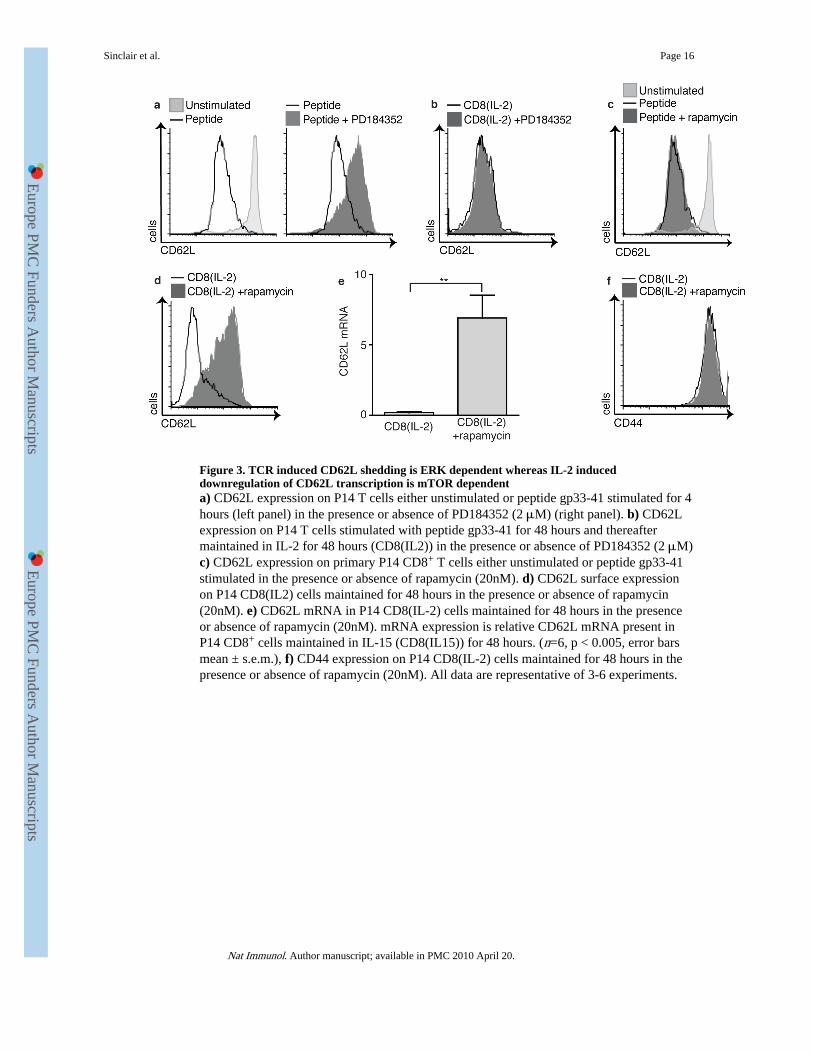
Figure 3. TCR induced CD62L shedding is ERK dependent whereas IL-2 induceddownregulation of CD62L transcription is mTOR dependenta) CD62L expression on P14 T cells either unstimulated or peptide gp33-41 stimulated for 4hours (left panel) in the presence or absence of PD184352 (2 μM) (right panel). b) CD62Lexpression on P14 T cells stimulated with peptide gp33-41 for 48 hours and thereaftermaintained in IL-2 for 48 hours (CD8(IL2)) in the presence or absence of PD184352 (2 μM)c) CD62L expression on primary P14 CD8+ T cells either unstimulated or peptide gp33-41stimulated in the presence or absence of rapamycin (20nM). d) CD62L surface expressionon P14 CD8(IL2) cells maintained for 48 hours in the presence or absence of rapamycin(20nM). e) CD62L mRNA in P14 CD8(IL-2) cells maintained for 48 hours in the presenceor absence of rapamycin (20nM). mRNA expression is relative CD62L mRNA present inP14 CD8+ cells maintained in IL-15 (CD8(IL15)) for 48 hours. (n=6, p < 0.005, error barsmean ± s.e.m.), f) CD44 expression on P14 CD8(IL-2) cells maintained for 48 hours in thepresence or absence of rapamycin (20nM). All data are representative of 3-6 experiments.
Sinclair et al. Page 16
Nat Immunol. Author manuscript; available in PMC 2010 April 20.
Europe PM
C Funders A
uthor Manuscripts
Europe PM
C Funders A
uthor Manuscripts
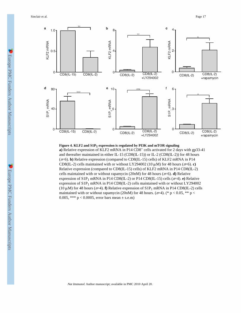
Figure 4. KLF2 and S1P1 expression is regulated by PI3K and mTOR signalinga) Relative expression of KLF2 mRNA in P14 CD8+ cells activated for 2 days with gp33-41and thereafter maintained in either IL-15 (CD8(IL-15)) or IL-2 (CD8(IL-2)) for 48 hours(n=6). b) Relative expression (compared to CD8(IL-15) cells) of KLF2 mRNA in P14CD8(IL-2) cells maintained with or without LY294002 (10 μM) for 48 hours (n=6). c)Relative expression (compared to CD8(IL-15) cells) of KLF2 mRNA in P14 CD8(IL-2)cells maintained with or without rapamycin (20nM) for 48 hours (n=6). d) Relativeexpression of S1P1 mRNA in P14 CD8(IL-2) or P14 CD8(IL-15) cells (n=4). e) Relativeexpression of S1P1 mRNA in P14 CD8(IL-2) cells maintained with or without LY294002(10 μM) for 48 hours (n=4). f) Relative expression of S1P1 mRNA in P14 CD8(IL-2) cellsmaintained with or without rapamycin (20nM) for 48 hours. (n=4). (* p < 0.05, ** p <0.005, *** p < 0.0005, error bars mean ± s.e.m)
Sinclair et al. Page 17
Nat Immunol. Author manuscript; available in PMC 2010 April 20.
Europe PM
C Funders A
uthor Manuscripts
Europe PM
C Funders A
uthor Manuscripts

Figure 5. Loss of PTEN is sufficient to downregulate CD62L expressiona) CD62L surface expression on PTEN KO(T) versus wild-type (WT) thymocytes (n=3). b)CD62L mRNA expression in sorted DP and CD4 SPs from PTEN KO(T) and WT controls.Expression of CD62L mRNA is shown relative to WT DP cells (n=3). c) Amount of CD62Lshed (pg/105 cells) over 1 hour from PTEN KO(T) or WT thymocytes. The data show anaverage of 2 experiments performed in triplicate (p < 0.0005, error bars mean ± s.e.m.). d)CD62L surface expression on PTEN KO(T) or WT CD8(IL-15) cells. e) Amount of CD62Lshed (pg/105 cells) during 1 hour from PTEN KO(T) or WT CD8(IL-15) cells The datashow an average of 2 experiments performed in triplicate. (p < 0.0005, error bars mean ±s.e.m.) f) Relative expression of CD62L mRNA from PTEN KO(T) or WT CD8(IL-15)cells. (n=3, p < 0.0005, error bars mean ± s.e.m.). g) Relative expression of KLF2 mRNAfrom PTEN KO(T) or WT CD8(IL-15) cells (n=3, p < 0.0005, error bars mean ± s.e.m.). h)CD24 expression on CD4 or CD8 single positive PTEN KO(T) or WT thymocytes (% ofCD24 low expressing cells is indicated). (n=6)
Sinclair et al. Page 18
Nat Immunol. Author manuscript; available in PMC 2010 April 20.
Europe PM
C Funders A
uthor Manuscripts
Europe PM
C Funders A
uthor Manuscripts

Figure 6. CCR7 downregulation on activated T cells is dependent on PI3K and mTORa) CCR7 surface expression on P14 CD8+ cells activated for 2 days with gp33-41 andthereafter maintained for 48 hours in either IL-15 (CD8(IL-15)) or IL-2 (CD8(IL-2)). b)CCR7 surface expression on CD8(IL-2) cells cultured for 48 hours with or withoutLY294002 (10 μM). c) CCR7 surface expression on PTEN KO(T) or wild-type (WT)CD8(IL-15) cells. d) CCR7 surface expression on CD8(IL-2) cells cultured with or withoutrapamycin (20nM) for 48 hours. e) Transwell migration to CCL19 of CD8(IL-15) orCD8(IL-2) cells maintained for 48 hours in the presence or absence of either LY294002 (10μM) (CD8(IL-2+LY294002)) or rapamycin (20nM) (CD8(IL-2+rapamycin)) (n=3, * p <0.05, ** p < 0.005, *** p < 0.0005, error bars mean ± s.e.m). f) P14 CD8(IL-2) cells wereeither untreated or treated with rapamycin for 48 hours, labeled with CFSE or CMTMRrespectively, and mixed at a ratio of 1:1 prior to injection into C57Bl/6 hosts. The data showthe percentage of CD8(IL-2) versus CD8(IL-2+rapamycin) cells as a percentage of the totalnumber of transferred cells recovered in the blood, lymph nodes or spleen 24 hours aftertransfer (n= 9, *** p < 0.0005, bar shows mean).
Sinclair et al. Page 19
Nat Immunol. Author manuscript; available in PMC 2010 April 20.
Europe PM
C Funders A
uthor Manuscripts
Europe PM
C Funders A
uthor Manuscripts


