Embed Size (px)
Citation preview

Phosphorylation-independent dual-site binding of theFHA domain of KIF13 mediates phosphoinositidetransport via centaurin α1Yufeng Tonga, Wolfram Tempela, Hui Wanga, Kaori Yamadab, Limin Shena, Guillermo A. Senisterraa, Farrell MacKenziea,Athar H. Chishtib,d, and Hee-Won Parka,c,1
aStructural Genomics Consortium, University of Toronto, Toronto, ON M5G 1L7, Canada; cDepartment of Pharmacology and Toxicology,University of Toronto, Toronto, ON M5S 1A8, Canada; bDepartment of Pharmacology, University of Illinois, College of Medicine, Chicago, IL 60612; anddDepartment of Physiology, Tufts University School of Medicine, Boston, MA 02111
Edited by Linda A. Amos, MRC Laboratory of Molecular Biology, Cambridge, United Kingdom, and accepted by the Editorial Board September 21, 2010(received for review June 30, 2010)
Phosphatidylinositol 3,4,5-triphosphate (PIP3) plays a key role inneuronal polarization and axon formation. PIP3-containing vesiclesare transported to axon tips by the kinesin KIF13B via an adaptorprotein, centaurin α1 (CENTA1). KIF13B interacts with CENTA1through its forkhead-associated (FHA) domain. We solved the crys-tal structures of CENTA1 in ligand-free, KIF13B-FHA domain-bound,and PIP3 head group (IP4)-bound conformations, and the CENTA1/KIF13B-FHA/IP4 ternary complex. The first pleckstrin homology(PH) domain of CENTA1 specifically binds to PIP3, while the secondbinds to both PIP3 and phosphatidylinositol 3,4-biphosphate(PIð3,4ÞP2). The FHA domain of KIF13B interacts with the PH1 do-main of one CENTA1 molecule and the ArfGAP domain of a secondCENTA1 molecule in a threonine phosphorylation-independentfashion. We propose that full-length KIF13B and CENTA1 formheterotetramers that can bind four phosphoinositide moleculesin the vesicle and transport it along the microtubule.
kinesin ∣ forkhead-associated domain ∣ vesicle transport ∣phosphatidylinositol triphosphate ∣ neuronal development
Neurons are highly polarized cells that typically feature onelong axon and several shorter dendrites. Newborn neurons
initially develop immature neurites that undergo constant, ran-dom growth and retraction regulated by positive and negativefeedback signals (1), and become polarized when this balanceis shifted so that one neurite becomes an axon and the othersbecome dendrites. Accumulation of phosphatidylinositol (3,4,5)-trisphosphate (PIP3), which is synthesized by phosphoinositide3-kinase (PI3K) and catabolized by the phosphatase PTEN (2)at the distal end of one neurite, induces axonal development(2, 3). However, an alternative pathway for PIP3 accumulationis the transport of PIP3-containing vesicles by KIF13B and thePIP3-binding protein centaurin α1 (CENTA1) (4).
KIF13B is a kinesin-3 subfamily member containing anN-terminal motor domain with ATPase activity that drives move-ment along the microtubule and a forkhead-associated (FHA)domain, the only known phosphothreonine (pThr)-specific recog-nition domain (5–8). KIF13B also contains a long coiled-coil do-main and a C-terminal glycine-rich microtubule-binding domain(9), both of which function in dimer formation (10). KIF13B doesnot interact with PIP3-containing vesicles directly; rather, it bindsvia its FHA domain to CENTA1 (4), which contains an ArfGAPdomain that inactivates Arf6 (11) and two pleckstrin homology(PH) domains that interact with PIP3 (12–15). CENTA1 is asso-ciated with presynaptic vesicle structures (16), and its expressionis elevated in the neurons of Alzheimer’s patients (17).
To understand how CENTA1 and PIP lipids interact, we solvedthe structures of lipid-free and lipid-bound forms of CENTA1 andconfirmed binding specificity using a PIP array assay. We foundthat the first PH domain of CENTA1 binds PIP3, while the secondbinds phosphatidylinositol (3,4)-bisphophate (PIð3;4ÞP2) and
PIP3. We also solved the structure of the KIF13B-FHA domainbound to full-length CENTA1, which interacts with KIF13B-FHA mainly through its first PH domain in a phosphorylation-independent manner. Our result provides structural evidence thatFHA can function as a non-pThr-binding module. The structuralfindings suggest that KIF13B-mediated cargo transport happensby two KIF13B and two CENTA1 molecules forming a heterote-tramer that binds up to four PIP molecules in each vesicle.
ResultsCrystal Structure of Free and IP4-Bound Centaurin α1. We solved thecrystal structures of CENTA1 in lipid-free and PIP3 head group(IP4)-bound conformations (Fig. 1A and Table S1). CENTA1 isan L-shaped protein with three domains linked by two short he-lices: an N-terminal ArfGAP domain and PH1 and PH2 domains.The lipid-binding pockets of the two PH domains are on thesame, nearly flat surface (Fig. 1 A and B). The ArfGAP domaincontains a C4-type zinc finger, and has a contact interface of517 Å2 with the PH1 domain, but is far from the PH2 domain.The PH1 and PH2 domains both adopt a typical PH fold (18)consisting of a β-barrel of orthogonal three- and four-strandβ-sheets, with one end blocked by a C-terminal α-helix and theother end open for binding phosphoinositides. The PH1 andPH2 domains have an average contact area of 523 Å2, with multi-ple hydrophobic and polar interactions. Residues 360–370 areinvisible in the lipid-free structure, but appear as a three-turnα-helix in the IP4-bound structure (Fig. 1A), the C terminus ofwhich points to the IP4-binding surface. The Cα atoms of theArfGAP domain have an rmsd difference of 0.19 Å betweenthe lipid-free and the IP4-bound structures, while the rmsds ofPH1 and PH2 are 0.38 Å and 0.53 Å, respectively. The flexibilityof the PH2 domain is consistent with its high B-factor (Fig. S1).
In the CENTA1-PIP3 structure, the IP4 moiety of the diocta-noyl-PIP3 occupies the phosphoinositide-binding pocket of bothPH domains, but the diacylglycerol group is not visible in the elec-tron density map. We hereinafter refer to IP4 as the ligand boundto CENTA1. The phosphate groups of IP4 make polar contactswith similar sets of residues around the binding pockets of bothPH domains (Fig. 1 C–D). However, while PH2 contains the
Author contributions: Y.T. and H.-W.P. designed research; Y.T., H.W., L.S., G.A.S., K.Y., andF.M. performed research; Y.T., W.T., and H.-W.P. analyzed data; and Y.T., W.T., K.Y., A.H.C.,and H.-W.P. wrote the paper.
The authors declare no conflict of interest.
This article is a PNAS Direct Submission. L.A.A. is a guest editor invited by theEditorial Board.
Data deposition: The atomic coordinates and structure factors have been deposited inthe Protein Data Bank, www.pdb.org.1To whom correspondence should be addressed. E-mail: [email protected].
This article contains supporting information online at www.pnas.org/lookup/suppl/doi:10.1073/pnas.1009008107/-/DCSupplemental.
20346–20351 ∣ PNAS ∣ November 23, 2010 ∣ vol. 107 ∣ no. 47 www.pnas.org/cgi/doi/10.1073/pnas.1009008107

canonical KXnðK∕RÞXR motif in the β1-β2 loop important forhigh-affinity 3-phosphoinositide-binding (18–20), PH1 has aleucine (Leu147) in place of the (K/R) residue, which is notseen in any other known phosphoinositide-binding PH domains.The invariant lysine residue in all canonical polyphosphoinosi-tide-binding PH domains, Lys138 in PH1 and Lys261 in PH2, in-teracts with both the 3- and 4- phosphates of IP4. The invariantarginine residue, Arg149 in PH1 and Arg273 in PH2, interactswith the 3-phosphate of IP4. In both cases, the axial 2-hydroxylgroup points to the β1-β2 loop and forms a water bridge withresidues in the sequence motif, thus fixing the inositol ring inthe β-barrel cleft.
Variability among other residues results in differential phos-phoinositide-binding specificity of the PH domains. The guanidi-nium group of Arg271 in PH2 interacts directly with the1-phosphate, and indirectly through a water bridge (Fig. 1D).This water molecule also forms a strong hydrogen bond withthe main chain carboxyl group of Gly269 and with the 2-hydroxylgroup of the inositol ring. In contrast, Leu147 of PH1 (the posi-
tion equivalent to Arg271) does not contact IP4, and the 1-phos-phate of IP4 is not visible. Tyr162 and Arg206 in PH1 and theirequivalent residues, Tyr284 and Arg332, in PH2 interact directlywith a 4-phosphate group. Strikingly, the 5-phosphate of PH2-bound IP4 does not directly contact PH2, while that of PH1-bound IP4 forms two strong hydrogen bonds with the Arg141and Lys172 side chains of PH1. The equivalent residues inPH2 are Pro264 and Arg294. While the side and main chainsof Arg294 interact with the 3- and 4-phosphates of IP4 bridgedby two water molecules, Pro264 is oriented such that the sidechain of Lys265 is far from the IP4 5-phosphate, preventing5-phosphate interaction with the PH2 domain. These structuraldata (Fig. 1 C–D) suggest that the two PH domains have differentphosphoinositide-binding specificity, with PH1 binding PIP3 andPH2 interacting with both PIP3 and PIð3;4ÞP2.
PIPArray Assay and Thermostability.To test the binding of CENTA1to all eight naturally occurring phosphoinositides, we performeda membrane-based phosphoinositide array assay. Consistentwith the structure-based prediction, CENTA1 interacts with bothPIP3 and PIð3;4ÞP2, though more weakly with the latter (Fig. 2A),and does not bind to other phosphoinositides.
To test the effect of phosphoinositides on CENTA1 stabilityand of key protein residues on lipid-binding affinity, we used dif-ferential static light scattering (21, 22) to measure the thermalaggregation of wild-type and mutant CENTA1 in the presenceof phosphoinositides (Fig. 2 B–C). Phosphoinositide-interactingresidues were mutated: Arg149 in PH1, and Arg271, Arg273 inPH2. Three single-site (R149C, R271C, and R273C) and onedouble-site (R149CR271C) mutants were generated, and thethermal aggregation temperatures (Tagg) of the proteins weremeasured.
Not surprisingly, Tagg decreases rapidly with increasing saltconcentration (Fig. S2A). We measured the Tagg of wild-typeand mutant CENTA1 with varying IP4 concentrations in lowsalt buffer [20 mM Tris, pH 8.0, 50 mM sodium chloride,1 mM tri(2-carboxyethyl)phosphine], then fitted the IP4 titrationdata with a pseudo one-site binding model to extrapolate maxi-mal aggregation temperature increase (ΔTmax) (Fig. 2B).Although both PH1 and PH2 interact with three phosphategroups of the phosphoinositides (Fig. 1 C–D), PH1 has a slightlyhigher affinity for IP4 than does PH2 (ΔTmax½R149C� ¼ 10.2 °C,ΔTmax½R273C� ¼ 11.0 °C), consistent with the observation thatIP4 appears to be bound to PH1 but not PH2 in the CENTA1/KIF13B-FHA/IP4 complex (see below). In the PH2 domain,Arg273 contributes more than Arg271 to protein stabilizationupon IP4 binding, because the ΔTmax½R273C� (11.0 °C) is lessthan ΔTmax½R271C� (12.3 °C). Double-point mutations in bothPH1 and PH2 (R149CR271C) greatly reduced IP4 binding(ΔTmax ¼ 2.6 °C).
Unexpectedly, we observed a decrease in the Tagg of CENTA1mutants with phosphoinositides bearing long aliphatic sidechains, such as dioctanyl-PIP3 and PIð3;4ÞP2, so simple models
Fig. 1. Structures of CENTA1 in apo- and IP4-bound forms. (A) Cartoondiagram of CENTA1 in apo-form (gray) aligned with IP4-bound form: ArfGAP(1–119, green), PH1 (131–238, magenta), PH2 (253–359, salmon red), interdo-main linkers (120–130, 239–252, yellow), C-terminal helix (360–370, red). IP4molecules and CENTA1 zinc-finger cysteines are shown in sticks. Zinc is shownas an orange sphere. (B) Bottom-up view of the �10.0 kT∕e electrostaticpotential surface of CENTA1 in the IP4-bound structure. (C) and (D) Ligplotrepresentations of the interaction between IP4 and PH1, PH2 domains respec-tively. Residues from the KXnðK∕RÞXR motif are marked with stars.
Fig. 2. Biochemical and biophysical characterization of phosphoinositides-CENTA1 interaction. (A) PIP array assay. Lanes A–G, each spot contains 100, 50, 25,12.5, 6.25, 3.13, and 1.56 pmol of corresponding phosphoinositides on the membrane. (B) Tagg titration curve of wild-type and mutant CENTA1 at different IP4concentrations. (C) Relative aggregation temperature of wild-type and mutant CENTA1 to that of R149CR271C double mutant. Values for IP4 are fitted ΔTmax
results from titration. Values for PIP3 and PIð3;4ÞP2 are ΔTagg in the presence of 100 μg∕mL phosphoinositides (109 and 123 μM, respectively).
Tong et al. PNAS ∣ November 23, 2010 ∣ vol. 107 ∣ no. 47 ∣ 20347
BIOPH
YSICSAND
COMPU
TATIONALBIOLO
GY

do not fit the data (Fig. S2B). To determine the effect of phos-phoinositides on the thermal stability of PH domains, we calcu-lated the relative aggregation temperature (Tr;agg) of wild-typeand mutant CENTA1, to the double-mutant R149CR271C,which shows near-complete loss of inositol phosphate binding.Tr;agg offsets the effect of phosphoinositide aliphatic chain bind-ing to thermally denatured protein, and is thus a measure of thebinding affinity of the head groups to PH domains. When Arg149is mutated (R149C), phosphoinositide binding occurs mainly inthe PH2 domain. The Tr;agg of R149C with PIP3 and PIð3;4ÞP2
is similar, indicating that PH2 binds equally well to both. In con-trast, proteins with mutation in PH2 (R271C and R273C) showhigher Tr;agg in the presence of PIP3 than of PIð3;4ÞP2, indicatingthat PH1 binds preferentially to PIP3. Thus, the PIP array assayand thermostability measurements confirmed our structure-based prediction that PH1 is PIP3-specific, while PH2 binds toboth PIP3 and PIð3;4ÞP2.
Crystal Structures of CENTA1/FHA and CENTA1/KIF13B/IP4 Complexes.KIF13B has previously been shown by yeast two-hybrid andGST-pull-down assays to interact directly through its FHA do-main with CENTA1 (4, 23). We verified the direct interactionof the KIF13B-FHA domain (aa 440–545) with CENTA1 bygel filtration (Fig. S3A). A longer KIF13B construct containingboth the motor and FHA domains (aa 1–550) also interactsdirectly with CENTA1 by GST-pull-down and gel filtration(Fig. S3B), consistent with an earlier observation that the KIF13Bmotor domain does not interfere with KIF13B-FHA interactionswith CENTA1 (4).
We solved the crystal structure of the CENTA1/KIF13B-FHAcomplex at 2.3 Å resolution (Table S1). The asymmetric unit con-tains two CENTA1 and two KIF13B-FHAmolecules (Fig. 3), andthe CENTA1 in the complex has an overall Cα rmsd of 0.74 Åcompared to the lipid-free CENTA1 structure, mainly due to in-terdomain movement of the PH2 domain upon FHA binding(Table S2). Other significant conformational changes in the com-plex include disordering of the PH1 β4-β5 and PH2 β5-β6 loopsand the appearance of the C-terminal α-helix (aa 360–370). TheFHA domain of KIF13B is a typical 11-stranded β-fold commonin other FHA structures (5, 24, 25). In the asymmetric unit, FHAresidues located mainly on strands β7 and β10 and in the β1-β2loop interact with a surface of PH1 orthogonal to the phosphoi-nositide-binding pocket, which consists of residues at the N ter-minus of β2, the β5-β6 loop, and the β7−α-helix loop. The FHAdomain and PH1 interface has an average buried surface area of663 Å2 and contains multiple polar and hydrophobic interactions(Fig. S4A). In addition, the interface contains three bridgingwater molecules. The KIF13B FHA domain also interacts withthe ArfGAP domain of the second CENTA1 molecule in theasymmetric unit through its Asn536 and Met486 side chains(Fig. S4A). Thus, KIF13B-FHA interacts mainly with the PH1
domain of one CENTA1 molecule, and less extensively withthe ArfGAP domain of the second CENTA1 molecule. TheCENTA1-CENTA1 interface has a buried surface of 667 Å2 andcontains multiple polar and hydrophobic interactions, and threebridging water molecules (Fig. S4B). The interactions mainlyhappen between the first interdomain linker of one CENTA1molecule with the first interdomain linker and PH1 domain ofthe second CENTA1 molecule.
The structure of the CENTA1/KIF13B-FHA/IP4 ternary com-plex was also solved at 2.95 Å resolution (Table S1). The overallstructure is almost the same as that of the binary complex, withan rmsd of 0.21 Å for all the Cα atoms of both KIF13B andCENTA1. In contrast to the CENTA1/IP4 binary complex whereboth the PH1 and PH2 domains were occupied by IP4 ligands,however, a IP4 ligand occupies only one of the two PH1 domainsof the heterotetramer in the ternary complex, but not the otherPH1 domain and the PH2 domains. Compared to the structureof CENTA1/IP4, the most obvious conformational change isthe visibility of the 1-phosphate of the IP4 bound to PH1 in theternary complex. Thus, no major conformational change is seenwhen IP4 is bound to the CENTA1/KIF13B-FHA complex.
CENTA1/KIF13B Interaction Specificity. To confirm that the interfacebetween KIF13B-FHA and CENTA1 observed in the crystalstructure is biologically relevant, we introduced single-site muta-tions in both CENTA1 and KIF13B-FHA and measured the ther-modynamic parameters using isothermal titration calorimetry(ITC). Wild-type proteins interact at a Kd of 0.8 μM, while singleconservative mutations result in enthalpy-entropy compensation(Table S3 and Fig. S5 A and B). For example, the Y211FCENTA1 mutation eliminates interactions between the Tyr211hydroxyl group and the Asn454 and Asn537 side chains ofKIF13B-FHA, leading to a ΔH increase of 3.4 kcal∕mol due toloss of the polar interactions, and an entropy increase (−TΔS)of 2.98 kcal∕mol. The net free energy change is only 0.4 kcal∕mol.Mutation of Tyr211 to glycine or ariginine caused CENTA1 toprecipitate at the temperature used for ITC measurement. Theinteractions of CENTA1 Y211G, and Y211R mutants withKIF13B-FHA were thus tested using size exclusion chromatogra-phy, and their complex formation was not detectable (Fig. S5C).
To test the binding specificity between CENTA1 and theFHA domains of different kinesin-3 family members, we carriedout GST-pull-down experiments. CENTA1 interacts with a GST-tagged KIF13B motor+FHA construct and a GST-taggedKIF13A motor+FHA construct, but not with the GST control(Fig. S6). Furthermore, the FHA domains of KIF1B, KIF1C,KIF14, and KIF16B do not interact with GST-CENTA1, whileKIF13B-FHA does (Table S4). Thus, CENTA1 interacts withKIF13A and 13B, but not with other kinesin-3 family members.
DiscussionCENTA1 Binds to both PIP3 and PIð3,4ÞP2. Our data indicate thatthe PH1 domain of CENTA1 preferentially binds PIP3, whilethe PH2 domain binds both PIP3 and PIð3;4ÞP2. Although the3- and 4- but not the 5-phosphate groups are recognized bythe PH2 domain of lipid-bound CENTA1, the binding affinityof PH2 to PIP3 is comparable to that of PH1, due to coordinationof the 1-phosphate by the Arg271 side chain and by solvent-mediated interactions with Gly269.
PIð3;4ÞP2 and PIP3 are two major PI3K kinase products thatregulate intracellular signaling (26, 27). While PIP3 is mainlyin the plasma membrane, PIð3;4ÞP2 is found in the plasma mem-brane, endoplasmic reticulum, and multivesicular endosomes(28, 29). In developing neurons, the balance of PIP3 generationand dephosphorylation into PIð4;5ÞP2 leads to constant out-growth and collapse of immature neurites (1). The role ofPIð3;4ÞP2 is not clear, but it is a direct, positive regulator ofAkt/PKB (30, 31) and is insensitive to PTEN inactivation (32, 33).
Fig. 3. Structure of CENTA1 bound to KIF13B-FHA domain in the asymmetricunit.
20348 ∣ www.pnas.org/cgi/doi/10.1073/pnas.1009008107 Tong et al.

Our data suggest that the CENTA1 PH2 domain may mediatetransport of PIð3;4ÞP2-containing vesicles to the distal end of de-veloping neurons. Thus, delivery of PIð3;4ÞP2 to neurite tips mayprovide an alternative signaling pathway to tip the balance ofPIP3-mediated feedback loops and regulate neurite outgrowth.
Conformational Change of CENTA1 upon Ligand Binding. The bindingpockets of both PH domains are located on the same surface ofCENTA1, allowing them to bind the same vesicle. Upon PIP3 orFHA binding, previously invisible residues C-terminal to the PH2domain (aa 360–370) of CENTA1 appeared as a short α-helix.The C terminus of the helix points to the lipid-binding surfaceof the molecule, bringing the last four residues of the molecule(KHKP) close to the vesicle. The basic lysine residues can thenbind nonspecifically to the head group of the phosphoinositide-containing vesicle (34), further enhancing the affinity of CENTA1for the vesicle.
The most significant conformational change of CENTA1 uponPIP3 binding occurs in the PH2 domain. The rmsd of Cα atoms inPH2 with and without lipid-binding is larger than that of thermsd of the three domains combined (Table S2). However, inthe CENTA1/KIF13B-FHA and CENTA1/KIF13B-FHA/IP4complexes, the rmsds of the Cα atoms in the individual CENTA1domains are all much smaller than the overall rmsd of the threedomains combined when compared to the apo-form of CENTA1.Upon binding of the FHA domain, PH1 shows the largestconformational change among the three domains in the binarycomplex. In the ternary complex, even though only one IP4 isbound to PH1, PH2 undergoes additional conformation changes(Table S2), suggesting that not only phosphoinositide bindingcauses local conformational changes in PH domains, but thatKIF13B-FHA binding to PH1 of CENTA1 causes PH2 to adjustits orientation, possibly promoting assembly of the PIP3-vesicle/CENTA1/KIF13B transport machinery.
CENTA1 and KIF13-FHA Interaction Is Specific and Phospho-Threonine-Independent. The discovery that KIF13B interacts with CENTA1via its FHA domain and transports PIP3 vesicles suggested a car-go-binding role for the FHA domain in kinesins (4). Phosphor-ylation-independent binding of CENTA1 to KIF13B-FHA wasfirst proposed based on GST-pull-down experiments (4). We havealso individually mutated each of the serines, threonines, andtyrosines in the ArfGAP domain of CENTA1 to alanine, testedthe binding of the mutants to KIF13B-FHA by GST-pull-down,and found no effect. The structure of KIF13B-FHA-boundCENTA1 and our ITC measurements of CENTA1 and KIF13B-FHA mutant interactions confirmed that KIF13B-FHA binds toboth the PH1 and ArfGAP domains of CENTA1 via a uniqueinterface in a phosphorylation-independent manner. Indeed,KIF13B-FHA lacks the conserved phosphate-interacting resi-dues (Fig. S7) and the positively charged surface (Fig. S8) ofpThr-binding FHA domains.
KIF13A is a close homolog of KIF13B and differs in theCENTA1-interacting regions by only two residues. Phe514 andMet486 of KIF13B, which interact hydrophobically with Pro192,and the ArfGAP domain of CENTA1 respectively, are a cysteineand a hydrophobic isoleucine in KIF13A (Fig. S4 and Fig. S7).Our GST-pull-down data suggest that CENTA1 binds to KIF13A-FHA and KIF13B-FHA with similar affinity, but does not bindthe FHA domain of other kinesin-3 family members. As coimmu-noprecipitation data from COS-7 cells (4) showed no interactionbetween KIF13A and CENTA1, alternative phosphorylation orKIF13A regulation mechanisms may exist in intact cells. KIF13Ais known to transport the mannose-6-phosphate receptor viaAP1/β1-adaptin complex through its C-terminal tail domain(35) and is expressed in the central nervous system during earlymouse development (36). The binding of the FHA domain of
KIF13A to CENTA1 suggests that KIF13A may also play a rolein PIP3 vesicle transport and neuronal development.
Based on the structure of FHA bound to a pThr-containingpeptide (24) and evolutionary trace analysis (37), pThr-bind-ing-independent functions of FHA were proposed. Phosphoryla-tion-independent interactions of FHA with c-Myc have also beenreported for SNIP1, a candidate transcriptional regulator (38).The interface residues in our CENTA1/KIF13B-FHA structurelie on strands β7 and β10 of KIF13B-FHA, consistent with theevolutionary trace analysis (37). Structure-based sequencealignment (Fig. S7) suggests that a glycine (Gly474 in KIF13B)following strand β3 is the only invariant residue in FHA domains.All known pThr-binding FHA domains have an arginine after thisglycine, while the KIF13B-FHA has a serine. Other proteins,such as MLLT4, PHLDB1, and RADIL have an FHA domainwithout residues necessary for phosphate group binding. OurCENTA1/KIF13B-FHA structure provides atomic details of howthe FHA domain interacts with another protein in a pThr-inde-pendent manner. Our results indicate that the FHA domaincould function through mechanisms other than phosphothreo-nine recognition.
PIP3 Vesicle Transport and Cargo Loading. KIF13B was proposed tofunction as a dimer based on chemical cross-linking and fragmentpull-down data (10). KIF13B dimerizes through its C-terminaldomain, unlike conventional kinesins, which dimerize throughthe coiled-coil domain following the motor domain. In the asym-metric unit of the CENTA1/KIF13B-FHA crystal structure, twoCENTA1 molecules and two FHA domains formed a heterote-tramer. The two FHA domains do not interact with each other.Instead, each FHA domain interacts with both the PH1 domainof one CENTA1molecule and the ArfGAP domain of the secondCENTA1 molecule, as fewer interactions were seen with the Arf-GAP domain (Fig. S4A). In addition, the two CENTA1moleculesform a homodimer with an interface containing multiple hydro-gen bonds and salt bridges (Fig. S4B), although CENTA1 homo-dimer is not detectable in solution (Fig. S3). Together, we predictthat each of the two FHA domains of full-length KIF13B dimermay interact with two CENTA1, promoting CENTA1 homodi-merization and thereby forming the heterotetramer.
The crystallographic heterotetramer is likely biologicallyrelevant for the following reasons: (i) interaction between theArfGAP domain of CENTA1 with the N terminus of theFHA-containing KIF13B stalk domain has been verified bothby pull-down and in vivo colocalization data (23); (ii) gel filtrationof KIF13B-motor-FHA with CENTA1 (Fig. S3B) shows that thecomplex size approaches heterotetramer size with increased saltconcentration, suggesting the asymmetric unit of CENTA1/KIF13B-FHA is primed for heterotetramerization in the contextof full-length KIF13B; (iii) the distance between the N termini ofthe two FHA domains in the asymmetric unit is 80 Å, similar tothe distance between adjacent β-tubulin subunits in microtubules,suggesting that the configuration of the crystallographic hetero-tetramer is compatible with a model of two motor heads bound totwo neighboring β-tubulins.
Heterooligomerization of KIF13B with CENTA1 doubles thenumber of PH domains exposed to the PIP3 vesicle, thus increas-ing the affinity between the cargo and KIF13B. However, PIP3-containing lipid vesicles may also help to cluster the CENTA1/KIF13B complex into a heterotetramer (Fig. 4A). Dimerizationof another kinesin-3 family member, UNC-104, with the aid ofPIð4;5ÞP2-containing liposomes renders the kinesin highly pro-cessive and triggers membrane transport (39, 40). EndogenousKIF13B exists in an autoinhibited state until activated by mem-brane-associated guanylate kinase family member hDlg (10).CENTA1 can shuttle easily between the cytosol and PIP-contain-ing lipid-membrane (41). We propose that CENTA1 is recruitedto the membrane, independent of KIF13B activation. Once
Tong et al. PNAS ∣ November 23, 2010 ∣ vol. 107 ∣ no. 47 ∣ 20349
BIOPH
YSICSAND
COMPU
TATIONALBIOLO
GY
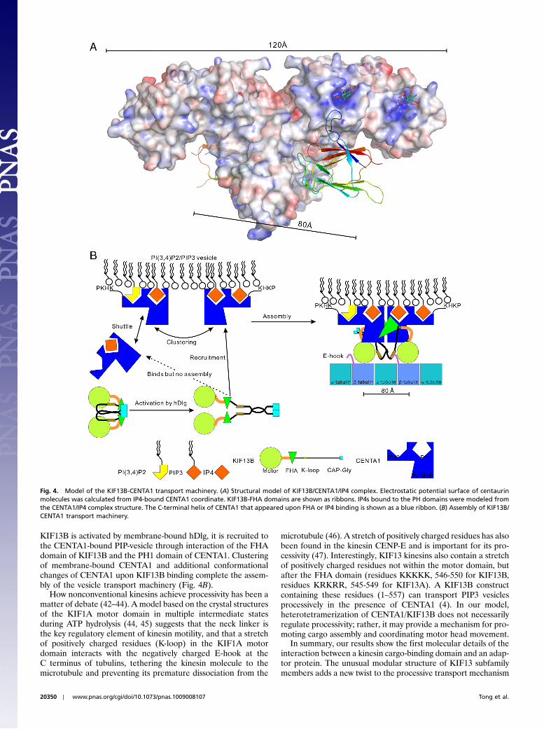
KIF13B is activated by membrane-bound hDlg, it is recruited tothe CENTA1-bound PIP-vesicle through interaction of the FHAdomain of KIF13B and the PH1 domain of CENTA1. Clusteringof membrane-bound CENTA1 and additional conformationalchanges of CENTA1 upon KIF13B binding complete the assem-bly of the vesicle transport machinery (Fig. 4B).
How nonconventional kinesins achieve processivity has been amatter of debate (42–44). A model based on the crystal structuresof the KIF1A motor domain in multiple intermediate statesduring ATP hydrolysis (44, 45) suggests that the neck linker isthe key regulatory element of kinesin motility, and that a stretchof positively charged residues (K-loop) in the KIF1A motordomain interacts with the negatively charged E-hook at theC terminus of tubulins, tethering the kinesin molecule to themicrotubule and preventing its premature dissociation from the
microtubule (46). A stretch of positively charged residues has alsobeen found in the kinesin CENP-E and is important for its pro-cessivity (47). Interestingly, KIF13 kinesins also contain a stretchof positively charged residues not within the motor domain, butafter the FHA domain (residues KKKKK, 546-550 for KIF13B,residues KRKRR, 545-549 for KIF13A). A KIF13B constructcontaining these residues (1–557) can transport PIP3 vesiclesprocessively in the presence of CENTA1 (4). In our model,heterotetramerization of CENTA1/KIF13B does not necessarilyregulate processivity; rather, it may provide a mechanism for pro-moting cargo assembly and coordinating motor head movement.
In summary, our results show the first molecular details of theinteraction between a kinesin cargo-binding domain and an adap-tor protein. The unusual modular structure of KIF13 subfamilymembers adds a new twist to the processive transport mechanism
Fig. 4. Model of the KIF13B-CENTA1 transport machinery. (A) Structural model of KIF13B/CENTA1/IP4 complex. Electrostatic potential surface of centaurinmolecules was calculated from IP4-bound CENTA1 coordinate. KIF13B-FHA domains are shown as ribbons. IP4s bound to the PH domains were modeled fromthe CENTA1/IP4 complex structure. The C-terminal helix of CENTA1 that appeared upon FHA or IP4 binding is shown as a blue ribbon. (B) Assembly of KIF13B/CENTA1 transport machinery.
20350 ∣ www.pnas.org/cgi/doi/10.1073/pnas.1009008107 Tong et al.
of cargo by kinesin motor proteins. The confirmation ofphosphorylation-independent binding of the FHA domain toCENTA1 reveals another potential function of what has longbeen considered a pThr recognition structural module. Finally,the unexpected finding that CENTA1 can recognize PIð3;4ÞP2
raises questions about the role of this previously largely ignoredphosphoinositide in signaling and axon development.
Materials and MethodsWe made DNA constructs of CENTA1 and KIF13B with an N-terminal His6 taginto pET28-MHL and purified the expressed proteins by affinity and sizeexclusion chromatography. Diffraction datasets of the crystals of CENTA1in various conformational states were collected at the Advanced PhotonSource and their structures were determined by using the crystallographyprograms. Assays of PIP array (Echelon Biosciences) and differential staticlight scattering using StarGazer (Harbinger Biotechnology and Engineering)were performed by following manufacture’s instructions. ITC measurementwas performed by using a VP-ITC MicroCalorimeter by following manufac-
ture’s instructions. We used glutathione Sepharose beads (Novagen) forGST-pull-down assays. The detailed methods are described in the onlineSI Text.
ACKNOWLEDGMENTS. We thank Dr. Haizhong Zhu for initial work onKIF13B-FHA domain, and Dr. Toshihiko Hanada for the first proposal anddemonstration of phosphorylation-independent binding of KIF13B FHAdomain with CENTA1. This work was supported by a Natural Sciences andEngineering Research Council of Canada (NSERC) Discovery Grant 371633-09 (to H.-W.P.) and an NIH Grant CA094414 (to A.H.C.). The Structural Geno-mics Consortium is a registered charity (number 1097737) that receives fundsfrom the Canadian Institutes for Health Research, the Canadian Foundationfor Innovation, Genome Canada through the Ontario Genomics Institute,GlaxoSmithKline, Karolinska Institutet, the Knut and Alice WallenbergFoundation, the Ontario Innovation Trust, the Ontario Ministry for Researchand Innovation, Merck and Co., Inc., the Novartis Research Foundation,the Swedish Agency for Innovation Systems, the Swedish Foundation forStrategic Research, and the Wellcome Trust.
1. Arimura N, Kaibuchi K (2007) Neuronal polarity: from extracellular signals to intracel-lular mechanisms. Nat Rev Neurosci 8:194–205.
2. Shi SH, Jan LY, Jan YN (2003) Hippocampal neuronal polarity specified by spatiallylocalized mPar3/mPar6 and PI 3-kinase activity. Cell 112:63–75.
3. Menager C, Arimura N, Fukata Y, Kaibuchi K (2004) PIP3 is involved in neuronalpolarization and axon formation. J Neurochem 89:109–118.
4. Horiguchi K, Hanada T, Fukui Y, Chishti AH (2006) Transport of PIP3 by GAKIN, akinesin-3 family protein, regulates neuronal cell polarity. J Cell Biol 174:425–436.
5. Mahajan A, et al. (2008) Structure and function of the phosphothreonine-specificFHA domain. Science Signaling 1:re12 http://www.ncbi.nlm.nih.gov/pubmed/19109241.
6. Liang X, Van D, Sr. (2008) Mechanistic insights into phosphoprotein-binding FHAdomains. Acc Chem Res 41:991–999.
7. Hammet A, et al. (2003) FHA domains as phospho-threonine binding modules in cellsignaling. IUBMB Life 55:23–27.
8. Durocher D, Jackson SP (2002) The FHA domain. FEBS Lett 513:58–66.9. Steinmetz MO, Akhmanova A (2008) Capturing protein tails by CAP-Gly domains.
Trends Biochem Sci 33:535–545.10. Yamada KH, Hanada T, Chishti AH (2007) The effector domain of human Dlg tumor
suppressor acts as a switch that relieves autoinhibition of kinesin-3 motor GAKIN/KIF13B. Biochemistry 46:10039–10045.
11. Venkateswarlu K, Brandom KG, Lawrence JL (2004) Centaurin-α1 is an in vivo phos-phatidylinositol 3,4,5-trisphosphate-dependent GTPase-activating protein for ARF6that is involved in actin cytoskeleton organization. J Biol Chem 279:6205–6208.
12. Hammonds-Odie LP, et al. (1996) Identification and cloning of centaurin-α. A novelphosphatidylinositol 3,4,5-trisphosphate-binding protein from rat brain. J Biol Chem271:18859–18868.
13. Tanaka K, et al. (1997) A target of phosphatidylinositol 3,4,5-trisphosphate with a zincfinger motif similar to that of the ADP-ribosylation-factor GTPase-activating proteinand two pleckstrin homology domains. Eur J Biochem 245:512–519.
14. Shirai T, et al. (1998) Specific detection of phosphatidylinositol 3,4,5-trisphosphatebinding proteins by the PIP3 analogue beads: an application for rapid purificationof the PIP3 binding proteins. Biochim Biophys Acta 1402:292–302.
15. Venkateswarlu K, Oatey PB, Tavare JM, Jackson TR, Cullen PJ (1999) Identification ofcentaurin-α1 as a potential in vivo phosphatidylinositol 3,4,5-trisphosphate-bindingprotein that is functionally homologous to the yeast ADP-ribosylation factor (ARF)GTPase-activating protein, Gcs1. Biochem J 340:359–363.
16. Kreutz MR, et al. (1997) Expression and subcellular localization of p42IP4∕centaurin-α,a brain-specific, high-affinity receptor for inositol 1,3,4,5-tetrakisphosphate andphosphatidylinositol 3,4,5-trisphosphate in rat brain. Eur J Neurosci 9:2110–2124.
17. Reiser G, Bernstein HG (2004) Altered expression of protein p42IP4∕centaurin-α1 in Alz-heimer's disease brains and possible interaction of p42IP4 with nucleolin. Neuroreport15:147–148.
18. DiNitto JP, Lambright DG (2006) Membrane and juxtamembrane targeting by PHand PTB domains. Biochim Biophys Acta 1761:850–867.
19. Isakoff SJ, et al. (1998) Identification and analysis of PH domain-containing targetsof phosphatidylinositol 3-kinase using a novel in vivo assay in yeast. EMBO J17:5374–5387.
20. Lemmon MA (2008) Membrane recognition by phospholipid-binding domains. NatRev Mol Cell Biol 9:99–111.
21. Vedadi M, et al. (2006) Chemical screening methods to identify ligands that promoteprotein stability, protein crystallization, and structure determination. Proc Natl AcadSci USA 103:15835–15840.
22. Senisterra GA, et al. (2006) Screening for ligands using a generic and high-throughputlight-scattering-based assay. J Biomol Screen 11:940–948.
23. Venkateswarlu K, Hanada T, Chishti AH (2005) Centaurin-α1 interacts directly withkinesin motor protein KIF13B. J Cell Sci 118:2471–2484.
24. Durocher D, et al. (2000) The molecular basis of FHA domain: phosphopeptide bindingspecificity and implications for phospho-dependent signaling mechanisms. Mol Cell6:1169–1182.
25. Wang P, et al. (2000) II. Structure and specificity of the interaction between the FHA2domain of Rad53 and phosphotyrosyl peptides. J Mol Biol 302:927–940.
26. Leevers SJ, Vanhaesebroeck B, Waterfield MD (1999) Signalling through phosphoino-sitide 3-kinases: the lipids take center stage. Curr Opin Cell Biol 11:219–225.
27. Toker A (2002) Phosphoinositides and signal transduction. Cell Mol Life Sci 59:761–779.28. Watt SA, et al. (2004) Detection of novel intracellular agonist responsive pools of
phosphatidylinositol 3,4-bisphosphate using the TAPP1 pleckstrin homology domainin immunoelectron microscopy. Biochem J 377:653–663.
29. Ivetac I, et al. (2005) The type Iα inositol polyphosphate 4-phosphatase generatesand terminates phosphoinositide 3-kinase signals on endosomes and the plasmamembrane. Mol Biol Cell 16:2218–2233.
30. Scheid MP, et al. (2002) Phosphatidylinositol (3,4,5)P3 is essential but not sufficient forprotein kinase B (PKB) activation; phosphatidylinositol (3,4)P2 is required for PKBphosphorylation at Ser-473: studies using cells from SH2-containing inositol-5-phos-phatase knockout mice. J Biol Chem 277:9027–9035.
31. Franke TF, Kaplan DR, Cantley LC, Toker A (1997) Direct regulation of the Akt proto-oncogene product by phosphatidylinositol-3,4-bisphosphate. Science 275:665–668.
32. Leslie NR, Batty IH, Maccario H, Davidson L, Downes CP (2008) Understanding PTENregulation: PIP2, polarity and protein stability. Oncogene 27:5464–5476.
33. Downes CP, Perera N, Ross S, Leslie NR (2007) Substrate specificity and acute regulationof the tumor suppressor phosphatase, PTEN. Biochem Soc Symp 74:69–80.
34. Roth MG (2004) Phosphoinositides in constitutive membrane traffic. Physiol Rev84:699–730.
35. Nakagawa T, et al. (2000) A novel motor, KIF13A, transports mannose-6-phosphatereceptor to plasma membrane through direct interaction with AP-1 complex. Cell103:569–581.
36. Jamain S, Quach H, Fellous M, Bourgeron T (2001) Identification of the humanKIF13A gene homologous to Drosophila kinesin-73 and candidate for schizophrenia.Genomics 74:36–44.
37. Lee GI, Ding Z, Walker JC, Van D, Sr. (2003) NMR structure of the forkhead-associateddomain from the Arabidopsis receptor kinase-associated protein phosphatase. ProcNatl Acad Sci USA 100:11261–11266.
38. Fujii M, et al. (2006) SNIP1 is a candidate modifier of the transcriptional activity ofc-Myc on E box-dependent target genes. Mol Cell 24:771–783.
39. Tomishige M, Klopfenstein DR, Vale RD (2002) Conversion of Unc104/KIF1A kinesininto a processive motor after dimerization. Science 297:2263–2267.
40. Klopfenstein DR, Tomishige M, Stuurman N, Vale RD (2002) Role of phosphatidylino-sitol(4,5)bisphosphate organization in membrane transport by the Unc104 kinesinmotor. Cell 109:347–358.
41. Stricker R, et al. (2003) Oligomerization controls in tissue-specific manner ligand bind-ing of native, affinity-purified p42IP4∕centaurin α1 and cytohesins-proteins with highaffinity for the messengers D-inositol 1,3,4,5-tetrakisphosphate/phosphatidylinositol3,4,5-trisphosphate. Biochim Biophys Acta 1651:102–115.
42. Valentine MT, Gilbert SP (2007) To step or not to step? How biochemistry andmechanics influence processivity in Kinesin and Eg5. Curr Opin Cell Biol 19:75–81.
43. Gennerich A, Vale RD (2009) Walking the walk: how kinesin and dynein coordinatetheir steps. Curr Opin Cell Biol 21:59–67.
44. HirokawaN, Nitta R, Okada Y (2009) Themechanisms of kinesinmotormotility: lessonsfrom the monomeric motor KIF1A. Nat Rev Mol Cell Biol 10:877–884.
45. Nitta R, Okada Y, Hirokawa N (2008) Structural model for strain-dependent microtu-bule activation of Mg-ADP release from kinesin. Nat Struct Mol Biol 15:1067–1075.
46. Kikkawa M, Okada Y, Hirokawa N (2000) Resolution model of the monomeric kinesinmotor, KIF1A. Cell 100:241–252.
47. Rosenfeld SS, et al. (2009) The ATPase cycle of the mitotic motor CENP-E. J Biol Chem284:32858–32868.
Tong et al. PNAS ∣ November 23, 2010 ∣ vol. 107 ∣ no. 47 ∣ 20351
BIOPH
YSICSAND
COMPU
TATIONALBIOLO
GY


