Embed Size (px)
Citation preview

Partially Unfolded Forms and Non-Two-State Folding of a β-Sandwich: FHA Domain from Arabidopsis Receptor Kinase-Associated Protein Phosphatase
Xiangyang Liang, Gui-in Lee†, and Steven R. Van Doren*Department of Biochemistry, 117 Schweitzer Hall, University of Missouri, Columbia, MO 65211 USA
AbstractFHA domains adopt a β-sandwich fold with 11 strands. The first evidence of partially unfolded formsof a β-sandwich is derived from native-state hydrogen exchange (NHX) of the FHA domain fromKinase-Associated Protein Phosphatase from Arabidopsis. The folding kinetics of this FHA domainindicate that EX2 behavior prevails at pH 6.3. In the chevron plot, rollover in the folding arm andbends in the unfolding arm suggest folding intermediates. NHX of this FHA domain suggests a coreof six most stable β-strands and two loops, characterized by rare global unfolding events. Flankingthis stable core are β-strands and recognition loops with less stability, termed subglobal motifs. Thesesuggest partially unfolded forms (near-native intermediates) with two levels of stability. The spatialseparation of the subglobal motifs on the flanks suggests possible parallelism in their folding asadditional β-strands align with the stable core of six strands. Intermediates may contribute todifferences in stabilities and m-values suggested by NHX or kinetics relative to chemicaldenaturation. Residual structure in the unfolded regime is suggested by superprotection of β-strand6 and by GdmCl-dependence of adjustments in amide NMR spectra and residual optical signal. Theglobal folding stability depends strongly on pH, with at least 3 kcal/mol more stability at pH 7.3 thanat pH 6.3. This FHA domain is hypothesized to fold progressively with initial hydrophobic collapseof its stable 6-stranded core followed by addition of less stable flanking β-strands and ordering ofrecognition loops.
IntroductionFHA domains share with SMAD MH2 domains and interferon regulatory factor (IRF)transactivation domains the following: a β-sandwich fold with 11 strands 1; 2, a set ofhydrophobic folding core residues 3, and the ability to recognize proteins phosphorylated onthreonine or serine. However, these proteins diverge greatly in sequence and signaling contexts.Among these widely important modules, the Kinase-Interacting FHA domain (KI-FHA) fromArabidopsis is the only one for which both detailed structure and solution dynamics have beendescribed, in addition to hydrodynamics and phosphopeptide binding effects 3; 4. As such, KI-FHA is an attractive representative of these protein families for further biophysical and proteinfolding studies. The sole report of folding measurements of an FHA domain or SMAD MH2domain is the stability to chemical or thermal denaturation of about 3 kcal/mol of the FHAdomain from kinesin subfamily UNC104 5.
*To whom correspondence should be addressed. E-mail: [email protected]., Phone: 1 (573) 882-5113, FAX: 1 (573) 884-4812†Current address: Department of Chemistry, 225 Prospect St., Yale University, New Haven, CT 06520 USAPublisher's Disclaimer: This is a PDF file of an unedited manuscript that has been accepted for publication. As a service to our customerswe are providing this early version of the manuscript. The manuscript will undergo copyediting, typesetting, and review of the resultingproof before it is published in its final citable form. Please note that during the production process errors may be discovered which couldaffect the content, and all legal disclaimers that apply to the journal pertain.
NIH Public AccessAuthor ManuscriptJ Mol Biol. Author manuscript; available in PMC 2007 November 24.
Published in final edited form as:J Mol Biol. 2006 November 24; 364(2): 225–240.
NIH
-PA Author Manuscript
NIH
-PA Author Manuscript
NIH
-PA Author Manuscript

KI-FHA is the phosphoThr recognition module from Kinase-Associated Protein Phosphatase(KAPP) in plants 6; 7. KAPP attenuates some receptor-like kinase (RLK) signaling pathwaysby dephosphorylating activated RLKs in plant development, environmental response, anddefense mechanisms 8. KI-FHA and its protein superfamily are formed of a sandwich of a 5-stranded mixed β-sheet with a 6-stranded anti-parallel β-sheet. The phosphoprotein recognitionsurface is formed by the 3/4, 4/5, 6/7, 8/9, and 10/11 loops 3. Consistent with the principle ofN-to C-terminal contact 9, β-strands 1 and 11 are adjacent, bringing the N- and C-termini ofthe domain close together on the edge of the sandwich opposite the phosphoprotein-bindingsurface. A dozen key side chains from the two β-sheet pack in the proposed folding core ofthis protein superfamily 3.
In the early stages of protein folding, global hydrophobic collapse forms clusters of non-polargroups, reducing the conformational possibilities and guiding the folding pathway 10. Thesecompact intermediates can form rapidly (in microseconds) 11 and contain extensive secondarystructure. Intermediates have been thought to lack the fixed and near crystalline tertiary sidechain contacts characteristic of the native state. However, the equilibrium atomic structures offolding intermediates or partially unfolded forms were determined for Rd-apocytochromeb562 12; 13.
Native-state hydrogen exchange (NHX), detected at residue-specific amide groups by NMR,enables exploration of the equilibrium energy landscape between native, intermediate andunfolded states. NHX exploits the dependence on low denaturant concentrations. NHX isdistinctive among protein folding techniques in that it can detect rarely populatedintermediates, known as partially unfolded forms (PUFs) visited transiently from the nativestate 14. This unique ability results from the transient intermediate states being populatedsignificantly relative to the rare, higher energy unfolded state U that is open for hydrogenexchange 15. Since NHX identifies minutely populated intermediates at equilibrium, it doesnot rely upon special arrangements of activation barriers required for kinetic trapping of foldingintermediates 14. NHX evidence has identified PUFs in the folding of cytochrome c 14; 16,RNase H 17, T4 lysozyme 18, CD2 19, redesigned apocytochrome b562 20, OspA 21, barnase22 and PDZ-3 from PSD-95 23. The ability of NHX to identify folding intermediates had beendisputed 24. Failures of NHX to identify PUFs were since attributed to the lower stability andinsufficient dynamic range of those proteins 14; 25. PUFs have been demonstrated tocorrespond to kinetic folding intermediates in seminal studies of cytochrome c 16; 25 andRNase H 17; 26, and subsequently in other proteins listed above.
The Linderstrøm-Lang model states that a hydrogen-bonded “closed” state equilibrates withthe “open” state where amide hydrogen exchange occurs with characteristics of a freeamide27; 28; 29; 30:
NH(closed) ⇌kcl
kopNH(open) →
exchange
krcND (1)
where kop is the rate constant for the opening step, kcl is the rate constant for the closing step,and krc is the intrinsic rate of chemical exchange of the exposed amide hydrogen from therandom coil state. To represent each amide in its random coil state, krc is calculated fromsystematic results for model peptides 31; 32. Salient aspects of obtaining thermodynamics ofprotein stability from HX 33 are repeated here. Under native-state conditions, kcl ≫ kop, theexperimental exchange rate kex is given by equation 2:
kex =kopkrc
kcl + krc(2)
Liang et al. Page 2
J Mol Biol. Author manuscript; available in PMC 2007 November 24.
NIH
-PA Author Manuscript
NIH
-PA Author Manuscript
NIH
-PA Author Manuscript

There are two limits for this kinetic exchange. In the EX2 limit, the closing step is much fasterthan the exchange step (kcl ≫ krc), making the intrinsic hydrogen exchange rate krc the rate-limiting step. In EX2, equation 2 can be simplified:
kex = Kopkrc =kopkcl
krc (3)
where Kop is the equilibrium constant for the reaction between the “closed” and “open” states.The EX1 limit describes unfolding under conditions far from physiological, such as high pH,high temperature or high denaturant concentration. In the EX1 limit, kcl ≪ krc, making theopening step the rate-limiting step. In the EX2 limit, the free energy of exchange of the amideprotons can be calculated from the equation:
ΔGHX = − RTln (kex/ krc) (4)
The most stable among a protein’s ΔGHX values, with a proline correction, usually provide aglobal stability estimate in excellent agreement with traditional unfolding methods 34.
Two processes for opening and exchange, i.e. rare global unfolding events and frequent localfluctuations, became widely accepted since their proposal 35; 36. The frequency of the globalunfolding events is increased by denaturant-induced shifts of the unfolding equilibrium. Localfluctuations are scarcely sensitive to the presence of denaturant. In order to distinguish globalfrom local fluctuations, NHX measures ΔGHX as a function of low denaturant concentrationwell below the unfolding transition 16; 37.
Observation of a stable core of six most hydrogen-exchange-protected β-strands, among 11,led us to investigate whether there could be a progression in the folding of KI-FHA. NHXindicates that KI-FHA samples two partially unfolded forms in which the subglobal motifscomprise β-strands and loops that flank the stable core of six strands of the β-sandwich. Theequilibrium energy landscape of the PUFs and location of the subglobal motifs suggests afolding progression hypothesis in which events occur in parallel on opposite ends of the β-sandwich. Differences in apparent stabilities between NHX and denaturant melts are shownand discussed. Three lines of evidence suggest subtle residual structure in the unfolded regimeof KI-FHA.
ResultsEvidence for EX2 hydrogen exchange regime
The native-state hydrogen exchange approach used requires EX2 behavior. We tested whetherthe hydrogen exchange kinetics of KI-FHA lie in the EX2 regime. A pH-dependence in thestability of KI-FHA described below interfered in testing EX2 by changing pH. EX2 can berecognized when residues of equivalent ΔGHX, calculated by equation 4, have widely varyingintrinsic random coil exchange rates krc 38. This diagnostic suggests that EX2 prevails for KI-FHA at pH 6.3 and 7.3. For example, at pH 6.3, residues with ΔGHX = 6.0 kcal/mol show 17-fold variation in krc. At pH 7.3, residues with ΔGHX = 6.57 kcal/mol show 11-fold variationin krc. This approach suggests EX2 behavior persists to 0.425 M GdmCl at pH 6.3 where 18.5-fold variation in krc is observed at a stability of ~5.4 kcal/mol and 35-fold variation in krc isobserved at about ~5.1 kcal/mol. The EX2 property of variation of krc at shared stability canbe illustrated by log-log plotting of kex vs. krc for residues of similar stability 21. Such plotsare general enough to reveal EX1 behavior as well. EX2 behavior has a slope of one in theselog-log plots. The krc-independence of EX1 gives a slope of zero 21. For KI-FHA at pH 6.3in the absence and presence of 0.425 M GdmCl, lines with slope of 1.0 on the log-log scale
Liang et al. Page 3
J Mol Biol. Author manuscript; available in PMC 2007 November 24.
NIH
-PA Author Manuscript
NIH
-PA Author Manuscript
NIH
-PA Author Manuscript

(EX2) fit the log-log scatter plots much better than lines with slope of 0.0 (EX1); see figure inSupplementary Data. This is the case both for the majority of residues sharing similar globalstability and for a smaller group of residues sharing less stability. We tested further thehypothesis of an EX2 regime.
Kinetics of folding and unfolding of the FHA domainWe estimated kcl as folding rate kf from kinetics of intrinsic tryptophan fluorescence changesafter denaturant jump using a stopped-flow fluorimeter. This established that kf and kcl exceed85 s−1 at pH 6.3 from zero through 425 mM GdmCl at 20 ºC in D2O (Figure 1). These are theconditions used for NHX measurements described below. Clearly, kcl ≫ krc globally, evenfor the fastest intrinsic exchange rates krc of ~3 s−1 at pH 6.3 for His196, Ser248 and Gly251.Consequently, KI-FHA folding appears to lie far into the EX2 regime at pH 6.3.
Most of the folding arm is linear. The slope of the linear phase, m‡f, is −1.7 kcal·mol−1·M−1,
using the relationship kij = kij,H2O ·exp(m‡ij ·D/RT) 39. Extrapolating the linear phase from
0.25 to 0.7 M GdmCl back to 0 M would suggest kf,H20 of about 318 s−1 at 0 M. However,rollover is present at < 0.25 M GdmCl (Figure 1). We examined whether transient aggregationcould account for this rollover, by looking for a dependence of folding kinetics on proteinconcentration 40; 41. At final [GdmCl] of 0.2 and 0.35 M, we observed no dependence ofapparent folding rate on protein concentrations varying from 0.5 to 20 μM. This implies thatthe rollover in the folding arm results from a near-native intermediate, but without identifyingit as on- or off-pathway 39. The folding and unfolding kinetics of KI-FHA suggest behaviormore complex than two states.
The unfolding arm has a steep phase between 0.7 and 0.85 M GdmCl. The unfolding rate fittedto the steep phase in Figure 1 is based on supposing ku = kf,H20 · exp(−ΔGg,NHX/RT), usingthe estimate of ΔGNHX,g reported below (see Discussion). Rough estimates of the slopem‡
u,steep of the steep unfolding phase range from 8.4 kcal·mol−1·M−1 for the fit shown downto 7 kcal·mol−1·M−1. The steep phase in the unfolding arm is followed by a pronounced bendaround 0.85 to 0.9 M, a more gradual linear slope from 1 to 1.75 M and a flat phase from about1.75 M to about 2.75 M (Figure 1). Another gradual slope in the unfolding arm extends fromabout 3.25 M to about 5.5 M GdmCl; this coincides with a region of subtle changes inequilibrium spectra shown below. The m-values for the two shallow unfolding phases are 0.14and 0.15 kcal·mol−1·M−1. A shift of the transition state ensemble can cause broad curvature injust the unfolding arm of a chevron plot 42; this might be relevant to the gradual bend around3 M GdmCl. However, a shift across a broad transition state appears inadequate to explain thesharpness of the bend in the unfolding arm at 0.85 M GdmCl (Figure 1). This abrupt changein the unfolding arm is better explained by a multi-state model in which the rate-limiting barriershifts among the barriers as a function of denaturant concentration, as illustrated for Arcrepressor and cyt c2 43; 44. Those authors pointed out that the shallowness of the denaturant-dependence of unfolding rate ku at high [GdmCl] in those cases suggests the shift to a rate-limiting transition state that is compact and near the native state. Such a rationale suggests acompact, near-native state of KI-FHA, as does native-state hydrogen exchange data presentedbelow.
The folding and unfolding kinetics of KI-FHA suggest both near-native and partly foldedintermediates (Figure 1), but are not enough to conclude whether the intermediates are on- oroff-pathway. Four-state folding with a near-native intermediate and partially foldedintermediates were proposed as a linear, on-pathway scheme for Rhodobacter cyt c2 44. In likemanner, we propose at least four states:
U ⇔ I ⇔ C ⇔ N (5)
Liang et al. Page 4
J Mol Biol. Author manuscript; available in PMC 2007 November 24.
NIH
-PA Author Manuscript
NIH
-PA Author Manuscript
NIH
-PA Author Manuscript

U represents the unfolded state and N the native state. I represents the intermediate thatspectroscopy at equilibrium suggests to be an ensemble with only residual structure (seebelow). Whether placement of I on-pathway is correct is speculative. C represents a compact,near-native intermediate. Placement of C on-pathway is consistent with NHX evidencepresented below. Linear fit to the shallow phase of the unfolding arm from 1 to 1.75 M GdmClsuggests the rate of unfolding of N to C, kNC 43; 44 to be around 110 sec−1, by extrapolationto 0 M GdmCl (Figure 1).
pH-dependence of stabilityBased on the EX2 behavior of KI-FHA, ΔGHX was calculated from kex and krc using equation4 33 for several GdmCl concentrations below the unfolding transition at pH 6.3 . β-strands 1,5, 6, 10 and 11 plus the 9/10 loop show the most stability (Figure 2). EX2 is likely to prevailalso at pH 7.3 where KI-FHA is more stable by chemical denaturation. (Even in the unlikelyevent kcl were 10-fold slower at pH 7.3, kcl would still be rapid enough to exceed most 10-foldfaster krc values at pH 7.3 to establish EX2). Estimating ΔGHX at pH 7.3 is then valid andmeaningful. KI-FHA’s greater stability at pH 7.3 is evident from its decreased kex values at itsmost stable residues, coupled with the 10-fold increase in krc values from stepping up 1 pHunit. Why ΔGHX values are systematically higher at pH 7.3 (Figure 2) remains unknown. Thiscould result either from higher kf or lower ku. A protein’s global stability can be estimated byaveraging ΔGHX of the three most stably protected sites. This is corrected for prolineisomerization that is too slow to occur in the transiently open, unfolded state accessible fromthe native state (at low denaturant) 34; 38. The small correction for the five trans prolines inthe folded domain of KI-FHA (see 1mzk.pdb) is ~0.38 kcal/mol at pH 6.3. Subtracting thecorrection, the global stability estimates for KI-FHA at 20 °C are 7.0 kcal/mol at pH 6.3 and10.9 kcal/mol at pH 7.3.
Native state hydrogen exchange of KI-FHAWe explored the nature of compact folding intermediate C further using the NHX approach tonear-native intermediates14; 16. We used pH 6.3 where krc random coil exchange rates areslower and where the NMR structure 3 and dynamics 4 are available, though pH 7.3 has agreater dynamic range of stability. The exchange rates of 44 backbone amide hydrogens weremeasured at 20 ºC at pH 6.3 using15N TROSY spectra. Among the most stably protected amidegroups in or near β-strands 1, 5, 6, 10 and 11 (Figure 2) are 23 residues with a very slowhydrogen exchange rate of 7.8 × 10⁻⁵ min−1 ≤ kex < 3.5 × 10⁻⁴ min−1, in the absence ofdenaturant. Among these 23, their protection factors or log10P = log (kex/krc) values are at least4.6 for residues 182 – 184, 186 and 188 at β-strand 1; for 230 - 233 from β5; for 240, 242 and243 from β6; for 275, 276 and 278 from the 9/10 loop; for 279 – 282 from β10 and for 288 –291 from β11. Among the 21 less protected residues, the exchange rates kex vary between 3.4× 10⁻⁴ min−1 and 2.1 × 10⁻² min−1 in absence of denaturant. Sites in β-strands 2, 3, 6, 7 and9 as well as 6/7 and 10/11 phosphoprotein recognition loops are protected with intermediatedegrees of stability, with log10P varying from 4 to 4.5 (Figure 2). Sites in the 4/5 and 8/9phosphoprotein recognition loops, 1/2 loop, and distal 2/3 and 5/6 loops are protected moremodestly with log10P varying from 3 to 4 (Figure 2). Most of the other amide NMR peaks ofKI-FHA are not observed after several minutes in D2O due to their rapid exchange with solventdue to their high exposure in loops or at the edge of the β-sandwich.
GdmCl concentration was varied from 0 to 0.425 M, below the unfolding transition (see below).TROSY spectra indicate that KI-FHA retains its native structure up to 0.425 M GdmCl butdenatures from around 0.5 M GdmCl upward. ΔGHX as a function of [GdmCl] for the 44protected residues is displayed in Figure 3 and listed in the table in Supplementary Data. Theacceleration of amide hydrogen exchange rates with low [GdmCl] in the native state is evidentin Figure 2 and 3. Most of the residual amide proton signals exchange away within 2 to 8 days.
Liang et al. Page 5
J Mol Biol. Author manuscript; available in PMC 2007 November 24.
NIH
-PA Author Manuscript
NIH
-PA Author Manuscript
NIH
-PA Author Manuscript
Three types of exchange behavior of amide protons can be observed: from global, subglobaland local opening events 16. Local openings lack any denaturant-dependent slope. The largestgroup of amide protons exhibits NHX with a phase of steep dependence on [GdmCl],attributable to rare opening events of global unfolding that expose substantial non-polar surfacearea. The 27 most stable residues with steepest denaturant dependence are fit best by anexpression with global and local terms (equation 6B and Table 1). The majority of these clusteraround an average global stability ΔGg of 7.53 ± 0.44 kcal/mol and m-value mg of −5.5 ± 0.76kcal/mol/M (Figure 3D); these are colored blue in Figures 3 and 4. (The m-value is thedenaturant-dependence of the free energy.)
For residues with intermediate stability, NHX is sensitive not only to global fluctuations, butalso to subglobal fluctuations. Lower m-values obtained from these curves represent exchangeduring a partial opening reaction exposing a small nonpolar surface of the subglobal motif16. The subglobal behavior of KI-FHA can be partitioned into two groups. The residues ofintermediate stability are fit best by an expression with a subglobal term (equations 6C and6D, Table 1). Thirteen of these residues cluster thermodynamically to form the largestsubglobal motif averaging ΔGsg of 5.62 ± 0.34 kcal/mol with msg of −2.01 ± 0.91 kcal/mol/M(green in Figure 3C,D and 4). The small subglobal motif of lowest stability averages ΔGsg of4.16 ± 0.11 kcal/mol with msg of −1.00 ± 0.14 kcal/mol/M for these three residues colored redin Figures 3C,D and 4. Either one or both of the partially unfolded forms suggested by thesesubglobal motifs could correspond to the kinetic evidence for a compact intermediate C.
Table 2 and Figure 4 display the stability of the elements of secondary structure of KI-FHA.The β-strands characterized by rare global unfolding fluctuations are β-strands 5 and 6 fromthe 5-stranded sheet (Figure 3A) and β-strands 1, 10 and 11 from the 6-stranded sheet (Figure3B). β-strand 9, the 9/10 loop and the 1/2 loop are nearly as stable. Flanking the most stablecore, β-strands 2, 3 and 7 as well as the 6/7 and 10/11 recognition loops display an intermediatelevel of stability (Table 2 and Figure 4). β-strands 4 and 8, which lie on the edges of oppositeends of the β-sandwich, lack HX protection on a time scale of several minutes, rendering theirstability too low to estimate. Combining the thermodynamic clustering of Figure 3D with thestructural mapping of Figure 4B locates the residues marked by collective subglobal or globalopening events. The least stable subglobal group is red and comprises Val225 of the 4/5recognition loop, Val254 of β7 and Leu264 of the 8/9 recognition loop (Figure 4). The formertwo are hydrophobic positions shared throughout the superfamily of FHA domains and SMADMH2 domains 3. The small msg of −1 kcal/mol/M is consistent with limited extent of openingof either the 4/5 loop or the segment extending from the end of β7 through the 8/9 loop. Welabel the partially unfolded form with these limited openings as PUF1. Collective behavior ismore evident in the green subglobal motifs. The packing of His196 of β2 with Val208 of β3suggests the pair of β-strands 2 and 3 forms a foldon. Collective unfolding fluctuations of theβ2/β3 pair at the edge of the β-sandwich may account for its msg of around −1.8 kcal/mol/M,judging from the correlation of m-value with ΔASA and number of residues 45. The adjoining6/7 and 10/11 phosphoprotein recognition loops show the green level of stability (top in Figure4B). The neighboring 6/7 and 10/1l loops may form another foldon at this green level ofstability. PUF2 is our label for the rare forms with subglobal unfolding of the β2/3 hairpin orthe pair of 6/7 and 10/11 recognition loops.
In contrast to the structural separation of groups of FHA domain residues with similar subglobalstability, the residues in blue reporting the rare global unfolding events form a single cohesivestructural core. β-hairpins are known to fold quickly 46 and the heart of the blue foldon is theβ5/6 hairpin sandwiched directly against the β10/11 hairpin (Figure 4B). Joining the β10/11hairpin to complete the most stable core are β1 on one side and the 9/10 loop and end of β9 onthe opposite side (foreground in Figure 4B). Glu242 and Leu243 of β-strand 6 (colored blackin Figure 3A,D and 4) stand out as having notably higher apparent global stability and mg
Liang et al. Page 6
J Mol Biol. Author manuscript; available in PMC 2007 November 24.
NIH
-PA Author Manuscript
NIH
-PA Author Manuscript
NIH
-PA Author Manuscript

(Figure 3D and Tables 1 and 2), albeit with greater uncertainty. This raises the question ofwhether the center of β-strand 6 experiences residual hydrogen-bonding and residual structurein the unfolded state accessed rarely from the native state.
Stability upon GdmCl denaturation: pH-dependence and intermediateWe further explored the nature of intermediate I and the extra high HX protection in βstrand6 using chemical denaturation. Addition of GdmCl shifts the negative CD peak at 216 nm toshorter wavelength and diminishes its amplitude, consistent with increased random coilcontribution at the expense of the β-structure. After the predominant unfolding transition atcomparatively low denaturant concentration, a subtle transition of much smaller amplitude isobserved by CD at higher GdmCl concentrations. The near-native intermediate C observedkinetically was not resolved in equilibrium melts. Consequently, the unfolding data were fittedto a three-state equilibrium expression among states N, I and U described in equation 7. Thesenon-linear fits of ΔG and m-value of the main transition equal those from the linearextrapolation method. At pH 6.3 in D2O at 20 °C, the midpoint of the predominant N to Itransition (~90% of GdmCl-dependent optical change) is at 0.84 M GdmCl. The coincidenceof the denaturation and renaturation curves (Figure 5) indicates the reversibility of theequilibrium. Each of five individual CD scans acquired at each point were consistently virtuallyidentical, indicating that the 2.5 minutes of mixing and settling sufficed for reachingequilibrium. In H2O at pH 6.3, the mid-point of this transition is at 0.95 M GdmCl. The stabilityand m-value are similar between D2O and H2O (Table 3). At pH 7.3 in D2O at 20 °C, themidpoint of the main transition (~90% of the optical change) lies at 1.58 M GdmCl (Figure 5).Whether monitored by CD or intrinsic tryptophan fluorescence, the main transition at pH 6.3displays an apparent ΔGNI of about 2.9 kcal/mol and m-value of −3.4 kcal/(mol·M) (Table 3).The main transition at pH 7.3 shows clearly higher ΔGNI of about 6.2 kcal/mol and m-valueof −3.9 kcal/mol/M (Table 3). The lower stability at pH 6.3 may correlate with low solubilityof KI-FHA at pH 6.0 and below.
About 10% of the denaturant-dependent optical signal change is attributed to a meltingtransition between I and U. The subtlety of the transition is subject to lower signal-to-noise,resulting in greater uncertainty in ΔGIU (Table 3). The I-U transition has clearly smaller m-value than the lower melting main transition (Table 3). This indicates a smaller exposure ofsurface area in melting the residual structure of I than in the main N-I transition. The smallamplitude of the optical transition (Figure 5) and relatively small size of mIU suggest I to bemostly unfolded.
NMR investigation of residual structure of GdmCl-unfolded KI-FHA ensemble15N TROSY spectra in H2O were acquired to monitor [GdmCl] effects on residual structureof intermediate I. From 1.5 M GdmCl upward where intermediate I is populated,15N TROSYspectra show all KI-FHA peaks to lie at essentially random coil positions, as evident in allpanels of Figure 6. The random coil chemical shifts of I give the appearance of an unfoldedensemble of states. At 1.5 M GdmCl, line broadening is evident (Figure 6A). This linebroadening is attributed to chemical exchange with the native state, populated to about 3 to 4%of the intermediate state population at 1.5 M, judging from the CD-detected GdmCl unfoldingcurve in H2O (Table 3). By 1.75 M GdmCl (not shown), this line broadening is gone and theNMR lines continue to be sharp up to 3 M GdmCl (Figure 6B), consistent with essentially100% population of the I state according to the CD-detected melt. The amide line widthsbroaden at 3.5 M GdmCl (Figure 6C) and are sharp again from 4 M through 5 M GdmCl (Figure6D), consistent with chemical exchange between I and U states suggested by the slighttransition in the CD-detected GdmCl melt (c.f. Figure 5). Shifts in relative amide peakpositions, as [GdmCl] is raised from 1.5 M through 5.5 M, are more pronounced for part ofthe denatured KI-FHA peaks (Figure 6). This is consistent with effects on locales of residual
Liang et al. Page 7
J Mol Biol. Author manuscript; available in PMC 2007 November 24.
NIH
-PA Author Manuscript
NIH
-PA Author Manuscript
NIH
-PA Author Manuscript
equilibrium structure. A locale suggested to have residual structure by NHX resides at andaround β-strand 6.
DiscussionThe folding of KI-FHA is clearly more complex than two states. Subtle equilibrium evidencesuggests a non-compact intermediate I with minimal structure. The chevron plot (Figure 1) isconsistent with the presence of I. Rollover in the folding arm at low denaturant concentrationcannot be attributed to aggregation and suggest a compact, near-native intermediate C. Native-state hydrogen exchange suggests two partially unfolded forms near the native state.Intermediate C from kinetics might correspond to one or both of these PUFs from NHX.
Apparent discrepancy of stability by NHX and kinetics from chemical denaturationFor KI-FHA at pH 6.3, the average ΔGg,NHX of 7.53 ± 0.44 kcal/mol and mg,NHX of −5.5 ±0.76 kcal/mol/M (Table 2) exceed ΔGNI,GdmCl of 2.8 kcal/mol and mNI,GdmCl = −3.4 kcal/mol/M of the predominant optically-detected transition in GdmCl (Table 3). The degree ofagreement between stabilities of KI-FHA suggested by NHX studies and the chevron plot(Figure 1) can be evaluated from the kinetics of the main folding transition as ΔG = RTln(kf/ku). Suppose ΔG equals the ΔGg, NHX of 7.53 kcal/mol (without proline correction), based onthe blue residues of Figure 3 and Table 2. Suppose kf,H20 is about 318 s−1 when extrapolatingthe folding arm of the chevron plot to 0 M GdmCl (Figure 1). These estimates with a two-stateassumption then suggest a ku,H2O of 7.7 x 10⁻⁴ at 0 M GdmCl. This estimate of ku,H2O givesan acceptable fit (R2 = 0.94) of the principal steep phase of the unfolding arm of the chevronplot (Figure 1). This estimate also falls in the middle of the range of possible values ofku,H2O that could fit this main phase of the unfolding arm. This implies agreement betweenthat stability estimated from NHX and the chevron plot. By contrast, a similar comparison ofstability by optical melt and chevron plot yields a seemingly unacceptable fit. Suppose thatΔG instead equals the ΔGNI of 2.8 kcal/mol of the main optical transition in equilibrium meltsat pH 6.3 (Table 3). This assumption and a kf of 318 s−1 imply a ku,H2O of 2.6 s−1. Such a largeku results in only a poor fit (R2 = 0.45) of the steep, main phase of the unfolding arm. Theoverall m-value suggested by the stopped-flow kinetic data can be estimated by summing them‡
ij -values for the individual phases 44. Estimates of overall kinetic m-value range from 7.0to 5.6 kcal·mol−1·M−1, depending on the estimate of m‡
u,steep used (see Results). Like stability,the kinetic m-value also agrees better with estimates of global m-value from NHX (Table 2)than m-value from chemical denaturation (Table 3).
Why might the NHX and kinetics agree in supporting a somewhat higher stability than fittedto the main optical transition of equilbrium melts? We entertain two possible contributions tothe discrepancy. First, a leading consideration is the evidence for near-native, compactintermediates. These are termed PUF1 and PUF2 based on NHX, or C based on kinetics. Weomitted C from our fits of the main equilibrium melting transition since C does not appear tobe resolved at equilibrium (Figure 5). RNase A and cytochrome c were also reported to havesignificantly greater stability by NHX than by denaturation 33; 47. Particularly relevant is thatcytochrome c has global stability by NHX (proline corrected) of 12.7 kcal/mol with mg of −4.6kcal/mol/M. Optical detection and two-state fitting of denaturant melting of cyt c indicatesΔGNU,GdmCl of 10.0 kcal/mol with m-value of −3.6 kcal/mol/M 47. Partially unfolded formsof cytochrome c were proposed to populate up to 20% at the midpoint of its melting transition,accounting for underestimation of both m-value and free energy of two-state fits of cytochromec melting 47. That suggests that thermodynamic fitting of NHX data (EX2 regime) for proteinswith PUFs may estimate global stability more accurately than two-state fitting of denaturantmelts. Possible population of PUFs during the denaturant melting transition of KI-FHA mightalso systematically decrease estimates of ΔG and m-value from such melts.
Liang et al. Page 8
J Mol Biol. Author manuscript; available in PMC 2007 November 24.
NIH
-PA Author Manuscript
NIH
-PA Author Manuscript
NIH
-PA Author Manuscript

There is a second major consideration regarding the appearance of discrepancies in KI-FHAstability. Comparing mg,NHX = −5.5 ± 0.8 kcal/mol/M with mNI,GdmCl = −3.5 kcal/mol/M(Table 2) suggests the N → I transition of chemical denaturation fails to unfold as much surfacearea as the rare global unfolding events monitored by NHX; two-state fitted ΔGNI,GdmCl doesnot suffice to account for the unfolding behavior of KI-FHA. The history of PUFs beingreported only in proteins with stabilities of about 8 kcal/mol or more 14; 25 is consistent withΔGNI,GdmCl (Table 3) being an underestimate of the global stability of KI-FHA. For barnasewhich folds through an intermediate, it was concluded that the open state for rapid hydrogenexchange is the unfolded state U for all variants, based upon correlation of ΔΔGg,NHX withΔΔGNU between each mutant and wild-type 48. Provided the open state of KI-FHA forhydrogen exchange is also U (see below), it is more appropriate to compare ΔGg,NHX withΔGNI,GdmCl + ΔGIU,GdmCl = ΔGNU, GdmCl ≈ 6.5 ± 1.1 kcal/mol, since KNU = KNI·KIU (equation9). ΔGg,NHX needs a correction for the five trans prolines of folded KI-FHA (cf. 1mzk.pdb)undergoing cis-trans isomerization in the unfolded ensemble, a process too slow to occurduring the transient openings monitored by NHX33; 34. Applying the proline correction 34,ΔGg,NHX is corrected downward by 0.38 kcal/mol to 7.15 ± 0.44 kcal/mol. This agrees, withinthe uncertainties, with ΔGNU, GdmCl of 6.5 ± 1.1 kcal/mol. This suggests that super-protectionneed not be invoked to account for the global stability of the folding core of KI-FHA.
However, Glu242 and Leu243 of β-strand 6 average a more stable 8.7 kcal/mol (prolinecorrected). This suggests about 1.5 kcal/mol of super-protection of β6. Residual structure orhydrogen bonding that impedes hydrogen exchange of these β6 residues in the open state U toinflate their estimate ΔGg,NHX is possible. Such super-protection and proposed residualstructure has been reported for cytochrome c 33. Residual β-barrel structure in denaturedstaphylococcal nuclease was reported 49.
At higher [GdmCl] above the main unfolding transition in denaturant melts, further loss of~10% of the UV signal across the I ↔ U transition suggests melting of residual structure. TheNMR peaks of I being at random coil peak positions implies that I is mostly unfolded (Figure6A,B). This suggests I could be open for hydrogen exchange. However, since ΔGg, NHX clearlyexceeds ΔGNI, optical, the I state may be unlikely to be the open state accessed in the infrequent,transient global opening events detected by NHX. The open state reported by ΔGg,NHX mayinstead be the higher energy unfolded state U.
Why global stability is markedly higher at pH 7.3 relative to 6.3 is unresolved, but it mightdepend on histidine protonation. Possibilities are that histidine deprotonation might eitherstabilize the folded structure or destabilize residual structure. The Nε of a conserved histidinenear the binding site was proposed to accept a hydrogen bond from the amide of the i−3conserved serine 1. However, this Ser226 in KI-FHA is not protected from HX at pH 7.3,dismissing a role in global stability.
Cooperative folding-unfolding units and hierarchy of states of KI-FHAThermodynamic and spatial clustering suggests a stable, central core and two flanking foldonsin KI-FHA. Large ΔGg,NHX and mg identify the central core of β1, 1/2 loop, β5, β6, β9, 9/10loop, β10 and β11 as the main foldon with global stability (blue, Figures 3 and 4). The mg-values of the principal foldon (blue) vary from −3.4 to −6.7 kcal·mol−1·M−1 when excludingthe two super-protected residues (Table 1). Raising [GdmCl] promotes the rare openingreactions that expose large surface area of the blue foldon. Two less stable green foldons flankthe stable core. β-strands 2 and 3 form one of the green foldons. This is supported by boththermodynamics and structure. The msg-values are similar for His196 from β2 and Val208from β3, namely −1.9 and −1.5 kcal·mol−1·M−1 (Table 1), respectively. β2 and β3 pack togetherfrom opposite faces of the β-sandwich (see Figure 2 in ref 3) . The 6/7 and 10/11 phosphopeptiderecognition loops appear to form the other green foldon. The msg-values of these two adjacent
Liang et al. Page 9
J Mol Biol. Author manuscript; available in PMC 2007 November 24.
NIH
-PA Author Manuscript
NIH
-PA Author Manuscript
NIH
-PA Author Manuscript

loops range from −1.2 to −2.1 kcal·mol−1·M−1 (Table 1). Compared to the blue core foldon,the modest msg-values of the green foldons (Tables 1 and 2) suggest that they under subglobalopening events that expose perhaps roughly 35% or less of the surface exposed by globalunfolding events 45.
Figure 7 presents a model of the states of KI-FHA accessed by infrequent to rare unfoldingfluctuations from the native state N at equilibrium. Red-colored sites in the 4/5 and 8/9recognition loops and one end of β-strand 7 (Figures 4 and 7) seem to undergo the smallestscale subglobal openings of the most accessible PUF1 state. The 8/9 loop affected has a paucityof NOEs and large RMSD in its solution structure 3 and is enriched in nanosec-scale motions4. PUF1 has a similarly low stability as the least stable PUF reported in apo cyt b562 20. Nextmost accessible PUF2 has the two green foldons melted. This includes (i) β-strands 2 and 3 onan edge of the β-sandwich and (ii) neighboring 6/7 and 10/11 recognition loops (Figure 7). Thestructural separation of these two foldons suggests their independence. Yet prior unfolding ofunstable β4 and the 4/5 loop may facilitate unfolding of both green foldons. The largest andrare global fluctuations open a large surface area of the core of H-bonded β-strands 1, 10, 11and 9/10 loop sandwiched against the hairpin of β-strands 5, 6 and 9 (blue in Figure 7) . Super-protection of Glu242 and Leu243 suggests a residual structure of β6 (dark gray in Figure 7).Subtle, residual structure that melts with greater stability is consistent with spectroscopicchanges of KI-FHA observed over higher [GdmCl] (Figures 5 and 6).
Implications of decreased NHX stability towards edges of β-sandwichThe stabilities of β-strands drop at three of the four edges of the β-sandwich of KI-FHA. Thestability progressions of these edges are β1 > β2, β5 > β3 > β4, and β10 > β7> β8 (Table 2 andFigure 4). (H/D exchange protection is absent for β4 and β8; see Figures 2 and 4). The drop atthe edges raises two major issues. First, it accounts for structural separation of subglobal motifsof similar (green) stability (Figure 4B). The spatial separation of subglobal motifs at each levelof stability (Figure 4B and 7) may suggest a succession of parallel subglobal opening events,a possibility that has been anticipated 14. Some parallelism in openings with shared stabilitiescontrasts the more strictly linear progressions demonstrated in the well-characterized foldingpathways of all-helical cytochrome c and apo cytochrome b562
12; 14. (Typical foldons of cytc and cyt b562 are helices that H-bond internally).
The second major issue raised by decreased stabilities of β-strands towards the edges of theβ-sandwich is a paradox that can complicate categorizing the strands into foldons. That is, thestrands on the edge could reasonably be predicted to share the same stability and foldoncategory with the more interior strands that they H-bond with, due to their shared network ofH-bonds. However, this is observed at only one of four edges of the β-sandwich of KI-FHA(β6-β9; Figure 4B). The lower stabilities and m-values at the other three edges of the sandwichclearly suggest them to be enriched in subglobal and local openings compared to their H-bonding partners in the interior. Why might this be? The edges (β-strands 2, 4, 8 and 9) of theβ-sandwich possess clearly greater surface accessibility than their H-bonding partners towardsthe interior (β1, β3, β7 and β6, respectively). Perhaps the greater surface exposure of the edgesaccounts for their greater frequency of smaller scale openings than their more buried partners.All four strands on the edges of the β-sandwich appear to be more mobile on the psec scale inthat their S2 order parameters are lower than S2 of the other β-strands 4. The first example ofthe paradox in the KI-FHA sequence is β-strand 1 of the blue foldon H-bonded to β2 on theedge. The H-bonding network could suggest that β2 should join β1 in the blue foldon. Yet,β2 has clearly less denaturant dependence and less stability (Table 1 and green in Figure 4).(His196 is the sole residue of β2 with HX protection observed). The thermodynamicsdistinguish β2 from β1 in our opinion. In the structure, β2 can instead be grouped with β3 toform a green foldon since their side chains pack together in the interior. β2’s lower sensitivity
Liang et al. Page 10
J Mol Biol. Author manuscript; available in PMC 2007 November 24.
NIH
-PA Author Manuscript
NIH
-PA Author Manuscript
NIH
-PA Author Manuscript

to denaturant than that of β1 seems to correlate with (i) the greater surface accessibility of 24%for β2 than the 11% of β1 and (ii) slightly greater flexibility in β2 (S2 = 0.846 for His196) thanin β1 (S2 of 0.914) (Ding et al., 2005). The NHX results on OspA show that some of the strandson the edges of its β-sheets (β-strands 1, 4 and 21 for example) also have lower m-values andstability than their neighbors to the interior of the sheet 21. This suggests that the apparentparadox of lower stability on exposed edges of some β-sheets could turn out to becommonplace.
The minimal stability of β-strand 8, lacking H/D exchange protection, is relevant to FHAdomain structure. The presumed local fluctuations of β8 might allow for its rearrangement toform the segment-swapped dimer observed in crystals of the FHA domain of human Chfr 50.
Reversible, rapid folding of transiently visited states back to N (Figure 7) is implicit in theequilibrium NHX measurements. This suggests the possibility of these PUFs being visited ina non-equilibrium kinetic folding progression from U to N. PUFs were found to correspond tothe kinetic progression of near-native folding intermediates in cyt c 16; 25, RNase H 17; 26,CD2 19, OspA 21; 51, barnase 22 and Rd-apocyt b562 52, but not T4 lysozyme 18.Consequently, it is reasonable to hypothesize that PUF1 or PUF2 (Figure 7) is related to near-native folding intermediate C of KI-FHA. The blue core of six stable β-strands would be formedearly in the folding process (PUF2). β-hairpins have been observed to zip up as rapidly as themicrosec-millisec scale46; 53, apparently due to low contact order 54. In the blue stable core,the hairpin of β-strands 5 and 6 packs against the hairpin of β-strands 10 and 11 (Figure 4Band 7). Early zipping up and stacking of these two β-hairpins of the blue foldon is likely tonucleate the formation of the β-sandwich. N-terminal β-strand 1 hydrogen bonds to C-terminalβ-strand 11 in the blue foldon proposed to form early. This is consistent with the highprevalence of the contact of N- and C-termini, proposed to promote initial folding andsubsequent stability of proteins 9. The stable blue foldon (PUF2, Figure 7) includes the 1/2loop that could poise β2 and its β3 neighbor for formation (green in PUF1, Figure 7). Perhapsthe foldon of the 6/7 and 10/11 recognition loops (green in PUF1, Figure 7) form in parallel.The last elements to form in reaching the native state are hypothesized to be the 4/5 and 8/9recognition loops, part of β7. Lastly, unstable β4 and β8 and the other loops are hypothesizedto become ordered in reaching the native state.
SummaryThe folding of the FHA domain from Arabidopsis KAPP is clearly more complex than two-states. Both rollover in folding arm kinetics and subglobal motifs of NHX protection of theEX2 regime suggest near-native, compact intermediate(s) or transiently sampled PUFs. Kinksin unfolding arm kinetics, a minor component at high denaturant in equilibrium melts and itsNMR spectra suggest a merely partly folded state I of KI-FHA. Superprotection of β6 isconsistent with subtle residual structure in the denaturant-unfolded ensemble. KI-FHA is morethan 3 kcal/mol more stable at pH 7.3.
The NHX approach suggests two partially unfolded forms of KI-FHA. The stable core foldon(blue) comprises β-strands 1, 5, 6, 9, 10 and 11, as well as 1/2 and 9/10 loops. Two foldons ofintermediate stability (green) appear to be (i) β-strands 2 and 3 and (ii) the 6/7 and 10/11recognition loops. The structural separation and shared stability of these two green foldonssuggest possible parallel (un)folding events. Subglobal motifs of modest stability and localizedopening are found at the 4/5 and 8/9 recognition loops and β7 (red). The NHX results suggestthe hypothesis of a folding process in which the central nascent core of the β-sandwich (blue)may accrue flanking β-strands and loops progressively outward (Figure 7).
Liang et al. Page 11
J Mol Biol. Author manuscript; available in PMC 2007 November 24.
NIH
-PA Author Manuscript
NIH
-PA Author Manuscript
NIH
-PA Author Manuscript

Materials and MethodsFolding kinetics by stopped-flow fluorescence
The kinetics of unfolding and refolding of KI-FHA, as a function of final [GdmC], weremonitored using a KinTek Model SF-2001 stopped-flow spectrofluorimeter. Intrinsictryptophan fluorescence emission was collected at 20 °C using a 340 nm cutoff filter withexcitation at 280 nm. In both unfolding and refolding experiments, 6-fold changes in [GdmCl]were obtained from the 1:5 ratio of volumes mixed. After rapid dilution, the final concentrationof KI-FHA was 2.5 μM in 20 mM sodium phosphate at pH 6.3 and 120 mM NaCl in 99.9%D2O. The KI-FHA solutions containing GdmCl were pre-incubated from two hours to amaximum of six hours in fresh GdmCl prior to experiments. The kinetic traces, an average of30 transients each, were fitted with single exponentials using KinTek’s software version 6.0.Residuals indicate that single exponential terms sufficient to fit the traces well. Segments ofthe chevron plot were fitted with exponential terms using Origin 7.5 (Microcal).
NMR spectroscopy of KI-FHA for NHXKI-FHA was expressed and purified as described previously 55, using M9 minimal mediumaugmented with Celtone N (Spectral Stable Isotopes, Inc.). Pure KI-FHA was dialyzed againstthe NMR buffer of 20 mM sodium phosphate (pH 6.3) and 120 mM NaCl, concentrated byultrafiltration and lyophilized. The GdmCl used was deuterated by dissolving it in D2O,adjusting its pH to 6.3 using 0.1 M NaOH, lyophilizing, dissolving the powder in D2O andlyophilized again. No attempt was made to correct pH correction for the isotope effect.Deuterium oxide (D2O; D, 99.96%) was purchased from Cambridge Isotope Laboratories, Inc.and guanidinium hydrochloride (Ultrapure Bioreagent) from Mallinckrodt Baker, Inc.
NMR spectra were collected on a Varian INOVA 600 with a cryogenically cooled probe andsamples set to 20 °C. The NMR peak assignments of KI-FHA were reported 55. Prior toinitiating H/D exchange, shims were first adjusted on a similar sample. H/D exchange wasinitiated by dissolving lyophilized KI-FHA in D2O containing appropriate concentrations ofthe deuterated GdmCl and centrifuging briefly to remove undissolved protein.15N TROSYspectra were recorded as an average of 8 transients over 80 t1 increments. The spectra wereprocessed using NMRPipe 56 and analyzed with Sparky 57.
Thermodynamic analysis of hydrogen exchange ratesThe exchange rates kex were obtained by fitting a decaying exponential of TROSY NMR peakheights A(t) vs. duration t of hydrogen exchange: A(t) = A0 exp(−kext). The intrinsic rate ofrandom coil HX, krc, was calculated by an established method 32; 58. Manual and automatedestimations of krc using the program SPHERE from the Roder laboratory at the Fox ChaseCancer Center, Philadelphia at www.fccc.edu/research/labs/roder/agreed. The protectionfactor P for each amide proton in the protein is defined as krc/kex. The free energy of hydrogenexchange was calculated from equation 4 33:
ΔGHX is influenced by opening transitions resulting from global unfolding, subglobalfluctuations, and local fluctuations 16; 17; 59. ΔGHX can thus be expressed 59:
ΔGHX = − RTln e−(ΔGg+mgD)/RT
+ e−(ΔGsg+msgD)/RT
+ e−ΔGloc/RT
(6A)
where D refers to denaturant concentration, the m-value is the slope of the dependence on D,and subscripts g, sg and loc refer to global, subglobal and local fluctuations, respectively. EachΔG is the free energy in absence of denaturant. Slope m reports on the surface area exposed inthe unfolding reaction 45. Fitting of all five parameters ΔGg, mg, ΔGsg, msg ΔGloc of equation
Liang et al. Page 12
J Mol Biol. Author manuscript; available in PMC 2007 November 24.
NIH
-PA Author Manuscript
NIH
-PA Author Manuscript
NIH
-PA Author Manuscript

6A was not warranted for any residue of KI-FHA. For a majority of residues of KI-FHA, non-linear fits of the global and local terms proved sufficient (see Table 1):
ΔGHX = − RTln e−(ΔGg+mgD)/RT
+ e−ΔGloc/RT
(6B)
For nine residues with subglobal behavior, another simplification of equation 6A was used inwhich only the subglobal term was fitted and the values of ΔGg and mg were fixed to the averageamong residues with global behavior (see Table 1):
ΔGHX = − RTln e−(ΔGg,avg+mg,avgD)/RT
+ e−(ΔGsg+msgD)/RT
(6C)
For six residues with a linear, subglobal dependence on D, a simpler linear fit of only thesubglobal term was sufficient:
ΔGHX = ΔGsg + msgD (6D)
For two residues lacking any dependence on D, only the local term was fitted: ΔGHX =ΔGloc. The most appropriate and parsimonious fitting equation was selected by optimizingcorrelation coefficient R, minimizing χ2/dof where dof represents degrees of freedom, andvisually identifying the fit with smallest residuals. All curve-fitting was done using Origin 7.5(Microcal).
Circular dichroism (CD) spectroscopy of denaturant-dependent unfolding and foldingCD measurements were carried out using an AVIV Model 62DS circular dichroismspectrometer. Denaturant melts were plotted using the ellipticity at 216 nm, taken from spectrascanned from 200 to 250 nm. All CD spectra were recorded at 20 °C using a 0.1 cm path-lengthquartz cuvette. Each protein sample contained 450 μg/ml KI-FHA in 20 mM sodium phosphateat pH 6.3 and 120 mM NaCl, usually with a solvent of 99.9% D2O. No isotope correction wasmade in reading pH of solutions containing D2O. In both denaturation and renaturationexperiments, KI-FHA equilibrated for two hours in fresh, high GdmCl was mixed with KI-FHA in low or zero denaturant to reach the desired [GdmCl] and maintain [KI-FHA] constant.At each [GdmCl], mixing lasted two minutes, followed by 30 sec. without mixing, and thenfive CD spectral scans of 1.5 min. each. Each of the five CD scans at each concentration wereessentially identical, indicating that the solutions had reached equilibrium within the 2.5minutes of mixing and resting. Unfolding transitions were non-linear least squares fitted inOrigin 7.5 (Microcal). Individual transitions were fitted initially to a two-state expression 60that agrees well with the linear extrapolation method. The CD-detected unfolding curves inGdmCl were then fitted to a three-state expression, one transition at a time. We obtained theexpression by simplifying a reported four-state expression 61. The three state expression is:
Ffold,app = 1 −KNI (ZI + KIU )
(1 + KNI + KNI ⋅ KIU ) (7)
where Ffold, app is the apparent fraction of folded protein, KNI and KIU are the equilibriumconstants for the native to intermediate and intermediate to unfolded transitions. ZI is theapparent unfolded fraction, approaching a value of 1 at U or 0 at N. The equilibrium constantsare defined as:
KNI = e
−(ΔGNI+mNI D)RT (8A)
Liang et al. Page 13
J Mol Biol. Author manuscript; available in PMC 2007 November 24.
NIH
-PA Author Manuscript
NIH
-PA Author Manuscript
NIH
-PA Author Manuscript

KIU = e
−(ΔGIU +mIU D)RT (8B)
KNU = KNI ⋅ KIU (9)
Supplementary MaterialRefer to Web version on PubMed Central for supplementary material.
Acknowledgements
We thank Professors C. R. Matthews, S. Marqusee and T. Sosnick for insightful comments at early stages of the work.We thank Prof. P. Tipton for orienting us to the stopped-flow fluorimeter. This work was supported in part by NSFgrant MCB 0111589 to S.R.V. NSF grant DBI0070359 and University of Missouri funds purchased the 600 MHzNMR system. NIH grant R01GM57289 contributed to purchase of the cryogenic NMR probe.
References1. Durocher D, Taylor IA, Sarbassova D, Haire LF, Westcott SL, Jackson SP, Smerdon SJ, Yaffe MB.
The molecular basis of FHA domain:phosphopeptide binding specificity and implications for phospho-dependent signaling mechanisms. Mol Cell 2000;6:1169–82. [PubMed: 11106755]
2. Qin BY, Liu C, Lam SS, Srinath H, Delston R, Correia JJ, Derynck R, Lin K. Crystal structure of IRF-3reveals mechanism of autoinhibition and virus-induced phosphoactivation. Nat Struct Biol2003;10:913–21. [PubMed: 14555996]
3. Lee GI, Ding Z, Walker JC, Van Doren SR. NMR structure of the forkhead-associated domain fromthe Arabidopsis receptor kinase-associated protein phosphatase. Proc Natl Acad Sci U S A2003;100:11261–6. [PubMed: 14500786]
4. Ding Z, Lee GI, Liang X, Gallazzi F, Arunima A, Van Doren SR. PhosphoThr Peptide Binding GloballyRigidifies much of the FHA Domain from Arabidopsis Receptor Kinase-Associated ProteinPhosphatase. Biochemistry 2005;44:10119–34. [PubMed: 16042389]
5. Westerholm-Parvinen A, Vernos I, Serrano L. Kinesin subfamily UNC104 contains a FHA domain:boundaries and physicochemical characterization. FEBS Letters 2000;486:285–90. [PubMed:11119720]
6. Stone JM, Colinge MA, Smith RD, Horn MA, Walker JC. Interaction of a protein phosphatase withan Arabidopsis Serine-Threonine Receptor Kinase. Science 1994;266:793–795. [PubMed: 7973632]
7. Li J, Smith GP, Walker JC. Kinase interation domain of kinase-associated protein phosphatase, aphosphoptotein-binding domain. Proc Natl Acad Sci USA 1999;96:7821–7826. [PubMed: 10393905]
8. Williams RW, Wilson JM, Meyerowitz EM. A possible role for kinase-associated protein phosphatasein the Arabidopsis CLAVATA1 signaling pathway. Proc Natl Acad Sci USA 1997;94:10467–10472.[PubMed: 9294234]
9. Krishna MM, Englander SW. The N-terminal to C-terminal motif in protein folding and function. ProcNatl Acad Sci U S A 2005;102:1053–8. [PubMed: 15657118]
10. Ptitsyn OB. Structures of folding intermediates. Curr Opin Struct Biol 1995;5:74–8. [PubMed:7773749]
11. Pandya MJ, Williams PB, Dempsey CE, Shewry PR, Clarke AR. Direct kinetic evidence for foldingvia a highly compact, misfolded state. J Biol Chem 1999;274:26828–37. [PubMed: 10480890]
12. Feng H, Zhou Z, Bai Y. A protein folding pathway with multiple folding intermediates at atomicresolution. Proc Natl Acad Sci U S A 2005;102:5026–31. [PubMed: 15793003]
13. Feng H, Vu ND, Bai Y. Detection and structure determination of an equilibrium unfoldingintermediate of Rd-apocytochrome b562: native fold with non-native hydrophobic interactions. JMol Biol 2004;343:1477–85. [PubMed: 15491625]
14. Maity H, Maity M, Krishna MM, Mayne L, Englander SW. Protein folding: the stepwise assemblyof foldon units. Proc Natl Acad Sci U S A 2005;102:4741–6. [PubMed: 15774579]
Liang et al. Page 14
J Mol Biol. Author manuscript; available in PMC 2007 November 24.
NIH
-PA Author Manuscript
NIH
-PA Author Manuscript
NIH
-PA Author Manuscript

15. Bai Y, Englander SW. Future directions in folding: the multi-state nature of protein structure. Proteins1996;24:145–51. [PubMed: 8820481]
16. Bai Y, Sosnick TR, Mayne L, Englander SW. Protein folding intermediates: native-state hydrogenexchange. Science 1995;269:192–7. [PubMed: 7618079]
17. Chamberlain AK, Handel TM, Marqusee S. Detection of rare partially folded molecules in equilibriumwith the native conformation of RNaseH. Nat Struct Biol 1996;3:782–7. [PubMed: 8784352]
18. Llinas M, Gillespie B, Dahlquist FW, Marqusee S. The energetics of T4 lysozyme reveal a hierarchyof conformations. Nat Struct Biol 1999;6:1072–8. [PubMed: 10542101]
19. Parker MJ, Marqusee S. A kinetic folding intermediate probed by native state hydrogen exchange. JMol Biol 2001;305:593–602. [PubMed: 11152615]
20. Chu R, Pei W, Takei J, Bai Y. Relationship between the native-state hydrogen exchange and foldingpathways of a four-helix bundle protein. Biochemistry 2002;41:7998–8003. [PubMed: 12069590]
21. Yan S, Kennedy SD, Koide S. Thermodynamic and kinetic exploration of the energy landscape ofBorrelia burgdorferi OspA by native-state hydrogen exchange. J Mol Biol 2002;323:363–75.[PubMed: 12381326]
22. Vu ND, Feng H, Bai Y. The folding pathway of barnase: the rate-limiting transition state and a hiddenintermediate under native conditions. Biochemistry 2004;43:3346–56. [PubMed: 15035606]
23. Feng H, Vu ND, Bai Y. Detection of a hidden folding intermediate of the third domain of PDZ. J MolBiol 2005;346:345–53. [PubMed: 15663949]
24. Clarke J, Itzhaki LS, Fersht AR. Hydrogen exchange at equilibrium: a short cut for analysing protein-folding pathways? Trends Biochem Sci 1997;22:284–7. [PubMed: 9270297]
25. Rumbley J, Hoang L, Mayne L, Englander SW. An amino acid code for protein folding. Proc NatlAcad Sci U S A 2001;98:105–12. [PubMed: 11136249]
26. Raschke TM, Kho J, Marqusee S. Confirmation of the hierarchical folding of RNase H: a proteinengineering study. Nat Struct Biol 1999;6:825–31. [PubMed: 10467093]
27. Linderstrøm-Lang, K. Chem Soc (London) Spec Publ. 2. 1955. Deuterium exchange between peptidesand water; p. 1-20.
28. Hvidt A, Nielsen SO. Hydrogen exchange in proteins. Adv Protein Chem 1966;21:287–386.[PubMed: 5333290]
29. Li R, Woodward C. The hydrogen exchange core and protein folding. Protein Science 1999;8:1571–1590. [PubMed: 10452602]
30. Clarke J, Itzhaki LS. Hydrogen exchange and protein folding. Curr Opin Struct Biol 1998;8:112–8.[PubMed: 9519304]
31. Bai Y, Milne JS, Mayne L, Englander SW. Primary Structure Effects on Peptide Group HydrogenExchange. Proteins 1993;17:75–86. [PubMed: 8234246]
32. Zhang, Y-Z. Protein and peptide structure and interactions studied by hydrogen exchange and NMR.Ph.D., University of Pennsylvania
33. Bai Y, Englander JJ, Mayne L, Milne JS, Englander SW. Thermodynamic parameters from hydrogenexchange measurements. Methods Enzymol 1995;259:344–56. [PubMed: 8538461]
34. Huyghues-Despointes BM, Scholtz JM, Pace CN. Protein conformational stabilities can bedetermined from hydrogen exchange rates. Nature Structural Biology 1999;6:910–2.
35. Woodward C, Carulla N, Barany G. Native state hydrogen-exchange analysis of protein folding andprotein motional domains. Methods Enzymol 2004;380:379–400. [PubMed: 15051346]
36. Woodward CK, Hilton BD. Hydrogen isotope exchange kinetics of single protons in bovine pancreatictrypsin inhibitor. Biophys J 1980;32:561–75. [PubMed: 7248461]
37. Bai Y, Milne JS, Mayne L, Englander SW. Protein stability parameters measured by hydrogenexchange. Proteins 1994;20:4–14. [PubMed: 7824522]
38. Huyghues-Despointes BM, Pace CN, Englander SW, Scholtz JM. Measuring the conformationalstability of a protein by hydrogen exchange. Methods in Molecular Biology 2001;168:69–92.[PubMed: 11357629]
39. Baldwin RL. On-pathway versus off-pathway folding intermediates. Fold Des 1996;1:R1–8.[PubMed: 9079355]
Liang et al. Page 15
J Mol Biol. Author manuscript; available in PMC 2007 November 24.
NIH
-PA Author Manuscript
NIH
-PA Author Manuscript
NIH
-PA Author Manuscript

40. Oliveberg M. Alternative Explanations for "Multistate" Kinetics in Protein Folding: TransientAggregation and Changing Transition-State Ensembles. Acc Chem Res 1998;31:765–772.
41. Silow M, Oliveberg M. Transient aggregates in protein folding are easily mistaken for foldingintermediates. Proc Natl Acad Sci U S A 1997;94:6084–6. [PubMed: 9177173]
42. Otzen DE, Kristensen O, Proctor M, Oliveberg M. Structural changes in the transition state of proteinfolding: alternative interpretations of curved chevron plots. Biochemistry 1999;38:6499–511.[PubMed: 10350468]
43. Jonsson T, Waldburger CD, Sauer RT. Nonlinear free energy relationships in Arc repressor unfoldingimply the existence of unstable, native-like folding intermediates. Biochemistry 1996;35:4795–802.[PubMed: 8664269]
44. Sauder JM, MacKenzie NE, Roder H. Kinetic mechanism of folding and unfolding of Rhodobactercapsulatus cytochrome c2. Biochemistry 1996;35:16852–62. [PubMed: 8988024]
45. Myers JK, Pace CN, Scholtz JM. Denaturant m values and heat capacity changes: relation to changesin accessible surface areas of protein unfolding. Protein Sci 1995;4:2138–48. [PubMed: 8535251]
46. McCallister EL, Alm E, Baker D. Critical role of beta-hairpin formation in protein G folding. NatureStructural Biology 2000;7:669–73.
47. Mayne L, Englander SW. Two-state vs. multistate protein unfolding studied by optical melting andhydrogen exchange. Protein Sci 2000;9:1873–7. [PubMed: 11106159]
48. Perrett S, Clarke J, Hounslow AM, Fersht AR. Relationship between equilibrium amide protonexchange behavior and the folding pathway of barnase. Biochemistry 1995;34:9288–98. [PubMed:7626599]
49. Wrabl J, Shortle D. A model of the changes in denatured state structure underlying m value effectsin staphylococcal nuclease. Nat Struct Biol 1999;6:876–83. [PubMed: 10467101]
50. Stavridi ES, Huyen Y, Loreto IR, Scolnick DM, Halazonetis TD, Pavletich NP, Jeffrey PD. CrystalStructure of the FHA Domain of the Chfr Mitotic Checkpoint Protein and Its Complex with Tungstate.Structure 2002;10:891–899. [PubMed: 12121644]
51. Yan S, Gawlak G, Smith J, Silver L, Koide A, Koide S. Conformational heterogeneity of anequilibrium folding intermediate quantified and mapped by scanning mutagenesis. J Mol Biol2004;338:811–25. [PubMed: 15099747]
52. Zhou Z, Huang Y, Bai Y. An on-pathway hidden intermediate and the early rate-limiting transitionstate of Rd-apocytochrome b562 characterized by protein engineering. J Mol Biol 2005;352:757–64. [PubMed: 16125200]
53. Varley P, Gronenborn AM, Christensen H, Wingfield PT, Pain RH, Clore GM. Kinetics of foldingof the all-beta sheet protein interleukin-1 beta. Science 1993;260:1110–3. [PubMed: 8493553]
54. Baker D. A surprising simplicity to protein folding. Nature 2000;405:39–42. [PubMed: 10811210]55. Lee, G.; Li, J.; Walker, JC.; Van Doren, SR. J Biomol NMR. 25. 2003. 1H,13C and15N resonance
assignments of kinase-interacting FHA domain from Arabidopsis phosphatase KAPP; p. 253-254.56. Delaglio F, Grzesiek S, Vuister GW, Zhu G, Pfeifer J, Bax A. NMRPipe: a multidimensional spectral
processing system based on UNIX pipes. J Biomol NMR 1995;6:277–93. [PubMed: 8520220]57. Goddard, TD.; Kneller, DG. SPARKY 3 edit. University of California, San Francisco; San Francisco:
2000.58. Bai Y, Milne JS, Mayne L, Englander SW. Primary structure effects on peptide group hydrogen
exchange. Proteins 1993;17:75–86. [PubMed: 8234246]59. Wildes D, Marqusee S. Hydrogen-exchange strategies applied to energetics of intermediate processes
in protein folding. Methods Enzymol 2004;380:328–49. [PubMed: 15051344]60. Santoro MM, Bolen DW. Unfolding free energy changes determined by the linear extrapolation
method. 1. Unfolding of phenylmethanesulfonyl alpha-chymotrypsin using different denaturants.Biochemistry 1988;27:8063–8. [PubMed: 3233195]
61. Gualfetti PJ, Bilsel O, Matthews CR. The progressive development of structure and stability duringthe equilibrium folding of the alpha subunit of tryptophan synthase from Escherichia coli. ProteinSci 1999;8:1623–35. [PubMed: 10452606]
Liang et al. Page 16
J Mol Biol. Author manuscript; available in PMC 2007 November 24.
NIH
-PA Author Manuscript
NIH
-PA Author Manuscript
NIH
-PA Author Manuscript
AbbreviationsHX
hydrogen exchange
NHX native-state HX
FHA forkhead-associated
KAPP kinase-associated protein phosphatase
KI-FHA kinase-interacting FHA domain
EX1 monomolecular exchange
EX2 bimolecular exchange
PUF partially unfolded form
Gdm guanidinium
N native state
U unfolded state
foldon cooperative folding–unfolding unit
Liang et al. Page 17
J Mol Biol. Author manuscript; available in PMC 2007 November 24.
NIH
-PA Author Manuscript
NIH
-PA Author Manuscript
NIH
-PA Author Manuscript

Figure 1.Chevron plot of [GdmCl]-dependence of folding and unfolding kinetics of KI-FHA suggestsnon-two-state behavior. The inset is an expansion of the low denaturant region. Standarddeviations of triplicate measurements are plotted as error bars. Single exponential fits to thefolding arm and sections of the unfolding arm are marked with broken lines. The fit to the firstand steep phase of the unfolding arm is an estimate of ku(0M) of 7.7·10−4 s−1 derived fromsupposing two states, kf(0M)=318 s−1 and ΔG = ΔGg,NHX = 7.53 kcal/mol.
Liang et al. Page 18
J Mol Biol. Author manuscript; available in PMC 2007 November 24.
NIH
-PA Author Manuscript
NIH
-PA Author Manuscript
NIH
-PA Author Manuscript

Figure 2.Residue-specific stability estimates of KI-FHA by hydrogen exchange indicate (i) fivesegments are most stable, (ii) stability is greater at pH 7.3 than at pH 6.3, and (iii) the sensitivityto low [GdmCl] additions below the unfolding transition. ΔGHX was estimated from kex values,observed at 20 °C using15N TROSY spectra after plunging lyophilized KI-FHA into D2O,coupled with krc estimates (equation 4) 31; 33. Open bars represent pH 7.3 and all other barspH 6.3. Black designates 0 M GdmCl, medium gray for 0.15 M GdmCl, light gray for 0.275M, and hatched bars for 0.425 M. Residue numbering reflects the location of KI-FHA in thelarger Kinase-Associated Protein Phosphatase sequence.
Liang et al. Page 19
J Mol Biol. Author manuscript; available in PMC 2007 November 24.
NIH
-PA Author Manuscript
NIH
-PA Author Manuscript
NIH
-PA Author Manuscript

Figure 3.[GdmCl]-dependence of ΔGHX at pH 6.3 and 20 °C fitted with variations of equation 6. Thecolor code designates four categories of stability and m-value: blue for global stability; blackfor higher-than-global stability, suggestive of superprotection; green for intermediate,subglobal stability; and red for the low subglobal stability. Dashed lines mark the average moststable term for each color category. Panel A plots isotherms from β-strands 5 and 6 of the 5-stranded sheet, plus the 9/10 loop, fits to equation 6B. Panel B plots isotherms, fitted to equation6B, from βstrands 1, 9, 10, and 11 plus the 1/2 loop, all from the 6-stranded sheet. Panel Cplots isotherms of residues with subglobal stability fitted to equation 6C or 6D (Table 1). Thesymbol legend refers to the residue number. Panel D plots fitted ΔGsg (if needed for fits) orfitted ΔGg versus fitted absolute value of msg or mg, respectively. The subscript “sg” refers tosubglobal and “g” to global openings.
Liang et al. Page 20
J Mol Biol. Author manuscript; available in PMC 2007 November 24.
NIH
-PA Author Manuscript
NIH
-PA Author Manuscript
NIH
-PA Author Manuscript

Figure 4.Mapping of NHX stability properties onto sequence and structure of KI-FHA. The color codeof Figure 3 is used in both panels A and B here, plus pink for local stability. (A) The moststable fitted NHX free energy variable, ΔGg or ΔGsg, is plotted against sequence position usingthe numbering within Arabidopsis KAPP. (B) The backbone ribbon of the KI-FHA NMRstructure 3 is colored by NHX-derived stability. Gold side chains were proposed to be criticalto the folding core throughout the superfamily of FHA domains and SMAD MH2 domains 3.“L” in the labels refers to a loop. Stereo viewing of the stereo pair clarifies the labeling.
Liang et al. Page 21
J Mol Biol. Author manuscript; available in PMC 2007 November 24.
NIH
-PA Author Manuscript
NIH
-PA Author Manuscript
NIH
-PA Author Manuscript

Figure 5.Chemical denaturation suggests pH-dependence and a partly folded intermediate of KI-FHA.The [GdmCl]-dependent changes in far UV CD spectra were monitored at 216 nm at 20°Cwith the same buffer used for NHX, i.e. 99.9% D2O, 120 mM NaCl and 20 mM sodiumphosphate. Red triangles and blue circles represent denaturing and refolding data, respectively,at pH 6.3. Black squares represent denaturing data at pH 7.3. The fitted curves use the three-state equilibrium expression of equation 7 with fitted parameters listed in Table 3.
Liang et al. Page 22
J Mol Biol. Author manuscript; available in PMC 2007 November 24.
NIH
-PA Author Manuscript
NIH
-PA Author Manuscript
NIH
-PA Author Manuscript

Figure 6.15N TROSY indicates random-coil peak positions that shift and broaden subtly from 1.5 to 5M GdmCl. The GdmCl concentrations shown are (A) 1.5 M, (B) 2.5 M, (C) 3.5 M, and (D)5.0 M. The standard buffer was used, but using H2O as solvent. Two of the regions with shiftingpatterns of peaks are boxed.
Liang et al. Page 23
J Mol Biol. Author manuscript; available in PMC 2007 November 24.
NIH
-PA Author Manuscript
NIH
-PA Author Manuscript
NIH
-PA Author Manuscript
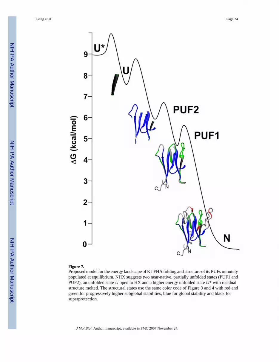
Figure 7.Proposed model for the energy landscape of KI-FHA folding and structure of its PUFs minutelypopulated at equilibrium. NHX suggests two near-native, partially unfolded states (PUF1 andPUF2), an unfolded state U open to HX and a higher energy unfolded state U* with residualstructure melted. The structural states use the same color code of Figure 3 and 4 with red andgreen for progressively higher subglobal stabilities, blue for global stability and black forsuperprotection.
Liang et al. Page 24
J Mol Biol. Author manuscript; available in PMC 2007 November 24.
NIH
-PA Author Manuscript
NIH
-PA Author Manuscript
NIH
-PA Author Manuscript

NIH
-PA Author Manuscript
NIH
-PA Author Manuscript
NIH
-PA Author Manuscript
Liang et al. Page 25Ta
ble
1Fi
tted
glob
al, s
ubgl
obal
and
loca
l ter
ms d
escr
ibin
g th
e H
X b
ehav
ior.
Res
idue
Glo
balA
Subg
loba
lL
ocal
ΔG, k
cal/m
olm
g, kc
al m
ol−1
M−1
ΔGsg
, kca
l/mol
msg
, kca
l mol−1
M−1
ΔG, k
cal/m
ol
E242
9.24
−8.7
36.
94L2
438.
92−8
.64
6.98
K24
08.
43−6
.28
7.67
L184
8.23
−6.4
37.
04F1
838.
20−6
.61
7.04
I281
7.88
−6.7
36.
52A
188
7.75
−5.5
87.
05K
288
7.72
−5.4
27.
70T2
337.
72−6
.20
7.07
Q23
17.
71−5
.60
7.64
R29
27.
63−5
.18
7.53
V29
17.
62−6
.33
6.99
V28
97.
57−5
.66
6.95
Y29
07.
53−5
.44
7.34
L182
7.53
−5.9
46.
75D
279
7.51
−5.5
57.
07L2
757.
50−6
.23
6.74
D27
87.
49−5
.11
7.17
T282
7.39
−5.5
37.
01I2
327.
35−5
.26
6.89
I280
7.30
−5.0
97.
18A
230
7.25
−4.0
07.
16A
276
7.16
−4.5
77.
03V
186
7.03
−4.7
06.
64L1
946.
74−4
.47
5.49
V27
36.
46−4
.12
6.20
G19
35.
98−3
.44
5.34
S248
C6.
01−1
.77
H19
6C6.
00−1
.94
T287
C5.
93−1
.98
G28
4B5.
81−1
.17
W24
1B5.
78−4
.36
V20
8B5.
71−1
.47
G25
1C5.
63−1
.32
L253
B5.
42−1
.53
D24
5B5.
38−1
.25
F239
C5.
23−2
.01
K20
9B5.
12−1
.85
T252
C5.
05−2
.09
L264
B4.
27−0
.92
V25
4B4.
17−0
.92
V22
5B4.
05−1
.17
I192
4.01
L206
3.79
A Fitte
d to
equ
atio
n 6B
.
J Mol Biol. Author manuscript; available in PMC 2007 November 24.

NIH
-PA Author Manuscript
NIH
-PA Author Manuscript
NIH
-PA Author Manuscript
Liang et al. Page 26B Fi
tted
to e
quat
ion
6C, u
sing
an
estim
ated
ave
rage
ΔG
g an
d m
g of
7.5
8 kc
al/m
ol a
nd −
5.60
kca
l mol−1
M−1
, res
pect
ivel
y, fo
r res
idue
s with
glo
bal s
tabi
lity
(blu
e in
Fig
ure
3 an
d 4)
.
CFi
tted
to e
quat
ion
6D.
J Mol Biol. Author manuscript; available in PMC 2007 November 24.
NIH
-PA Author Manuscript
NIH
-PA Author Manuscript
NIH
-PA Author Manuscript
Liang et al. Page 27
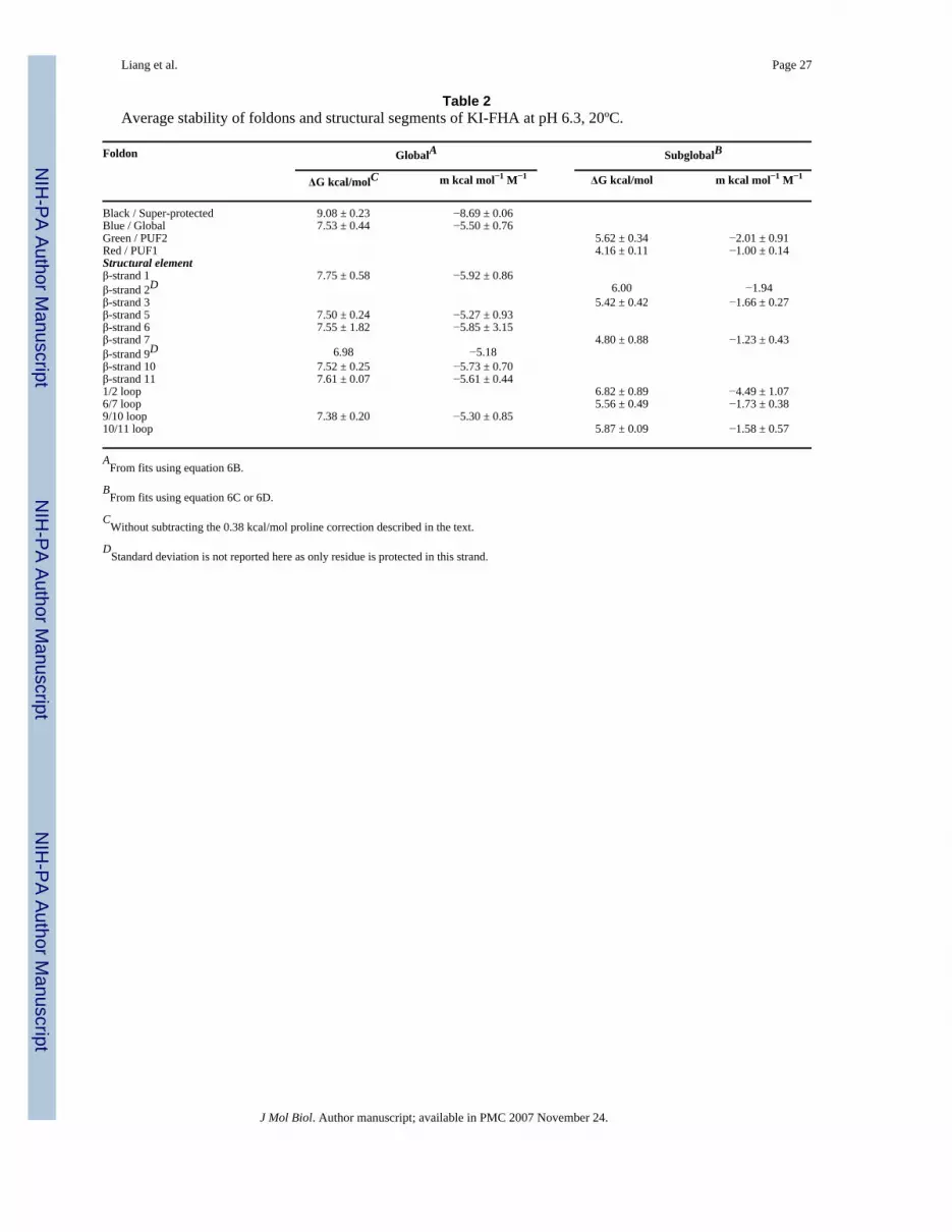
Table 2Average stability of foldons and structural segments of KI-FHA at pH 6.3, 20ºC.
Foldon GlobalA SubglobalB
ΔG kcal/molC m kcal mol−1 M−1 ΔG kcal/mol m kcal mol−1 M−1
Black / Super-protected 9.08 ± 0.23 −8.69 ± 0.06Blue / Global 7.53 ± 0.44 −5.50 ± 0.76Green / PUF2 5.62 ± 0.34 −2.01 ± 0.91Red / PUF1 4.16 ± 0.11 −1.00 ± 0.14Structural elementβ-strand 1 7.75 ± 0.58 −5.92 ± 0.86β-strand 2D 6.00 −1.94β-strand 3 5.42 ± 0.42 −1.66 ± 0.27β-strand 5 7.50 ± 0.24 −5.27 ± 0.93β-strand 6 7.55 ± 1.82 −5.85 ± 3.15β-strand 7 4.80 ± 0.88 −1.23 ± 0.43β-strand 9D 6.98 −5.18β-strand 10 7.52 ± 0.25 −5.73 ± 0.70β-strand 11 7.61 ± 0.07 −5.61 ± 0.441/2 loop 6.82 ± 0.89 −4.49 ± 1.076/7 loop 5.56 ± 0.49 −1.73 ± 0.389/10 loop 7.38 ± 0.20 −5.30 ± 0.8510/11 loop 5.87 ± 0.09 −1.58 ± 0.57
AFrom fits using equation 6B.
BFrom fits using equation 6C or 6D.
CWithout subtracting the 0.38 kcal/mol proline correction described in the text.
DStandard deviation is not reported here as only residue is protected in this strand.
J Mol Biol. Author manuscript; available in PMC 2007 November 24.

NIH
-PA Author Manuscript
NIH
-PA Author Manuscript
NIH
-PA Author Manuscript
Liang et al. Page 28Ta
ble
3St
abili
ty o
f KI-
FHA
aga
inst
den
atur
atio
n by
Gdm
Cl a
t 20
°C, d
etec
ted
optic
ally
.
pHSo
lven
tD
etec
tion
Tra
nsiti
onC
m-v
alue
D k
cal/(
mol
.M)
ΔG k
cal/m
olD
6.3
D2O
CD
AΔG
NI,
90%
−3.3
7 ±
0.06
2.79
± 0
.05
6.3
D2O
CD
AΔG
IU, 1
0%−1
.15
± 0.
313.
71 ±
1.1
6.3
D2O
Trp
fluor
esce
nceB
ΔGN
I−3
.51
± 0.
253.
08 ±
0.2
2
6.3
H2O
CD
AΔG
NI,
94%
−3.3
1 ±
0.07
3.11
± 0
.07
6.3
H2O
CD
AΔ
GIU
, 6%
−1.1
2 ±
0.35
3.22
± 1
.25
7.3
D2O
CD
AΔ
GN
I, 90
%−3
.91
± 0.
086.
17 ±
0.1
3
7.3
D2O
CD
AΔ
GIU
, 10%
−1.2
6 ±
0.19
5.0
± 0.
7
A Mon
itore
d at
216
nm
.
B Mon
itore
d as
a d
iffer
ence
bet
wee
n em
issi
on p
eak
at 3
54 n
m a
nd b
asel
ine
at 2
95 n
m. S
igna
l-to-
nois
e w
as in
suff
icie
nt to
cha
ract
eriz
e a
min
or tr
ansi
tion
at h
igh
[Gdm
Cl].
CPr
opor
tion
of to
tal o
ptic
al c
hang
e m
easu
red
is g
iven
as a
per
cent
age
whe
re d
eter
min
ed.
DTh
e un
certa
intie
s are
the
fittin
g un
certa
intie
s rep
orte
d by
Orig
in 7
.5.
J Mol Biol. Author manuscript; available in PMC 2007 November 24.


