Embed Size (px)
Citation preview

17
Copyright 2009 CBS-KNAW Fungal Biodiversity Centre, P.O. Box 85167, 3508 AD Utrecht, The Netherlands.
You are free to share - to copy, distribute and transmit the work, under the following conditions:Attribution: You must attribute the work in the manner specified by the author or licensor (but not in any way that suggests that they endorse you or your use of the work).Non-commercial: You may not use this work for commercial purposes. Noderivativeworks:You may not alter, transform, or build upon this work. For any reuse or distribution, you must make clear to others the license terms of this work, which can be found at http://creativecommons.org/licenses/by-nc-nd/3.0/legalcode. Any of the above conditions can be waived if you get permission from the copyright holder. Nothing in this license impairs or restricts the author’s moral rights.
StudieS in Mycology 64: 17–47. 2009.doi:10.3114/sim.2009.64.02
INTRODUCTION
The Dothideomycetes encompasses plant and human pathogens, endophytes, saprobes and epiphytes. The class presently contains two subclasses, namely Pleosporomycetidae and Dothideomycetidae (Schoch et al. 2006, 2009a). Although the main orders, Pleosporales and Dothideales correlate with the presence or absence of pseudoparaphyses and other centrum characteristics, many orders remain unresolved. The Dothideomycetidae include the orders Dothideales, Capnodiales and Myriangiales, which lack paraphyses, pseudoparaphyses and periphysoids. Based on a multi-gene phylogeny, and the presence of ostiolar periphyses as possible synapomorphy, the Capnodiales were recognised as the order incorporating the Capnodiaceae, Davidiellaceae, Mycosphaerellaceae and Piedraiaceae (Schoch et al. 2006). However, several studies (Hunter et al. 2006, Crous et al. 2007a, b) showed the Mycosphaerellaceae to be polyphyletic, and to contain additional variation at the familial level, leading to the circumscriptions of the Teratosphaeriaceae and Schizothyriaceae. Crous et al. (2009b, c) again revealed Teratosphaeriaceae to be too widely defined, including some further unresolved families.
The present study focuses on the Capnodiales, which is based on the Capnodiaceae, representing a group of leaf epiphytes associated with honeydew of insects, usually visible as a black growth on leaf surfaces, fruit and twigs. Members of the Capnodiaceae form superficial ascomata with fasciculate asci, and hyaline to dark, septate ascospores. Anamorphs are dematiaceous, and include mycelial (phragmo- to dictyoconidia), spermatial and
pycnidial synanamorphs (Hughes 1976, Cheewangkoon et al. 2009).
The Mycosphaerellaceae was treated as a family in the Dothideales by Hawksworth et al. (1995), while Kirk et al. (2001) introduced a separate order, the Mycosphaerellales for this family, and Kirk et al. (2008) again placed it in the Capnodiales. The Mycosphaerellaceae is recognised by having characteristic pseudothecial ascomata that can be immersed or superficial, embedded in host tissue or erumpent, having ostiolar periphyses, but lacking interascal tissue at maturity. Ascospores are hyaline, but in some cases slightly pigmented (Barr 1987), and predominantly 1-septate, although some taxa with 3-septate ascospores have been recorded (Crous et al. 2003). Although up to 30 anamorph genera have been linked to Mycosphaerella (Crous et al. 2000, 2001, 2007a–c, 2009a–c, Crous 2009), recent studies have shown this to be incorrect, and that the family in fact consists of numerous genera with morphologically conserved Mycosphaerella-like teleomorphs, and distinct anamorphs (Crous et al. 2007a, b, 2009b, c).
Families tentatively placed in the Capnodiales (Lumbsch & Huhndorf 2007, Kirk et al. 2008) include epiphytes (Antennulariellaceae, Capnodiaceae, Metacapnodiaceae) (Hughes 1976), saprobes and plant pathogens (Davidiellaceae, Dissoconiaceae, Mycosphaerellaceae, Schizothyriaceae, Teratosphaeriaceae) (Aptroot 2006, Crous 2009), and colonisers or hair shafts of mammals (Piedraiaceae) (de Hoog et al. 2000). To address the status of the Capnodiales as an order, and the intrafamilial relationships within this order, DNA sequences of
PhylogeneticlineagesintheCapnodiales
P.W. Crous1, 2*, C.L. Schoch3, K.D. Hyde4, A.R. Wood5, C. Gueidan1, G.S. de Hoog1 and J.Z. Groenewald1
1CBS-KNAW Fungal Biodiversity Centre, P.O. Box 85167, 3508 AD, Utrecht, The Netherlands; 2Wageningen University and Research Centre (WUR), Laboratory of Phytopathology, Droevendaalsesteeg 1, 6708 PB Wageningen, The Netherlands; 3National Center for Biotechnology Information, National Library of Medicine, National Institutes of Health, 45 Center Drive, MSC 6510, Bethesda, Maryland 20892-6510, U.S.A.; 4School of Science, Mae Fah Luang University, Tasud, Muang, Chiang Rai 57100, Thailand; 5ARC – Plant Protection Research Institute, P. Bag X5017, Stellenbosch, 7599, South Africa
*Correspondence: Pedro W. Crous, [email protected]
Abstract: The Capnodiales incorporates plant and human pathogens, endophytes, saprobes and epiphytes, with a wide range of nutritional modes. Several species are lichenised, or occur as parasites on fungi, or animals. The aim of the present study was to use DNA sequence data of the nuclear ribosomal small and large subunit RNA genes to test the monophyly of the Capnodiales, and resolve families within the order. We designed primers to allow the amplification and sequencing of almost the complete nuclear ribosomal small and large subunit RNA genes. Other than the Capnodiaceae (sooty moulds), and the Davidiellaceae, which contains saprobes and plant pathogens, the order presently incorporates families of major plant pathological importance such as the Mycosphaerellaceae, Teratosphaeriaceae and Schizothyriaceae. The Piedraiaceae was not supported, but resolves in the Teratosphaeriaceae. The Dissoconiaceae is introduced as a new family to accommodate Dissoconium and Ramichloridium. Lichenisation, as well as the ability to be saprobic or plant pathogenic evolved more than once in several families, though the taxa in the upper clades of the tree lead us to conclude that the strictly plant pathogenic, nectrotrophic families evolved from saprobic ancestors (Capnodiaceae), which is the more primitive state.
Keywords: Ascomycetes, Brunneosphaerella, Capnodiales, DNA sequence comparisons, Mycosphaerella, novel primers, systematics.Taxonomicnovelties: Brunneosphaerella Crous, gen. nov., B. jonkershoekensis (Marinc., M.J. Wingf. & Crous) Crous, comb. nov., B. protearum (Syd. & P. Syd.) Crous, comb. nov., Devriesia hilliana Crous & U. Braun, sp. nov., D. lagerstroemiae Crous & M.J. Wingf., sp. nov., D. strelitziicola Arzanlou & Crous, sp. nov., Dissoconiaceae Crous & de Hoog, fam. nov., Hortaea thailandica Crous & K.D. Hyde, sp. nov., Passalora ageratinae Crous & A.R. Wood, sp. nov., P. armatae Crous & A.R. Wood, sp. nov., Rachicladosporium cboliae Crous, sp. nov.
available online at www.studiesinmycology.org

18
crouS et al.
the 18S, 5.8S and 28S nrRNA genes were generated for a set of specifically selected taxa. A further aim was to clarify genera within these families, and resolve anamorph-teleomorph relationships for the taxa investigated.
MATeRIAlsANDMeThODs
IsolatesIsolates were selected (Table 1 - see online Supplementary Information) that are representative of the Mycosphaerellaceae (Crous 1998, Crous et al. 2004a, c, 2006a, b, 2007a), Schizothyriaceae (Batzer et al. 2005, 2007), Teratosphaeriaceae (Crous et al. 2007a, 2008b, c, 2009a–c), Piedraiaceae (Kruys et al. 2006), Davidiellaceae (Braun et al. 2003, Schubert et al. 2007a, b), Capnodiaceae (Schoch et al. 2006), as well as numerous other genera for which the familial relationships have remained unclear, such as the Phaeophleospora complex (Crous et al. 1997, 2007a, 2009b, c, Andjic et al. 2007), Polythrincium (Simon et al. 2009), the Dissoconium complex (Crous et al. 2004c, 2007c, 2008b, Arzanlou et al. 2008b), and several less well-known genera represented by one or two species only. For fresh material excised leaf spots bearing ascomata were soaked in water for approximately 2 h, after which they were placed in the bottom of Petri dish lids, with the top half of the dish containing 2 % malt extract agar (MEA; Crous et al. 2009d). Ascospore germination patterns were examined after 24 h, and single-ascospore and conidial cultures established as described by Crous et al. (1991). Colonies were sub-cultured onto synthetic nutrient-poor agar (SNA), potato-dextrose agar (PDA), oatmeal agar (OA), MEA (Crous et al. 2009d), and incubated at 25 °C under continuous near-ultraviolet light to promote sporulation. Other cultures were obtained from the culture collection of the Centraalbureau voor Schimmelcultures (CBS-KNAW) in Utrecht, the Netherlands or the working collection of Pedro Crous (CPC).
DNAisolation,amplificationandmolecularphylog-enyGenomic DNA was extracted from mycelium taken from fungal colonies on MEA using the UltraCleanTM Microbial DNA Isolation Kit (Mo Bio Laboratories, Inc., Solana Beach, CA, U.S.A.). A part of the nuclear rDNA operon spanning the 3’ end of the 18S rRNA gene (SSU), the first internal transcribed spacer (ITS1), the 5.8S rRNA gene, the second ITS region (ITS2) and the first 900 bp at the 5’ end of the 28S rRNA gene (LSU) was amplified and sequenced as described by Cheewangkoon et al. (2008) standard for all strains included (Table 1). For selected strains (see Table 1), the almost complete SSU and LSU (missing the first and last 20–30 nucleotides) were amplified and sequenced using novel and previously published primers (Table 2; see below).
Novel primers were designed using a variety of complete SSU and LSU sequences obtained from the GenBank sequence database (www.ncbi.nlm.nih.gov/). The selection was not limited only to fungi belonging to the Dothideomycetes but encompassed as many as possible full sequences in order to make the primers as robust as possible. We aimed to keep the melting temperature (Tm) of the novel primers at 40–45 °C and the GC content to approximately 50 % to keep them as compatible as possible to existing published primers. Primer parameters were calculated using the OligoAnalyzer tool on the web site of Integrated DNA Technologies (http://eu.idtdna.com/analyzer/Applications/
OligoAnalyzer/) with the “Oligo Conc” parameter set at 0.2 mM and the “Na+ Conc” parameter set at 16 mM. A framework of existing and novel primers was then aligned onto the sequence of Magnaporthe grisea (GenBank accession AB026819) to derive primer positions (Table 2) and evaluate coverage over the gene regions. These primers were amplified and sequenced in the following overlapping sections to cover the almost complete SSU and LSU for the selected strains (Table 2): SSU1Fd or SSU6Fm with SSU2Rd, SSU2Fd with SSU3Rd, SSU7Fm with SSU4Rd or SSU6Rm, SSU4Fd with 5.8S1Rd, V9G or LSU1Fd with LSU3Rd, LSU8Fd with LSU8Rd, LSU4Fd with LSU5Rd, and LSU5Fd with LSU7Rd. For some strains (Table 3) it was necessary to add an additional overlap for SSU4Fd with 5.8S1Rd (using SSU4Fd with SSU7Rm and SSU8Fm with 5.8S1Rd), for LSU8Fd with LSU8Rd (using LSU8Fd with LSU3Rd and LSU3Fd with LSU8Rd), and for LSU5Fd with LSU7Rd (using LSU5Fd with LSU6Rd and LSU6Fd with LSU7Rd) to complete the gaps due to large insertions.
The internal transcribed spacer regions, as well as all insertions (Table 3) were excluded from all analyses. Sequence data were deposited in GenBank (Table 1) and alignments in TreeBASE (www.treebase.org). Two separate analyses were performed: The first using only partial LSU data due to the limited number of complete LSU sequences available and the second using the almost complete SSU, 5.8S nrDNA and LSU alignment.
Maximum likelihood analyses (ML) were conducted in RAxML v. 7.0.4 (Stamatakis 2006) for the partial LSU alignment. A general time reversible model (GTR) with a discrete gamma distribution and four rate classes was applied. A tree was obtained by simultaneously running a fast bootstrap search of 1000 pseudoreplicates (Stamatakis et al. 2008) followed by a search for the most likely tree. Maximum Likelihood bootstrap value (MLBP) equal or greater than 70 % are given at the nodes (Fig. 1).
Maximum likelihood analyses (ML) were conducted in RAxML v. 7.0.4 (Stamatakis 2006) for the almost complete SSU, 5.8S nrDNA and LSU alignment. A general time reversible model (GTR) with a discrete gamma distribution and four rate classes was applied to each partition (SSU, 5.8S nrDNA and LSU). A tree was obtained by simultaneously running a fast bootstrap search of 500 pseudoreplicates (Stamatakis et al. 2008) followed by a search for the most likely tree. Maximum Likelihood bootstrap value (MLBP) equal or greater than 70 % are given at the nodes (Fig. 2).
TaxonomyFungal structures were mounted in lactic acid, and 30 measurements (× 1000 magnification) obtained per structure type. The range obtained is presented, except for spore measurements, where the 95 % confidence intervals are given with the extremes in parentheses. Colony colours (surface and reverse) were assessed after 1–2 wk on MEA at 25 °C in the dark, using the colour charts of Rayner (1970). All cultures obtained in this study are maintained in the culture collection of the Centraalbureau voor Schimmelcultures (CBS-KNAW) in Utrecht, the Netherlands (Table 1). Nomenclatural novelties and descriptions were deposited in MycoBank (Crous et al. 2004b). Names for which the taxonomy has not been resolved, but need to be allocated to another genus, are placed in inverted commas, e.g. “Mycosphaerella” iridis.

19www.studiesinmycology.org
Phylogenetic lineageS in the Capnodiales
Table2.Details of primers used for this study and their relation to selected published primers. Primer names ending with a "d" denotes a degenerate primer whereas those ending with a "m" denotes specific primers designed based on the partial novel sequences generated. The start and end positions of the primers are derived using Magnaporthe grisea GenBank accession AB026819 as reference in the 5'–3' direction.
Name sequence(5’–3’) Orientation %GC Tm(oC) start end Reference5.8S1Fd CTC TTG GTT CBV GCA TCG Forward 57.4 49.8 – 54.2 – 56.8 2333 2350 This study5.8S1Rd WAA TGA CGC TCG RAC AGG CAT G Reverse 52.3 57.6 – 58.9 – 60.2 2451 2472 This studyF377 AGA TGA AAA GAA CTT TGA AAA
GAG AAForward 26.9 40.3 3005 3030 www.lutzonilab.net/primers/
page244.shtmlITS1 TCC GTA GGT GAA CCT GCG G Forward 63.2 49.5 2162 2180 White et al. (1990)ITS1F CTT GGT CAT TTA GAG GAA GTA A Forward 36.4 39.0 2124 2145 Gardes & Bruns (1993)ITS1Fd CGA TTG AAT GGC TCA GTG AGG C Forward 54.5 48.0 2043 2064 This studyITS1Rd GAT ATG CTT AAG TTC AGC GGG Reverse 47.6 43.1 2671 2691 This studyITS4 TCC TCC GCT TAT TGA TAT GC Reverse 45.0 41.6 2685 2704 White et al. (1990)ITS4S CCT CCG CTT ATT GAT ATG CTT
AAG Reverse 41.7 42.9 2680 2703 Kretzer et al. (1996)
ITS5 GGA AGT AAA AGT CGT AAC AAG G Forward 40.9 40.8 2138 2159 White et al. (1990)LR0R GTA CCC GCT GAA CTT AAG C Forward 52.6 43.2 2668 2686 Rehner & Samuels (1994)LR2 TTT TCA AAG TTC TTT TC Reverse 23.5 28.5 3009 3025 www.lutzonilab.net/primers/
page244.shtmlLR2R AAG AAC TTT GAA AAG AG Forward 29.4 30.4 3012 3028 www.lutzonilab.net/primers/
page244.shtmlLR3 GGT CCG TGT TTC AAG AC Reverse 52.9 40.5 3275 3291 Vilgalys & Hester (1990)LR3R GTC TTG AAA CAC GGA CC Forward 52.9 40.5 3275 3291 www.lutzonilab.net/primers/
page244.shtmlLR5 TCC TGA GGG AAA CTT CG Reverse 52.9 41.0 3579 3595 Vilgalys & Hester (1990)LR5R GAA GTT TCC CTC AGG AT Forward 47.1 37.8 3580 3596 www.biology.duke.edu/fungi/
mycolab/primers.htmLR6 CGC CAG TTC TGC TTA CC Reverse 58.8 43.5 3756 3772 Vilgalys & Hester (1990)LR7 TAC TAC CAC CAA GAT CT Reverse 41.2 35.3 4062 4078 Vilgalys & Hester (1990)LR8 CAC CTT GGA GAC CTG CT Reverse 58.8 44.3 4473 4489 www.lutzonilab.net/primers/
page244.shtmlLR8R AGC AGG TCT CCA AGG TG Forward 58.8 44.3 4473 4489 www.lutzonilab.net/primers/
page244.shtmlLR9 AGA GCA CTG GGC AGA AA Reverse 52.9 43.6 4799 4815 www.lutzonilab.net/primers/
page244.shtmlLR10 AGT CAA GCT CAA CAG GG Reverse 52.9 41.6 5015 5031 www.lutzonilab.net/primers/
page244.shtmlLR10R GAC CCT GTT GAG CTT GA Forward 52.9 41.6 5013 5029 www.lutzonilab.net/primers/
page244.shtmlLR11 GCC AGT TAT CCC TGT GGT AA Reverse 50.0 43.9 5412 5431 www.lutzonilab.net/primers/
page244.shtmlLR12 GAC TTA GAG GCG TTC AG Reverse 52.9 39.4 5715 5731 Vilgalys & Hester (1990)LR12R CTG AAC GCC TCT AAG TCA GAA Forward 47.6 43.7 5715 5735 www.biology.duke.edu/fungi/
mycolab/primers.htmLR13 CAT CGG AAC AAC AAT GC Reverse 47.1 38.8 5935 5951 www.lutzonilab.net/primers/
page244.shtmlLR14 AGC CAA ACT CCC CAC CTG Reverse 61.1 47.6 5206 5223 www.lutzonilab.net/primers/
page244.shtmlLR15 TAA ATT ACA ACT CGG AC Reverse 35.3 32.5 2780 2796 www.lutzonilab.net/primers/
page244.shtmlLR16 TTC CAC CCA AAC ACT CG Reverse 52.9 42.1 3311 3327 Moncalvo et al. (1993)LR17R TAA CCT ATT CTC AAA CTT Forward 27.8 31.2 3664 3681 www.lutzonilab.net/primers/
page244.shtmlLR20R GTG AGA CAG GTT AGT TTT ACC
CTForward 43.5 43.6 5570 5592 www.lutzonilab.net/primers/
page244.shtmlLR21 ACT TCA AGC GTT TCC CTT T Reverse 42.1 41.7 3054 3072 www.lutzonilab.net/primers/
page244.shtmlLR22 CCT CAC GGT ACT TGT TCG CT Reverse 55.0 46.8 2982 3001 www.lutzonilab.net/primers/
page244.shtml

20
crouS et al.
Name sequence(5’–3’) Orientation %GC Tm(oC) start end ReferenceLSU1Fd GRA TCA GGT AGG RAT ACC CG Forward 55.0 41.8 – 44.0 – 46.3 2655 2674 This studyLSU1Rd CTG TTG CCG CTT CAC TCG C Reverse 63.2 49.6 2736 2754 This studyLSU2Fd GAA ACA CGG ACC RAG GAG TC Forward 57.5 45.5 – 46.5 – 47.6 3280 3299 This studyLSU2Rd ATC CGA RAA CWT CAG GAT CGG
TCGReverse 52.1 48.3 – 49.0 – 49.8 3379 3402 This study
LSU3Fd GTT CAT CYA GAC AGC MGG ACG Forward 57.1 44.7 – 47.4 – 50.2 3843 3863 This studyLSU3Rd CAC ACT CCT TAG CGG ATT CCG
ACReverse 56.5 49.1 3876 3898 This study
LSU4Fd CCG CAG CAG GTC TCC AAG G Forward 68.4 51.2 4469 4487 This studyLSU4Rd CGG ATC TRT TTT GCC GAC TTC
CCReverse 54.3 47.4 – 48.7 – 50.0 4523 4545 This study
LSU5Fd AGT GGG AGC TTC GGC GC Forward 70.6 51.6 3357 / 5072
3373 / 5088
This study
LSU5Rd GGA CTA AAG GAT CGA TAG GCC ACA C
Reverse 52.0 48.3 5355 5379 This study
LSU6Fd CCG AAG CAG AAT TCG GTA AGC G Forward 54.5 48.1 5499 5520 This studyLSU6Rd TCT AAA CCC AGC TCA CGT TCC C Reverse 54.5 48.6 5543 5564 This studyLSU7Fd GTT ACG ATC TRC TGA GGG TAA
GCC Forward 52.1 46.0 – 47.4 – 48.8 5943 5966 This study
LSU7Rd GCA GAT CGT AAC AAC AAG GCT ACT CTA C
Reverse 46.4 47.9 5927 5954 This study
LSU8Fd CCA GAG GAA ACT CTG GTG GAG GC
Forward 60.9 51.2 3469 3491 This study
LSU8Rd GTC AGA TTC CCC TTG TCC GTA CC
Reverse 56.5 48.9 4720 4742 This study
LSU9Fm GGT AGC CAA ATG CCT CGT CAT C Forward 54.5 47.9 4882 4903 This studyLSU9Rm GAT TYT GCS AAG CCC GTT CCC Reverse 59.5 49.2 – 50.0 – 50.9 4979 4999 This studyLSU10Fm GGG AAC GTG AGC TGG GTT TAG A Forward 54.5 48.6 5543 5564 This study
LSU10Rm CGC TTA CCG AAT TCT GCT TCG G Reverse 54.5 48.1 5499 5520 This studyLSU11Fm TTTGGTAAGCAGAACTGGCGATGC Forward 50.0 49.4 3753 3776 This studyLSU12Fd GTGTGGCCTATCGATCCTTTAGTCC Forward 52.0 48.3 5355 5379 This studyNS1 GTA GTC ATA TGC TTG TCT C Forward 42.1 36.9 413 431 White et al. (1990)NS1R GAG ACA AGC ATA TGA CTA C Reverse 42.1 36.9 413 431 www.lutzonilab.net/primers/
page244.shtmlNS2 GGC TGC TGG CAC CAG ACT TGC Reverse 66.7 53.8 943 963 White et al. (1990)NS3 GCAAGTCTGGTGCCAGCAGCC Forward 66.7 53.8 943 963 White et al. (1990)NS4 CTT CCG TCA ATT CCT TTA AG Reverse 40.0 38.2 1525 1544 White et al. (1990)NS5 AAC TTA AAG GAA TTG ACG GAA G Forward 36.4 40.1 1523 1544 White et al. (1990)NS6 GCA TCA CAG ACC TGT TAT TGC
CTC Reverse 50.0 47.5 1806 1829 White et al. (1990)
NS7 GAG GCA ATA ACA GGT CTG TGA TGC
Forward 50.0 47.5 1806 1829 White et al. (1990)
NS8 TCC GCA GGT TCA CCT ACG GA Reverse 60.0 50.4 2162 2181 White et al. (1990)NS17 CAT GTC TAA GTT TAA GCA A Forward 31.6 34.2 447 465 Gargas & Taylor (1992)NS18 CTC ATT CCA ATT ACA AGA CC Reverse 40.0 38.0 887 906 Gargas & Taylor (1992)NS19 CCG GAG AAG GAG CCT GAG AAA C Forward 59.1 49.3 771 792 Gargas & Taylor (1992)NS20 CGT CCC TAT TAA TCA TTA CG Reverse 40.0 37.3 1243 1262 Gargas & Taylor (1992)NS21 GAA TAA TAG AAT AGG ACG Forward 33.3 30.5 1193 1210 Gargas & Taylor (1992)NS22 AAT TAA GCA GAC AAA TCA CT Reverse 30.0 36.4 1687 1706 Gargas & Taylor (1992)NS23 GAC TCA ACA CGG GGA AAC TC Forward 55.0 45.5 1579 1598 Gargas & Taylor (1992)NS24 AAA CCT TGT TAC GAC TTT TA Reverse 30.0 36.2 2143 2162 Gargas & Taylor (1992)SR11R GGA GCC TGA GAA ACG GCT AC Forward 60.0 47.8 779 798 Spatafora et al. (1995)SR1R TAC CTG GTT GAT TCT GC Forward 47.1 38.5 394 410 Vilgalys & Hester (1990)SR3 GAA AGT TGA TAG GGC T Reverse 43.8 34.8 696 711 www.biology.duke.edu/fungi/
mycolab/primers.htm
Table2.(Continued).

21www.studiesinmycology.org
Phylogenetic lineageS in the Capnodiales
Name sequence(5’–3’) Orientation %GC Tm(oC) start end ReferenceSSU1Fd CTG CCA GTA GTC ATA TGC TTG
TCT CForward 48.0 46.5 407 431 This study
SSU1Rd CTT TGA GAC AAG CAT ATG AC Reverse 40.0 48.7 416 435 This studySSU2Fd GAA CAA YTR GAG GGC AAG Forward 50.0 47.8 – 50.7 – 53.5 930 947 This studySSU2Rd TAT ACG CTW YTG GAG CTG Reverse 47.2 48.4 – 49.9 – 51.2 974 991 This studySSU3Fd ATC AGA TAC CGT YGT AGT C Forward 44.7 48.4 – 49.5 – 50.5 1389 1407 This studySSU3Rd TAY GGT TRA GAC TAC RAC GG Reverse 47.5 49.0 – 52.5 – 56.0 1397 1416 This studySSU4Fd CCG TTC TTA GTT GGT GG Forward 52.9 50.0 1670 1686 This studySSU4Rd CAG ACA AAT CAC TCC ACC Reverse 50.0 50.3 1682 1699 This studySSU5Fd TAC TAC CGA TYG AAT GGC Forward 47.2 48.9 – 50.1 – 51.2 2037 2054 This studySSU5Rd CGG AGA CCT TGT TAC GAC Reverse 55.6 52.5 2148 2165 This studySSU6Fm GCT TGT CTC AAA GAT TAA GCC
ATG CAT GTC Forward 43.3 49.0 423 452 This study
SSU6Rm GCA GGT TAA GGT CTC GTT CGT TAT CGC
Reverse 51.9 50.1 1707 1733 This study
SSU7Fm GAG TGT TCA AAG CAG GCC TNT GCT CG
Forward 55.8 51.0 – 52.2 – 53.3 1153 1178 This study
SSU7Rm CAA TGC TCK ATC CCC AGC ACG AC
Reverse 58.7 49.5 – 50.8 – 52.1 1921 1943 This study
SSU8Fm GCA CGC GCG CTA CAC TGA C Forward 68.4 52.2 1848 1866 This studyV9G TTA CGT CCC TGC CCT TTG TA Forward 45.0 42.8 2002 2021 de Hoog & Gerrits van den Ende
(1998)
Table2.(Continued).
Table3. Isolates containing group I intron sequences. The insertion positions of these introns are derived using Magnaporthe grisea GenBank accession AB026819 as reference in the 5'–3' direction.
Isolate Insertionbetween
18sor28snrDNA
Intronsize(bp)
Blastresult
Batcheloromyces leucadendri CBS 110892 1559 – 1560 18S nrDNA 350 No significant similarity1820 – 1821 18S nrDNA 399 190/252 of AY545722 Hydropisphaera erubescens 18S nrDNA
4875 – 4876 28S nrDNA 328 211/264 of DQ246237 Teratosphaeria mexicana 28S nrDNA
5424 – 5425 28S nrDNA 538 No significant similarity
5538 – 5539 28S nrDNA 383 218/283 of EU181458 Trichophyton soudanense 28S nrDNA
Batcheloromyces proteae CBS 110696 1559 – 1560 18S nrDNA 325 No significant similarity1820 – 1821 18S nrDNA 399 191/254 of AY545722 Hydropisphaera erubescens 18S nrDNA
4875 – 4876 28S nrDNA 328 211/263 of DQ246237 Teratosphaeria mexicana 28S nrDNA
5424 – 5425 28S nrDNA 535 75/90 of DQ442697 Arxula adeninivorans 26S nrDNA
5538 – 5539 28S nrDNA 372 34/36 of GQ120133 Uncultured marine fungus 18S nrDNA
Catenulostroma macowanii CBS 110756 1559 – 1560 18S nrDNA 395 297/379 of DQ848302 Mycosphaerella latebrosa 18S nrDNA5424 – 5425 28S nrDNA 914 No significant similarity
Catenulostroma macowanii CBS 111029 1559 – 1560 18S nrDNA 395 303/379 of DQ848302 Mycosphaerella latebrosa 18S nrDNA5424 – 5425 28S nrDNA 914 No significant similarity
Cercospora apii CBS 118712 1820 – 1821 18S nrDNA 733 288/363 of EU167577 Mycosphaerella milleri 18S nrDNACercospora capsici CPC 12307 1820 – 1821 18S nrDNA 732 287/363 of EU167577 Mycosphaerella milleri 18S nrDNACercospora janseana CBS 145.37 1820 – 1821 18S nrDNA 350 295/365 of EU167577 Mycosphaerella milleri 18S nrDNADevriesia staurophora CBS 375.81 3560 – 3561 28S nrDNA 309 No significant similarityMiuraea persicae CPC 10069 1820 – 1821 18S nrDNA 603 399/443 of DQ848342 Mycosphaerella populorum 18S nrDNAMycosphaerella latebrosa CBS 652.85 1559 – 1560 18S nrDNA 370 234/296 of DQ848311 Septoria betulae 18S nrDNA
1820 – 1821 18S nrDNA 933 Matches same species
2168 – 2169 18S nrDNA 494 377/449 of DQ848326 Septoria alnifolia 18S nrDNA
4875 – 4876 28S nrDNA 481 No significant similarity
missing 5018 – 5019
28S nrDNA Not present Not present

22
crouS et al.
Isolate Insertionbetween
18sor28snrDNA
Intronsize(bp)
Blastresult
5424 – 5425 28S nrDNA 680 No significant similarity
5538 – 5539 28S nrDNA 471 No significant similarity
Mycosphaerella latebrosa CBS 687.94 1559 – 1560 18S nrDNA 370 231/295 of DQ848310 Septoria betulae 18S nrDNA1820 – 1821 18S nrDNA 918 Matches same species
2168 – 2169 18S nrDNA 494 377/449 of DQ848326 Septoria alnifolia 18S nrDNA
4875 – 4876 28S nrDNA 480 No significant similarity
5018 – 5019 28S nrDNA 417 144/181 of AF430703 Beauveria bassiana 28S nrDNA
5424 – 5425 28S nrDNA 680 No significant similarity
5538 – 5539 28S nrDNA 471 No significant similarity
Mycosphaerella marksii CBS 110942 1559 – 1560 18S nrDNA 341 332/355 of DQ848296 Mycosphaerella musae 18S nrDNAMycosphaerella marksii CPC 11222 1559 – 1560 18S nrDNA 341 332/355 of DQ848296 Mycosphaerella musae 18S nrDNAPassalora-like genus CPC 11876 5538 – 5539 28S nrDNA 580 No significant similarityPassalora bellynckii CBS 150.49 1559 – 1560 18S nrDNA 409 147/191 of DQ848296 Mycosphaerella musae 18S nrDNAPassalora dodonaea CPC 1223 5424 – 5425 28S nrDNA 738 No significant similarityPhacellium paspali CBS 113093 4875 – 4876 28S nrDNA 340 161/197 of DQ248314 Symbiotaphrina kochii 28S nrDNAPhaeophleospora eugeniicola CPC 2557 missing 5424 –
542528S nrDNA Not present Not present
5538 – 5539 28S nrDNA 744 No significant similarity
Phaeophleospora eugeniicola CPC 2558 5424 – 5425 28S nrDNA 1846 No significant similarity5538 – 5539 28S nrDNA 744 No significant similarity
Pseudocercospora angolensis CBS 112933 5018 – 5019 28S nrDNA 379 No significant similarityPseudocercospora angolensis CBS 149.53 5018 – 5019 28S nrDNA 379 No significant similarityPseudocercospora punctata CBS 113315 5424 – 5425 28S nrDNA 723 No significant similarity
5538 – 5539 28S nrDNA 725 67/73 of AF430699 Beauveria bassiana 28S nrDNAPseudocercospora punctata CPC 10532 5424 – 5425 28S nrDNA 731 No significant similarity
5538 – 5539 28S nrDNA 725 67/73 of AF430699 Beauveria bassiana 28S nrDNARamularia coleosporii CPC 11516 1559 – 1560 18S nrDNA 445 No significant similarityRamularia grevilleana CPC 656 5538 – 5539 28S nrDNA 546 No significant similaritySeptoria apiicola CBS 400.54 5424 – 5425 28S nrDNA 763 No significant similaritySeptoria obesa CBS 354.58 1820 – 1821 18S nrDNA 575 No significant similarity
2168 – 2169 18S nrDNA 548 394/454 of DQ848326 Septoria alnifolia 18S nrDNA
4875 – 4876 28S nrDNA 430 No significant similarity
Septoria pyricola CBS 222.31 5424 – 5425 28S nrDNA 723 No significant similaritySeptoria quercicola CBS 663.94 1559 – 1560 18S nrDNA 334 241/308 of DQ848303 Mycosphaerella latebrosa 18S nrDNA
1820 – 1821 18S nrDNA 442 379/452 of DQ848335 Mycosphaerella latebrosa 18S nrDNA
4875 – 4876 28S nrDNA 345 No significant similarity
5018 – 5019 28S nrDNA 367 122/155 of DQ518980 Lipomyces spencermartinsiae 28S nrDNA
5424 – 5425 28S nrDNA 526 No significant similarity
5538 – 5539 28S nrDNA 603 No significant similarity
Septoria rosae CBS 355.58 1820 – 1821 18S nrDNA 496 No significant similaritySonderhenia eucalypticola CPC 11252 1559 – 1560 18S nrDNA 408 339/404 of DQ848314 Mycosphaerella populorum 18S nrDNA
4875 – 4876 28S nrDNA 337 229/289 of AB044641 Cordyceps sp. 28S nrDNA
5424 – 5425 28S nrDNA 705 No significant similarity
Stigmina platani CBS 110755 1559 – 1560 18S nrDNA 379 40/44 of AB007686 Exophiala calicioides 18S nrDNA5018 – 5019 28S nrDNA 376 No significant similarity
Stigmina synanamorph CPC 11721 5018 – 5019 28S nrDNA 371 No significant similarityTeratosphaeria aff. nubilosa CBS 114419 4871 – 4872 28S nrDNA 141 No significant similarity; high identity to Teratosphaeria nubilosa
5538 – 5539 28S nrDNA 580 No significant similarity; high identity to Teratosphaeria nubilosa
Table3.(Continued).

23www.studiesinmycology.org
Phylogenetic lineageS in the Capnodiales
Isolate Insertionbetween
18sor28snrDNA
Intronsize(bp)
Blastresult
Teratosphaeria aff. nubilosa CBS 116283 4871 – 4872 28S nrDNA 141 No significant similarity; high identity to Teratosphaeria nubilosa
5538 – 5539 28S nrDNA 580 No significant similarity; high identity to Teratosphaeria nubilosa
Teratosphaeria juvenalis CBS 110906 1559 – 1560 18S nrDNA 403 52/61 of DQ471010 Rutstroemia firma 18S nrDNA4875 – 4876 28S nrDNA 345 224/290 of EF115309 Cordyceps bassiana 28S nrDNA
5424 – 5425 28S nrDNA 478 47/50 of EF115313 Cordyceps bassiana 28S nrDNA
5538 – 5539 28S nrDNA 402 No significant similarity
Teratosphaeria juvenalis CBS 111149 1559 – 1560 18S nrDNA 403 52/61 of DQ471010 Rutstroemia firma 18S nrDNA4875 – 4876 28S nrDNA 345 224/290 of EF115309 Cordyceps bassiana 28S nrDNA
5424 – 5425 28S nrDNA 478 47/50 of EF115313 Cordyceps bassiana 28S nrDNA
5538 – 5539 28S nrDNA 402 No significant similarity
Teratosphaeria mexicana CBS 110502 954 – 955 18S nrDNA 316 129/158 of DQ518980 Lipomyces spencermartinsiae 26S nrDNA
1559 – 1560 18S nrDNA 360 No significant similarity
1820 – 1821 18S nrDNA 388 128/168 of AF281670 Cryptendoxyla hypophloia 18S nrDNA
3560 – 3561 28S nrDNA 383 124/151 of EF647754 Thecaphora thlaspeos 28S nrDNA
4875 – 4876 28S nrDNA 327 99/114 of L81104 Gaeumannomyces graminis var. tritici 28S nrDNA
5018 – 5019 28S nrDNA 315 No significant similarity
5424 – 5425 28S nrDNA 553 No significant similarity
Teratosphaeria mexicana CBS 120744 954 – 955 18S nrDNA 318 130/158 of DQ518980 Lipomyces spencermartinsiae 26S nrDNA
1559 – 1560 18S nrDNA 360 No significant similarity
1820 – 1821 18S nrDNA 389 85/109 of AF281670 Cryptendoxyla hypophloia 18S nrDNA
3560 – 3561 28S nrDNA 378 119/155 of AY298780 Lentinellus castoreus 18S nrDNA
4875 – 4876 28S nrDNA 327 162/200 of AB033530 Penicillium sabulosum 18S nrDNA
5018 – 5019 28S nrDNA 309 No significant similarity
5424 – 5425 28S nrDNA 659 No significant similarity
Teratosphaeria nubilosa CBS 115669 4871 – 4872 28S nrDNA 141 No significant similarity; high identity to Teratosphaeria aff. nubilosa
5538 – 5539 28S nrDNA 580 No significant similarity; high identity to Teratosphaeria aff. nubilosa
Teratosphaeria nubilosa CBS 116005 4871 – 4872 28S nrDNA 141 No significant similarity; high identity to Teratosphaeria aff. nubilosa
5538 – 5539 28S nrDNA 580 No significant similarity; high identity to Teratosphaeria aff. nubilosa
Teratosphaeria ohnowa CBS 112896 954 – 955 18S nrDNA 325 28/28 of DQ848329 Botryosphaeria quercuum 18S nrDNA3560 – 3561 28S nrDNA 294 168/227 of FJ358267 Chaetothyriales sp. 28S nrDNA
5424 – 5425 28S nrDNA 607 47/48 of EF115313 Cordyceps bassiana 28S nrDNA
Teratosphaeria ohnowa CBS 112973 954 – 955 18S nrDNA 324 28/28 of DQ848329 Botryosphaeria quercuum 18S nrDNA3560 – 3561 28S nrDNA 294 168/227 of FJ358267 Chaetothyriales sp. 28S nrDNA
5424 – 5425 28S nrDNA 607 47/48 of EF115313 Cordyceps bassiana 28S nrDNA
Teratosphaeria pseudosuberosa CBS 118911 3560 – 3561 28S nrDNA 324 28/28 of DQ848329 Botryosphaeria quercuum 18S nrDNA4875 – 4876 28S nrDNA 364 No significant similarity
Teratosphaeria sp. CBS 208.94 954 – 955 18S nrDNA 342 No significant similarity3560 – 3561 28S nrDNA 309 59/70 of AY207244 Mycena pura 28S nrDNA
4875 – 4876 28S nrDNA 296 44/51 of EF551317 Tremella globispora 28S nrDNA
Teratosphaeria suberosa CPC 11032 5424 – 5425 28S nrDNA 313 159/197 of AB033529 Penicillium oblatum 18S nrDNA5538 – 5539 28S nrDNA 596 80/99 of AB044639 Cordyceps kanzashiana 28S nrDNA
Thedgonia-like genus CPC 12304 1820 – 1821 18S nrDNA 444 262/331 of EU167577 Mycosphaerella milleri 18S nrDNA
Table3.(Continued).

24
crouS et al.
0.1
Dothidea insculpta DQ247802Phaeotheca triangularis EU019279
Comminutispora agavaciensis Y18699Phaeotheca fissurella EU981289
Racodium rupestre EU048576Racodium rupestre EU048575Racodium rupestre EU048577
Graphiopsis chlorocephala EU009458Rachicladosporium luculiae EU040237Rachicladosporium cboliae CPC 14034
Verrucocladosporium dirinae EU040244Toxicocladosporium rubrigenum FJ790305Toxicocladosporium irritans EU040243
Toxicocladosporium chlamydosporum FJ790301Davidiella allicina CBS 723.79Davidiella macrospora DQ008148Sphaerulina polyspora CBS 354.29Cladosporium bruhnei EU019261
Cladosporium sp. FJ790289Melanodothis caricis CBS 860.72
Cladosporium sp. CPC 15513Cladosporium sp. CPC 15516Cladosporium cladosporioides FJ890369
Scorias spongiosa DQ678075Fumagospora capnodioides EU019269
Antennariella placitae GQ303299Microxyphium theae GU301849Polychaeton citri CBS 116435
Leptoxyphium madagascariensis GQ303308Leptoxyphium fumago CBS 123.26Microxyphium citri AY004337
Capnodium coffeae DQ247800Microxyphium aciculiforme GU301847Condioxyphium gardeniorum GU301807
Devriesia strelitziicola CBS 122480Anisomeridium consobrinum GU323215
Mycosphaerella eurypotami GU301852Cystocoleus ebeneus EU048571 Cystocoleus ebeneus EU048573
Hortaea acidophila CBS 113389Sporidesmium pachyanthicola DQ408557
Capnodiales sp. GU323217Ramichloridium brasilianum EU041854Capnodiales sp. GU323218
Staninwardia suttonii DQ923535Capnodiales sp. GU323220
Teratosphaeria sp. GQ852712Devriesia lagerstroemiae CPC 14403
Devriesia strelitziae EU436763Teratosphaeria knoxdavesii EU707865
Capnodiales sp. GU323223Tripospermum myrti GU323216
Passalora sp GQ852622Devriesia hilliana CBS 123187Penidiella strumelloidea EU019277Capnobotryella renispora EU019248Capnobotryella renispora CBS 215.90
Phacellium paspali GQ852627Penidiella tasmaniensis DQ246233Penidiella pseudotasmaniensis GQ852625
Capnodiales sp. GU323219Teratosphaeria parva EU707875
Teratosphaeria jonkershoekensis GU301874Catenulostroma protearum CPC 15370Catenulostroma protearum CPC 15368Catenulostroma protearum CPC 15369
Catenulostroma elginense EU019252Capnodiales sp. GU323222
Capnodiales sp. GU323221Xenomeris juniperi EF114709
Devriesia staurophora EF137359Aulographina pinorum GU296138Aulographina pinorum CBS 302.71Catenulostroma microsporum EU167572Catenulostroma abietis EU019249Catenulostroma microsporum EU019255Catenulostroma germanicum EU019253
Phaeothecoidea minutispora GQ852629Phaeothecoidea eucalypti EU019280Phaeothecoidea intermedia GQ852628
Teratosphaeria mexicana DQ246237Teratosphaeria pseudosuberosa EU019256
Teratosphaeria suberosa CPC 11032Teratosphaeria suberosa DQ246235
Readeriella menaiensis GQ852670Readeriella mirabilis GQ852662
Readeriella novaezelandiae DQ246239Readeriella dendritica EU019271Readeriella patrickii GQ852664Readeriella tasmanica GQ852669
Readeriella eucalyptigena GQ852667Readeriella dimorphospora EU019258Readeriella angustia GQ852665Readeriella nontingens EU019260Readeriella minutispora EU019259
Readeriella callista GQ852655Readeriella pseudocallista GQ852668Readeriella eucalypti EU019289Readeriellaa readeriellophora DQ246238
Bootstrap support values: = 95 % and higher = 90 % to 94 % = 80 % to 89 % = 70 % to 79 %
Davidiellaceae
Incertae sedis
Capnodiaceae
Ter
ato
sph
aeri
acea
e
Fig.1. RAxML tree using only the partial LSU alignment with bootstrap values after 1 000 pseudorepetitions on the nodes. Type strains and novel species described in this study are indicated in bold.

25www.studiesinmycology.org
Phylogenetic lineageS in the Capnodiales
0.1
Batcheloromyces proteae EU019247Teratosphaeria sp. EU019307Teratosphaeria ohnowa EU019305Teratosphaeria secundaria EU019306Teratosphaeria flexuosa FJ493216
Penidiella columbiana EU019274Penidiella eucalypti EU882145Penidiella tenuiramis GQ852626
Penidiella venezuelensis EU019278Hortaea werneckii EU019270Hortaea werneckii GU301818Hortaea werneckii GU301817
Stenella araguata EU019250Hortaea thailandica CPC 16651
Baudoinia compniacensis GQ852580Piedraia quintanilhae CBS 327.63
Piedraia hortae var. hortae CBS 374.71Piedraia hortae var. paraguayensis CBS 276.32Piedraia hortae var. hortae CBS 375.71Piedraia hortae var. hortae CBS 480.64Piedraia hortae AY016366
Teratosphaeria cryptica CBS 110975Teratosphaeria cryptica DQ246222
Teratosphaeria ovata FJ493218Teratosphaeria sp. GQ852713
Teratosphaeria alboconidia FJ493221Teratosphaeria brunneotingens EU019286Teratosphaeria complicata GQ852714Teratosphaeria miniata GQ852711Teratosphaeria hortaea FJ790299Teratosphaeria hortaea FJ790300
Teratosphaeria angophorae CBS 120493Teratosphaeria verrucosa EU019293
Teratosphaeria juvenalis FJ493217Teratosphaeria considenianae DQ923527Teratosphaeria majorizuluensis GQ852710
Teratosphaeria corymbiae FJ493203Teratosphaeria gauchensis EU019290
Teratosphaeria blakelyi DQ923526Teratosphaeria zuluensis EU019296Teratosphaeria stellenboschiana GQ852716Teratosphaeria stellenboschiana EU019295
Teratosphaeria veloci FJ493223Teratosphaeria suttonii EU019288
Teratosphaeria toledana DQ246230Teratosphaeria nubilosa DQ246228
Teratosphaeria eucalypti DQ246225Teratosphaeria viscidus FJ493204Teratosphaeria destructans EU019287
Teratosphaeria sp. FJ493202Teratosphaeria molleriana EU019292
Teratosphaeria profusa FJ493220Teratosphaeria dimorpha FJ493215
Teratosphaeria macowanii EU019254Teratosphaeria maxii DQ885899
Teratosphaeria maculiformis EU707867Teratosphaeria proteae-arboreae EU707883
Teratosphaeria fibrillosa EU019282Teratosphaeria fibrillosa GU323213
Ramichloridium apiculatum EU041848Ramichloridium apiculatum EU041851
Dissoconium aciculare CBS 204.89Dissoconium aciculare EU019266
Dissoconium australiensis GQ852588Dissoconium dekkeri DQ204768Dissoconium dekkeri CBS 111282Dissoconium commune EU019267Dissoconium commune CBS 110747
Zygophiala cryptogama FJ147157Schizothyrium pomi EF134947Zygophiala sp. FJ147159
Ramichloridium pini EU041859Mycosphaerella parkii GQ852616
Pseudocercosporella sp. FJ031995Mycosphaerella madeirae DQ204756
Polythrincium trifolii EU167612Polythrincium trifolii EU167613Polythrincium trifolii EU167610
Passalora vaginae GQ852624Zasmidium aerohyalinosporum GQ852736
Mycosphaerella rosigena CBS 330.51Mycosphaerella intermedia DQ246248
Mycosphaerella marksii GQ852612Zasmidium nabiacense GQ852734
Phaeophleospora concentrica FJ493205Brunneosphaerella protearum CPC 13905Brunneosphaerella protearum CPC 16338Brunneosphaerella protearum CPC 13914Brunneosphaerella protearum CPC 15231
Periconiella arcuata EU041836Verrucisporota daviesiae GQ852730
Periconiella velutina EU041838Rasutoria tsugae EF114705Rasutoria pseudotsugae EF114704Verrucisporota proteacearum GQ852731
Penidiella nectandrae EU019275Ramichloridium biverticillatum EU041853
Ramichloridium musae EU041857Ramichloridium australiense EU041852Ramichloridium strelitziae EU041860
Zasmidium citri GQ852733Zasmidium anthuriicola GQ852732
Ramichloridium cerophilum EU041855Zasmidium cellare EF137362
Zasmidium nocoxi GQ852735Mycosphaerella aleuritidis EU167594
Bootstrap support values: = 95 % and higher = 90 % to 94 % = 80 % to 89 % = 70 % to 79 %
Ter
ato
sph
aeri
acea
e (c
on
tin
ued
)Dissoconiaceae
Schizothyriaceae
Mycosphaerellaceae
Fig.1. (Continued).

26
crouS et al.
0.1
Ramularia nagornyi EU019257Ramularia aplospora EU040238
Ramularia endophylla EU167569Ramularia miae DQ885902Ramularia pratensis var. pratensis EU019284
Ramularia brunnea EU167605Mycosphaerella graminicola DQ678084
Mycosphaerella graminicola CBS 115943Phaeophleospora eugeniicola FJ493209
Lecanosticta acicola GQ852598Phaeophleospora eugeniae FJ493206
Mycosphaerella endophytica DQ246255Mycosphaerella endophytica GQ852603Mycosphaerella gregaria EU167580Mycosphaerella pseudoendophytica DQ246253Mycosphaerella stromatosa EU167598
Passalora daleae EU040236Passalora sp. GQ852623Passalora ageratinae CPC 15365Passalora fulva DQ008163Dothistroma septosporum GU350739Dothistroma septosporum GQ852597Dothistroma septosporum GU301853Dothistroma pini GQ852596
Passalora bellynckii GQ852618Mycosphaerella microsora EU167599Mycosphaerella keniensis GQ852610
Passalora brachycarpa GQ852619Mycosphaerella africana GQ852601Mycosphaerella aurantia DQ246256Mycosphaerella ellipsoidea GQ852602
Passalora graminis GQ852621Passalora dalbergiae CPC 15419
Cercosporella virgaureae GQ852585Ramulispora sorghi GQ852653
Cercospora beticola DQ678091Cercospora zebrinae GQ852584Cercospora apii GQ852583
Mycosphaerella coacervata EU167596Mycosphaerella linorum EU167590Septoria cucubali GQ852676
Septoria rubi EU167589Septoria senecionis GQ852678Septoria convolvuli GQ852675Septoria apiicola GQ852674
Septoria leucanthemi GQ852677Septoria populicola EU167578
Septoria aceris GQ852673Mycosphaerella latebrosa CBS 687.94
Mycosphaerella flageoletiana EU167597Mycosphaerella harthensis EU167602Septoria berberidis EU167603Mycosphaerella ribis EU167588
Sonderhenia eucalypticola DQ267574Sonderhenia eucalyptorum DQ923536
Phaeophleospora atkinsonii CBS 124565Phaeophleospora atkinsonii CBS 124566Mycosphaerella colombiensis DQ204745
Phaeocryptopus gaeumannii EF114698Mycosphaerella irregulariramosa GQ852609Mycosphaerella acaciigena GQ852599Mycosphaerella holualoana GQ852608Mycosphaerella crystallina DQ204746Mycosphaerella heimii GQ852604Mycosphaerella heimioides GQ852607Mycosphaerella konae GQ852611
Passalora eucalypti GQ852620Pseudocercospora vitis DQ073923
Pseudocercospora sphaerulinae GQ852652Mycosphaerella bixae GQ852630Pseudocercospora gracilis DQ204750Pseudocercospora robusta DQ204767Pseudocercospora eucalyptorum DQ204762Pseudocercospora pseudoeucalyptorum GQ852636Pseudocercospora basitruncata DQ204759Pseudocercospora platani GQ852635
Pseudocercospora fori DQ204748Pseudocercospora crousii GQ852631Pseudocercospora natalensis DQ267576
Pseudocercospora punctata GQ852645Mycosphaerella milleri EU167577
Mycosphaerella pyri GQ852617Pseudocercospora griseola f. griseola GU348997Pseudocercospora griseola f. griseola GQ852633Pseudocercospora fijiensis GQ852632
Pseudocercospora schizolobii GQ852646Pseudocercospora tereticornis GQ852649Pseudocercospora paraguayensis GQ852634Pseudocercospora basiramifera DQ204761Pseudocercospora sp. GQ852651
Bootstrap support values: = 95 % and higher = 90 % to 94 % = 80 % to 89 % = 70 % to 79 %
Myc
osp
hae
rella
ceae
(co
nti
nu
ed)
Fig.1. (Continued).

27www.studiesinmycology.org
Phylogenetic lineageS in the Capnodiales
0.1
Magnaporthe grisea AB026819 Stomiopeltis betulae CBS 114420
Neofusicoccum australe CPC 10899Graphiopsis chlorocephala CPC 11969Toxicocladosporium irritans CBS 185.58
Davidiella tassiana CBS 723.79Cladosporium bruhnei CBS 115683Cladosporium bruhnei CBS 188.54Cladosporium cladosporioides CBS 401.80Cladosporium uredinicola ATCC 46649Cladosporium cladosporioides CBS 109.21
Scorias spongiosa CBS 325.33Leptoxyphium fumago CBS 123.26
Capnodium coffeae CBS 147.52Pseudocercosporella fraxini CPC 11509
Mycosphaerella tasmaniensis CBS 111687Phacellium paspali CBS 113093Pseudocercospora-like genus CPC 10712
Capnobotryella renispora CBS 215.90Capnobotryella renispora CBS 214.90
Pseudotaeniolina globosa CBS 109889Devriesia staurophora CBS 375.81
Catenulostroma microsporum CBS 110890Catenulostroma germanicum CBS 539.88
Readeriella mirabilis CBS 116293Readeriella dimorphospora CBS 120034Teratosphaeria mexicana CBS 110502Teratosphaeria mexicana CPC 12349Teratosphaeria suberosa CPC 11032Teratosphaeria pseudosuberosa CBS 118911
Stenella araguata CBS 105.75Teratosphaeria bellula CBS 111700Catenulostroma chromoblastomycosum CBS 597.97Catenulostroma elginense CBS 111030
Batcheloromyces proteae CBS 110696Batcheloromyces leucadendri CPC 1837Penidiella columbiana CBS 486.80Teratosphaeria secundaria CBS 115608
Teratosphaeria sp. CBS 208.94Teratosphaeria ohnowa CBS 112896Teratosphaeria ohnowa CBS 112973
Teratosphaeria cryptica CBS 110975Teratosphaeria stellenboschiana CBS 116428
Teratosphaeria fibrillosa CPC 1876Teratosphaeria macowanii CBS 110756Teratosphaeria macowanii CBS 111029
Teratosphaeria molleriana CPC 4577Teratosphaeria molleriana CBS 111164Teratosphaeria molleriana CBS 116370
Teratosphaeria alcornii CBS 313.76Teratosphaeria verrucosa CPC 18Teratosphaeria juvenalis CBS 110906Teratosphaeria juvenalis CBS 111149
Teratosphaeria nubilosa CBS 116005Teratosphaeria nubilosa CBS 115669Teratosphaeria aff. nubilosa CBS 114419Teratosphaeria aff. nubilosa CBS 116283Teratosphaeria destructans CBS 111370Teratosphaeria destructans CBS 111369
Teratosphaeria suttonii CPC 11279Teratosphaeria suttonii CPC 12352
Teratosphaeria toledana CBS 113313Teratosphaeria toledana CBS 115513
Bootstrap support values: = 95 % and higher = 90 % to 94 % = 80 % to 89 % = 70 % to 79 %
Davidiellaceae
Capnodiaceae
Teratosphaeriaceae
Fig.2. RAxML tree using the SSU, 5.8S nrDNA and LSU alignment with bootstrap values after 500 pseudorepetitions on the nodes.

28
crouS et al.
0.1
Staninwardia suttonii CBS 120061Passalora-like genus CPC 11876
Schizothyrium pomi CBS 228.57Schizothyrium pomi CBS 486.50Schizothyrium pomi CBS 406.61
Ramichloridium apiculatum CPC 12310Dissoconium aciculare CBS 342.82Dissoconium aciculare CBS 204.89Dissoconium aciculare CBS 201.89
Dissoconium commune CBS 114238Dissoconium commune CBS 110747Dissoconium commune CBS 114239
Dissoconium dekkeri CBS 567.89Dissoconium dekkeri CBS 111272Dissoconium dekkeri CBS 110748Dissoconium dekkeri CBS 111169Dissoconium dekkeri CBS 111282
Passalora zambiae CBS 112971Passalora zambiae CBS 112970
Passalora vaginae CBS 140.34Ramichloridium-like genus CPC 10672Mycosphaerella parkii CBS 387.92Mycosphaerella marksii CBS 110942Mycosphaerella marksii CPC 11222Lecanosticta acicola CBS 871.95Phaeophleospora eugeniicola CPC 2557
Phaeophleospora eugeniicola CPC 2558Mycosphaerella stromatosa CBS 101953Mycosphaerella endophytica CBS 114662
Mycosphaerella sp. CBS 111166Mycosphaerella sp. CBS 111167Mycosphaerella marasasii CBS 110790Verrucisporota daviesiae CBS 116002Verrucisporota proteacearum CBS 116003Ramichloridium musae CBS 190.63
Ramichloridium cerophilum CBS 103.59Zasmidium citri CBS 116366Zasmidium anthuriicola CBS 118742Thedgonia-like genus CPC 12304
Passalora sp. CPC 12319Mycosphaerella lupini CPC 1661Septoria-like genus CBS 102377
Passalora dodonaeae CPC 1223Phloeospora maculans CBS 115123Passalora perplexa CBS 116364Passalora sequoiae CPC 11258
Passalora fulva CBS 119.46Pseudocercospora opuntiae CBS 117708Dothistroma septosporum CBS 112498Dothistroma pini CBS 116487Passalora bellynckii CBS 150.49Mycosphaerella keniensis CBS 111001Passalora sp. CPC 3951Passalora brachycarpa CBS 115124Mycosphaerella africana CBS 116154Mycosphaerella ellipsoidea CBS 110843Mycosphaerella graminicola CBS 100335
Ramularia rufomaculans CPC 10852Mycosphaerella graminicola CBS 115943Mycosphaerella graminicola CBS 110744
Ramularia sp. CBS 324.87Ramularia endophylla CBS 113265Ramularia grevilleana CPC 656Ramularia sp. CPC 11297Ramularia nagornyi CBS 120253Ramularia acroptili CBS 120252Ramularia brunnea CPC 4903Ramularia pratensis var. pratensis CPC 11294Ramularia sp. CPC 10066Ramularia coleosporii CPC 11516Ramularia uredinicola CPC 10813
Bootstrap support values: = 95 % and higher = 90 % to 94 % = 80 % to 89 % = 70 % to 79 %
Schizothyriaceae
Dissoconiaceae
Mycosphaerellaceae
Incertae sedis
Fig.2. (Continued).
29www.studiesinmycology.org
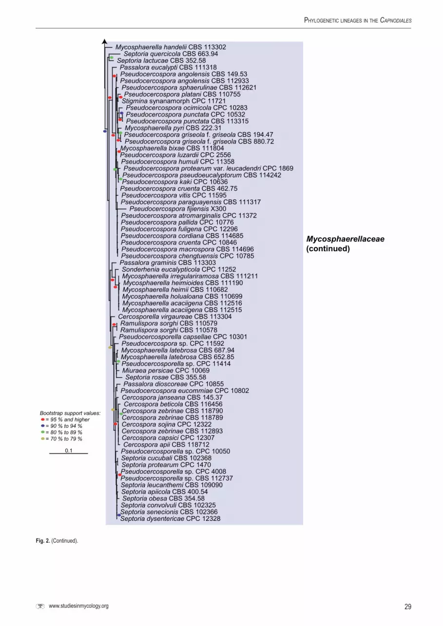
Phylogenetic lineageS in the Capnodiales
0.1
Mycosphaerella handelii CBS 113302Septoria quercicola CBS 663.94
Septoria lactucae CBS 352.58Passalora eucalypti CBS 111318Pseudocercospora angolensis CBS 149.53Pseudocercospora angolensis CBS 112933Pseudocercospora sphaerulinae CBS 112621Pseudocercospora platani CBS 110755
Stigmina synanamorph CPC 11721Pseudocercospora ocimicola CPC 10283Pseudocercospora punctata CPC 10532Pseudocercospora punctata CBS 113315Mycosphaerella pyri CBS 222.31Pseudocercospora griseola f. griseola CBS 194.47Pseudocercospora griseola f. griseola CBS 880.72
Mycosphaerella bixae CBS 111804Pseudocercospora luzardii CPC 2556Pseudocercospora humuli CPC 11358Pseudocercospora protearum var. leucadendri CPC 1869Pseudocercospora pseudoeucalyptorum CBS 114242Pseudocercospora kaki CPC 10636
Pseudocercospora cruenta CBS 462.75Pseudocercospora vitis CPC 11595Pseudocercospora paraguayensis CBS 111317
Pseudocercospora fijiensis X300Pseudocercospora atromarginalis CPC 11372Pseudocercospora pallida CPC 10776Pseudocercospora fuligena CPC 12296Pseudocercospora cordiana CBS 114685Pseudocercospora cruenta CPC 10846Pseudocercospora macrospora CBS 114696Pseudocercospora chengtuensis CPC 10785Passalora graminis CBS 113303Sonderhenia eucalypticola CPC 11252Mycosphaerella irregulariramosa CBS 111211Mycosphaerella heimioides CBS 111190Mycosphaerella heimii CBS 110682Mycosphaerella holualoana CBS 110699Mycosphaerella acaciigena CBS 112516Mycosphaerella acaciigena CBS 112515
Cercosporella virgaureae CBS 113304Ramulispora sorghi CBS 110579Ramulispora sorghi CBS 110578Pseudocercosporella capsellae CPC 10301Pseudocercospora sp. CPC 11592Mycosphaerella latebrosa CBS 687.94Mycosphaerella latebrosa CBS 652.85Pseudocercosporella sp. CPC 11414Miuraea persicae CPC 10069Septoria rosae CBS 355.58Passalora dioscoreae CPC 10855
Pseudocercospora eucommiae CPC 10802Cercospora janseana CBS 145.37Cercospora beticola CBS 116456
Cercospora zebrinae CBS 118790Cercospora zebrinae CBS 118789Cercospora sojina CPC 12322Cercospora zebrinae CBS 112893Cercospora capsici CPC 12307Cercospora apii CBS 118712
Pseudocercosporella sp. CPC 10050Septoria cucubali CBS 102368Septoria protearum CPC 1470Pseudocercosporella sp. CPC 4008Pseudocercosporella sp. CBS 112737Septoria leucanthemi CBS 109090Septoria apiicola CBS 400.54Septoria obesa CBS 354.58Septoria convolvuli CBS 102325Septoria senecionis CBS 102366Septoria dysentericae CPC 12328
Bootstrap support values: = 95 % and higher = 90 % to 94 % = 80 % to 89 % = 70 % to 79 %
Mycosphaerellaceae(continued)
Fig.2. (Continued).

30
crouS et al.
ResUlTs
DNAamplificationandphylogenyAmplification products of approximately 1 700 bases were obtained for the standard amplification of the isolates listed in Table 1. The LSU region of these sequences was used to obtain additional sequences from GenBank that were added to the partial LSU alignment. We expected a total size of approximately 5 500 bp for the concatenated SSU, ITS1, 5.8S nrDNA, ITS2 and LSU at the start of the study; however, our alignment totalled about 12 000 bp due to numerous insertions (most likely group 1 introns) encountered for several strains (Table 3). These insertions frequently resulted in products too large to amplify or sequence effectively and sometimes required us to design additional novel primers in extra overlapping steps to complete these gaps (see Materials and Methods for details). Searching the GenBank database using these insertions had varied success (Table 3). Sequences of the 18S nrDNA are more abundant in the database whereas sequences of the second half to two-thirds of the 28S nrDNA are mostly absent. This also evident in Table 3, where insertions in the SSU more frequently found with similarity sequences in the database and insertions in the LSU (e.g. those between positions 5018–5019 and 5424–5425) frequently did not retrieve any significant similarity. Although there were some exceptions (e.g. the insertion between 1820 and 1821 in the SSU of Batcheloromyces leucadendri), most of the insertions in the SSU obtained hits with SSU sequences of species of Capnodiales in the database. In one case, between 954 and 955 for the SSU sequence of Teratosphaeria mexicana (both strains), a partial hit was obtained with an LSU sequence of Lipomyces spencermartinsiae (GenBank DQ518980). Many of the insertions in the LSU sequences did not retrieve significant hits in the database and those that did were with unrelated taxa. It is quite possible that this is an artifact of the poor representation of full-length LSU sequences in the database, especially for members of the Capnodiales. In some cases, an LSU insertion retrieved a hit with SSU sequences in the database, e.g. the insertion between 5538 and 5539 in Batcheloromyces proteae and between 3560 and 3561 and 4875 and 4876 in Teratosphaeria mexicana strain CBS 120744. In two cases (Mycosphaerella latebrosa and Phaeophleospora eugeniicola), an insertion was either lost or gained between two strains of the same species. The primers designed in this study allowed us to effectively amplify and sequence the SSU and LSU for the selected isolates. Althought these primers were not tested on taxa outside of the Capnodiales (except for one of the outgroups, Neofusicoccum australe), we attempted to design them as robust as possible using degeneracy if needed. We therefore expect that these primers will have wider applicability than just the Capnodiales in cases where other published primers fail to amplify or amplify poorly.
The RAxML search of the partial LSU alignment yielded a most likely tree (Fig. 1) with a log likelihood -13397.994021. The matrix had 395 distinct alignment patterns, with 6 % completely undetermined characters in the alignment. The manually adjusted alignment contained 295 sequences (including the outgroup sequence, Dothidea insculpta GenBank DQ247802) and 763 characters including alignment gaps. The RAxML search of the almost complete SSU, 5.8S nrDNA and LSU alignment yielded a most likely tree (Fig. 2) with a log likelihood -39022.881140. The matrix had 1211 alignment patterns with 0.01 % of the characters consisting of gaps or undetermined characters. The manually adjusted alignment contained 205 sequences (including the
outgroup sequences, Neofusicoccum australe CPC 10899 and Magnaporthe grisea GenBank AB026819) and 5110 characters including alignment gaps. The obtained phylogenies (Figs 1–2) are discussed in the Taxonomy section below.
TaxonomySeveral well-supported clades could be distinguished in the present study (Figs 1–2), correlating to families in the Capnodiales. These families, and several new genera and species, are treated below.
TreatmentofphylogeneticcladesCapnodiales Woron. Ann. Mycol. 23: 177. 1925.Data obtained from multi-gene phylogenies prompted Schoch et al. (2006) to merge Mycosphaerellales with Capnodiales. Although the present study included numerous additional isolates, the orders remain problematic. Although there is support for the Mycosphaerellales as an order, additional families such as the Schizothyriaceae and Dissoconiaceae (see below) would have to also be elevated to order level, which would result in orders containing a single family, while Teratosphaeriaceae appears to comprise unresolved lineages. For this reason it was decided to retain these families within Capnodiales, but noting that as more families are added and better circumscribed, it is quite possible that the Mycosphaerellales would again be resurrected.
MycosphaerellaceaeLindau, In: Engler & Prantl, Nat. Pflan-zenfamilien 1(1): 421. 1897.Type species: Mycosphaerella punctiformis (Pers. : Fr.) Starbäck, Bih. Kongl. Svenska Vetensk.-Akad. Handl. 15(3, 2): 9. 1889.
Notes: The Mycosphaerellaceae contains numerous genera, 20 of which are listed by Crous (2009), with many names under consideration (Crous et al. 2009b, c). From these data it is clear that genera such as Passalora, Pseudocercospora, Pseudocercosporella, Septoria, Zasmidium and Ramichloridium are paraphyletic (Hunter et al. in prep.). Well-resolved genera include Cercospora, Cercosporella, Ramularia, Ramulispora, Sonderhenia and Polythrincium. One particularly problematic clade contains Periconiella, Ramichloridium, Verrucisporota and Zasmidium, along with “Mycosphaerella” and Rasutoria teleomorphs. Barr (1987) erected Rasutoria for species with brown ascospores occurring on Gymnospermae. Rasutoria clusters in a clade adjacent to “Mycosphaerella” species with hyaline ascospores, such as M. aleuritidis and Mycosphaerella daviesiicola (Verrucisporota daviesiae) (Beilharz & Pascoe 2002).
The genus Phaeophleospora (1916) clusters with Lecanosticta acicola. The genus Lecanosticta (1922) has typical Phaeophleospora-like conidia, except that its conidiomata are acervular, and not pycnidial. If the type of Lecanosticta, L. pini also clusters in this clade, the generic concept Phaeophleospora may have to be widened to include Lecanosticta, as was done with Kirramyces to include Colletogloeopsis (Cortinas et al. 2006a, b).
Considerable controversy has surrounded the coelomycetes that Crous et al. (1997) placed in Phaeophleospora. Based on DNA phylogenetic data, it has now been shown that Kirramyces anamorphs (Walker et al. 1992), including those accommodated in Colletogloeopsis (Crous & Wingfield 1996, Crous et al. 2004c, 2006c, Cortinas et al. 2006a, b), are linked to Teratosphaeria (Andjic et al. 2007, Crous et al. 2009b, c). Crous et al. (2007a)

31www.studiesinmycology.org
Phylogenetic lineageS in the Capnodiales
showed Phaeophleospora to reside in the Mycosphaerellaceae and Kirramyces in the Teratosphaeriaceae, respectively. However, most taxa investigated to date were collected from Eucalyptus. As shown in the present study, Phaeophleospora atkinsonii, a pathogen of Hebes spp. (Wu et al. 1996, Pennycook & McKenzie 2002), clusters distant from Phaeophleospora s. str., while the same is true for Phaeophleospora concentrica, which is a pathogen of Protea spp. (Taylor et al. 2001a), and Phaeophleospora stonei, a pathogen of Eucalyptus (Crous et al. 2007c, 2009c). These taxa thus clearly represent yet another two genera in the Phaeophleospora complex. An older name that would potentially be available is Scoleciasis. However, when B. Sutton examined exsiccati of the type species, S. aquatica, only ascomata of a Leptosphaeria species were found (Crous et al. 1997). The association of S. aquatica with the Leptosphaeria was also noted in the original description, and this may indicate that Scoleciasis is allied to taxa in the Phaeosphaeriopsis/Phaeoseptoria complex (Arzanlou & Crous 2006). Both P. atkinsonii and P. concentrica have a typical Kirramyces morphology, namely brown, percurrently proliferating conidiogenous cells, and brown, obclavate, verruculose, transversely euseptate conidia. Further species thus need to be included in analyses before these generic concepts can be clarified.
During the course of this study several fresh collections of Leptosphaeria protearum were obtained. Leptosphaeria protearum is a major leaf spot and blight pathogen of Protea spp. (Knox-Davies et al. 1987), and causes severe losses in plantations of South African Protea spp. in Hawaii, and has been recorded in many countries where South African proteas are cultivated (Taylor & Crous 1998, Taylor et al. 2001b, Crous et al. 2004a). Cultures of this pathogen were found to cluster in the Mycosphaerellaceae, where they represent an undescribed genus, characterised by having bitunicate asci without pseudoparaphyses, brown, 3-septate ascospores, and a Coniothyrium-like anamorph. Its close phylogenetic relationship to Phaeophloeospora concentrica (Fig. 1) suggests that they could be congeneric, and that in future more Phaeophloeospora-like anamorphs may be found to cluster in this clade. We propose a new genus to accommodate Leptosphaeria protearum below.
BrunneosphaerellaCrous,gen.nov.MycoBank MB514694.Etymology: Brunneus + Sphaerella = is after its brown ascospores and Sphaerella-like morphology.
Mycosphaerellae similis, sed ascosporis brunneis, 3-septatis.
Ascomata amphigenous, immersed to semi-immersed, black, single, gregarious, substomatal, pyriform or globose with a papillate, periphysate ostiole. Peridium consisting of three strata of slightly compressed textura angularis, an outer stratum of dark brown, thick-walled cells, becoming paler in the central stratum, and hyaline, thin-walled in the inner stratum. Asci clavate to cylindro-clavate, often curved, tapering to a pedicel, narrowing slightly to a rounded apex with an indistinct ocular chamber, 8-spored, bitunicate with fissitunicate dehiscense. Pseudoparaphyses absent. Ascospores biseriate, fusiform, broader at the apical end, initially hyaline and 1-septate, becoming yellow-brown and 3-septate at maturity, slightly constricted at median to supra-median septum.
Type species: Brunneosphaerella protearum (Syd. & P. Syd.) Crous, comb. nov.
Brunneosphaerella jonkershoekensis (Marinc., M.J. Wingf. & Crous) Crous, comb.nov. MycoBank MB514695. Fig. 3.Basionym: Leptosphaeria jonkershoekensisMarinc., M.J. Wingf. & Crous, In: Marincowitz et al., Microfungi occurring on Proteaceae in the fynbos: 62. 2008.
Ascomata pseudothecial, subepidermal, immersed, obpyriform, papillate, 180–205 × 160–235 µm. Peridium 20–30 µm thick, composed of relatively large cells, 11–15 × 2.5–5.5 µm; cells arranged in three strata; outer stratum consisting of 3–5 layers of dark brown, very thick-walled cells; middle stratum transient, consisting of a few layers of pale brown, thick-walled, compressed cells; inner stratum consisting of 1–2 layers of thin-walled, very compressed cells. Pseudoparaphyses absent. Asci bitunicate, inflated cylindrical to clavate, 81–95 × 13–15 µm, ocular chamber dome-shaped, indistinct. Ascospores pale brown, fusoid to ellipsoidal, tapering towards the base, (25–)29–34(–36) × (5–)6–7(–9) µm (av. 31.4 × 6.7 µm), apical cell the shortest, upper hemispore slightly larger than lower, at times slightly curved, 3-septate, smooth, guttulate (adapted from Marincowitz et al. 2008).
Host range and geographic distribution: Protea repens (South Africa, Western Cape) (Marincowitz et al. 2008).
Specimen examined: southAfrica, Western Cape Province, Jonkershoek Nature Reserve, leaf litter of Protea repens, 6 Jun. 2000, S. Marincowitz, PREM 59447 holotype.
Notes: Although no culture is presently available for this species, it clearly represents a species of Brunneosphaerella, characterised by its bitunicate asci, and brown, 3-septate ascospores, as well as the absence of pseudoparaphyses. Brunneosphaerella jonkershoekensis can easily be distinguished from B. protearum based on its much larger ascospores (Crous et al. 2004a).
Brunneosphaerella protearum (Syd. & P. Syd.) Crous, comb.nov. MycoBank MB514696. Fig. 4.Basionym: Leptosphaeria protearum Syd. & P. Syd., Ann. Mycol. 10: 441. 1912. Anamorph: “Coniothyrium” protearum Joanne E. Taylor & Crous, IMI Descriptions of Fungi and Bacteria No. 1343.1998.
Leaf spots circular to irregular, discrete to confluent, variable in size, under conditions favourable to disease symptoms more similar to a blight than a leaf spot, necrotic, sunken with a raised dark brown margin and with conspicuous black ascomata in the dead tissue, 4–30 mm diam. Ascomata pseudothecial, substomatal, amphigenous, immersed to semi-immersed, not erumpent, black, single, gregarious, 180–320 µm diam; in section, substomatal, subepidermal, pyriform or globose with a papillate, periphysate ostiole, immersed in a stroma consisting of deteriorated host mesophyll cells filled with fungal hyphae, (210–)230–264(–288) µm high, (180–)200–255(–300) µm diam. Peridium consisting of three strata of slightly compressed textura angularis, an outer stratum of dark brown, thick-walled cells, becoming paler in the central stratum, and hyaline, thin-walled in the inner stratum, altogether (20–)24.5–37.5(–50) µm thick. Asci clavate to cylindro-clavate, often curved, tapering to a pedicel, narrowing slightly to a rounded apex with an indistinct ocular chamber, 8-spored, bitunicate with fissitunicate dehiscense, (70–)80–87.5(–105) × (13.5–)14.5–16(–21.5) µm. Pseudoparaphyses absent. Ascospores biseriate, fusiform, broader

32
crouS et al.
Fig.3. Brunneosphaerella jonkershoekensis. A–B. Vertical sections through ascomata showing wall structure. C–D, G. Bitunicate asci. E–F. Ascospores. Scale bars: A, C = 50 µm, B = 20 µm, D, G = 10 µm, E–F = 5 µm (from Marincowitz et al. 2008).
at the apical end, initially hyaline and 1-septate, becoming yellow-brown and 3-septate at maturity, slightly constricted at median to supra-median septum, (21.5–)27.5–29.5(–37.5) × (6.3–)7.5–8(–10) µm in water mounts, (21–)25.5–27.5(–31) × (5.5–)6–7(–8) µm in lactophenol. Conidiomata barely visible and interspersed between ascomata, pycnidial, subepidermal, substomatal, separate, globose to pyriform, occasionally with well-developed papilla, dark brown, < 200 µm diam. Conidiophores reduced to conidiogenous cells. Conidiogenous cells discrete, smooth, hyaline, doliiform to ampulliform, holoblastic, proliferating 1–2 times percurrently, 4–6 × 3–4 µm. Conidia pale brown to medium brown, thick-walled on maturity, smooth to finely verruculose, eguttulate, ellipsoidal to globose, often truncate at one end, 5–10 × 3–7 µm (adapted from Crous et al. 2004a).
Host range and geographic distribution: Protea cynaroides, P. ‘Susara’ (Portugal, Madeira) (Moura & Rodrigues 2001); P. caffra, P. compacta, P. cynaroides, P. gaguedi, P. grandiceps, P. lacticolor, P. laurifolia, P. lepidocarpodendron, P. lorifolia, P. magnifica, P. nitida, P. punctata, P. repens, P. ‘Sheila’, Protea spp. (South Africa); P. cynaroides, P. laurifolia, P. neriifolia, P. ‘Ivory Musk’, P. ‘Mink’, P. ‘Pink Ice’, P. ‘Rose Mink’, P. susannae, Protea sp. (U.S.A., Hawaii) (Taylor et al. 2001b); P. cynaroides, P. gaguedi, P. neriifolia, Protea sp. (Zimbabwe, Inyanga) (Masuka et al. 1998).
Specimens examined: south Africa, Western Cape Province, Bettys’ Bay, leaf litter of Protea magnifica, 11 Jul. 2000, S. Marincowitz, PREM 59448; Helderberg Nature Reserve, leaf litter of Protea laurifolia, 14 Aug. 2000, S. Marincowitz, PREM 59482; Helderberg Nature Reserve, leaf litter of Protea obtusifolia, 14 Aug. 2000, S. Marincowitz, PREM 59495; Jonkershoek Nature Reserve, leaf litter of Protea
nitida, 6 Jun. 2000, S. Marincowitz, PREM 59442; Jonkershoek Nature Reserve, leaf litter of Protea repens, 6 Jun. 2000, S. Marincowitz, PREM 59450; Jonkershoek Nature Reserve, S33°59’11.2” E18°57’14.7” leaves of Protea sp., 1 Apr. 2007, P.W. Crous, CBS H-20330, cultures CPC 13914–13916; Jonkershoek Nature Reserve, S33°59’26.1” E18°57’59.5” leaves of Protea repens, 1 Apr. 2007, P.W. Crous, CBS H-20331, cultures CPC 13911–13913; Jonkershoek Nature Reserve, leaves of Protea sp., 1 Apr. 2007, P.W. Crous, CBS H-20332, cultures CPC 13908–13910; Jonkershoek Nature Reserve, “Tweede Waterval”, leaves of Protea sp., 1 Apr. 2007, P.W. Crous, CBS H-20333, cultures CPC 13902–13907; Jonkershoek Nature Reserve, leaves of Protea nitida, 12 Apr. 2008, L. Mostert, CBS H-20334, cultures CPC 15231–15233; Kirstenbosch Botanical Garden, leaves of Protea sp., 13 Jan. 2009, P.W. Crous, CBS H-20335, culture CPC 16338.
Notes: Although Taylor & Crous (1998) reported a Coniothyrium-like anamorph to develop in culture, none of the cultures examined in the present study on MEA, PDA or OA could be induced to sporulate, though spermatogonia and ascomatal initials were commonly observed.
The fact that cultures of Leptosphaeria protearum, which represents a well-known and serious pathogen of Proteaceae, clustered in the Mycosphaerellaceae, was totally unexpected. A further surprise was the fact that this species appears to represent a complex of several cryptic taxa. Whether these taxa can be correlated with differences in host range and geographic distribution can only be resolved once more collections have been obtained for study. Although the genus Sphaerulina, which represents Mycosphaerella-like taxa with 3-septate, hyaline ascospores, is part of the Mycosphaerellaceae (Crous et al., unpubl data), the type species, S. myriadea, clusters in the Septoria clade, and is thus unavailable for the species occurring on Proteaceae. Morphologically Brunneosphaerella is also distinct in

33www.studiesinmycology.org
Phylogenetic lineageS in the Capnodiales
Fig.4. Brunneosphaerella protearum. A–D. Leaf spots on different Protea spp. E. Close up of leaf spot showing ascomata. F. Substomatal ascomata. G–H. Vertical sections though ascomata, showing wall structure. I–K. Germinating ascospores on MEA. L–M, R. Bitunicate asci. N–Q, S. Juvenile to mature ascospores. Scale bars: G = 75 µm, H = 10 µm.

34
crouS et al.
that ascospores are always brown at maturity, and anamorphs have brown, percurrently proliferating conidiogenous cells, appearing Phaeophleospora-like. The recognition of Brunneosphaerella as a distinct genus in the Mycosphaerellaceae also raises the intriguing possibility that many phytopathogenic species of the Leptosphaeria-complex with brown, 3-septate ascospores, but lacking paraphyses, actually belong to Brunneosphaerella.
Passalora ageratinae Crous & A.R. Wood, sp.nov. Myco-Bank MB514697. Fig. 5.Etymology: Named after the host on which it occurs, Ageratina adenophora.
Passalorae assamensis similis, sed coloniis amphigenis, sine mycelio externo, conidiophoris brevioribus, 15–40 × 3–4.5 µm.
Leaf spots amphigenous, angular to irregular, 2–8 mm diam, medium brown, frequently with pale to grey-brown central part, and raised, dark brown border; pale to medium brown in reverse, with raised, dark brown border. Mycelium internal, consisting of smooth, branched, pale brown, 2–3 µm wide hyphae. Caespituli fasciculate, amphigenous, medium brown, arising from a brown, erumpent stroma, up to 80 µm wide, 40 µm high. Conidiophores subcylindrical, straight to geniculous-sinuous, unbranched, medium brown, finely verruculose, 1–3-septate, 15–40 × 3–4.5 µm. Conidiogenous cells terminal, pale to medium brown, finely verruculose with terminal, sympodial conidiogenous loci that are 1–2 µm diam, slightly thickened, darkened and refractive, 10–20 × 3–4 µm. Conidia in unbranched chains, pale brown, smooth, finely to prominently guttulate, subcylindrical to narrowly obclavate, apex obtuse, base long obconically subtruncate, (0–)1–3(–5)-septate, (20–)30–60(–80) × (3–)4(–4.5) µm; hila 1–1.5 µm wide, somewhat thickened, darkened and refractive.
Culture characteristics: On MEA erumpent, with uneven, folded surface, lobate margin, and moderate aerial mycelium; centre pale mouse-grey with patches of cinnamon, outer margin olivaceous-grey; reverse olivaceous-grey with patches of cinnamon; reaching 15 mm diam; on PDA spreading, with cinnamon to cream patches in centre, becoming umber towards smooth margins, with diffuse red pigment in agar; reverse olivaceous-grey, with patches of red, reaching 15 mm diam; on OA flat, spreading, up to 30 mm diam, iron-grey, with white, solitary mycelia strands, though aerial mycelium generally absent, reaching 30 mm diam.
Host range and geographic distribution: Ageratina adenophora, Australia, South Africa.
Specimen examined: southAfrica, KwaZulu-Natal Province, Hilton, on leaves of Ageratina adenophora, 28 May 2008, A.R. Wood, CBS H-20336 holotype, cultures ex-type CPC 15365 = CBS 125419, CPC 15366, 15367.
Notes: Ageratina adenophora (crofton weed; Asteraceae), which is indigenous to Mexico, has invaded many countries as a rapidly growing weed, forming dense thickets (Morris 1989, Parsons & Cuthbertson 1992, Wagner et al. 1999, Zhu et al. 2007, Muniappan et al. 2009). It is considered a serious weed in agriculture and forestry (Bess & Haramoto 1958, Sharma & Chhetri 1977, Kluge 1991), often replacing more-desired vegetation or native species.
A leaf spot pathogen, originally misidentified as Cercospora eupatorii (this species is currently known as Pseudocercospora eupatorii), was found to infect plants in Australia where a stem galling fly (Procecidochares utilis; Tephritidae) was introduced from Hawaii as a biological control agent (Dodd 1961). Presumably the fungus was introduced together with the flies originally from Mexico to Hawaii and then to Australia. Subsequently this same fungus was obtained from Australia and released in South Africa after host specificity testing indicated it was restricted to A. adenophora
Fig.5. Passalora ageratinae. A. Leaf spots. B. Close up of leaf spot with fruiting structures. C–D. Conidiophores. E–J. Conidia. Scale bars = 10 µm.

35www.studiesinmycology.org
Phylogenetic lineageS in the Capnodiales
(Morris 1989). The fungus causes partial defoliation of mature plants (Dodd 1961, Auld 1969), though the impact depends on environmental conditions (Dodd 1961). Seedlings are however killed rapidly (Wang et al. 1997).
This fungus, which has hitherto been known simply as “Phaeoramularia” sp., still lacks a name and proper description. The genus Phaeoramularia is treated as a synonym of Passalora (Crous & Braun 2003), and hence the species is named in the latter genus as P. ageratinae. Interestingly, this species appears to be closely related to Passalora fulva, which is a serious pathogen of tomato (Solanaceae) (Thomma et al. 2005).
Passalora armatae Crous & A.R. Wood, sp.nov. MycoBank MB514698. Fig. 6.Etymology: Named after the host on which it occurs, Dalbergia armata.
Passaloraea dalbergiicolae similis, sed conidiophoris in synnematibus densis, conidiis ad basim obconice truncatis, apice rostrato.
Leaf spots amphigenous, on upper surface visible as red-brown, irregular to subcircular spots with indistinct margins, 0.5–2 mm diam; in reverse indistinct, chlorotic to medium or red-brown. Mycelium internal, consisting of smooth, branched, pale brown, 2–3 µm wide hyphae. Caespituli hypophyllous, fasciculate to synnematous, up to 200 µm high and 250 µm wide, situated on a prominently erumpent, pale brown stroma, up to 100 µm high and wide. Conidiophores subcylindrical, unbranched, flexuous, guttulate, pale to medium brown, smooth, 120–180 × 4–6 µm, 2–6-septate. Conidiogenous cells terminal, subcylindrical,
guttulate, pale to medium brown, finely verruculose, becoming somewhat swollen, appearing slightly clavate, 25–70 × 6–8 µm; conidiogenous loci 4–20 per conidiogenous cell, sympodial, round, darkened, thickened, refractive, prominent, 2–3 µm wide, up to 1 µm high. Conidia (27–)30–40(–45) × 9–10(–12) µm, pale to medium brown, smooth to finely verruculose, granular to guttulate, thin-walled, ellipsoidal to obovoid, transversely 2–4-euseptate, widest in middle of basal cell, or middle of conidium, tapering to an obconically truncate base; hilum thickened, darkened and refractive; apical cell conical, elongating to an apical beak up to 20 µm long. When cultivated conidia remain attached to conidiogenous cells, giving conidiophores the appearance of small tufts which is very characteristic, and not commonly observed in Passalora.
Culture characteristics: On MEA slow growing, erumpent, with dense white aerial mycelium, which becomes mouse-grey, reaching 5 mm diam after 1 wk; on PDA mouse-grey (surface), iron-grey (reverse), with diffuse red pigment in agar; on OA similar to PDA, also with diffuse red pigment in agar.
Host range and geographic distribution: Dalbergia armata, South Africa.
Specimen examined: southAfrica, KwaZulu-Natal Province, South Coast, Mpenjati Nature Reserve, between Ramsgate and Port Edward, on leaves of Dalbergia armata, 28 May 2008, A.R. Wood, CBS H-20337 holotype, cultures ex-type CPC 15419 = CBS 125420, CPC 15420, 15421.
Notes: Passalora dalbergiae, which occurs on Dalbergia sissoo (Fabaceae) in India, is distinct from P. armatae in having superficial mycelium and solitary conidiophores (Hernández-Gutiérrez &
Fig.6. Passalora armatae. A. Fruiting in vivo. B–C. Caespituli with prominent basal stroma. D. Sporulation on MEA. E. Conidiogenous cells giving rise to conidia. F–G. Conidia. Scale bars: B = 125 µm, C–E = 10 µm.

36
crouS et al.
Dianese 2009). The previously described Passalora dalbergiicola is similar to P. armatae in conidial dimensions (3-septate, 25–45 × 7–10 µm; Ellis 1976), but distinct in that conidiophores are not in dense synnemata, conidiogenous cells can have single apical loci, and conidia have a less prominent basal taper, and lack the apical beaks typical of P. armatae (in vivo and in vitro).
Schizothyriaceae Höhn. ex Trotter, Sacc., D. Sacc. & Tra-verso, In: Saccardo, Syll. Fung. 24(2): 1254. 1928.Type species: Schizothyrium acerinum Desm., Ann. Sci. Nat. Bot. 11: 360. 1849.
Notes: Members of the Schizothyriaceae are associated with flyspeck symptoms on apples and pear fruit. The fungi grow superficially on the epicuticular wax, thereby reducing the marketability of the fruit, but do not penetrate the cuticle (Belding et al. 2000). Batzer et al. (2005, 2007) reported a range of diverse fungi to be associated with flyspeck symptoms on apples, the most prominent being species of Schizothyrium.
DissoconiaceaeCrous & de Hoog, fam. nov.MycoBank MB514699.Ascomata pseudotheciales, immerse, globosa, uniloculares. Sine pseudoparaphysibus. Asci fasciculati, octospori, bitunicati. Ascosporae ellipsoideae-fusiformes, 1-septatae, hyalinae. Conidiophora separata, ex hyphis oriunda, subcylindrica, subulata, lageniformia vel cylindrica, apicem versus attenuata, apice obtuse rotundato vel truncate, recta vel semel geniculata, laevia, modice brunnea, 0–pluriseptata, locis terminalibus vel lateralibus, rhachidi cum cicatricibus leniter incrassates, fuscatis. Conidia solitaria, pallide olivaceo-brunnea, laevia, ellipsoidea, obclavata vel globosa, 0–1-septata, hilis aliquantum fuscatis. Conidia secundaria nulla vel formata ad conidia primaria, pallide olivacea vel subhyalina, aseptata, pyriformia; conidiis impigre vel passive emittentibus.
Ascomata pseudothecial, immersed, globose, unilocular, papillate, ostiolate, canal periphysate; wall consisting of 3–4 layers of brown textura angularis; inner layer of flattened, hyaline cells. Pseudoparaphyses absent. Asci fasciculate, 8-spored, bitunicate. Ascospores ellipsoid-fusoid, 1-septate, hyaline, with or without mucoid sheath. Mycelium internal and external, consisting of branched, septate, smooth, hyaline to pale brown hyphae. Conidiophores separate, arising from hyphae, subcylindrical, subulate or lageniform to cylindrical, tapering to a bluntly rounded or truncate apex, straight to once geniculate, smooth, medium brown, 0–multi-septate; loci terminal and lateral, visible as slightly thickened, darkened scars on a rachis. Conidia solitary, pale olivaceous-brown, smooth, ellipsoid to obclavate or globose, 0–1-septate; hila somewhat darkened. Secondary conidia present or absent; developing adjacent to primary conidia, pale olivaceous to subhyaline, aseptate, pyriform; conidium discharge active or passive.
Type species: Dissoconium aciculare de Hoog, Oorschot & Hijwegen, Proc. K. Ned. Akad. Wet., Ser. C, Biol. Med. Sci. 86(2): 198. 1983.
Notes: Species of Dissoconium have Mycosphaerella-like teleomorphs (Crous et al. 2004c). The genus is characterised by forming conidia in pairs that are forcefully discharged, which is quite unique in the Capnodiales (de Hoog et al. 1983). Although D. aciculare, the type species of Dissoconium, was originally assumed to be hyperparasitic on powdery mildew (de Hoog et al. 1983), Jackson et al. (2004) revealed that another species, D. dekkeri,
could act as a foliar pathogen of Eucalyptus. Dissoconium dekkeri is, however, most commonly found in leaf spots in association with other species of Teratosphaeria and Mycosphaerella. Species of Dissoconium remain commensalists, and frequently occur asexually on lesions associated with pathogenic species of Capnodiales (Crous unpubl. data). They are ecologically and morphologically quite distinct from other members of the Capnodiales, and hence a separate family, the Dissoconiaceae, is herewith introduced to accommodate them. Ramichloridium forms brown, solitary conidiophores with a rachis and apical loci similar to that observed on Dissoconium, and primary conidia that are pale brown, 0–1-septate, with slightly thickened hila, but lacks secondary conidia (Arzanlou et al. 2008b). Both Dissoconium and Ramichloridium have in the past been reported as hyperparasitic on powdery mildews on various hosts (Hijwegen & Buchenauer 1984), which suggests that they share a similar ecology.
Teratosphaeriaceae Crous & U. Braun, Stud. Mycol. 58: 8. 2007. Type species: Teratosphaeria fibrillosa Syd. & P. Syd., Ann. Mycol. 10: 40. 1912.
Notes: Since the family was established by Crous et al. (2007a) it has been shown to be too widely defined, incorporating many diverse genera (Crous et al. 2009b, c), and even families such as the Piedraiaceae (Fig. 1). The node as such is not well supported, suggesting that as more taxa are added, further families remain to be separated from the Teratosphaeriaceae. Presently it incorporates diverse elements, and even lichens such as Cystocoleus ebeneus and Anisomeridium consobrinum. The identity of the latter strain (CBS 101364) needs to be confirmed, as its position in the tree appears doubtful.
The genus Catenulostroma, which is associated with numerous diverse substrates and habitats (Crous et al. 2007a), is typified by C. protearum, for which an epitype is designated in the present study. Several strains isolated from rock surfaces (Guiedan et al. 2008, Ruibal et al. 2008, 2009, this volume) cluster with Catenulostroma (Fig. 1), and appear to represent undescribed species of the latter. Of interest is the fact that the type species of Aulographina, A. pinorum (CBS 302.71, 174.90), which has hysterothecia, clusters in a clade with Catenulostroma microsporum, which has a Teratosphaeria-like teleomorph with pseudothecia (Taylor & Crous 2000, Crous et al. 2004a, 2007a). Isolates of A. pinorum were found to produce a Catenulostroma anamorph in culture. This raises two possibilities, namely that either the incorrect fungus was originally isolated from pine needles (namely Catenulostroma abietis), or that this is a species complex, in which A. pinorum resides. If these strains are indeed confirmed to represent A. pinorum, then it reveals the genus Aulographina to be heterogeneous, as A. eucalypti, which is a major leaf spot pathogen of Eucalyptus (Crous et al. 1989, Park et al. 2000, Carnegie & Keane 2003), clusters distant from A. pinorum. The taxonomy of these taxa is currently being addressed, and will be reported on elsewhere (Cheewangkoon et al., in prep.). During the course of this study some new members of the Teratosphaeriaceae were collected, which are described below:
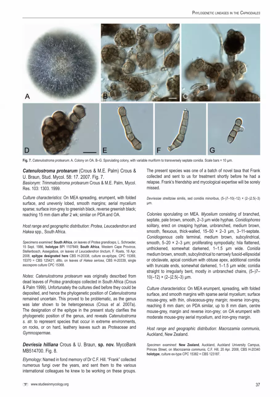
37www.studiesinmycology.org
Phylogenetic lineageS in the Capnodiales
Catenulostroma protearum (Crous & M.E. Palm) Crous & U. Braun, Stud. Mycol. 58: 17. 2007. Fig. 7.Basionym: Trimmatostroma protearum Crous & M.E. Palm, Mycol. Res. 103: 1303. 1999.
Culture characteristics: On MEA spreading, erumpent, with folded surface, and unevenly lobed, smooth margins; aerial mycelium sparse; surface iron-grey to greenish black, reverse greenish black; reaching 15 mm diam after 2 wk; similar on PDA and OA.
Host range and geographic distribution: Protea, Leucadendron and Hakea spp., South Africa.
Specimens examined: southAfrica, on leaves of Protea grandiceps, L. Schroeder, 15 Sept. 1986, holotype BPI 1107849; southAfrica, Western Cape Province, Stellenbosch, Assegaibos, on leaves of Leucadendron tinctum, F. Roets, 16 Apr. 2008, epitype designated here CBS H-20338, culture ex-epitype, CPC 15369, 15370 = CBS 125421; ditto, on leaves of Hakea sericea, CBS H-20339, single ascospore culture CPC 15368.
Notes: Catenulostroma protearum was originally described from dead leaves of Protea grandiceps collected in South Africa (Crous & Palm 1999). Unfortunately the cultures died before they could be deposited, and hence the phylogenetic position of Catenulostroma remained uncertain. This proved to be problematic, as the genus was later shown to be heterogeneous (Crous et al. 2007a). The designation of the epitype in the present study clarifies the phylogenetic position of the genus, and reveals Catenulostroma s. str. to represent species that occur in extreme environments, on rocks, or on hard, leathery leaves such as Proteaceae and Gymnospermae.
Devriesia hilliana Crous & U. Braun, sp. nov. MycoBank MB514700. Fig. 8.Etymology: Named in fond memory of Dr C.F. Hill. “Frank” collected numerous fungi over the years, and sent them to the various international colleagues he knew to be working on these groups.
The present species was one of a batch of novel taxa that Frank collected and sent to us for treatment shortly before he had a relapse. Frank’s friendship and mycological expertise will be sorely missed.
Devriesiae strelitziae similis, sed conidiis minoribus, (5–)7–10(–12) × (2–)2.5(–3) µm.
Colonies sporulating on MEA. Mycelium consisting of branched, septate, pale brown, smooth, 2–3 µm wide hyphae. Conidiophores solitary, erect on creaping hyphae, unbranched, medium brown, smooth, flexuous, thick-walled, 15–50 × 2–3 µm, 3–11-septate. Conidiogenous cells terminal, medium brown, subcylindrical, smooth, 5–20 × 2–3 µm; proliferating sympodially; hila flattened, unthickened, somewhat darkened, 1–1.5 µm wide. Conidia medium brown, smooth, subcylindrical to narrowly fusoid-ellipsoidal or obclavate, apical conidium with obtuse apex, additional conidia with truncate ends, somewhat darkened, 1–1.5 µm wide; conidia straight to irregularly bent, mostly in unbranched chains, (5–)7–10(–12) × (2–)2.5(–3) µm.
Culture characteristics: On MEA erumpent, spreading, with folded surface, and smooth margins with sparse aerial mycelium; surface mouse-grey, with thin, olivaceous-grey margin; reverse iron-grey, reaching 8 mm diam; on PDA similar, up to 8 mm diam, centre mouse-grey, margin and reverse iron-grey; on OA erumpent with moderate mouse-grey aerial mycelium, and iron-grey margin.
Host range and geographic distribution: Macrozamia communis, Auckland, New Zealand.
Specimen examined: New Zealand, Auckland, Auckland University Campus, Princes Street, on Macrozamia communis, C.F. Hill, 20 Apr. 2008, CBS H-20340 holotype, culture ex-type CPC 15382 = CBS 123187.
Fig.7. Catenulostroma protearum. A. Colony on OA. B–G. Sporulating colony, with variable muriform to transversely septate conidia. Scale bars = 10 µm.
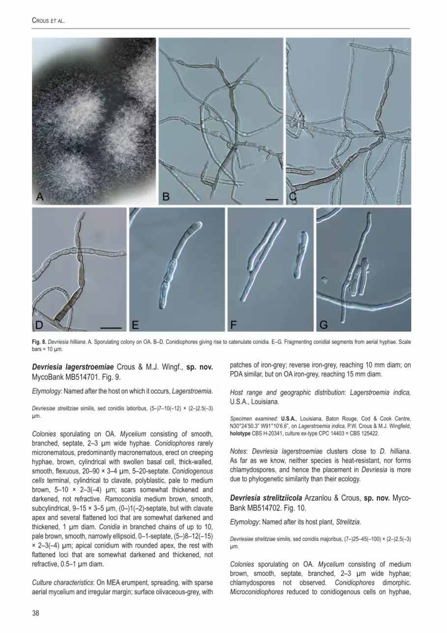
38
crouS et al.
Fig.8. Devriesia hilliana. A. Sporulating colony on OA. B–D. Conidiophores giving rise to catenulate conidia. E–G. Fragmenting conidial segments from aerial hyphae. Scale bars = 10 µm.
Devriesia lagerstroemiae Crous & M.J. Wingf., sp. nov. MycoBank MB514701. Fig. 9.Etymology: Named after the host on which it occurs, Lagerstroemia.
Devriesiae strelitziae similis, sed conidiis latioribus, (5–)7–10(–12) × (2–)2.5(–3) µm.
Colonies sporulating on OA. Mycelium consisting of smooth, branched, septate, 2–3 µm wide hyphae. Conidiophores rarely micronematous, predominantly macronematous, erect on creeping hyphae, brown, cylindrical with swollen basal cell, thick-walled, smooth, flexuous, 20–90 × 3–4 µm, 5–20-septate. Conidiogenous cells terminal, cylindrical to clavate, polyblastic, pale to medium brown, 5–10 × 2–3(–4) µm; scars somewhat thickened and darkened, not refractive. Ramoconidia medium brown, smooth, subcylindrical, 9–15 × 3–5 µm, (0–)1(–2)-septate, but with clavate apex and several flattened loci that are somewhat darkened and thickened, 1 µm diam. Conidia in branched chains of up to 10, pale brown, smooth, narrowly ellipsoid, 0–1-septate, (5–)8–12(–15) × 2–3(–4) µm; apical conidium with rounded apex, the rest with flattened loci that are somewhat darkened and thickened, not refractive, 0.5–1 µm diam.
Culture characteristics: On MEA erumpent, spreading, with sparse aerial mycelium and irregular margin; surface olivaceous-grey, with
patches of iron-grey; reverse iron-grey, reaching 10 mm diam; on PDA similar, but on OA iron-grey, reaching 15 mm diam.
Host range and geographic distribution: Lagerstroemia indica, U.S.A., Louisiana.
Specimen examined: U.s.A., Louisiana, Baton Rouge, Cod & Cook Centre, N30°24’50.3” W91°10’6.6”, on Lagerstroemia indica, P.W. Crous & M.J. Wingfield, holotype CBS H-20341, culture ex-type CPC 14403 = CBS 125422.
Notes: Devriesia lagerstroemiae clusters close to D. hilliana. As far as we know, neither species is heat-resistant, nor forms chlamydospores, and hence the placement in Devriesia is more due to phylogenetic similarity than their ecology.
Devriesia strelitziicola Arzanlou & Crous, sp.nov.Myco-Bank MB514702. Fig. 10.Etymology: Named after its host plant, Strelitzia.
Devriesiae strelitziae similis, sed conidiis majoribus, (7–)25–45(–100) × (2–)2.5(–3) µm.
Colonies sporulating on OA. Mycelium consisting of medium brown, smooth, septate, branched, 2–3 µm wide hyphae; chlamydospores not observed. Conidiophores dimorphic. Microconidiophores reduced to conidiogenous cells on hyphae,

39www.studiesinmycology.org
Phylogenetic lineageS in the Capnodiales
erect, cylindrical, medium brown, smooth with truncate ends, proliferating sympodially, 4–7 × 2–3 µm. Macroconidiophores erect, cylindrical, straight to geniculate-sinuous, medium brown, smooth, unbranched or branched above, 30–100 × 2.5–3 µm, 3–10-septate. Conidiogenous cells terminal or lateral on branched conidiophores, medium brown, smooth, cylindrical, proliferating sympodially, 7–15 × 2.5–3 µm; loci truncate, inconspicuous, 1–1.5 µm wide. Conidia medium brown, smooth, guttulate, subcylindrical to narrowly obclavate, apex obtuse to truncate, base truncate, occurring in branched chains, widest at the basal septum, (7–)25–45(–100) × (2–)2.5(–3) µm, (0–)3–6(–13)-septate; hila inconspicuous to somewhat darkened and thickened, not refractive, 1–1.5 µm wide.
Culture characteristics: On MEA erumpent, slow growing, with moderate aerial mycelium and smooth margins; surface mouse-grey, reverse iron-grey, reaching 8 mm diam after 2 wk; similar on PDA and OA.
Host range and geographic distribution: Strelitzia sp., South Africa.
Specimen examined: south Africa, KwaZulu-Natal, Durban, Botanical Garden near Reunion, on leaves of Strelitzia sp., 5 Feb. 2005, W. Gams & H. Glen, CBS H-20342, holotype, culture ex-type X1045 = CBS 122480.
Notes: Devriesia strelitziicola is the second Devriesia species to be described from this host (Arzanlou et al. 2008a). The genus Devriesia was originally established to accommodate a group of heat-resistant, Cladosporium-like fungi (Seifert et al. 2004), and it
appears that a different generic name will have to be introduced to accommodate those taxa occurring on plants. Further collections are required, however, to clarify the generic boundaries of Devriesia (Crous et al. 2007b).
Hortaea thailandica Crous & K.D. Hyde, sp. nov.Myco-Bank MB514703. Fig. 11.Etymology: Named after the country where it was collected, Thailand.
Hortaeae werneckii similis, sed conidiis brunneis, verruculosis, majoribus, (9–)10–13(–15) × (4–)5–6(–7) µm.
Colonies sporulating on MEA. Mycelium consisting of pale brown, smooth, septate, branched, 3–4 µm wide hyphae that become darker and thick-walled in the conidiogenous region. Conidiogenous cells integrated, intercalary on hyphae, reduced to short cylindrical loci, 2–2.5 µm wide, 1–4 µm tall; collarettes inconspicuous to minute; proliferating 1–2 times percurrently at apex. Conidia ellipsoid, aseptate, pale to medium brown, (4–)5–7(–9) × (2.5–)3 µm, verruculose, apex obtuse, base subtruncate with minute collarette; becoming swollen and elongate at maturity, with 1–4 transverse and 1–2 oblique septa; (9–)10–13(–15) × (4–) 5–6(–9) µm; hila inconspicuous, up to 2 µm wide, frequently with visible marginal frill; microcyclic conidiation commonly observed on OA, MEA and PDA.
Fig.9. Devriesia lagerstroemiae. A. Leaves and flowers of Lagerstroemia indica. B. Leaf spots. C. Colony on OA. D–H. Conidiophores giving rise to branched conidial chains. Scale bars = 10 µm.

40
crouS et al.
Culture characteristics: On MEA erumpent, spreading; surface irregular, folded, greenish black, with sparse olivaceous-grey aerial mycelium and smooth, lobed, margins; reverse greenish black; reaching 12 mm diam after 2 wk; similar on OA and PDA.
Host range and geographic distribution: Syzygium siamense, Thailand.
Specimen examined: Thailand, Khao Yai National Park, N14°14’42.6” E101°22’15.7”, on leaves of Syzygium siamense, in lesions with a cercosporoid fungus, 27 Mar. 2009, P.W. Crous & K.D. Hyde, holotype in BBH, isotype CBS H-20343, culture ex-type CPC 16652, 16651 = CBS 125423, also in BCC.
Notes: Similar to Hortaea werneckii, which is also frequently isolated from lesions in association with plant pathogenic fungi, H. thailandica occurred in leaf spots in association with a cercosporoid fungus. It is distinct from H. werneckii by forming larger conidia that turn medium brown and verruculose with age.
Several other taxa are newly placed in the Teratosphaeriaceae in the present study that require further evaluation. Xenomeris juniperi, a bitunicate ascomycete on Jupinerus with pseudothecia associated with a stroma, and pigmented, 1-septate ascospores, clusters close to Teratosphaeria species occurring on Protea and Eucalyptus, where the ascomata are also associated with stromatic tissue (Taylor & Crous 2000, Crous et al. 2006c). Fresh collections of this fungus would be required, however, to resolve its status. The occurrence of Sporidesmium species in the Teratosphaeriaceae should be interpreted with care, as the genus is polyphyletic, and further studies are required to resolve its status (Shenoy et al. 2006, Crous et al. 2008a, Yang et al., in prep.).
Fig.10. Devriesia strelitziicola. A. Strelitzia sp. with dead leaves. B. Colony on OA. C–G. Conidiophores giving rise to conidia. H–M. Conidia. Scale bars = 10 µm.

41www.studiesinmycology.org
Phylogenetic lineageS in the Capnodiales
Davidiellaceae C.L. Schoch, Spatafora, Crous & Shoemak-er, Mycologia 98: 1048. 2006.Type species: Davidiella tassiana (De Not.) Crous & U. Braun, Mycol. Progr. 3: 8. 2003.
Notes: The Davidiellaceae was introduced for the genus Davidiella, which has Cladosporium anamorphs. As shown in the present analysis, however, allied genera such as Toxicocladosporium, Verrucocladosporium, Rachicladosporium and Graphiopsis also belong in this family. Of interest is the position of Melanodothis caricis in Cladosporium s. str. This fungus, which infects florets of Carex and Kobresia, forms a stroma that gives rise to several immersed ascomata with bitunicate, oblong asci that are aparaphysate, and 0–(2)-septate, hyaline, 9–14.5 × 2–4 µm ascospores. In culture, a hyaline, Ramularia-like anamorph developed, with sympodial proliferation, catenulate conidia, with thickened, darkened loci (Arnold 1971). Although these characteristics are atypical of the Davidiella/Cladosporium species in this clade, the position of Melanodothis caricis in this family cannot simply be disregarded. However, the ex-type culture of this fungus (CBS 860.72) proved to be sterile.
A further unconfirmed sequence (CBS 354.29, culture sterile, but fast growing, grey-brown, Cladosporium-like), is that submitted as Sphaerulina polyspora. The culture was accessioned in 1929, deposited by A.E. Jenkins, and there is reason to believe that it was derived from BPI 623724!, which is authentic for the species,
and collected by F.A. Wolf in May 1924. Wolf (1925) described this fungus from twigs of Oxydendron arboretum with die-back disease symptoms, collected in Raleigh, North Carolina. Sphaerulina polyspora (623723 = Type!) has pseudothecia with aparaphysate, bitunicate asci, and ascospores that are hyaline, 3–5-septate, 20–24 × 6–7 µm. On the host it was linked to a Phoma-like anamorph, which also grew similar in culture (yeast-like budding), and has hyaline conidia which are ellipsoidal, 7–8 × 3.8–4 µm.
Colonies were reported as slow-growing, grey, appressed, with germinating ascospores forming yeast-like budding cells, and rarely having hyphae that extended from the margin of the colonies. The link between Sphaerulina-like species, with Selenophoma and Aureobasidium synanamorphs was recently illustrated by Cheewangkoon et al. (2009). Although members of the Dothideomycetes, these taxa do not cluster in the Davidiellaceae, and hence it seems a fair assumption that CBS 354.29 is not representative of Sphaerulina polyspora.
Rachicladosporium cboliae Crous, sp. nov. MycoBank MB514704. Fig. 12.Etymology: Named after the Consortium for the Barcode of Life, CBOL, who organised a Fungal Barcoding Symposium, during which this fungus was collected.
Rachicladosporio americano similis, sed conidiophoris dense fasciculatis et conidiis minoribus.
Fig.11. Hortaea thailandica. A. Cercosporoid leaf spots on Syzygium siamense, in which H. thailandica occurred. B. Colonies on OA. C–E. Hyphae with conidiogenous loci (arrows). F–H. Conidia. Scale bars = 10 µm.

42
crouS et al.
Colonies sporulating on OA. Mycelium consisting of branched, septate hyphae, pale brown, smooth, 1.5–3 µm wide, frequently constricted at septa, forming hyphal coils, but characteristically also forming intercalary and terminal clusters of chlamydospores that are brown, thick-walled, up to 6 µm diam. Conidiophores forming laterally on creeping hyphae, erect, visible as densely branched tufts on agar surface; conidiophores medium brown, smooth, thick-walled with bulbous base, lacking rhizoids, cylindrical, unbranched, flexuous, up to 250 µm long, 4–6 µm wide, 10–20-septate. Conidiogenous cells terminal, medium brown, smooth, polyblastic, subcylindrical, 10–20 × 3–4 µm; loci terminal, thickened, darkend, refractive, 1 µm diam. Ramoconidia 0(–1)-septate, subcylindrical, medium brown, smooth, 7–12 × 3–4 µm. Conidia 0(–1)-septate, in branched chains of up to 10, ellipsoid, pale brown, smooth, (6–)7–8(–10) × (2–)2.5(–3) µm; hila thickened, darkened and refractive, up to 1 µm diam.
Culture characteristics: On MEA spreading with sparse aerial mycelium and smooth margins; surface folded, centre pale mouse-grey to mouse-grey, margin iron-grey; reverse greenish black, reaching 15–20 mm diam after 2 wk; on PDA spreading with
moderate aerial mycelium and smooth margins; surface olivaceous-grey, margin mouse-grey, reverse olivaceous-grey; reaching 30 mm diam; on OA spreading, folded with moderate aerial mycelium; surface pale mouse-grey (centre) to olivaceous-grey at margin, reaching 20 mm diam.
Host range and geographic distribution: Twig litter, Virginia, U.S.A.
Specimen examined: U.S.A., Virginia, Front Royal, N38°53’35” W78°10’50”, on twig debris, 14 May 2007, P.W. Crous, holotype CBS H-20344, cultures ex-type CPC 14034 = CBS 125424, CPC 14035, 14036.
Notes: Rachicladosporium cboliae is a cryptic species close to R. americanum, which was collected at the same site. They can be distinguished on the litter in that R. cboliae has conidiophores with densely branched tufts of conidia, in contrast to the more sparsely branched conidiophores of R. americanum. Furthermore, R. cboliae also forms prominent chains of chlamydospores in culture, which lacks in R. americanum. Finally, R. cboliae has smaller ramoconidia and conidia than those found in R. americanum (ramoconidia 13–23 × 3–4 µm; conidia 10–18 × 3–4 µm; Cheewangkoon et al. 2009).
Fig.12. Rachicladosporium cboliae. A. Front Royal collection site in Virginia. B–E, G. Conidiophores with branched conidial chains. F. Hyphal coil. H–I. Chlamydospores in chains. J. Conidia. Scale bars = 10 µm.

43www.studiesinmycology.org
Phylogenetic lineageS in the Capnodiales
DIsCUssION
The class Dothideomycetes incorporates fungal taxa exhibiting a wide range of nutritional modes, and results in these fungi being found in many diverse niches (Fig. 13). The two largest orders Pleosporales (Zhang et al. 2009; this volume) and Capnodiales encapsulate this diversity. Here we continue to expand sampling within the Capnodiales in order to provide a well founded phylogenetic scaffold for taxonomic classification, informative genomic sampling, ecological studies and evolutionary evaluations.
CapnodialesThe Capnodiales currently contain nine families (Lumbsch & Huhndorf 2007, Kirk et al. 2008), a selection of which are included in this study, namely Capnodiaceae, Davidiellaceae, Mycosphaerellaceae, Piedraiaceae, and Teratosphaeriaceae. Unfortunately, no cultures were available of the Antennulariellaceae and Metacapnodiaceae, while Coccodiniaceae was again shown to cluster outside the order, in Chaetothyriales (Crous et al. 2007a). Families supported within Capnodiales (Fig. 1) include Capnodiaceae, Davidiellaceae, Teratosphaeriaceae, Dissoconiaceae, Schizothyriaceae and Mycosphaerellaceae. No support was obtained for Piedraiaceae, which appeared to cluster within Teratosphaeriaceae.
One of the main aims of the present study was to resolve the status of the Capnodiales and Mycosphaerellales. Although we were able to distinguish a clear, well resolved node for the Mycosphaerellales (incl. Mycosphaerellaceae), this node was not well supported, and elevating it to ordinal level would mean that additional orders need to be introduced to accommodate several families outside the Capnodiales s. str. This finding led us to conclude that it is best to retain all families within a single, diverse order, namely the Capnodiales.
evolution of nutritional modes and ecologicalgrowthhabitsThe ancestral state of the present assemblage of taxa is likely to be saprobic, as Phaeotheca (Sigler et al. 1981, de Hoog et al. 1997, Tsuneda et al. 2004), and Comminutispora (Ramaley 1996) represent the earliest diverging lineages. This was similarly found for a majority of lineages in the larger context of Ascomycota (Schoch et al. 2009a, b). These taxa were not only all isolated from dead materials or substrates, but they also share the same unique mode of conidiogenesis, namely endoconidia, and a “black-yeast” appearance in culture. Phaeotheca, which is strongly halophilic (Zalar et al. 1999) is closely related to the lichen Racodium rupestre, which forms an association with Trentepohlia algae, in which the filamentous algae is enclosed by melanised hyphae of the fungus. This feature is also shared by another lichen, namely Cystocoleus ebeneus (Teratosphaeriaceae) (Muggia et al. 2008). The Capnodiaceae (sooty molds) that also cluster in a basal position in the tree are epiphytes, growing on insect exudates (honey dew). The Capnodiaceae are related to the Davidiellaceae, which represent Cladosporium and allied genera. This family contains a wide range of ecological adaptations, from primary plant pathogens, such as Graphiopsis chlorocephala on Paeonia (Schubert et al. 2007a, Braun et al. 2008), “Mycosphaerella” iridis on Iris (David 1997), to taxa opportunistic on humans, Cladosporium bruhnei (Schubert et al. 2007b), to halotolerant taxa, Cladosporium sphaerospermum
(Zalar et al. 2007, Dugan et al. 2008), to saprobes, C. herbarum, C. cladosporioides (Schubert et al. 2007b).
The Teratosphaeriaceae contains several disjunct elements, many of which may still eventually be removed from the family as more taxa and additional sequence data are added, providing a better resolution to some of these clades. In its widest sense, the family contains lichens (Anisomeridium, Cystocoleus), saprobes (Catenulostroma spp.), and halophilic, hyperhydrotic or lipophilic species that have been reported from humans (Piedraia, Hortaea, Penidiella, Stenella) (de Hoog et al. 2000, Bonifaz et al. 2008, Plemenitaš et al. 2008), with the most derived clades tending to contain plant pathogens (Readeriella, Teratosphaeria).
Dissoconiaceae is an early diverging lineage to the Mycosphaerellaceae and Schizothyriaceae. Whereas most members of Dissoconiaceae appear to be commensalists, there is evidence that some species could be plant pathogenic (Jackson et al. 2004), while the Schizothyriaceae contains epiphytes (Batzer et al. 2007). The Mycosphaerellaceae contains species that are biotrophic (Polythrincium; Simon et al. 2009), necrotrophic plant pathogens (Brunneosphaerella, Cercospora, Dothistroma, Pseudocercospora, Pseudocercosporella, Ramularia, and Septoria), as well as some species that are saprobic (Passalora, Pseudocercospora, Ramichloridium and Zasmidium; Arzanlou et al. 2007), or endophytic (Pseudocercosporella endophytica; Crous 1998).
Within the Capnodiales, the positioning of saprobes such as Phaeotheca and Comminutispora and the sooty moulds (Capnodiaceae) may represent the more primitive state, from where transitions occurred to more lichenised, saprobic, biotrophic and nectrotrophic, plant pathogenic members of the order (Fig. 13). This appears to mirror the other large and diverse order in the class, the Pleosporales (Zhang et al. 2009; this volume). Lichenisation, as well as the ability to be saprobic or plant pathogenic evolved more than once, though the taxa in the later diverging clades of the tree tend to be strictly nectrotrophic plant pathogens. This should be interpreted with care, however, as Polythrincium is presently the only biotrophic member included in this analysis, and other biotrophic members of the Capnodiales may end up clustering here, among the presently dominant nectrotropic plant pathogens. One important and recent addition to Capnodiales diversity is the rock-inhabiting fungi (Ruibal et al. 2008, 2009; this volume). Although so far mainly isolated from sources in Antarctica and the Mediterranean area, it is clear that they are a ubiquitous group of fungi likely found throughout the globe. Their genetic diversity is underscored by the fact that rock inhabiting fungi of convergent morphology are also placed in other ascomycotan classes and orders (Gueidan et al. 2008). The fact that many of these species have reduced morphologies and are slow growers make their taxonomy challenging, but their phylogenetic placement within Teratosphaeriaceae and several other lineages within Capnodiales makes their inclusion in subsequent phylogenetic assessments of this order essential.
For this study, we designed novel primers to supplement primers presently available in literature. Although primers are usually designed for the genus or family of interest, they frequently tend to have a wider application. Therefore, we attempted to design our primers using a wide range of sequences from the GenBank sequence database, in the hope that these primers will eventually find application outside of the Capnodiales as well. Although this remains to be tested, we expect it to be the case. Our sequencing of the complete SSU and LSU for the selected members of the Capnodiales had a surprisingly large number of insertions present
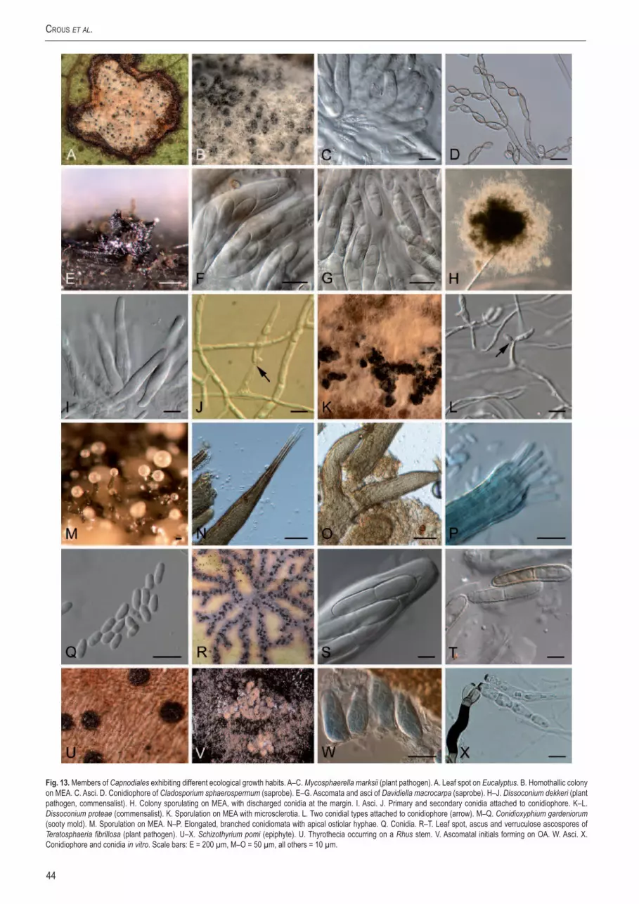
44
crouS et al.
Fig.13. Members of Capnodiales exhibiting different ecological growth habits. A–C. Mycosphaerella marksii (plant pathogen). A. Leaf spot on Eucalyptus. B. Homothallic colony on MEA. C. Asci. D. Conidiophore of Cladosporium sphaerospermum (saprobe). E–G. Ascomata and asci of Davidiella macrocarpa (saprobe). H–J. Dissoconium dekkeri (plant pathogen, commensalist). H. Colony sporulating on MEA, with discharged conidia at the margin. I. Asci. J. Primary and secondary conidia attached to conidiophore. K–L. Dissoconium proteae (commensalist). K. Sporulation on MEA with microsclerotia. L. Two conidial types attached to conidiophore (arrow). M–Q. Conidioxyphium gardeniorum (sooty mold). M. Sporulation on MEA. N–P. Elongated, branched conidiomata with apical ostiolar hyphae. Q. Conidia. R–T. Leaf spot, ascus and verruculose ascospores of Teratosphaeria fibrillosa (plant pathogen). U–X. Schizothyrium pomi (epiphyte). U. Thyrothecia occurring on a Rhus stem. V. Ascomatal initials forming on OA. W. Asci. X. Conidiophore and conidia in vitro. Scale bars: E = 200 µm, M–O = 50 µm, all others = 10 µm.

45www.studiesinmycology.org
Phylogenetic lineageS in the Capnodiales
for numerous strains. Although some of these insertions were anticipated based on data already present in GenBank’s database, the insertions in the LSU were not expected based on the sequences used for primer design. However, this could be a result of the fewer complete LSU sequences available in the database rather than a deviation on the part of members of the Capnodiales. More complete LSU sequences are needed from diverse orders to test whether this is the case or not. Some of the taxa sequenced during this study had insertions present at almost all of the possible insertion positions, e.g. Mycosphaerella latebrosa, Septoria quercicola and Teratosphaeria mexicana. These taxa are distributed throughout the tree, and do not only cluster in a basal position, and therefore it is difficult to predict why so many insertions were present. If these insertions were all present in a basal position, it would have been possible to argue that the higher number of insertions represents the ancestral condition, and that these insertions are lost during evolution. However, this proved not to be the case, and it could be that these taxa accumulated these insertions.
Although the present study adds significantly to our knowledge of the Capnodiales, the Capnodiaceae are still underrepresented, and probably consist of numerous diverse lineages that can be elevated to family level once our phylogenies become more resolved. Regardless of this fact, the Mycosphaerellaceae clade appears to be quite robust. It seems likely that further sampling of the diverse Teratosphaeriaceae will necessitate further taxonomic changes. The fact that the saprobic and plant pathogenic and endophytic modes have evolved several times in different families, suggest that many taxa can still easily adapt to changing environments. A focus on adding more lichenicolous taxa, and taxa occurring on non-plant substrates is crucial to provide further insight into the ecological adaptations occurring in the Capnodiales.
ACKNOwleDGeMeNTsSeveral colleagues have helped to collect material studied here, without which this work would not have been possible. Drs E.H.C. Mckenzie and S.R. Pennycook (Landcare New Zealand) are specifically thanked for recollecting Phaeophleospora atkinsonii. We are grateful to BIOTEC and Dr Lily Eurwilaichitr (Director, Bioresources unit, BIOTEC) for assisting with a collection trip in Thailand under the collaborative BIOTEC-CBS memorandum of understanding. We thank Miss Marjan Vermaas for preparing the photographic plates, and M. Starink-Willemse, and A. van Iperen, for assistance with DNA sequencing and fungal cultures. Work performed for this paper by the second author after 2008 was supported in part by the Intramural Research Program of the NIH, National Library of Medicine. Before 2008 work was funded by a grant from NSF (DEB-0717476). We are grateful to Drs Roland Kirschner, Alan J. Phillips and Treena I. Burgess for their critical comments on this script. The views expressed, however, are those of the authors.
ReFeReNCesAndjic V, Barber PA, Carnegie AJ, Hardy GEStJ, Wingfield MJ, Burgess TI (2007).
Phylogenetic reassessment supports accommodation of Phaeophleospora and Colletogloeopsis from eucalypts in Kirramyces. Mycological Research 111: 1184–1198.
Aptroot A (2006). Mycosphaerella and its Anamorphs: 2. Conspectus of Mycosphaerella. CBS Biodiversity Series 5, Utrecht, The Netherlands.
Arnold RH (1971). Melanodothis caricis, n. gen., n. sp. and “Hyalodothis? caricis”. Canadian Journal of Botany 49: 2187–2196.
Arzanlou M, Crous PW (2006). Phaeosphaeriopsis musae. In: Fungal Planet – A Global Initiative to promote the Study of Fungal Biodiversity (Crous PW, Seifert KA, Samson RA, Hawksworth DL eds). CBS, Utrecht, Netherlands. Fungal PlanetNo. 9.
Arzanlou M, Crous PW, Groenewald JZ (2008a). Devriesia strelitziae. In: Fungal Planet – A Global Initiative to promote the Study of Fungal Biodiversity (Crous PW, Seifert KA, Samson RA, Hawksworth DL eds). CBS, Utrecht, Netherlands. Fungal Planet No. 22.
Arzanlou M, Groenewald JZ, Fullerton RA, Abeln ECA, Carlier J, et al. (2008b). Multiple gene genealogies and phenotypic characters differentiate several novel species of Mycosphaerella and related anamorphs on banana. Persoonia 20: 19–37.
Arzanlou M, Groenewald JZ, Gams W, Braun U, Shin H-D, Crous PW (2007). Phylogenetic and morphotaxonomic revision of Ramichloridium and allied genera. Studies in Mycology 58: 57–93.
Auld BA (1969). Incidence of damage caused by organisms which attack crofton weed in the Richmond-Tweed region of New South Wales. Australian Journal of Science 32: 163.
Barr ME (1987). Prodomus to class Loculoascomycetes. Published by the author, Amherst, Massachusetts.
Batzer JC, Gleason ML, Harrington TC, Tiffany LH (2005). Expansion of the sooty blotch and flyspeck complex on apples based on analysis of ribosomal DNA gene sequences and morphology. Mycologia 97: 1268–1286.
Batzer JC, Mercedes Diaz Arias M, Harrington TC, Gleason ML, Groenewald JZ, Crous PW (2007). Four species of Zygophiala (Schizothyriaceae, Capnodiales) are associated with the sooty blotch and flyspeck complex on apple. Mycologia 100: 246–258.
Beilharz V, Pascoe I (2002). Two additional species of Verrucisporota, one with a Mycosphaerella teleomorph, from Australia. Mycotaxon 82: 357–365.
Belding RD, Sutton TB, Blankenship SM, Young E (2000). Relationship between apple fruit epicuticular wax and growth of Peltaster fructicola and Leptodontidium elatius, two fungi that cause sooty blotch disease. Plant Disease 84: 767–772.
Bess HA, Haramoto FH (1958). Biological control of pamakani, Eupatorium adenophorum, in Hawaii by a tephritid gall fly, Procecidochares utilis. 1. The life history of the fly and its effectiveness in the control of the weed. In: Proceedings of the Tenth International Congress of Entomology, Vol. 4 (Becker EC, ed.). Canada, Ottawa, Mortimer: 543–548.
Bonifaz A, Badali H, Hoog GS de, Cruz M, Araiza J, et al. (2008). Tinea nigra by Hortaea werneckii, a report of 22 cases from Mexico. Studies in Mycology 61: 77–82.
Braun U, Crous PW, Dugan F, Groenewald JZ, Hoog GS de (2003). Phylogeny and taxonomy of Cladosporium-like hyphomycetes, including Davidiella gen. nov., the teleomorph of Cladosporium s.str. Mycological Progress 2: 3–18.
Braun U, Crous PW, Schubert K (2008). Taxonomic revision of the genus Cladosporium s. lat. 8. Reintroduction of Graphiopsis (= Dichocladosporium) with further reassessments of cladosporioid hyphomycetes. Mycotaxon 103: 207–216.
Carnegie AJ, Keane PJ (2003). Variation in severity of target spot, caused by Aulographina eucalypti, in a eucalypt species and provenance trial in Victoria. Australasian Plant Pathology 32: 393–402.
Cheewangkoon R, Crous PW, Hyde KD, Groenewald JZ, To-anan C (2008). Species of Mycosphaerella and related anamorphs on Eucalyptus leaves from Thailand. Persoonia 21: 77–91.
Cheewangkoon R, Groenewald JZ, Summerell BA, Hyde KD, To-anun C, Crous PW (2009). Myrtaceae, a cache of fungal biodiversity. Persoonia 23: 55–85.
Cortinas M-N, Burgess T, Dell D, Xu D, Crous PW, Wingfield BD, Wingfield MJ (2006b). First record of Colletogloeopsis zuluense comb. nov., causing a stem canker of Eucalyptus in China. Mycological Research 110: 229–236.
Cortinas M-N, Crous PW, Wingfield BD, Wingfield MJ (2006a). Multi-gene phylogenies and phenotypic characters distinguish two species within the Colletogloeopsis zuluensis complex associated with Eucalyptus stem cankers. Studies in Mycology 55: 133–146.
Crous PW (1998). Mycosphaerella spp. and their anamorphs associated with leaf spot diseases of Eucalyptus. Mycologia Memoir 21: 1–170.
Crous PW (2009). Taxonomy and phylogeny of the genus Mycosphaerella and its anamorphs. Fungal Diversity 38: 1–24.
Crous PW, Aptroot A, Kang JC, Braun U, Wingfield MJ (2000). The genus Mycosphaerella and its anamorphs. Studies in Mycology 45: 107–121.
Crous PW, Braun U (2003). Mycosphaerella and its anamorphs. 1. Names published in Cercospora and Passalora. CBS Biodiversity Series 1: 1–571.
Crous PW, Braun U, Groenewald JZ (2007a). Mycosphaerella is polyphyletic. Studies in Mycology 58: 1–32.
Crous PW, Braun U, Schubert K, Groenewald JZ (2007b). Delimiting Cladosporium from morphologically similar genera. Studies in Mycology 58: 33–56.
Crous PW, Denman S, Taylor JE, Swart L, Palm ME (2004a). Cultivation and diseases of Proteaceae: Leucadendron, Leucospermum and Protea. CBS Biodiversity Series 2: 1–228.
Crous PW, Ferreira FA, Sutton BC (1997). A comparison of the fungal genera Phaeophleospora and Kirramyces (coelomycetes). South African Journal of Botany 63: 111–115.
Crous PW, Gams W, Stalpers JA, Robert V, Stegehuis G (2004b). MycoBank: an online initiative to launch mycology into the 21st century. Studies in Mycology 50: 19–22.
Crous PW, Groenewald JZ, Mansilla JP, Hunter GC, Wingfield MJ (2004c). Phylogenetic reassessment of Mycosphaerella spp. and their anamorphs occurring on Eucalyptus. Studies in Mycology 50: 195–214.

46
crouS et al.
Crous PW, Groenewald JZ, Summerell BA, Wingfield BD, Wingfield MJ (2009a). Co-occurring species of Teratosphaeria on Eucalyptus. Persoonia 22: 38–48.
Crous PW, Groenewald JZ, Wingfield MJ, Aptroot A (2003). The value of ascospore septation in separating Mycosphaerella from Sphaerulina in the Dothideales: a Saccardoan myth? Sydowia 55: 136–152.
Crous PW, Groenewald JZ, Wood AR (2008a). Sporidesmium knawiae. In: Fungal Planet – A Global Initiative to promote the Study of Fungal Biodiversity (Crous PW, Seifert KA, Samson RA, Hawksworth DL eds). CBS, Utrecht, Netherlands. Fungal PlanetNo. 29.
Crous PW, Kang JC, Braun U (2001). A phylogenetic redefinition of anamorph genera in Mycosphaerella based on ITS rDNA sequence and morphology. Mycologia 93: 1081–1101.
Crous PW, Knox-Davies PS, Wingfield MJ (1989). A summary of fungal leaf pathogens of Eucalyptus and the diseases they cause in South Africa. South African Forestry Journal 149: 9–16.
Crous PW, Liebenberg MM, Braun U, Groenewald JZ (2006a). Re-evaluating the taxonomic status of Phaeoisariopsis griseola, the causal agent of angular leaf spot of bean. Studies in Mycology 55: 163–173.
Crous PW, Palm ME (1999). Systematics of selected foliicolous fungi associated with leaf spots of Proteaceae. Mycological Research 103: 1299–1304.
Crous PW, Schroers HJ, Groenewald JZ, Braun U, Schubert K (2006b). Metulocladosporiella gen. nov. for the causal organism of Cladosporium speckle disease of banana. Mycological Research 110: 264–275.
Crous PW, Summerell BA, Carnegie AJ, Mohammed C, Himaman W, Groenewald JZ (2007c). Foliicolous Mycosphaerella spp. and their anamorphs on Corymbia and Eucalyptus. Fungal Diversity 26: 143–185.
Crous PW, Summerell BA, Carnegie AJ, Wingfield MJ, Groenewald JZ (2009b). Novel species of Mycosphaerellaceae and Teratosphaeriaceae. Persoonia 23: 119–146.
Crous PW, Summerell BA, Carnegie AJ, Wingfield MJ, Hunter GC, et al. (2009c). Unravelling Mycosphaerella: do you believe in genera? Persoonia 23: 99–118.
Crous PW, Summerell BA, Mostert L, Groenewald JZ (2008b). Host specificity and speciation of Mycosphaerella and Teratosphaeria species associated with leaf spots of Proteaceae. Persoonia 20: 59–86.
Crous PW, Verkley GJM, Groenewald JZ, Samson RA (eds) (2009d). Fungal Biodiversity. CBS Laboratory Manual Series. Centraalbureau voor Schimmelcultures, Utrecht, Netherlands.
Crous PW, Wingfield MJ (1996). Species of Mycosphaerella and their anamorphs associated with leaf blotch disease of Eucalyptus in South Africa. Mycologia 88: 441–458.
Crous PW, Wingfield MJ, Mansilla JP, Alfenas AC, Groenewald JZ (2006c). Phylogenetic reassessment of Mycosphaerella spp. and their anamorphs occurring on Eucalyptus. II. Studies in Mycology 55: 99–131.
Crous PW, Wingfield MJ, Park RF (1991). Mycosphaerella nubilosa a synonym of M. molleriana. Mycological Research 95: 628–632.
Crous PW, Wood AR, Okada G, Groenewald JZ (2008c). Foliicolous microfungi occurring on Encephalartos. Persoonia 21: 135–146.
David JC (1997). A contribution to the systematics of Cladosporium. Revision of the fungi previously referred to Heterosporium. Mycological Papers 172: 1–157.
Dodd AP (1961). Biological control of Eupatorium adenophorum in Queensland. Australian Journal of Science 23: 356–365.
Dugan FM, Braun U, Groenewald JZ, Crous PW (2008). Morphological plasticity in Cladosporium sphaerospermum. Persoonia 21: 9–16.
Ellis MB (1976). More dematiaceous hyphomycetes. CAB, International Mycological Institute, Surrey, Kew, U.K.
Gardes M, Bruns TD (1993). ITS primers with enhanced specificity for basidiomycetes - application to the identification of mycorrhizae and rusts. Molecular Ecology 2: 113–118.
Gargas A, Taylor JW (1992). Polymerase chain reaction (PCR) primers for amplifying and sequencing 18S rDNA from lichenized fungi. Mycologia 84: 589–592.
Gueidan C, Ruibal Villaseñor C, Hoog GS de, Gorbushina AA, Untereiner WA, Lutzoni F (2008). A rock-inhabiting ancestor for mutualistic and pathogen-rich fungal lineages. Studies in Mycology 61: 111–119.
Hawksworth DL, Kirk PM, Sutton BC, Pegler DN (1995). Ainsworth and Bisby’s Dictionary of the Fungi, 8th edn. CAB International, Wallingford, U.K.
Hernández-Gutiérrez A, Dianese JC (2009). New cercosporoid fungi from the Brazilian Cerrado 2. Species on hosts of the subfamilies Caesalpinioideae, Faboideae and Mimosoideae (Leguminosae s. lat.). Mycotaxon 107: 1–24.
Hijwegen T, Buchenauer H (1984). Isolation and identification of hyperparasitic fungi associated with Erysiphaceae. Netherlands Journal of Plant Pathology 90: 79–84.
Hoog GS de, Beguin H, Batenburg-van de Vegte WH (1997). Phaeotheca triangularis, a new meristematic black yeast from a humidifier. Antonie van Leeuwenhoek 71: 289–295.
Hoog GS de, Gerrits van den Ende AHG (1998). Molecular diagnostics of clinical strains of filamentous Basidiomycetes. Mycoses 41: 183–189.
Hoog GS de, Guarro J, Gené J, Figueras MJ (2000). Atlas of Clinical Fungi. 2nd Edn. Centraalbureau voor Schimmelcultures, Utrecht, Netherlands, and Universitat Rovira I Virgili, Reus, Spain.
Hoog GS de, Oorschot CAN van, Hijwegen T (1983). Taxonomy of the Dactylaria complex. II. Proceedings van de Koninklijke Nederlandse Akademie van Wetenschappen, Series C, 86(2): 197–206.
Hughes SJ. 1976. Sooty molds. Mycologia 68: 451–691.Hunter GC, Wingfield BD, Crous PW, Wingfield MJ (2006). A multi-gene phylogeny
for species of Mycosphaerella occurring on Eucalyptus leaves. Studies in Mycology 55:147–161.
Jackson SL, Maxwell A, Neumeister-Kemp HG, Dell B, Hardy GEStJ (2004). Infection, hyperparasitism and conidiogenesis of Mycosphaerella lateralis on Eucalyptus globulus in Western Australia. Australasian Plant Pathology 33: 49–53.
Kirk PM, Cannon PF, David JC, Stalpers JA (2001). Ainsworth and Bisby’s Dictionary of the Fungi, 9th edn. CAB International, Wallingford, U.K.
Kirk PM, Cannon PF, Minter DW, Stalpers JA (2008). Ainsworth and Bisby’s Dictionary of the Fungi, 10th edn. CAB International, Wallingford, U.K.
Kluge RL (1991). Biological control of crofton weed, Ageratina adenophora (Asteraceae), in South Africa. Agriculture, Ecosystems & Environment 37: 187–191.
Knox-Davies PS, Wyk PS van, Marasas WFO (1987). Diseases of Protea, Leucospermum and Leucadendron recorded in South Africa. Phytophylactica 19: 327–337.
Kretzer A, Li Y, Szaro T, Bruns TD (1996). Internal transcribed spacer sequences from 38 recognized species of Suillus sensu lato: phylogenetic and taxonomic implications. Mycologia 88: 776–785.
Kruys A, Eriksson OE, Wedin M (2006). Phylogenetic relationships of coprophilous Pleosporales (Dothideomycetes, Ascomycota), and the classification of some bitunicate taxa of unknown position. Mycological Research 110: 527–536.
Lumbsch HT, Huhndorf S (2007). Outline of Ascomycota. Myconet 13: 1–99.Marincowitz S, Crous PW, Groenewald JZ, Wingfield MJ (2008). Microfungi
occurring on Proteaceae in the fynbos. CBS Biodiversity Series 7: 1–166.Masuka J, Cole DL, Mguni C (1998). List of Plant Diseases in Zimbabwe.
Department of Research Specialist Services, Ministry of Lands and Agriculture, Harare, Zimbabwe.
Moncalvo J-M, Rehner SA, Vilgalys R (1993). Systematics of Lyophyllum section Difformia based on evidence from culture studies and ribosomal DNA sequences. Mycologia 85: 788–794.
Morris MJ (1989). Host specificity studies of leaf spot fungus, Phaeoramularia sp. for the biological control of crofton weed (Ageratina adenophorum) in South Africa. Phytophylactica 21: 281–283.
Moura MF, Rodrigues PF (2001). Fungal diseases on Proteas identified in Maderia Island. Acta Horticulturae 545: 265–268.
Muggia L, Hafellner J, Wirtz N, Hawksworth DL, Grube M (2008). The sterile microfilamentous lichens Cystocoleus ebeneus and Racodium rupestre are relatives of clinically important dothidealean fungi. Mycological Research 112: 50–56.
Muniappan R, Raman A, Reddy GVP (2009) Ageratina adenophora (Sprengel) King and Robinson (Asteraceae). In: Biological control of tropical weeds using arthropods. (Muniappan R, Reddy GVP, Raman A, eds). Cambridge University Press, Cambridge: 1–16.
Park RF, Keane PJ, Wingfield MJ, Crous PW (2000). Fungal diseases of eucalypt foliage. In: Diseases and pathogens of eucalypts. (Keane PJ, Kile GA, Podger FD, Brown BN, eds). CSIRO publishing, Australia: 153–239.
Parsons WT, Cuthbertson EG (1992). Noxious Weeds of Australia. Melbourne: Inkata Press.
Pennycook SR, McKenzie EHC (2002). Scoleciasis atkinsonii, an earlier name for Phaeophleospora hebes; and a note on G.H. Cunningham’s epithets hebe and pseudopanax. Mycotaxon 82: 145–146.
Plemenitaš A, Vaupotič T, Lenassi M, Kogej T, Gunde-Cimerman N (2008). Adaptation of extremely halotolerant black yeast Hortaea werneckii to increased osmolarity: a molecular perspective at a glance. Studies in Mycology 61: 67–75.
Ramaley AW (1996). Comminutispora gen. nov. and its Hyphospora gen. nov. anamorph. Mycologia 88: 132–136.
Rayner RW (1970). A mycological colour chart. CMI and British Mycological Society. Kew, Surrey, England.
Rehner SA, Samuels GJ (1994). Taxonomy and phylogeny of Gliocladium analysed from nuclear large subunit ribosomal DNA sequences. Mycological Research 98: 625–634.
Ruibal C, Platas G, Bills GF (2008). High diversity and morphological convergence among melanised fungi from rock formations in the Central Mountain System of Spain. Persoonia 21: 93–110.
Ruibal C, Gueidan C, Selbmann L, Gorbushina A, Crous PW, et al. (2009). Phylogeny of rock-inhabiting fungi related to Dothideomycetes. Studies in Mycology 64: 123–133.
Schoch CL, Shoemaker RA, Seifert KA, Hambleton S, Spatafora JW, Crous PW (2006). A multigene phylogeny of the Dothideomycetes using four nuclear loci. Mycologia 98: 1041–1052.

47www.studiesinmycology.org
Phylogenetic lineageS in the Capnodiales
Schoch CL, Crous PW, Groenewald JZ, Boehm EWA, Burgess TI, et al. (2009a). A class-wide phylogenetic assessment of Dothideomycetes. Studies in Mycology 64: 1–15.
Schoch CL, Sung GH, Lopez-Giraldez F, Townsend JP, Miadlikowska J, et al. (2009b). The Ascomycota Tree of Life: A Phylum-wide Phylogeny Clarifies the Origin and Evolution of Fundamental Reproductive and Ecological Traits. Systematic Biology 58: 224–239.
Schubert K, Braun U, Groenewald JZ, Crous PW (2007a). Cladosporium leaf-blotch and stem rot of Paeonia spp. caused by Dichocladosporium chlorocephalum gen. nov. Studies in Mycology 58: 95–104.
Schubert K, Groenewald JZ, Braun U, Dijksterhuis J, Starink M, et al. (2007b). Biodiversity in the Cladosporium herbarum complex (Davidiellaceae, Capnodiales), with standardisation of methods for Cladosporium taxonomy and diagnostics. Studies in Mycology 58: 105–156.
Seifert KA, Nickerson NL, Corlett M, Jackson ED, Louis-Seize G, Davies RJ (2004). Devriesia, a new hyphomycete genus to accommodate heat-resistant, cladosporium-like fungi. Canadian Journal of Botany 82: 914–926.
Sharma KC, Chhetri GKK (1977). Reports on studies on the biological control of Eupatorium adenophorum. Nepalese Journal of Agriculture 12: 135–157.
Shenoy BD, Jeewon R, Wu WP, Bhat DJ, Hyde KD (2006). Ribosomal and RPB2 DNA sequence analyses suggest that Sporidesmium and morphologically similar genera are polyphyletic. Mycological Research 110: 916–928.
Sigler L, Tsuneda A, Carmichael JW (1981). Phaeotheca and Phaeosclera, two new genera of dematiaceous hyphomycetes and a redescription of Sarcinomyces Lindner. Mycotaxon 12: 449–467.
Simon UK, Groenewald JZ, Crous PW (2009). Cymadothea trifolii, an obligate biotrophic leaf parasite of Trifolium, belongs to Mycosphaerellaceae as shown by nuclear ribosomal DNA analyses. Persoonia 22: 49–55.
Singh SK, Chaudhary RK, Morgan-Jones G (1995). Notes on Hyphomycetes: LXVII. Three new species of Phaeoramularia from Nepal. Mycotaxon 54: 57–66.
Spatafora JW, Mitchell TG, Vilgalys R (1995). Analysis of genes encoding for small-subunit rRNA sequences in studying phylogenetics of dematiaceous fungal pathogens. Journal of Clinical Microbiology 33: 1322–1326.
Stamatakis A (2006). RAxML-VI-HPC: maximum likelihood-based phylogenetic analyses with thousands of taxa and mixed models. Bioinformatics 22: 2688–2690.
Stamatakis A, Hoover P, Rougemont J (2008). A Rapid Bootstrap Algorithm for the RAxML Web Servers. Systematic Biology 57: 758–771.
Taylor JE, Cannon PF, David JC, Crous PW (2001a). Two new Phaeophleospora species associated with leaf spots of Proteaceae. South African Journal of Botany 67: 39–43.
Taylor JE, Crous PW (1998). Leptosphaeria protearum. IMI Descriptions of Fungi and Bacteria No. 1343.
Taylor JE, Crous PW (2000). Fungi occurring on Proteaceae. New anamorphs for Teratosphaeria, Mycosphaerella and Lembosia, and other fungi associated with leaf spots and cankers of Proteaceous hosts. Mycological Research104: 618–636.
Taylor JE, Crous PW, Palm ME (2001b). Foliar and stem fungal pathogens of Proteaceae in Hawaii. Mycotaxon78: 449–490.
Thomma BPHJ, van Esse PH, Crous PW, Wit PJGM de (2005). Cladosporium fulvum (syn. Passalora fulva), a highly specialized plant pathogen as a model for functional studies on plant pathogenic Mycosphaerellaceae. Molecular Plant Pathology 6: 379–393.
Tsuneda A, Tsuneda I, Currah RS (2004). Endoconidiogenesis in Endoconidioma populi and Phaeotheca fissurella. Mycologia 96: 1134–1140.
Vilgalys R, Hester M (1990). Rapid genetic identification and mapping of enzymatically amplified ribosomal DNA from several Cryptococcus species. Journal of Bacteriology 172: 4238–4246.
Wagner WL, Herbst DR, Sohmer SH (1999). Manual of the Flowering Plants of Hawaii. Revised edition. Honolulu, HI: University of Hawaii Press.
Walker J, Sutton BC, Pascoe IG (1992). Phaeoseptoria eucalypti and similar fungi on Eucalyptus with description of Kirramyces gen. nov. (coelomycetes). Mycological Research 96: 911–924.
Wang F, Summerell BA, Marshall D, Auld BA (1997). Biology and pathology of a species of Phaeoramularia causing a leaf spot of crofton weed. Australasian Plant Pathology 26: 165–172.
White TJ, Bruns T, Lee S, Taylor J (1990). Amplification and direct sequencing of fungal ribosomal RNA genes for phylogenetics. In: PCR Protocols: a guide to methods and applications (Innis MA, Gelfand DH, Sninsky JJ, White TJ, eds). Academic Press, San Diego, California: 315–322.
Wolf FA (1925). Some undescribed fungi on sourwood, Oxydendron arboretum (L.) DC. Journal of the Elisha Mitchell Scientific Society 41: 94–99.
Wu W, Sutton BC, Gange AC (1996). Revision of Septoria species on Hebe and Veronica and description of Kirramyces hebes sp. nov. Mycological Research 100: 1207–1217.
Yen JM, Lim G (1980). Cercospora and allied genera of Singapore and the Malay Peninsula. Gardens’ Bulletin Singapore 33: 151–263.
Zalar P, Hoog GS de, Gunde-Cimerman N (1999). Ecology of halotolerant dothideaceous black yeast. Studies in Mycology 43: 38–48.
Zalar P, Hoog GS de, Schroers H-J, Crous PW, Groenewald JZ, Gunde-Cimerman N (2007). Phylogeny and ecology of the ubiquitous saprobe Cladosporium sphaerospermum, with descriptions of seven new species from hypersaline environments. Studies in Mycology 58: 157–183.
Zhang Y, Schoch CL, Fournier J, Crous PW, Gruyter J de, et al. (2009). Multi-locus phylogeny of the Pleosporales: a taxonomic, ecological and evolutionary re-evaluation. Studies in Mycology 64: 85–102.
Zhu L, Sun OJ, Sang W, Li Z, Ma K (2007). Predicting the spatial distribution of an invasive plant species (Eupatorium adenophorum) in China. Landscape Ecology 22: 1143–1154.

47-S1www.studiesinmycology.org
Crous et al.
Species Accession number1 Host Country Collector GenBank Accession numbers18S nrDNA, 5.8S nrDNA, 28S nrDNA
Aulographina pinorum CBS 302.71; ETH 7129; UAMH 4037
Pinus maritima France E. Müller —, GU214622, GU214393
Batcheloromyces leucadendri CBS 110892; CPC 1837 Leucadendron sp. South Africa L. Swart GU214515, AY260100, EU019246Batcheloromyces proteae CBS 110696; CPC 1518 Protea cynaroides South Africa L. Viljoen AY251102, AY260099, EU019247Brunneosphaerella protearum
CPC 13905 Protea sp. South Africa P.W. Crous —, GU214623, GU214394
CPC 13914 Protea sp. South Africa P.W. Crous —, GU214624, GU214395
CPC 15231 Protea nitida South Africa L. Mostert —, GU214625, GU214396
CPC 16338 Protea sp. South Africa P.W. Crous —, GU214626, GU214397
Capnobotryella renispora CBS 214.90; CBS 176.88; IAM 13014; JCM 6932; TNS F-198506
Capnobotrys neessii Japan J. Sugiyama AY220612, AY220612, GU214398
CBS 215.90; IAM 13015 Capnobotrys neessii Japan J. Sugiyama AY220613, AY220613, GU214399
Capnodium coffeae CBS 147.52 Coffea robusta Zaire — DQ247808, AJ244239, GU214400Catenulostroma chromoblastomycosum
CBS 597.97 Man, chromoblastomycosis
Zaire V. de Brouwere GU214516, AJ244260, EU019251
Catenulostroma elginense CBS 111030; CPC 1958 Protea grandiceps South Africa J.E. Taylor GU214517, AY260093, EU019252Catenulostroma germanicum CBS 539.88 Stone Germany — GU214518, EU019253, EU019253Catenulostroma microsporum CBS 110890; CPC 1832 Protea cynaroides South Africa L. Swart GU214520, AY260097, EU019255Catenulostroma protearum CBS 125421; CPC 15370 Leucadendron tinctum South Africa F. Roets —, GU214627, GU214401
CPC 15368 Hakea sericea South Africa F. Roets —, GU214628, GU214402
CPC 15369 Leucadendron tinctum South Africa F. Roets —, GU214629, GU214403
Cercospora apii CBS 118712 — Fiji P. Tyler GU214653, GU214653, GU214653Cercospora beticola CBS 116456; CPC 11557 Beta vulgaris Italy V. Rossi AY840527, AY840527, GU214404Cercospora capsici CPC 12307 Capsicum annuum South Korea H.D. Shin GU214654, GU214654, GU214654Cercospora janseana CBS 145.37; CPC 4303;
IMI 303642Oryza sativa U.S.A. E.C. Tullis AY251103, AY260064, GU214405
Cercospora sojina CPC 12322 Glycine soja South Korea H.D. Shin GU214655, GU214655, GU214655Cercospora zebrinae CBS 112893; CPC 3955 Trifolium protense Canada K. Seifert AY251104, AY260078, GU214406
CBS 118789; WAC 5106 Trifolium subterraneum Australia M.J. Barbetti GU214656, GU214656, GU214656
CBS 118790; IMI 262766; WAC 7973
Trifolium subterraneum Australia M.J. Barbetti GU214657, GU214657, GU214657
Cercosporella virgaureae CBS 113304 Erigeron annueus South Korea H.D. Shin GU214658, GU214658, GU214658Cladosporium bruhnei CBS 115683; ATCC
66670; CPC 5101CCA-treated Douglas-fir pole
U.S.A. — AY251096, AY251078, GU214408
CBS 188.54; ATCC 11290; IMI 049638; CPC 3686
— — G.A. de Vries AY251098, AY251077, EU019263
Cladosporium cladosporioides
CBS 109.21; ATCC 11277; ATCC 200940; CPC 3682; IFO 6368; IMI 049625
Hedera helix U.K. G.A. de Vries AY251093, AY251073, EU019262
CBS 401.80; CPC 3683 Triticum aestivum Netherlands N.J. Fokkema AY251091, AY251074, GU214409
Cladosporium herbarum CBS 723.79 Allium porrum New Zealand A.C. Jamieson EU167558, EU167558, GU214410Cladosporium sp. CPC 15513 Rubus fruticosus Italy P.W. Crous —, GU214630, GU214411
CPC 15516 Pyrus communis Ukraine A. Akulov —, GU214631, GU214412Cladosporium uredinicola ATCC 46649; CPC 5390 Quercus nigra U.S.A. G. Morgan-Jones AY251097, AY251071, EU019264Davidiella rosigena CBS 330.51 Leaf spot in Rosa sp. Netherlands — —, GU214632, GU214413
Table 1. Details of the isolates for which novel sequences were generated. Samples without an 18S rDNA accession number were only used in the 28S rDNA analysis; sequences of CBS 723.79 and CBS 123.26 were used in both analyses. The accession number for 5.8S nrDNA also includes the flanking spacer regions.
SupplemeNTAry INformATIoN

47-S2
PhylogenetiC lineages in the Capnodiales
Species Accession number1 Host Country Collector GenBank Accession numbers18S nrDNA, 5.8S nrDNA, 28S nrDNA
Devriesia hilliana CBS 123187; CPC 15382 Macrozamia communis New Zealand C.F. Hill —, GU214633, GU214414Devriesia lagerstroemiae CBS 125422; CPC 14403 Lagerstroemia indica U.S.A. P.W. Crous &
M.J. Wingfield—, GU214634, GU214415
Devriesia staurophora CBS 375.81; ATCC 200934; CPC 3687
Páramo soil Colombia H. Valencia EF137359, AF393723, GU214416
Devriesia strelitziicola CBS 122480; X1045 Strelitzia sp. South Africa W. Gams & H. Glen
—, GU214635, GU214417
Dissoconium aciculare CBS 201.89 Brassica sp. Netherlands T. Hijwegen GU214522, AY725519, GU214418CBS 204.89 Astragalus sp. Germany T. Hijwegen GU214523, AY725520, GU214419
CBS 342.82; CPC 1534 Erysiphe, on Medicago lupulina
Germany T. Hijwegen GU214524, AF173308, EU019266
Dissoconium commune CBS 110747; CPC 831 Eucalyptus nitens South Africa P.W. Crous GU214525, AY725535, GU214420CBS 114238; CPC 10440 Eucalyptus globulus Spain J.P.M. Vazquez GU214526, AY725541, EU019267
CBS 114239; CPC 10492 Eucalyptus globulus New Zealand W. Gams GU214527, AY725542, GU214421
Dissoconium dekkeri CBS 110748; CMW 14906; CPC 825
Eucalyptus grandis South Africa G. Kemp GU214528, AF309625, GU214422
CBS 111169; CMW 5164; CPC 1232
Eucalyptus globulus Zambia — GU214529, AY725550, GU214423
CBS 111272; CPC 1188 Eucalyptus nitens South Africa M.J. Wingfield GU214530, AY725551, GU214424
CBS 111282; CPC 1233 Eucalyptus globulus Zambia — GU214531, AF173305, GU214425
CBS 567.89; CPC 1535 Juniperus chinensis Netherlands T. Hijwegen AY251101, AF173309, EU019268
Dothistroma pini CBS 116487; CMW 10951 Pinus nigra U.S.A. G. Adams GU214532, AY808302, GU214426Dothistroma septosporum CBS 112498; CPC 3779 Pinus radiata Ecuador — GU214533, AY293062, GU214427Graphiopsis chlorocephala CBS 121523; CPC 11969 Paeonia officinalis Germany K. Schubert GU214534, EU009458, EU009458Hortaea acidophila CBS 113389 Lignite, pH 1 Germany U. Hölker —, GU214636, GU214428Hortaea thailandica CBS 125423; CPC 16651 Syzygium siamense Thailand P.W. Crous &
K.D. Hyde—, GU214637, GU214429
Lecanosticta acicola CBS 871.95; MPFN 314 Pinus radiata France M. Morelet GU214663, GU214663, GU214663Leptoxyphium fumago CBS 123.26; ATCC
11925; IMI 089363; LSHB X13
Hibiscus tiliaceus Indonesia — GU214535, —, GU214430
Melanodothis caricis CBS 860.72; ATCC 24309; DAOM 116433
Carex sitchensis Canada — —, GU214638, GU214431
Miuraea persicae CPC 10069 Prunus persica South Korea H.D. Shin GU214660, GU214660, GU214660Mycosphaerella acaciigena CBS 112515; CPC 3837 Acacia mangium Venezuela M.J. Wingfield AY251116, AY752143, GU214432
CBS 112516; CPC 3838 Acacia mangium Venezuela M.J. Wingfield GU214661, GU214661, GU214661
Mycosphaerella africana CBS 116154; CMW 4945; CPC 794
Eucalyptus viminalis South Africa P.W. Crous GU214536, AF173314, GU214433
Mycosphaerella bixae CBS 111804; CPC 2554 Bixa orellana Brazil P.W. Crous & R.L. Benchimol
GU214557, AF362056, GU214455
Mycosphaerella ellipsoidea CBS 110843; CPC 850 Eucalyptus cladocalyx South Africa P.W. Crous GU214537, AY725545, GU214434Mycosphaerella endophytica CBS 114662; CPC 1193 Eucalyptus sp. South Africa P.W. Crous GU214538, DQ302953, GU214435Mycosphaerella graminicola CBS 100335; IPO
69001.61Triticum aestivum — G.H.J. Kema GU214539, EU019297, EU019297
CBS 110744; CPC 658 Triticum sp. South Africa P.W. Crous AY251117, AF362068, EU019298
CBS 115943; IPO323 Triticum aestivum Netherlands R. Daamen GU214540, AF181692, GU214436
Mycosphaerella handelii CBS 113302 Rhododendron sp. Netherlands P.W. Crous & U. Braun
EU167581, EU167581, GU214437
Mycosphaerella heimii CBS 110682; CMW 4942; CPC 760
Eucalyptus sp. Madagascar P.W. Crous GU214541, AF309606, GU214438
Mycosphaerella heimioides CBS 111190; CMW 3046; CPC 1312
Eucalyptus sp. Indonesia M.J. Wingfield GU214542, AF309609, GU214439
Mycosphaerella holualoana CBS 110699; CPC 2155 Leucospermum sp. U.S.A.: Hawaii P.W. Crous GU214543, AY260084, GU214440
Table 1. (Continued).
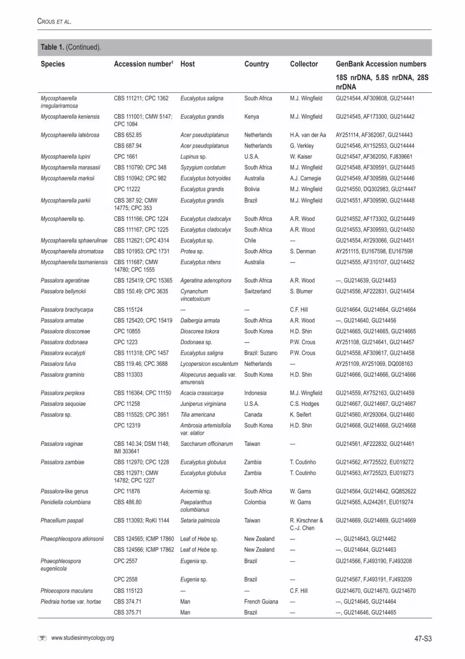
47-S3www.studiesinmycology.org
Crous et al.
Species Accession number1 Host Country Collector GenBank Accession numbers18S nrDNA, 5.8S nrDNA, 28S nrDNA
Mycosphaerella irregulariramosa
CBS 111211; CPC 1362 Eucalyptus saligna South Africa M.J. Wingfield GU214544, AF309608, GU214441
Mycosphaerella keniensis CBS 111001; CMW 5147; CPC 1084
Eucalyptus grandis Kenya M.J. Wingfield GU214545, AF173300, GU214442
Mycosphaerella latebrosa CBS 652.85 Acer pseudoplatanus Netherlands H.A. van der Aa AY251114, AF362067, GU214443CBS 687.94 Acer pseudoplatanus Netherlands G. Verkley GU214546, AY152553, GU214444
Mycosphaerella lupini CPC 1661 Lupinus sp. U.S.A. W. Kaiser GU214547, AF362050, FJ839661Mycosphaerella marasasii CBS 110790; CPC 348 Syzygium cordatum South Africa M.J. Wingfield GU214548, AF309591, GU214445Mycosphaerella marksii CBS 110942; CPC 982 Eucalyptus botryoides Australia A.J. Carnegie GU214549, AF309589, GU214446
CPC 11222 Eucalyptus grandis Bolivia M.J. Wingfield GU214550, DQ302983, GU214447
Mycosphaerella parkii CBS 387.92; CMW 14775; CPC 353
Eucalyptus grandis Brazil M.J. Wingfield GU214551, AF309590, GU214448
Mycosphaerella sp. CBS 111166; CPC 1224 Eucalyptus cladocalyx South Africa A.R. Wood GU214552, AF173302, GU214449CBS 111167; CPC 1225 Eucalyptus cladocalyx South Africa A.R. Wood GU214553, AF309593, GU214450
Mycosphaerella sphaerulinae CBS 112621; CPC 4314 Eucalyptus sp. Chile — GU214554, AY293066, GU214451Mycosphaerella stromatosa CBS 101953; CPC 1731 Protea sp. South Africa S. Denman AY251115, EU167598, EU167598Mycosphaerella tasmaniensis CBS 111687; CMW
14780; CPC 1555Eucalyptus nitens Australia — GU214555, AF310107, GU214452
Passalora ageratinae CBS 125419; CPC 15365 Ageratina adenophora South Africa A.R. Wood —, GU214639, GU214453Passalora bellynckii CBS 150.49; CPC 3635 Cynanchum
vincetoxicumSwitzerland S. Blumer GU214556, AF222831, GU214454
Passalora brachycarpa CBS 115124 — — C.F. Hill GU214664, GU214664, GU214664Passalora armatae CBS 125420; CPC 15419 Dalbergia armata South Africa A.R. Wood —, GU214640, GU214456Passalora dioscoreae CPC 10855 Dioscorea tokora South Korea H.D. Shin GU214665, GU214665, GU214665Passalora dodonaea CPC 1223 Dodonaea sp. — P.W. Crous AY251108, GU214641, GU214457Passalora eucalypti CBS 111318; CPC 1457 Eucalyptus saligna Brazil: Suzano P.W. Crous GU214558, AF309617, GU214458Passalora fulva CBS 119.46; CPC 3688 Lycopersicon esculentum Netherlands — AY251109, AY251069, DQ008163Passalora graminis CBS 113303 Alopecurus aequalis var.
amurensisSouth Korea H.D. Shin GU214666, GU214666, GU214666
Passalora perplexa CBS 116364; CPC 11150 Acacia crassicarpa Indonesia M.J. Wingfield GU214559, AY752163, GU214459Passalora sequoiae CPC 11258 Juniperus virginiana U.S.A. C.S. Hodges GU214667, GU214667, GU214667Passalora sp. CBS 115525; CPC 3951 Tilia americana Canada K. Seifert GU214560, AY293064, GU214460
CPC 12319 Ambrosia artemisifolia var. elatior
South Korea H.D. Shin GU214668, GU214668, GU214668
Passalora vaginae CBS 140.34; DSM 1148; IMI 303641
Saccharum officinarum Taiwan — GU214561, AF222832, GU214461
Passalora zambiae CBS 112970; CPC 1228 Eucalyptus globulus Zambia T. Coutinho GU214562, AY725522, EU019272CBS 112971; CMW 14782; CPC 1227
Eucalyptus globulus Zambia T. Coutinho GU214563, AY725523, EU019273
Passalora-like genus CPC 11876 Avicermia sp. South Africa W. Gams GU214564, GU214642, GQ852622Penidiella columbiana CBS 486.80 Paepalanthus
columbianusColombia W. Gams GU214565, AJ244261, EU019274
Phacellium paspali CBS 113093; RoKI 1144 Setaria palmicola Taiwan R. Kirschner & C.-J. Chen
GU214669, GU214669, GU214669
Phaeophleospora atkinsonii CBS 124565; ICMP 17860 Leaf of Hebe sp. New Zealand — —, GU214643, GU214462CBS 124566; ICMP 17862 Leaf of Hebe sp. New Zealand — —, GU214644, GU214463
Phaeophleospora eugeniicola
CPC 2557 Eugenia sp. Brazil — GU214566, FJ493190, FJ493208
CPC 2558 Eugenia sp. Brazil — GU214567, FJ493191, FJ493209
Phloeospora maculans CBS 115123 — — C.F. Hill GU214670, GU214670, GU214670Piedraia hortae var. hortae CBS 374.71 Man French Guiana — —, GU214645, GU214464
CBS 375.71 Man Brazil — —, GU214646, GU214465
Table 1. (Continued).

47-S4
PhylogenetiC lineages in the Capnodiales
Species Accession number1 Host Country Collector GenBank Accession numbers18S nrDNA, 5.8S nrDNA, 28S nrDNA
CBS 480.64; IHEM 3823; UAMH 4341
Man, hair Brazil — —, GU214647, GU214466
Piedraia hortae var. paraguayensis
CBS 276.32; VKM F-393 — — — —, GU214648, GU214467
Piedraia quintanilhae CBS 327.63; IMI 101644 Genetta tigrina Central African Republic
— —, —, GU214468
Polychaeton citri CBS 116435 Citrus aurantium, leaf, with Pseudococcus citri
Iran R. Zare & W. Gams
—, GU214649, GU214469
Pseudocercospora angolensis
CBS 112933; CPC 4118 Citrus sp. Zimbabwe — GU214568, AY260063, GU214470
CBS 149.53; ATCC 11669 Citrus sinensis Angola — AY251106, AF222847, GU214471
Pseudocercospora atromarginalis
CPC 11372 Solanum nigrum South Korea H.D. Shin GU214671, GU214671, GU214671
Pseudocercospora chengtuensis
CPC 10785 Lycium chinense South Korea H.D. Shin GU214672, GU214672, GU214672
Pseudocercospora cordiana CBS 114685; CPC 2552 Cordia goeldiana Brazil P.W. Crous & R.L. Benchimol
GU214569, AF362054, GU214472
Pseudocercospora cruenta CBS 462.75 Phaseolus sp. Fiji W. IJzermans-Lutgerhorst
AY251105, AF362065, GU214473
CPC 10846 Vigna sp. Trinidad H. Booker GU214673, GU214673, GU214673
Pseudocercospora eucommiae
CPC 10802 Eucommia ulmoides South Korea H.D. Shin GU214674, GU214674, GU214674
Pseudocercospora fijiensis X300 Musa sp. Tonga — GU214570, AY752150, GU214474Pseudocercospora fuligena CPC 12296 Lycopersicum sp. Thailand — GU214675, GU214675, GU214675Pseudocercospora griseola f. griseola
CBS 194.47; ATCC 22393 Phaseolus vulgaris Portugal — DQ289861, DQ289801, GU214475
CBS 880.72 Phaseolus vulgaris Netherlands H. A. v. Kesteren DQ289862, DQ289802, GU214476
Pseudocercospora humuli CPC 11358 Humulus japonicus South Korea H.D. Shin GU214676, GU214676, GU214676Pseudocercospora kaki CPC 10636 Diospyros lotus South Korea H.D. Shin GU214677, GU214677, GU214677Pseudocercospora luzardii CPC 2556 Hancornia speciosa Brazil A.C. Alfenas &
P.W. CrousGU214571, AF362057, GU214477
Pseudocercospora macrospora
CBS 114696; CPC 2553 Bertholletia excelsa Brazil P.W. Crous & R.L. Benchimol
GU214572, AF362055, GU214478
Pseudocercospora ocimicola CPC 10283 Ocimum basilicum Mexico M.E. Palm GU214678, GU214678, GU214678Pseudocercospora opuntiae CBS 117708; CPC 11772 Opuntia sp. Mexico M. De Jesus
YanezGU214679, GU214679, GU214679
Pseudocercospora pallida CPC 10776 Campsis grandiflora South Korea H.D. Shin GU214680, GU214680, GU214680Pseudocercospora paraguayensis
CBS 111317; CPC 1458 Eucalyptus nitens Brazil: Suzano P.W. Crous GU214573, AF309596, GU214479
Pseudocercospora protearum var. leucadendri
CPC 1869 Leucadendron sp. South Africa S. Denman & P.W. Crous
AY251107, AY260089, GU214480
Pseudocercospora pseudoeucalyptorum
CBS 114242; CMW 14908; CPC 10390
Eucalyptus globulus Spain J.P.M. Vazquez GU214574, AY725526, GU214481
Pseudocercospora punctata CBS 113315 Syzygium cordatum South Africa M.J. Wingfield EU167582, EU167582, GU214407CPC 10532 Syzygium cordatum South Africa M.J. Wingfield GU214659, GU214659, GU214659
Pseudocercospora sp. CPC 11592 Zelkova serrata South Korea H.D. Shin GU214575, DQ303085, GU214482Pseudocercospora vitis CPC 11595 Vitis vinifera South Korea H.D. Shin DQ073923, DQ073923, GU214483Pseudocercospora-like genus
CPC 10712 Quercus sp. Netherlands G. Verkley GU214681, GU214681, GU214681
Pseudocercosporella capsellae
CPC 10301 Brassica sp. U.K. R. Evans GU214662, GU214662, GU214662
Pseudocercosporella fraxini CPC 11509 Fraxinus rhynchophylla South Korea H.D. Shin GU214682, GU214682, GU214682Pseudocercosporella sp. CBS 112737; CPC 3959 Rhus typhina Canada K. Seifert GU214684, GU214684, GU214684
CPC 4008 Rhus typhina Canada K. Seifert GU214686, GU214686, GU214686
Table 1. (Continued).

47-S5www.studiesinmycology.org
Crous et al.
Species Accession number1 Host Country Collector GenBank Accession numbers18S nrDNA, 5.8S nrDNA, 28S nrDNA
CPC 10050 Rubus oldhamii South Korea H.D. Shin GU214685, GU214685, GU214685
CPC 11414 Vicia amurense South Korea H.D. Shin GU214683, GU214683, GU214683
Pseudotaeniolina globosa CBS 109889 Rock Italy C. Urzi GU214576, AY128700, EU019283Rachicladosporium cboliae CBS 125424; CPC 14034 Twig debris U.S.A. P.W. Crous —, GU214650, GU214484Ramichloridium apiculatum CPC 12310 Vicia amurensis South Korea H.D. Shin GU214687, GU214687, GU214687Ramichloridium cerophilum CBS 103.59; MUCL
10034Sasa sp. Japan — EU041798, EU041798, GU214485
Ramichloridium musae CBS 190.63; MUCL 9557 Musa sapientum — — GU214577, EU041800, EU041857Ramichloridium-like genus CPC 10672 Phellodendron amurense South Korea H.D. Shin GU214688, GU214688, GU214688Ramularia acroptili CBS 120252 Acroptilon repens Turkey R. Sobhian GU214689, GU214689, GU214689Ramularia brunnea CPC 4903 — — — GU214691, GU214691, GU214691Ramularia coleosporii CPC 11516 Plectranthus excisus South Korea H.D. Shin GU214692, GU214692, GU214692Ramularia endophylla CBS 113265 Quercus robur Netherlands G. Verkley AY490775, AY490763, AY490776Ramularia grevilleana CPC 656 Fragaria sp. South Africa P.W. Crous GU214578, AF173312, GU214486Ramularia nagornyi CBS 120253 Centaurea solstitiales Greece D. Berner GU214579, EU019257, EU019257Ramularia pratensis var. pratensis
CPC 11294 Rumex crispus South Korea H.D. Shin GU214580, EU019284, EU019284
Ramularia sp. CBS 324.87 leaf spot on Brassica sp., in Mycosphaerella sp.
Netherlands — GU214581, EU019285, EU019285
CPC 10066 Alangium plataniflium South Korea H.D. Shin GU214690, GU214690, GU214690CPC 11297 Stellaria aquatica South Korea H.D. Shin GU214693, GU214693, GU214693
Ramularia uredinicola CPC 10813 Salix sp. South Korea H.D. Shin GU214694, GU214694, GU214694Ramularia-like genus CPC 10852 Polygonum sp. South Korea H.D. Shin GU214695, GU214695, GU214695Ramulispora sorghi CBS 110578; CPC 905 Sorghum sp. South Africa D. Nowell AY251110, AY259131, GU214487
CBS 110579; CPC 906 Sorghum sp. South Africa D. Nowell AY251111, AY259132, GU214488
Readeriella dimorphospora CBS 120034; CPC 12636 Eucalyptus nitens Australia — GU214521, EF394850, EU019258Readeriella mirabilis CBS 116293; CPC 10506 Eucalyptus fastigata New Zealand W. Gams EU754110, AY725529, EU019291Schizothyrium pomi CBS 228.57 — Italy R. Ciferri EF134947, EF134947, EF134947
CBS 406.61 Rubus idaeus Netherlands — EF134949, EF134949, EF134949
CBS 486.50 Polygonum sachalinense Netherlands — EF134948, EF134948, EF134948
Scorias spongiosa CBS 325.33 Aphid — — GU214696, GU214696, GU214696Septoria apiicola CBS 400.54; IMI 092628 Apium graveolens Netherlands J.A. von Arx GU214584, AY152574, GU214490Septoria convolvuli CBS 102325 Calystegia sepium Netherlands G. Verkley GU214697, GU214697, GU214697Septoria cucubali CBS 102368 Cucubalus baccifer Netherlands G. Verkley GU214698, GU214698, GU214698Septoria dysentericae CPC 12328 Daucus carota Brazil N. Massola GU214699, GU214699, GU214699Septoria lactucae CBS 352.58 Lactuca sativa Germany — GU214585, AY489282, GU214491Septoria leucanthemi CBS 109090 Chrysanthemum
leucanthemumAustria G. Verkley GU214586, AY489277, GU214492
Septoria obesa CBS 354.58; BBA 8554; IMI 091324
Chrysanthemum indicum Germany — GU214587, AY489285, GU214493
Septoria protearum CPC 1470 Protea cynaroides South Africa L. Viljoen GU214588, AY260081, GU214494Septoria pyricola CBS 222.31; CPC 3677 Pyrus communis — — GU214589, AY152591, GU214495Septoria quercicola CBS 663.94 Quercus robur Netherlands — GU214590, AY490771, GU214496Septoria rosae CBS 355.58; ATCC
24311; PD 341; CPC 4302Rosa sp. — — AY251113, AY293065, GU214497
Septoria senecionis CBS 102366 Senecio fluviatilis Netherlands G. Verkley GU214591, AY489272, GU214498Septoria-like genus CBS 102377 Castanea sativa Netherlands G. Verkley GU214592, AY152588, GU214499Sonderhenia eucalypticola CPC 11252 Eucalyptus globulus Spain M.J. Wingfield GU214593, DQ303064, GU214500Sphaerulina polyspora CBS 354.29 — — — —, GU214651, GU214501Staninwardia suttonii CBS 120061; CPC 13055 Eucalyptus robusta Australia B.A. Summerell GU214594, DQ923535, DQ923535
Table 1. (Continued).

47-S6
PhylogenetiC lineages in the Capnodiales
Species Accession number1 Host Country Collector GenBank Accession numbers18S nrDNA, 5.8S nrDNA, 28S nrDNA
Stenella araguata CBS 105.75; ATCC 24788; FMC 245
Man Venezuela — GU214596, EU019250, EU019250
Stigmina platani CBS 110755; IMI 136770; CPC 4299
Platanus orientalis India — GU214598, AY260090, FJ839663
Stigmina synanamorph CPC 11721 Platanus occidentalis South Korea H.D. Shin GU214700, GU214700, GU214700Stomiopeltis betulae CBS 114420 Betula sp. Sweden K. & L. Holm GU214701, GU214701, GU214701Teratosphaeria aff. nubilosa CBS 114419; CPC 10497 Eucalyptus globulus New Zealand — GU214599, AY725574, EU019303
CBS 116283; CPC 10495 Eucalyptus globulus Spain W. Gams GU214600, AY725573, GU214503Teratosphaeria alcornii CBS 313.76; CPC 3632 Eucalyptus tessellaris Australia J.L. Alcorn GU214514, AF362061, EU019245Teratosphaeria angophorae CBS 120493; DAR 77452 Angophora floribunda Australia A.J. Carnegie —, GU214652, GU214504Teratosphaeria bellula CBS 111700; CPC 1821;
JT 196Protea eximia South Africa J.E. Taylor GU214601, EU019301, EU019301
Teratosphaeria cryptica CBS 110975; CMW 3279; CPC 936
Eucalyptus globulus Australia A.J. Carnegie GU214602, AF309623, GU214505
Teratosphaeria destructans CBS 111369; CPC 1366 Eucalyptus grandis Indonesia M.J. Wingfield GU214603, DQ267595, EU019287CBS 111370; CPC 1368 Eucalyptus sp. Indonesia P.W. Crous GU214702, GU214702, GU214702
Teratosphaeria fibrillosa CPC 1876 Protea nitida South Africa J.E. Taylor EU019282, EU019282, GU214506Teratosphaeria juvenalis CBS 110906; CMW
13347; CPC 40Eucalyptus cladocalyx South Africa P.W. Crous AY720715, AY725513, FJ493217
CBS 111149; CPC 23 Eucalyptus cladocalyx South Africa P.W. Crous AY720714, AY725514 , EU019294
Teratosphaeria macowanii CBS 110756; CPC 1872 Protea nitida South Africa J.E. Taylor GU214519, AY260095, EU019254CBS 111029; CPC 1488 Protea sp. South Africa P.W. Crous AY251118, AY260096, FJ493199
Teratosphaeria mexicana CBS 110502; CMW 14461 Eucalyptus globulus Australia — GU214604, AY725558, GU214507CBS 120744; CPC 12349 Eucalyptus sp. U.S.A.: Hawaii W. Gams GU214605, EU019302, EU019302
Teratosphaeria molleriana CBS 111164; CMW 4940; CPC 1214
Eucalyptus globulus Portugal M.J. Wingfield GU214606, AF309620, EU019292
CBS 116370; CPC 10397 Eucalyptus globulus Spain J.P.M. Vazquez GU214607, AY725561, GU214508
CPC 4577 Eucalyptus sp. Australia — GU214582, AY725524, GU214489
Teratosphaeria nubilosa CBS 115669; CPC 933 Eucalyptus nitens South Africa M.J. Wingfield GU214608, AY725548, GU214509CBS 116005; CMW 3282; CPC 937
Eucalyptus globulus Australia A.J. Carnegie GU214609, AY725572, GU214510
Teratosphaeria ohnowa CBS 112896; CMW 4937; CPC 1004
Eucalyptus grandis South Africa M.J. Wingfield AY251119, AF309604, EU019305
CBS 112973; CMW 4936; CPC 1005
Eucalyptus grandis South Africa M.J. Wingfield GU214610, AF309605, GU214511
Teratosphaeria pseudosuberosa
CBS 118911; CPC 12085 Eucalyptus sp. Uruguay M.J. Wingfield GU214611, DQ303011, EU019256
Teratosphaeria secundaria CBS 115608; CPC 504 Eucalyptus grandis Brazil A.C. Alfenas GU214612, DQ303018, EU019306Teratosphaeria sp. CBS 208.94; CPC 727 Eucalyptus grandis Indonesia A.C. Alfenas GU214613, AY626982, EU019307Teratosphaeria stellenboschiana
CBS 116428; CPC 10886 Eucalyptus sp. South Africa P.W. Crous GU214583, AY725518, EU019295
Teratosphaeria suberosa CPC 11032 Eucalyptus sp. Colombia M.J. Wingfield GU214614, DQ303044, GU214512Teratosphaeria suttonii CPC 11279 Eucalyptus tereticornis Bolivia M.J. Wingfield GU214615, DQ303055, FJ493222
CPC 12352 Eucalyptus sp. U.S.A.: Hawaii W. Gams GU214616, EU019288, EU019288
Teratosphaeria toledana CBS 113313; CMW 14457 Eucalyptus sp. Spain P.W. Crous & G. Bills
GU214617, AY725580, GU214513
CBS 115513; CPC 10840 Eucalyptus sp. Spain P.W. Crous & G. Bills
GU214618, FJ493198, FJ493225
Teratosphaeria verrucosa CPC 18 Eucalyptus cladocalyx South Africa P.W. Crous AY720713, AY725517, EU019293Thedgonia-like genus CPC 12304 Oplismenus
undulatifoliusSouth Korea H.D. Shin GU214703, GU214703, GU214703
Table 1. (Continued).

47-S7www.studiesinmycology.org
Crous et al.
Species Accession number1 Host Country Collector GenBank Accession numbers18S nrDNA, 5.8S nrDNA, 28S nrDNA
Toxicocladosporium irritans CBS 185.58 Mouldy paint Suriname M.B. Schol-Schwarz
GU214619, EU040243, EU040243
Verrucisporota daviesiae CBS 116002; VPRI 31767 Daviesia latifolia Australia V. Beilharz GU214620, FJ839633, FJ839669Verrucisporota proteacearum CBS 116003; VPRI 31812 Grevillea sp. Australia J.L. Alcorn GU214621, FJ839635, FJ839671Zasmidium anthuriicola CBS 118742 Anthurium sp. Thailand C.F. Hill GU214595, FJ839626, FJ839662Zasmidium citri CBS 116366; CMW
11730; CPC 10522Acacia mangium Thailand K. Pongpanich GU214597, AY752145, GU214502
1ATCC: American Type Culture Collection, Virginia, U.S.A.; BBA: Biologische Bundesanstalt für Land- und Forstwirtschaft, Berlin-Dahlem, Germany; CBS: Centraalbureau voor Schimmelcultures, Utrecht, The Netherlands; CMW: Culture Collection of the Forestry and Agricultural Biotechnology Institute (FABI) of the University of Pretoria, Pretoria, South Africa; CPC: Culture collection of Pedro Crous, housed at CBS; DAOM: Plant Research Institute, Department of Agriculture (Mycology), Ottawa, Canada; DAR: Plant Pathology Herbarium, Orange Agricultural Institute, Forest Road, Orange. NSW 2800, Australia; DSM: Deutsche Sammlung von Mikrorrganismen und Zellkulturen GmbH, Braunschweig, Germany; ETH: Swiss Federal Institute of Technology Culture Collection, Zurich, Switzerland; FMC: Venezuelan School of Medicine; IAM: IAM Culture Collection, Institute of Molecular and Cellular Biosciences, The University of Tokyo, Japan; ICMP: International Collection of Micro-organisms from Plants, Landcare Research, Private Bag 92170, Auckland, New Zealand; IFO: Institute for Fermentation, Osaka, Japan; IHEM: Collection of the Laboratorium voor Microbiologie en Microbiele Genetica, Rijksuniversiteit, Ledeganckstraat 35, B-9000, Gent, Belgium; IMI: International Mycological Institute, CABI-Bioscience, Egham, Bakeham Lane, U.K.; IPO: Culture collection of the Research Institute for Plant Protection, Wageningen, The Netherlands; JCM: Japan Collection of Microorganism, RIKEN BioResource Center, Japan; JT: Working collection of Joanne E. Taylor; LSHB: London School of Hygiene & Tropical Medicine, London, U.K.; MPFN: Culture collection at the Laboratoire de Pathologie Forestie`re, INRA, Centre de Recherches de Nancy, 54280 Champenoux, France; MUCL: Université Catholique de Louvain, Louvain-la-Neuve, Belgium; PD: Plant Protection Service, Wageningen, The Netherlands; RoKI: Private culture collection Roland Kirschner; TNS: Herbarium of the National Museum of Nature and Science of Japan, Tokyo, Japan; UAMH: University of Alberta Microfungus Collection and Herbarium, Edmonton, Alberta, Canada; VKM: All-Russian Collection of Microorganisms, Russian Academy of Sciences, Institute of Biochemistry and Physiology of Microorganisms, 142292 Pushchino, Moscow Region, Russia; VPRI: Victorian Department of Primary Industries, Knoxfield, Australia; WAC: Department of Agriculture Western Australia Plant Pathogen Collection, Perth, Australia; X: Working collection of Mahdi Arzanlou.
Table 1. (Continued).


