Embed Size (px)
Citation preview
PHYLOGENETIC RELATIONSHIPS OF ROTIFERS, AS DERIVl$D FROM PHOTORECEPTOR MORPHOLOGY AND OTHER ULTRASTRUCTURAL ANALYSES
Pierre CLEMENT
Laboratoire d’Histologie et de Biologie tissulaire, UniversitC Lyon 1 (Claude Bernard) 69622, Villeurbanne, France
Keywords: Phylogeny, evolution, adaptations, rotifers, ultrastructures, eyes, ocelli, photosensitivity, pigments, ovogenesis, muscles, protonephridia, integument, pseudocoel, collagen, glia
For the Summary to this contribution see pp. I 13-114.
I. Introduction
1963: disagreements on lower metazoan phylogeny ‘Are phylogenetic theories subjective views? Can any man propose his own phylogeny or can we get definite scientific solutions?’ asked Remane (1963) in criticism of Hadzi’s (1944, 1953), Steinbock’s (1952,1958,1963)and Hanson’s theories (I 958, I 963), stating that acoels turbellarians are derived from plasmodial ciliates and are the most primi- tive metazoa.
The first trap in constructing any phylogeny is to mis- take a convergent similarity for an homology. To recog- nize homologies, Remane (1955) defined precise criteria. However, even with these criteria (that I shall criticize in the next chapter), it is possible to see two ways in a phyletic line. Thus for Remane (1955, 1958, 1960, 1963), Marcus (1958) and Jagersten (1955, 1959), Platyhelminths and Nemathelminths stem from coelomates by regression.
According to these two groups of theories (lower meta- zoa come from acoels or from coelomates), Cnidaria would be less primitive than Platyhelminths: their radial symmetry would derive from the bilateral symmetry of the Anthozoa. ‘Emotion, too, sometimes, seems to substitute to reason’ says Hand (1963). Hyman (1959) and Hand (1959, 1963) brought some classical theories back into fashion: Cnidaria would be primitive and the first bilateral symmetry of the Anthozoa would be primitive too. They assert that ‘the early worm was a planula or a planuloi’d organism and the planula did not come from early worms’.
In 1963, the battle raged. In the book published by Dougherty, Ax regarded the Cnidaria as the most primi- tive metazoa, but he suggested that the other metazoa were derived from a primitive coelomate; Beklemishev favoured a polyphyletic origin for the metazoa from dif- ferent Coelenterate ancestors; and Remane repeated his ideas about the coelomate ancestor for pseudocoelomates. In the same book, Ruttner-Kolisko prudently ends her paper on the origin of rotifers by proposing two possibili-
ties: I) from the Turbellaria, 2) from ‘forms that might be traced back to the Turbellaria’, like Diurodrilus (Dino- philidae).
Today a relationship between Rotifera and Turbellaria is generally favoured. The anatomy and the embryology (De Beauchamp, 1907, 1909, 1965; Nachtwey, 1925) sug- gest the origin of rotifers ‘from some low grade creeping bilateral type suchasaprimitiveflatworm’(Hyman, 1951). ‘There is no fact indicating a case of reduction from more highly developed, coelomate worms (no rudimentary coel- om or mesoderm)’ (Ruttner-Kolisko, 1974).
Nevertheless, Koste (I 978) still favours the hypothesis of a coelomate ancestor of rotifers, thus supporting Re- mane et al., 1972, 1976.
Evolution of basis for arguments on phylogeny From the first observations of rotifers by Leeuwenhoek to the recent treatises on rotifers (De Beauchamp, 1965; Ruttner-Kolisko, 1972; Koste, 1978), the principal source of information has been morphological or histological observation under optical microscopy. The problem of homology is the main one. On this basis, different relation- ships were successively suggested for rotifers: Infusoria, Polypa, Crustacea, Annelida, Molluska, Turbellaria . . . (Hyman, I 951; De Beauchamp, 1965). This agitated histo- ry originates from:
I. Imprecision of the observations: rotifers were classi- fied as infusoria when their nuclei and cells were not yet observed; when Huxley (1853) saw their protonephridia, he put rotifers in Vermes.
2. Confusion between specialized rotifers (Hexarthra or Trochosphaera) and archetypes of the group.
Recent ultrastructural studies on rotifers (review in ClC- merit, 1977b) are of much better quality, and throw a new light on the problem of homologies. However, ultrastruc- tures of only a few species of rotifers are actually described, so that danger of confusion between specialized structures and archetypes remains present. In this work, I shall dis-
93
Hydrobiologia 73, 93-117(1980). 0018-8158/80/073~-0093$05.00. 0 Dr. W. Junk b.v. Publishers, The Hague. Printed in the Netherlands.

cuss the phylogeny of rotifers with the help of ultrr+struc- tural results on seven genera of rotifers: Trichocerca, No- tommata, Brachionus, Rhinoglena, Asplanchna, Philodina, Habrotrocha. Obviously, the size and the phylogenetic distribution of this sample must be taken into account when making generalizations. For some organs, we have ultrastructural information on only one or two species (an exception is the integument, known for the seven genera, and also in Mytilina, Keratella and Synchaeta).
Animal behaviour is very important to study evolution and to try to understand trans-specific evolution (Mayr, I 974). Unfortunately ethological work on rotifers is scarce, and only just beginning to grow.
Finally, recent progress in genetics and ecology of roti- fers (review in King, 1977) will help to understand specia- tion and evolution in this group. King (I 977) detected by electrophoresis a great variation in different clones of the same species. Nevertheless, we have no information about correlations between genetic variation and structural or behavioral variation. So, when I speak in this text about hypothetical ‘chromosomic segments’, characteristic of a precise ultrastructure in rotifers or other animal groups, this will be speculation. I know the danger of such specula- tions when some biologists write that individuals are nothing else than their gene pools (Wilson, 1979). We begin to know the origin of the variability of the responses of single rotifers which have the same genome: the reasons are in their own history, and in the history of their parents, grand-parents and otherascendants(Clement, rg77a; Cli- ment & Pourriot, rg7g and 1980). So, even in rotifers, it is impossible to reduce an individual to its (until now un- known) gene pool.
II. Photoreceptors and photosensitivities in rotifers
Are photoreceptors good indicators of phylogeny? Eakin (1965) proposed ‘a speculation as a catalyst of research’: some zoological groups, leading to deuterosto- mia, would have only ciliary-type photoreceptors; others groups (acoelomates, pseudocoelomates and protosomia) would have only rhabdomeric-type photoreceptors. In the first case, the photoreceptoral organelles are derived from the ciliary membrane; in the second one, from the distal cell membrane.
Eakin himself (rg68,rg72) proposed a number of excep- tions to his diphyletic theory. We now know that both types of photoreceptors are present in most zoological groups.
Vanfleteren & Coomans (1975) summarized these ex- ceptions and made a new, monophyletic theory: the photo- receptoral organelles would always be induced ‘by a ciliary information which, after initiating membrane prolifera- tion, may become more or less abortive (rhabdomeric type) or may develop further into a ciliary organelle (ciliary type)‘. They concluded that the photoreceptor structure is not useful to distinguish large phyla like protostomia and deuterostomia, but only to study closer phylogenetic rela- tionships.
In a more recent synthesis, Salvini-Plawen & Mayr (1977) proposed a different idea about the photoreceptor types: they described at least 40 (possibly up to 65 or more) independant phyletic lines, which can be grouped in a ganglionic or an epidermal category by their localization and embryology. The ganglionic (diverticular) type would be rhabdomeric and there would be three epidermal types: ciliary (enlargement of the ciliary membrane), rhabdom- eric (enlargement of the distal cell portion) and unpleated (surface enlargement through increase in cilia num,ber). For these authors, ‘similar photoreceptor types differen- tiated convergently several times’and ‘their distribution in various phyla of animals cannot safely be used as the basis for the construction of phylogenesis’.
These three theories (‘diphyletism’ of Eakin, ‘mono- phyletism’ of Vanfleteren 62 Coomans and ‘aphyletism’ of Salvini-Plawen & Mayr) use mainly morphological obser- vations, often neglecting biochemical, physiological and ethological aspects. Moreover the scarcity of ultrastruc- tural descriptions’ enhances a danger that I presented above: confusion between a specialized structure and an archetype of the zoological group. For instance, until now, the cerebral eye of Asplanchna (Eakin & Westfall, 1965) was considered to be the unique photoreceptor type of rotifers. Salvini-Plawen & Mayr (1977) considered a second ‘epidermal’ type with the anterior ocelli. The first ultrastructural observations of these ocelli and others photoreceptors in rotifers will allow me to discuss this precise point but also to propose a new (‘polyphyletic’) theory about the evolution of photoreceptors and photo- sensitivities. As much as possible I shall try not to limit my descriptions and conclusions to morphological features.
Photosensitivities and photopigments in rotifers The vision of rotifers is very primitive. No female, even if carnivorous, seems to see her food. No male seems to see the female he tries to fecundate. Instead, these meetings occur by random encounters facilitated by taxes.
Among these taxes, phototaxis was studied first (Jen-
94
nings, rgor; Viaud, rg4o-1943; Menzel & Roth, 1972; Preissler, 1977; Clement, tg77a-c; Hertel, 1979). It is a resultant of two components: phototaxis, sensu strictu, providing the orientation of the animal, and photokinesis, directing its movements.
Three cases are possible: I. Regular phototaxis: in planktonic species, in particu-
lar those only moving by swimming. A variability in this behaviour has been noted but not studied in different animals or clones.
2. Irregular phototaxis: in particular in rotifers which often settle or creep. In Notommata copeus, a species re- putedly not phototactic, the phototaxis is inhibited by contact with a filament and others factors (Clement, 19774.
3. Apparently non existant phototaxis: Synchaetapecti- nata (Menzel &Roth, rgT2);Resticulagelida (Viaud, 1943); perhaps many bdelloi’ds. More precise studies on these species are needed: are there particular inhibitions as for Notommata copeus or is there complete inhibition?
Phototaxis is characterized by a peak about 540 nm (Viaud, 1940-43; Menzel& Roth, 1972; Clement, rg77a-c), except in Filinia longiseta whose peak is about 450 nm (Menzel & Roth, 1972).
Photokinesis is characterized by a regular increase of speed (Brachionus calycifrorus) or of proportion of swim- ming animals (Notommata copeus) as the light changes from blue to red (Clement, rg77a-c).
A third photosensitivity was discovered by Pourriot (1963) and studied by Pourriot & Clement (review in Clement, rg77a and in Pourriot et al., in press): in three species (N. copeus, N. codonella and Trichocerca rattus) photoperiod controls the production of mictic females, and therefore the production of males and resting eggs. The action spectrum of N. capeus is different from that of the other two species; it has peaks at approximately 3 10, 360, and 450 nm, and there is no response to red light. So, the same animal shows three different photosensitivities.
It is possible to hypothesize that p-carotene or ribofla- vine or pterine is responsible for photoperiod influence, rhodopsin or porphyrin is responsible for phototaxis, and phytochrome is responsible for photokinesis (Clement, 19774.
Note that Champ (1976) and Wallace (1980) have pointed out a photoperiod influence on the hatching rhythm in Sinantherina socialis, and Pourriot & Rougier (1980) have demonstrated a light effect on the hatching of resting eggs inBrachionus rubens. These two effects of light have not been studied in detail and we do not know
whether they use one of the three preceding pigments. Lastly, we do not know if rotifers have a shadow re-
sponse. However, there have been studies of the influence of light intensity on photoperiod, phototaxis and photo- kinesis (Clement, rg77a); this influence can explain the avoidance of shores by some planktonic rotifers (Preissler, 1977).
Photoreceptors of rotifers The synthesis of Remane (1929-32) takes into account only the pigment cups. In the absence of pigment, it was thought that there were no eyes (the ‘blind’ Asplanchna of Viaud, I 940-43). However, with the electron microscope, it is possible to demonstrate that the red pigment is only an accessory epithelial cell associated with nervous structures (Eakin & Westfall, 1965; Clement, 1975). Other presumed photoreceptive structures can exist without the presence of a pigment cup, and probably correspond to the ‘derma- toptic sensibility’ of Viaud (I 940-43).
The following is an annotated list of photoreceptors that have been described in rotifers:
a/ Trichocerca rattus: As in Notommata copeus, there are three photosensitivities. Three presumed photoreceptors have been described:
-The cerebral eye (Fig. I to 4) (Clement, 1975): the red cup is located in a single epithelial cell. The lamellar photoreceptive neurites are piled up and embedded in the cytoplasm of the sensory neuron.
-Paired cerebral receptors (Fig. 16) (Clement, rg77a): located on both sides of the brain. Some neurons bear microvilli.
-Anterior ocelli (Fig. 13, -14, I 5) (Clement et al., 1980): these two complex apical sensory organs have specialized short, ampulla-shaped cilia containing electrondense ma- terial.
b/ Asplanchna brightwelli: Only the cerebral eye has been described (Eakin &West- fall, 1965). The pigment of the cup is flatter than in Tricho- cerca ruttus, and is arranged in several superposed layers. The sensory neurons bear lamellar photoreceptive rhab- domeres that pile up like onion leaves.
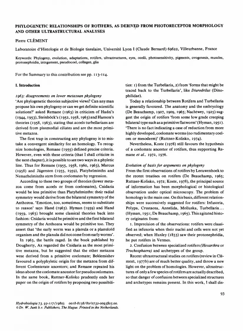
c/ Brachionus calyctjlorus: Only the cerebral eye has been described (Clement et al., in press) (Fig. 5, 6, 7). Two pigment cells form the red cup. The pigment resembles that of the cerebral eye of Tricho- cerca rattus. Two neurons form the sensory part. Cylindri-
95

96

cal neurites of the first neuron penetrate the cytoplasm of the second neuron.
d/ Rhinoglena frontalis: Only the anterior ocelli have been observed (Clement et
al., in press) (Fig. II and 12). The pigment cup is intra- epithelial. The sensory structures are piled dendritic la- mellae, coming from cerebral neuron processes. They are everse ocelli (the preceding cerebral eyes were inverse).
e/ Philodina roseola: -The cerebral eyes (Fig. 8, 9, and IO) (Clement et al., in press) are located on each side of the brain. The pigment is different from the pigment of the cup’s eyes of the Mono- gononta described above. In each eye, the photosensory strutures are ampullae-shaped cilia containing electron- dense material.
-An anterior receptor (Fig. 17, 18, and 19) (Clement et
al., in press): it is a median apical receptor, located in the pseudocoel. Beneath an epithelial anterior cell, a nerve process contains a spherical cavity filled with numerous flattened and piled lamellar cilia. Each cilium bears lateral lamellar expansions that are also piled up.
The polyphyletic origin of the photoreceptors of rotifers
Ampullae-shaped cilia containing electron-dense material are characteristic of a first phyletic line. In rotifers, I found these cilia in the cerebral eyes of Philodina roseola as well as in the anterior ocelli of Trichocerca rattus. Elsewhere, to my knowledge, this kind if cilia has only been described from the stigma of some phytoflagellates (Faurt-Fremiet & Rouillet, 1957; FaurC-Fremiet, 1961). They look like the parabasal apparatus of the phytoflagellates (Wolken, 1971).
The presumed anterior ocellus of Philodina roseola re- presents a second phyletic line. We find exactly the same structures and organization in the cercaria of Schistosoma mansoni (Short & Cagne, 1975). Very similar organs are found in:
-other Platyhelminths, in which an intraneural spheri- cal cavity contains some cilia with slightly modified axo- nems, but with piled lamellar expansions (Wilson, 1970;
Figs. 1-4. Cerebral eye of Trichocerca rattus. Fig. I. x ZIOOO. Axial section. The eye caps a retrocerebral gland (R). It is made of a dendritic blade (L), a sensory neuron (S and its nucleus NS), and a pigmented cell (NP: its nucleus, P: pigments of the cup). The sensory neurocytoplasm (S) contains dendritic lamellar expansions (the piled dendritic lamellae: Id) and some cylindrical expansions (arrows); Fig. 2. x 2500. The pigmented
Brooker, 1972; Lyons, 1972).
-some Annelida (Clark, 1967; Rohlich et al., 1970) and Pogonophora (Norrevang, 1974) in which an intraneural spherical cavity, called the ‘phaosome’, contains piled lamellar expansions, with sometimes regressed cilia or only ciliary rootlets. In no case is this very special organ associated with a pigmented epithelial structure. The func- tion of these organs is always presumed to be photorecep- tion, except by Wilson (1970) who proposed that it func- tions in gravity reception (Vanfleteren & Coomans, 1975,
disagree with Wilson). The impaired cerebral eyes of monogononts represent
at least one more phyletic line: cylindrical or lamellar rhabdomeres juxtaposed to a pigmented epithelial cup. Clement et al. (in press) detail the comparison of these eyes: primitive characeristics are noted in B. caIycijlorus and specializations in Asplanchna brightwelli. Rhabdom- eric structures, also issued from a cerebral neuron, and juxtaposed to a pigmented epithelial cup, are found in the anterior ocelli of Rhinoglena frontalis. As in the first phyl- etic line (ampullae-shaped cilia), we find here anterior ocelli as well as cerebral eyes in the same phyletic line. This phyletic line represents the ganglionic diverticular type of Salvini-Plawen & Mayr (I 977) in which the rhabdomeric structure seems to differentiate without cilia from a gangli- onic cell juxtaposed with a pigment cell. This photorecep- tor type is present in Platyhelminthes (see also Fournier & Combes, I 978, and a review in Fournier, in press), Aschel- minths, Polychaeta and some Arthropoda.
In conclusion, we know of at least three phyletic lines of photoreceptor types in lower metazoa. All three lines are present in rotifers and homologies can be established with photoreceptors of others zoological groups. On this basis, I propose a polyphyletic origin of rotifer photoreceptors.
Salvini-Plawen & Mayr (I 977) suppose no ciliary induc- tion in the photoreceptors of their ganglionic type. This hypothesis, which criticises the monophyletic theory of Vanfleteren & Coomans (1975), could be tested by an ultrastructural embryological study of some of these re- ceptors.
Our observations on rotifers are in contradiction with the classification of photoreceptor types proposed by Sal-
cup caps a retrocerebral gland (R); Fig. 3. x 500. The arrow points to the eye; Fig. 4. x 18500. Transversal section of the eye. The pigmented cup (P) surrounds the sensory neurone (S) and a part of the retrocerebral gland (R). The arrow shows the communica- tion between the dendritic blade (L) and a dendritic lamella (D). This lamella (D) is sectionned tangentially, as is the part of cytoplasm (C) located between two piled lamellae.
97
98

vini-Plawen & Mayr (1977). Their fundamental distinction between ganglionic and epidermal photoreceptors is not supported by our observations: the previously presumed ‘epidermal’ anterior ocelli are in fact feedings of cerebral neurons (see above). I think that it is not pertinent to propose photoreceptor types based on epidermal or ner- vous origin, because both have the same neuro-ectodermal origin. I therefore prefer more precise definitions of photo- receptor types. I have three other points of disagreement with Salvini-Plawen & Mayr:
I. In rotifers, some cerebral neurons bear photoreceptor cilia (eyes of Philodina roseola, ocelli of Trichocerca rattus); other cerebral neurons associated with eyes or ocelli bear rhabdomers. Because of this, I disagree with the rhabdom- eric ‘ganglionic type’ proposed by Salvini-Plawen & Mayr. As discussed earlier, the third phyletic line of rotifer photo- receptors has three characteristics: ganglionic, rhabdom- eric, and associated with an epithelial pigment cup.
2. Another point concerns the ampulla-shaped cilia (see above: first phyletic line). These cilia have no place in the receptor types defined by Salvini-Plawen & Mayr. In this phyletic line, there is no enlargement of membranes but accumulation of electron dense material inside the short cilia. The presence of the same material, closelyjuxtaposed to the pigment stigma in the parabasal apparatus of the phytoflagellates, suggests the photosensitivity of the mate- rial.
3. Our second phyletic line (‘ocellus’ of Philodina roseo- la) is defined to be preecise structure named ‘phaosome’ in Annelids. In this line, there is a progressive evolution from ciliary to rhabdomeric types.
Why, then, did Salvini-Plawen & Mayr find no phylo- genetic significance in the distribution of their photorecep- tor types? The reason is perhaps an insufficient precision in their definitions of these types.
I have tried to formulate precise definitions for the photoreceptors that can be observed in rotifers. However, completely satisfactory definitions must take into account both ultrastructural features and biochemical and physi- ological aspects.
Phylogeny and evolution of photoreceptors andphotosensi- tivities I have described (paragraph 2) at least three photosensi- tivities, and three photopigments, in rotifers. Rhodopsin is the only pigment which can be implied in phototaxis. Wolken (1970) states that rhodopsin is the only visual pigment found in invertebrates, but presents no data on lower metazoa. It is possible that in rotifers and in other lower metazoa, one of the photoreceptors is associated with rhodopsin and represents a primitive form of future visual organs. It would be interesting to construct a phyl- ogeny of the animal kingdom by comparing all photo- receptors associated with a rhodopsin.
The other pigments of rotifers are involved in photo- kinesis and in determinism ofmixis by photoperiod. Their localization is unknown. Yet, we can postulate that it is extraocular, in those presumed photoreceptors that do not possess a pigmented epithelial cup. It would be interesting to establish a correlation between a pigment, a photo- receptor, and a behaviour. If such a relationship could be found, its comparative evolution in the animal kingdom would have a real phylogenetic interest. Unfortunately, even in rotifers, we have no knowledge of these correla- tions.
For instance, in all animal groups, the receptor involved in the sensitivity to photoperiod is unknown. The action spectra found in other groups are often, but not always, the same as those in rotifers (review in Pourriot & Clement, I 973). Different mechanisms were proposed in Arthropo- da for the influence of photoperiod (review in Saunders, 1976). The mechanism present in Notommata copeus is a primitive one, without endogenous rhythm (Pourriot et aZ., in press). I am sure that the comparison of the photo- receptors and nervous and endocrine structures involved in these influences of photoperiod on animals, would have a phylogenetic sense.
In summary, I think that a phylogeny must simulta- neously consider the evolution of structures, pigments and functions of photoreceptors. In this sense, rotifers are primitive metazoa, having primitive responses to light, no
Figs. 5-7. Cerebral eye of Brachionus caIyciflorus. expansions originate in the sensory neuron NSI. G: golgi appara- Fig. 5. x 3600. Axial section of the brain. The neuropile is sur- tus. The arrows indicate a very peculiar piled cytoplasmic struc- rounded by small neurons (n: their nuclei). Towards the back of ture; Fig. 7. x 14000. The sensory neuron NSI gives expansions the brain, the two sensory neurons (NSI and NSZ their nuclei) (large arrow) between the pigmented cup(P) and the cytoplasm of occupy a large volume. The biggest (NSz), is capped by two the second sensory neuron (NSz); some of these dendritic expan- pigmented epithelial cells (EP and P) which contain the pigmented sions (I) go down in to the cytoplasm (2) of the second sensory cup of the eye; Fig. 6. x 22000. Detail of the large sensory neuron. Note that another dendritic expansion (fine arrow) of the cytoplasm; NS2: its nucleus, P: pigmented cup, (I) dendritic sensory neuron NSI goes towards the cerebral neuropile.
99

100

real vision, and photoreceptors with only one or two neurons.
One hypotheis is that the most primitive metazoa, like rotifers, evolved different photosensitivities, with a large number of pigments and different but simple structures of photoreceptors. With subsequent evolution, one of these adaptations was successful. The result was the use of one pigment (rhodopsin) for vision; however some variation was retained between different phyletic lines for the differ- ent kinds of rhodopsins, for photoreceptor structures, and for organisation of the eyes (multiplication of sensory cells and of accessory structures). Extraocular sensitivities within different phyletic lines can persist or disappear.
A remark about the phylogeny of lower metazoa We know that the genome of metazoa is considerably richer than suggested by the limited set of different pheno- types present at any one time. Since an important part of this genetic potential does not express itself.
It is well-known that the same genome can be expressed, for example, in a miracidium, in a cercaria, in a metacerca- ria, or in an adult form of a parasitic Platyhelminth. Each stage has unique structures and functions not expressed in other stages.
The classical criteria of homology (Remane, 1955) seem to be too rigid and not always justified when juxtaposed to this view of the genome. For instance, with Remane’s criteria, the cerebral eyes of Monogononta are homolo- gous, and can constitute a phyletic line; but this is not true of the ampullae-shaped cilia in the ocelli of Trichocerca rattus and in the eyes of Philodina roseola. These cilia are not found in Brachionus or in Rhinoglena. So, the classical point of view says that they represent a convergent analo- gy, as does the same cilium in a phytoflagellate. I do not agree with these conclusions. In my opinion, the hypothe- tical, chromosome segment involved in the differentiation of the ampullae-shaped photoreceptor cilia in some phyto- flagellates, is transmitted and is present in the primitive lower metazoa but expresses itself only in some of them.
The same argument is possible for the receptors with phaosome. The corresponding part of the genome is per- haps present in platyhelminths, rotifers and lower coel-
Figs. 8, 9, 16. Cerebral eyes of Philodina roseola. Fig. 8. x 30000. Tranversal section of the two photoreceptive cilia (arrows): the electron dense subStance is lateral to the cilium axonema. P: pigments in an epithelial pigmented cell. Ps: pseudo- coel; Fig. 9. x 13000. Tranversal section of the basis of two receptive cilia (arrows). The pigmented cell (P) is located at the periphery of the brain (left). Ps: pseudocoel, T: integument, ex:
omates; it expresses itself only in some cases such as in cercaria of some parasitic platyhelminths, P. roseola, some annelids and pogonophora, etc.
I do not mean to imply that each similarity can be an homology. The chromosome segments involved in the construction of the eyes of peridinians and cephalopods are surely different. But when the anterior ocelli of Rhino- glena have the same structures (rhabdomers borne by a cerebral neuron) and the same function (phototaxis) as the cerebral eye of a monogonont, are differences (such as localization, and perhaps one pigment more in the cerebral eyes) important enough to say that the same structures come from convergent independent mutations? I do not think so. Instead, it seems likely that the similarities be- tween these two photoreceptors come from the same ge- nome, and the differences from additional genetic infor- mation.
Finally, I think that there are two possibilities for ap- proaching the phylogeny of a zoological group. The first one is to understand their richness in behaviour and related structures. I began here with photoreception. The next chapter summarizes other possible approaches. One is to study the structures and functions which are constant within a group, and then to compare them with other groups. This I shall try in the last chapter.
III. Adaptations and evolution
The success of rotifers is probably due to their rapid parthenogenetic reproduction. Rapid reproduction is only possible if the animal can get enough food. Therefore I first discuss moving and feeding mechanisms. Next, I consider those adaptations that foster survival in unstable biotopes often colonized by rotifers (ponds, mosses, lichens.). After briefly discussing the cycle of reproduction, I end this chapter with some hypotheses about the mecha- nisms of evolution in rotifers.
Moving and feeding behaviour Recent studies on rotifers have substantially increased our knowledge of moving and feeding behaviours.
external medium; Fig. IO. x 26000. Axial section of one of the two photoreceptive cilia (wide arrow). Its extremity goes down into the pigmented cell (P) whose nucleus (NP) is visible. The insertion of the second cilium near the base of the first is indicated by a black arrow. The lower part of the picture is occupied by periph- eral cerebral neurons. Ps: pseudocoel, M: muscles.
101

102

The classical work of De Beauchamp (1907, rgog) on the modifications and lack of foot, pedal glands, retro- cerebral apparatus and different parts of the rotatory apparatus, is currently being expanded by two types of approaches.
First, the ultrastructural approach is used to study the different categories of cilia, muscles and sensory receptors involved in these behaviours (Clement, I g77a and b; Am- sellem & Clement, 1977; Clement et al. a, b, c in this volume). Second, behaviour is being studied directly by a variety of experimental and observational approaches: see Gilbert (rg77a, b), Gilbert & Starkweather (1977) and Starkweather (in this volume) for studies on feeding; Wallace (1980) for studies on sessile rotifers; and the preceding chapter for studies on phototaxis.
These results are too new and too voluminous to review them here. I only tried with photosensitivities (Chapter II) and the ultrastructural approach to feeding behaviour (this volume). About this last point, the classical work of De Beauchamp (agog) on the digestive tract of rotifers begins to be completed by electron microscopy, from which new questions arize: for instance, why are the pharyngeal cilia, which are the only cilia until now known to contain striated material, not exactly the same in Philo- dim and Bruchionus (the striated material is immediately under the cytomembrane in Brachionus and inside the axonema in Philodina)? These cilia probably have the same function, as the malleate and ramate mastax seem to have the same function.
The dietary specialization of each rotifer (review in Pourriot, I 965, I 977 and Starkweather in this volume) is a complex problem: it is dependent as much on the type of mastax and digestive tract as on the different specializa- tions of the sensory receptors and behaviours of the spe- cies. Photoreceptors and photosensitivities are only one part of these multifaceted problems.
Figs. I I and I 2. Anterior ocelli of Rhinoglena frontalis (in lateral sensory organs). Fig. I I. x 8000. Localization of one of the two ocelli. The pigments (P) of the red cup are located in the epithelial cell (El) under the photoreceptive part (0) Left, the syncitial integument. Right, the four sections of symmetrical ducts of retrocerebral organ (R). Two epithelial ciliary cells (EI and E2). In the cell EI, are em- bedded the neurites of the lateral sensory complex which contains the ocellus. The arrows indicate the apical sensory receptor neu- rites which are also symmetrical: they are located more centrally near the openings of the retrocerebral organ; Fig. 12. x zoooo. Detail of an ocellus. Many sensory neurites (n) form the lateral anterior sensory receptor. They are surrounded by only one
Ovogenesis and cycle of reproduction The reproduction of rotifers is more specialized than that of Platyhelmintha for two reasons: parthenogenesis and lack of scissiparity, and power of regeneration. The first forms of parthenogenesis appear in Platyhelmintha. In different primitive zoological groups, parthenogenesis exists in some individuals. With the exception of one genus (Seison), all rotifers can reproduce by parthenogenesis. For this reason, parthenogenesis appears to be a primitive trait of the entire group. Variations of the rate of repro- duction with temperature, feeding and other factors, probably express adaptations of the parthenogenetic re- production to precise biotopes.
The number of ovocytes of rotifers is determined prior to birth. These ovocytes are situated in the follicular epi- thelium (Fig. 20, Bentfeld, rg7ra, b; Clement, rg77a, b) which sometimes surrounds the whole female genital ap- paratus (Philodina roseola, Fig. 20) or sometimes sur- rounds only the ovocytes (Trichocerca rattus, Clement, I g77a, b). An ovocyte grows with substances which come from both the vitellarium and pseudocoel (sometimes via the follicular epithelium: Fig. 20). Then the ovule secretes its shell and is layed.
The different egg deposition behaviours also express adaptations to precise biotopes. The eggs of some plank- tonic species can float; others are carried by the mother, either internally(in which case the female is ovoviviparous) or externally after laying. In some periphytic species, such as Notommata copeus, the mother turns for ten minutes around the egg she has layed, and thus fastens it to a filamentous alga. This alga is one of the food species of N. copeus.
The capacity for anhydrobiosis of Bdelloi’dea is an ad- aptation to environments that frequently dry up (e.g. mosses and lichens). This ability perhaps explains the complete lack of males in this group.
epithelial cell (El). This cell (El) contains pigments(P) which form the pigment-cup of the ocellus. The photoreceptive parts are the piled branches originating from the neurite (no). Figs. 13-q-15. Anterior ocelli of Ttichocercu rattus (in apical anterior sensory organs). Fig. 13. x 28000 and Fig. 14. x 54000. Transversal sections of dense ampullae shaped cilia; Fig. 15. x 26000. Axial section of one of the two anterior sensory organs of T rattus. The ampullar-shaped cilia whose content is dense (ar- rows) are cut axially or obliquely; they are inserted on a neurite (n). This neurite is situated in an epithelial cell of the pseudo- trochus (E). (C) pseudotrochus cilia (in the external medium). (Cu) anterior fine cuticle.
103

104

The function of males and of sexual reproduction is indeed, in Monogononta, to produce resting eggs. These eggs retain viability after being frozen or dessicated. Males seem to be absent in some clones of Monogononta which live in stable environments, such as big lakes (Ruttner- Koklisko, 1974).
The factors controling mixis can sometimes, but not always, be understood. In some cases, there is continuous production of mictic females (Pourriot & Rougier, per- sonnal communication); in other cases, there are alterna- tive phases of parthenogenesis and sexual reproduction. In the latter cases, mixis is produced by a precise factor: photoperiod in Notommata copeus or cw-tocopherol in As- planchna (review in Clement, r977a and Gilbert, 1977C). In all cases, exogenous and endogenous factors control the percentage of mictic females (Clement et al., 1976; Cl4 ment, r977a).
The first appearance of such an heterogonic cycle and formation of resting eggs is in the Platyhelminthes. The influence of population density and photoperiod can al- ready be noticed. In more advanced animal groups, very similar cycles controlled by the same factors can also be observed (Cladocera).
Hypotheses on the mechanisms of evolution in rotifers The general scheme could be the following one:
-initially rich genome with both multiple and primitive potentialities;
-acquisition of the unchanging characteristics of the group (see chapter IV, and above about parthenogenesis);
-diversification of the forms which keep these charac- teristics, and the initial rich genome, but express a diversity of more or less different phenotypes.
The specialization of rotifers and of the main lines of their classification can probably be explained by modifica- tions of their genome during sexual reproduction: crossing- over, mutation.. . (King, 1977). Yet, other specializations of rotifers can be due to peculiar mechanisms.
The first of these mechanisms is mutation occurring during mitoses of the parthenogenesis. This parthenoge- nesis is probably mitotic (King, 1977) and not endomei’o-
Fig. 16. x 23000. Cerebral paired receptors of Trichocerca rattus. Detail of one of them. The sensory neurite (n) bears thin microvilli (arrows); (N) nuclei of cerebral neurons; (NE) nucleus of an epithelial cell located at the brain periphery. Fig. 17. x 68000; 18. x 2~000; 19. x 35000. Anterior unpaired receptor of Philodina roseola. The sensory neurite (n) forms a sort of sphere in which lamellar cilia are piled. These cilia are inserted on both sides of the neurite (Fig. 18). The cilia base shows a
tic. It seems to be primitive in rotifers (see above), and rapid reproduction increases the probability of mutations. The speciation of Bdelloi’da, and possibly of a lot of Mono- gononta, is perhaps due to his mechanism (Pourriot & Clement, in press).
The second mechanism may be maternal effects. I am using the term to include all reversible maternal influences expressed over some generations: Lansing effect (Lansing, 1947, 1954); influence of the age of the grand-mothers
(POUrriOt & Rougier, 1977; Cknent & POWriOt, 1979);
influence of substances sent out by Asplanchna on the appearance of tegumentary spines in Brachionus, Filina, Asplanchna (Gilbert, I 967, I 977; Pourriot, I 974); influence of substances related to crowding in the induction of mixis (Clement & Pourriot, in this volume). In the last case, mixis can disappear when females of N. copeus are in a crowded situation, but this is reversible. Nevertheless, could be apparently complete disappearance of mixis and males in planktonic rotifers of big lakes (Ruttner-Kolisko, 1972) have a similar origin?
IV. Ultrastructures and phylogeny
In the second and the third chapter, I discussed the diversi- ty of the phenotypes of rotifers: I criticized the often too arbitrary rigidity of Remane’s criteria for homology, and I made some hypothesis about the possible use of this diver- sity for phylogeny.
In this fourth chapter, I am considering some structures that are constant in all rotifers, in spite of adaptative modifications from an animal to an other. I have choosen five examples that seem to be of interest for comparing rotifers to other zoological groups. These are the integu- ment, the flame-cells, the body cavity, the thick myofila- ments, and the nervous system.
The syncytial integument The skeleton of rotifers is peripheral but not extracellular: the extracellular cuticle is always gelatinous and never skeletal.
classical axonema (Fig. 17). The two central tubules then dis- appear while the other tubules form the parallel ribs in the ciliar lamella (CT); (CL) an axial section of the base of a lamellar cilia; (E) anterior epithelial cell on which the neurite is fixed by a desmosome (arrow, Fig. 19); (R) retrocerebral organ. The large arrow (Fig. 19) indicates that the membrane of cilia extends in flattened villi.
105

Fig. 20. Ovogenesis in Philodina roseola. x 16400. The follicular follicular cell shows many infoldings: this indicates a probable epithelium (F) surrounds all the vitellarium (V) in which the entrance of substances from the pseudocoel (right arrow); Fig. 21. ovocytes (0 and OV) are found. The ovocyte which grows to form Musculuture-Tranversal section of the mastax striated muscle of an ovule (OV) communicates with the vitellarium by a cytoplas- Trichocerca rattus (x 200000). The ratio between thick and thin mic bridge (left arrow). At the level of this ovocyte (OV), the myofilaments is 1/3. See comments in chapter IV, par. 4.
106

The peripheral skeleton, which muscles attach to, is a dense intracytoplasmic lamina located inside the syncytial integument. This skeletal lamina is either thick and rigid, at the level of the trunk (Fig. 22,23,24,25,27) or supple, at the level of the articulations or at the front of the animal (Fig. 26).
Three kinds of skeletal lamina have been observed (Clement, 1969), However, four categories can be distin- guished now:
I. The Philodina type (Fig. 27, and Schramm, r978b for Habrotrocha), in which only the internal layer of the skele- tal lamina is thickened. In the three other types, only the external layer of the skeletal lamina is thickened.
2. The Trichocerca type (Fig. 24), in which the external layer of the lamina is uniformly dense (see also Kerutellu in Koelher, 1966 and in Hendelberg et al., 1979).
3. The Bruchionus type (Fig. 22, 23; Clement, 1969, 1977b; Starch & Welsh, 1969), in which the external layer of the lamina is made of juxtaposed vertical tubules (see also Mytilina in Clement, 1969).
4. The Notommata type (Fig. 25; Clement, 1969) in which the external layer of the lamina is made of stacked lamellae (see also Asplanchna in Koelher, 1965, and Syn- chata in Clement, 1969).
In the four cases, the function of this skeletal lamina remains the same. Variations of structure are therefore good indicators of the phylogeny among rotifers.
The internal layer of the skeletal lamina is thicker than the external layer in the bdelloi’ds; but this is also the case in some young monogononts (Bruchionus, Clement, 1 m’bh
An intracytoplasmic peripheral skeleton seems to exist in only one other zoological group, the Acanthocephala. In other Aschelminths, in Annelida, Mollusca, and Ar- thropoda, the external skeleton is cuticular, i.e. extracellu- lar.
The phyletic origin of the skeleton of rotifers is to be sought in animals with soft integument: for example in the terminal webb or in the infraciliature of a ciliary integu- ment. A soft non-cuticular and ciliary integument can be observed in Platyhelmints as well as in Cnidaria and Ctenaria.
The jlame-cells In 1853, Huxley emphasized the phylogenetic importance of the flame-cell of rotifers. Its study by electron micro- scopy enabled both a better definition of its characteristics and variations in rotifers, and the postulation of possible relationships with other animal groups.
In rotifers (Fig. 28 to 32) the flame cell has been studied in some Monogononta: Asplanchna (Pontin, 1964, 1966; Braun et al., 1966; Warner, 1969), Notommata (Fig. 28, Clement, 1967, 1968, 1969, r977a), Trichocerca (Fig. 29, Clement, r977a-b), Rhinoglena (Fig. 31), and in some BdelloTda: Pkilodina (Fig. 30, Mattem & Daniel, I 966) and Hubrotrocka (Schramm, r978a).
In all cases, it is a hollow, cylindrical or ampulla-shaped cell with a non-apical nucleus. The apical cap contains the bases of the cilia of the flame. The cavity of the cell communicates with the lumen of the protonephridial tube. The membranes of the filtering wall are situated between thin parallel cytoplasmic columns. This first grid sub- tended inside a second grid that has a more skeletal func- tion The second grid is formed by cytoplasmic extensions called pillars.
There are three types of variations: I. Size and number of flame-cells. The volume of the
animal and the surface of the filtering wall are correlated (Clement, I 977a). The number of cilia of the flame and the size of the cell are also related. In the large species of rotifers, there are two ways possible to increase the surface area of the filtering membranes: (I) to increase the size of the flame-cells (N. copeus Fig. 28), (2) to increase their number (Asplunchna Pontin, I 966).
2. Structure of the filtering-wall. In Trickocerca (Fig. 29) and in bdelloi’ds (Fig. 30) pillars and columns are often bound together. In the other Monogononta, pillars and columns are two distinct parallel grids.
3. Structure of the dense material of the pillars. It is cross-wise striated, as a ciliary root in bdelloi’ds, but not in monogononts (Fig. 31 and 32).
The flame-cell of Platyhelminths is very similar to that of rotifers (Ktimmel & Brandenburg, 1961; McKanna, 1968; Swiderski et al., 1975): flame with many cilia, and filtering-wall with pillars and columns. Nevertheless in Platyhelminths the nucleus is always apical, and the fil- tering-wall is a grid in which a column and a pillar con- taining electron dense material alternate regularly. The filtering membrane is located between each column and pillar.
In nematods and nematophores,flame-cellsdo not exist. In Priapulids, the protonephridial apparatus has groups
of typical solenocytes, quite similar to those of some An- nelida: apical nucleus, one cilium only, filtering wall with one grid only (Ktimmel & Brandenburg, 1961).
In Gastrotricha (Brandenburg, 1962; Teuchert, 1973),
the flame-cells are solenocytes too, going by pair or grouped.
I=‘7

108

In Kinorhincha, the flame-cell seems to have several nuclei and each flame is made of one or two cilia (Hyman, 195 I). The ancestor of the plurinucleated flame-cell could be the ciliary rosette of the Ctenaria (Franc, 1972).
In Cnidaria, no structure looks like a flame-cell. Some cells are more like the choanocytes of sponges whose function is more digestive than excretive, but which are fairly similar to that of the solenocytes.
These facts suggest some hypotheses about the phyl- ogeny of lower metazoa:
I. A close relationship between rotifers and Platyhel- minths; but the non-apical flame-cell nuclei distinguish rotifers from Platyhelminths.
2. An early separation between bdelloi’ds (striated pil- lars) and monogononts (non-striated pillars).
3. A great homogeneity of bdelloi’ds, perhaps reflecting a rapid disappearance of sexual reproduction in this order. In contrast, a relative heterogeneity in monogononts, and in Platyheminths, in which these variations of the flame- cell are good indicators for the phylogeny.
4. A possible relationship between gastrotrichs, priapu- lids, and annelids, which all possess solenocytes and no flame-cells.
5. A more speculative relationship between Ctenaria, Kinorhincha, and Nemertina, with only the basis of their pluricellular flame-cells.
The body cavity
Pseudocoel and mesenchyme
In rotifers, electron microscopy has demonstrated that the pseudocoel is directly limited by the integument on the outside and by the digestive epithelium on the inside. No thin membrane looking like a regressed coelomic wall has been observed, contrarily to what Remane suggested (1963). The presence of basal lamellae between a cell and the pseudocoel is very variable (compare Figs. 28,29 and 30 for a basal lamina around the flame-cell).
Free mesenchymatic cells do not seem to exist in the pseudocoel of rotifers. The starry cells of the pseudocoel described by Nachtwey (1925) and Remane (I 929-32) are
scarce and do not seem to be free. In electron microscopy, some very thin cellular expansions are sometimes observed in the pseudocoel, but they often seem to be expansions of muscular cells.
Many moving cells, associated with fibrous structures, are observed in the pseudocoel of Kinorhincha, Priapulida and Nematomorpha (Hyman, I 95 I). So, in these animals, the pseudocoel seems to be a mesenchyme less compact than that of Platyhelminths. In nematods, there is an intermediate situation. The pseudocoelocytes are fixed, they are neither phagocytic nor amoeboi’d and will not take up vital dyes (Hyman, 1951). Are they real mesen- chymatic cells? Lastly, in Gastrotricha, there are no free amoebol’d cells in the pseudocoel. (Hyman, 1951).
No ultrastructural morphological argument allows us to suggest that the pseudocoel of rotifers is a regressed coelom.
On the other hand, the presence of mesenchymatic cells in the pseudocoel of most of the Aschelminths raises another question: is this ‘cavity’ a mesenchymatic or a classical conjunctive space? The answer is perhaps yes for Priapulida, Nematomorpha, and Kinorhincha. In nema- tods, gastrotrichs, and rotifers, the question remains with- out answer. We are attempting to solve it by another criterium: the intercellular collagen which, in all the animal kingdom, characterizes the conjunctive spaces. First, however, I wish to stress a correlation between eutely and lack of characteristic mesenchyme with active and free amoebol’d cells.
Eutely
The number of nuclei is remarkably constant in rotifers: from birth to death and from one animal to another in the same species. Exceptions to this last point are rare and concern individual variations of the number of nuclei in polyploi’d syncytial organs: vitellarium and gastric glands (Birky & Field, 1966).
This perfect eutely in rotifers explains the absence of regeneration in these animals. So, it is usually admitted that on the genealogical tree of the animal kingdom, the rotifers are apparently out on a limb from which there is
Figs. 22-27. x 50000. The skeletal lamina of the integument. ex external medium Fig. 22. Brachionus calyciforus; Fig. 23. Brachionus calyciforus: 0 pseudocoel tangential section; Fig. 24. Trichocerca rattus; Fig. 25. Notommata B bulb copeus; Fig. 26. Philodinia roseola: anterior supple integument (C: P pore pseudotrochus cilia); Fig. 27. Philodina roseola: trunk integument I apical cytoplasmic membrane
2 and 3 the two layers of the skeletal lamina.
109

110

nowhere to go (Ruttner-Kolisko, 1963). Hyman (195 I) propounds that entoprocts come from rotifers. Most cer- tainly the similarities with the Collothecacea are impres- sive. But the entoprocts regenerate and multiply them- selves by asexual reproduction: these characters observ- able in Turbellaria also are primitive and do not exist in Collothecacea. They exclude any possibility of direct rela- tionships between the specialized sessile rotifers and the Entoprocta.
Such a cell constancy is observed neither in Acoelomates nor in other Pseudocoelomates, except for nematods (Hy- man, I 95 I). In nematods, however, eutely is less perfect; it does not apply to gonads and variations for other organs have also been observed in young stages. In particular, in Aschelminths, the Gastrotrichs, Kinorchinchs and Pria- pulids have no cell constancy (Lang, 1963).
Collagen and collagen fibrils
Collagen has been found in all pluricellular animals where it has been looked for (Adams, rg78), mostly as trans- versely striated librils. This is probably a primary animal characteristic (Pikkarainen & Kulonen, 1969).
However, some variation exists in primitive Inverte- brates (reviews in Bairati, 1972; Garonne, 1975; Adams, 1978). In platyhelminths and nematods, the fibrils seem less structured than in Porifera and in Anthozoa (Cnida- ria). In Ctenaria, there are no structured fibrils but a network of microfilaments which contain some hydroxy- prolin (Franc et al., 1976). After extraction, this collagen precipitates inthe shape of distinct fibrils. The collagen of a parasitic platyhelminth does the same: the fibrils are more distinct in vitro than in vivo. Garonne (1975) sug- gests the presence in vivo of a factor limiting the organiza- tion of the fibrils of collagen in Platyhelminths and in Ctenaria.
In rotifers, we see neither microfilaments nor fibrils of collagen in the pseudocoel. The basal lamellae that seem to have a type IV collagen in vertebrates are not always present according to the cells and the species of rotifer. Lastly, the gelatinous cuticle of rotifers does not show the
Figs. 28 to 32. x 30000. The flame-cell. Fig. 28, 2g and 30. Tranversal sections in the filtering wall; Fig. 28. Notommata copeus; Fig. 29. Trichocerca rattus; Fig. 30. Philodina roseola. Fig. 31 and 32. Axial sections of the cap and the filtering wall. Pig. 31. Rhinoglena frontalis; Fig. 32. Philodina roseola. The filtering wall is made of the pillars (P), the columns (C), the filter membrane between, the columns, and sometimes the basal
same aspect as that of nematods, nematomorphs or anne- lids which contain massive collagen (reviews in Bairati, 1972 and in Garonne, 1975; see also Eakin & Branden- burger, 1974 for the nematomorphs).
A biochemical assay of hydroxyprolin, aminoacid characteristic of collagen, would allow to see if non-fibril- lar collagen exists in rotifers. Nevertheless a large number of rotifers would be necessary for such an assay especially if their located is only in the basal lamellae.
Lastly, in comparison with Ctenaria, the presence of non-fibrillar collagen will represent either an homology or an analogy. More advanced biochemical analyses of the different chains of collagen, comparable with those being carried out in vertebrates, would be necessary in inverte- brates for the presence and the shape of collagen to become good phylogenetic indicators,
The body cavity and the relationships of rotifers
Rotifers are very different in their eutely and their lack of mesenchyma from Platyhelminths and most of the other Aschelminths apart from nematods. Nematods and roti- fers seem to represent endings of phyla having lost all plasticity while becoming more specialized.
From which type of organization the parallel and irre- versible acquisition of(~) eutely, (2) factors preventing the apparition of (visible) collagen, and (3) factors preventing the existence of little differentiated and labile cells, has been made?
Thick myojilaments All muscles of rotifers, slow or fast, smooth or striated, have two kinds of myofilaments characterized by their diameter (Fig. 21). The thin myofilaments have the same morphological characteristic than classical myofilaments of actin. The thick myofilaments look like the myofila- ments made of myosin of the arthropods, in particular of the Crustacea (Atwood, 1972; Pringle, 1972).
Rotifers are different from the zoological groups which have very thick myofilaments of paramyosin. These myo- filaments are characterized by their very large diameter
lamina (bl). The vibratile flame cilia are located in the flame-cell lumen (L) and inserted on the cap (arrows). The pillars show a transversal striation in P. roseola (Fig. 32) but not in the Mono- gononts (Fig. 31). In P. roseola, the pillars and small columns are nearly always fused (Fig. 30). They are sometimes fused in Tricho- cerca rattus (Fig. 29) and practically never in Notommata copeus (Fig. 28). (Ps) pseudocoel, (PR) protonephridial tubule.
III

and by their striation. Myofilaments of paramyosin are well known in Annelida and Mollusca but have also been observed in Platyhelminths (Reger, 1976; Kryvi, 1973; Fournier, pers. comm.), in nematomorphs (Eakin & Brandenburger, I 974; Lanzavecchia et al., 1979) etc.. .
In other respects, the thick myofilaments of the Ctenaria and Cnidaria have the same diameter as those of rotifers (Hernandez-Nicaise & Amsellem, in press).
Nervous system and endocrine secretions
A glia-free nervous system
Rotifers do not seem to have glial cells: it is epithelial cells and sometimes muscular ones which surround some nerves or ganglia and an imporant part of the brain.
Such a lack of glia is only seen in the most primitive invertebrates. Horridge (1968) reports that sea anemones, jelly fishes, siphonophores and ctenophores do not have glia. But contrary to these animals, rotifers do not possess nervous nets; the nervous system is very concentrated: brain, two main nerves, a few ganglia.
In Platyhelminths, groups of nervous cells are sur- rounded by thin glial sheaths (Morita & Best, 1966 and mainly Koopowitz & Chien, 1974), as in nematomorphs (Eakin & Brandenburger, 1974).
In nematods, Ward et al. (1975) Ware et al. (1975) and Wergin & Endo (I 976) describe a sheath structure around each anterior sensorial neurite. This sheath cell is likely to be epithelial. Such a disposition is also seen in most ante- rior sensorial receptors of rotifers (Clement, r977a). But Ware and ~011. (I 975) also report glial cells in the nerve ring of Caenorhabditis elegans. They locate the cells in a precise tridimensional reconstruction. They notice that the aggregation of cell bodies of neurons ‘are occasionally separated from the surrounding hypodermal cells by thin glial processes but more often are in direct contact with them’.
Neurosecretions and hormones
The neurons of rotifers show a variety of vesicles contain- ing neurotransmitters of at least four types (Villeneuve- Amsellem & Clement, 197 I ; Clement; r 977a). This variety is already seen in the most primitive invertebrates, in par- ticular in Platyhelminths (Lentz, I 968).
Among the vesicles with dense center, the biggest have the morphological features of neurosecretions (Clement, rg77a, p. 193-198). Neurosecretions have been described
in most primitive animal groups. A neurohormone has been isolated in Hydra (Schaller, 1976): it acts either on mitoses or by induction of the transformation of indiffer- entiated cells into neurons. The neurosecretion also plays a role in the regeneration and scissiparity of planarians (Lender, 1976). As there is not differentiation or cellular multiplication in rotifers, neurosecretions could control ovogenesis, in particular production of mictic females (Clement, 1977).
I have suggested (Clement, rg77a and b) the existence of an endocrine integumental secretion in T. rattus. However, ultrastructural features on which this hypothesis was based have not been found in other rotifers. This problem re- mains without answer.
The endocrine system of rotifers is little developed; this feature is probably related to eutely, to lack of moultings and plasticity of the structures.
V. Conclusions
Phylogeny in rotifers Inside the class of rotifers, I have argued for a very early separation between Bdelloi’ds and Monogononta. Beside the classical differences there are ultrastructural differ- ences in the skeletal lamina of the integument, pillars of the flame-cells, eyes and ocelli, pharyngeal cilia, stomach, etc.. .
I have also tried to show that: -the study of sensory organs and, at the same time, the
study of behaviour, is necessary to understand the evolu- tion of rotifers. For instance, what are the relations be- tween the photosensitivities and the biotope of a specific rotifer (cf. chapter II)?
-the mechanisms of evolution rotifers are not limited to classical meiotic mutations; there are also mutations during parthenogenesis and maternal effects to be con- sidered. The implications of these on phylogeny merit further study (cf. chapter III).
-Some structures are constant in all rotifers, and have the same functions. In these cases small variations are good indicators of the phylogeny of rotifers. Such struc- tures for instance, are the skeletal lamina of the integument and the flame-cells (cf. chapter IV). Nevertheless, it is difficult to use these new criteria for classification and phylogeny of rotifers because, to date, the number of ultrastructural studies is still very inadequate.
II2

Relationships of rotifers Comparing the embryogenesis of different groups of pseudocoelomates, Joffe (1979) distinguished three types: I/ Priapulida and Nematomorpha; a/ Rotifera and Acan- thocephala and 3/ Nematoda and Gastrotricha. He also emphasized the similarities between the embryology of Rotifera (and Acantocephala) and Turbellaria.
Our ultrastructural study of the integument of rotifers also shows a possible relationship between Rotifera, Acantocephala and Platyhelminths: the other pseudocoel- omates and the lower coelomates have a cuticular external skeleton. Other ultrastructural features seem to join roti- fers and Platyhelminths: the flame cells, the cerebral eyes, the ocellus which is a ciliary phaosome, etc.. .
Moreover, the pseudocoel of rotifers is not a regressed coelom. Neither embryological nor morphological evi- dence supports Remane’s theory of the coelomate origin of rotifers.
Nevertheless, I have shown in this paper some major differences between Rotifera and Platyhelmints. The most important of these is that rotifers have eutely and lack mesenchyme. These caracters are related to the absence of regeneration. Beside well-known differences (partheno- genesis, cloaca). I have pointed out new ones concerning for instance the thick myofilaments, the glia etc.. . These differences indicate that Rotifera do not directly stem from Platyhelminths. However, the two groups probably had a common ancestor.
The success of rotifers is based on their mechanisms of reproduction, well adapted to their aquatic biotope; at the same time, their cells are few, and constant in number, and very specialized. So, even with a great richness of receptors and with a centralized nervous system, their behaviour remains simple (taxis) and they lack capacity to learn.
Conversely, and although their ancestor was the same, the Platyhelminths remained a labile and totipotent group. The same species can be successively adapted, by different morphological forms, to different free or parasitic bio- topes. The structure of the sensory receptors and of the nervous system are not very different from the Rotifera; a lot of sensitivities and behaviours which have been studied in the two groups, seem to be the same in Platyhelminths and Rotifera. However, some behaviours seem to be more evolved in Platyhelminths, for instance, the possibility of learning, and are perhaps related to the presence of glia or of some pluricellular sensory receptors.
Finally, this work suggest an indirect relationship be- tween the Rotifera and Platyhelminths and a phyletic het- erogeneity of the Aschelminths. This last point must be
further clarified by syntheses on each pseudocoelomate group.
Another exciting point is the origin of the lower meta- zoa. In the second chapter, I suggested that Rotifera pos- sess at least a part of the genome of the Phytoflagellates. If this relationship is not direct and comes through the acoel- ornate groups, we should observe in some species of Pori- fera, Cnidaria or Ctenaria, some characteristics of Phyto- flagellates (for instance the photoreceptor ampulla-shaped cilia). Future ultrastructural work will perhaps give an answer to this question.
Summary
The first chapter summarizes the state of the disagree- ments about the phylogeny of rotifers and lower metazoa in 1963. The only arguments were morphological, and the only problem was the definition of homologies. There are today more diversified approaches of the evolution: elec- tron microscopy, ethology, genetics and ecology.
The second chapter shows, using an example, that phyl- ogeny is very complex. A synthesis is made on the photo- sensitivities and the photoreceptors of rotifers, with several original ultrastructural descriptions (ocelli of Rhinoglena frontalis and Philodina roseola; cerebral eyes of Brachionus calyciflorus and P. roseola). After a criticism of several theories on the use of photoreceptors in phylogeny, a new polyphyletic theory is proposed and the classical criteria of homology (Remane, 1955) are discussed.
The third chapter considers two major evolutionary features of rotifers: parthenogenetic reproduction, which is correlated with feeding, and special adaptations pro- moting survivorship in harsh environments (anhydrobio- sis in Bdello’idea, resting eggs production in Monogonon- ta). In addition to classical meiotic recombination, evolu- tionary mechanisms in the Rotatoria include mutation during parthenogenesis and maternal effects.
The forth chapter describes some constant ultrastruc- tural features in rotifers, and compares them to homolo- gous structures in related groups: skeletal integument, flame-cells, pseudocoel, thick myofilaments and a glia- free nervous system. Since some of these structures (in- tegument and flame-cell) have the same fonctions in all rotifers, their variations are good indicators of phylogeny.
In conclusion (V), not one argument corroborates Re- mane’s hypothesis of the coelomate origin of rotifers. The hypothesis of Josse (1979), founded on embryological works, is corroborated by several ultrastructural features

discussed herein, although rotifers have been placed in the phylum Aschelminthes, several aspects of their ultra- structural morphology suggest more relationships to the Acanthocephala and Platyhelminths than to the other classes of Aschelminths. Other ultrastructural observa- tions show that this relationship Rotatoria-Platyhelmintes is not direct: they have a common ancestor. The relation- ship Rotifera-Phytoflagellates is also discussed. Finally it is necessary to carry on other ultrastructural, ethological and genetic work on both rotifers and related groups.
Acknowledgements
Jacqueline Amsellem provided essential aid with the elec- tron microscopy, preparation of the micrographs, and the typing of this text. Charles E. King and Roger Pourriot discussed this work with fruitful1 criticism, as did John Stewart for the genetic part. Roger Pourriot and Claudia Ricci gave us the clones of rotifers that Anne Luciani and Annie Cornillac maintain in culture in Lyon. E. A. Kuti- kova and K. Plasota helped with the russian bibliography. Fiona Hemig, Pascal Rousset and Charles E. King partici- pated in the translation ofthis workfromfrench to english. Financial support came from the Laboratoire d’Histologie et de Biologie tissulaire, L.A. C.N.R.S. 244 (Universite Lyon I); electron microscopy was conducted in the C.M.E.A.B.G. (Universite Lyon I).
References
Adams, E. 1978. Invertebrate Collagen?.. Marked differences from vertebrate collagens appear in only a few invertebrate groups. Science, 302: 591-598.
Amsellem, J. t Cltment, P. 1977. Correlation between ultra- structural features and contraction rates in rotiferan muscles. I. Preliminary observations on longitudinal retractor muscles in Trichocerca rattus. Cell. Tiss. Res., 181: 81-90.
Atwood, H. L. 1972. Crustacean muscle. in ‘The structure and function of muscle’ 2nd ed., C. H. Bourne ed., Acad. Press, I: 422-490.
Ax, P. 1963, Relationships and phylogeny of the Turbellaria. in: ‘The Lower Metazoa’, E. C. Dougherty ed., Univ. Calif. Press, p. 191-224.
Bairati, A. 1972. Collagen: an analysis of phylogenetic aspects. Boll. Zool., 39: 205-248.
Beauchamp, P. de 1907. Morphologie et variations de l’appareil rotateur des Rotiferes. Arch. Zool. exp. g&n., 6: 1-29.
Beauchamp, P. de I 909. Recherches sur les Rotiferes: les forma- tions tegumentaires et I’appareildigestif. Arch. Zool. exp. g&n., 10: 1-410.
Beauchamp, P. de 1965. Classe des Rotiferes. in ‘Trait6 de Zoolo-
gie, Anatomie, Systematique, Biologie’ P. P. Grasse ed., IV, 3: 1225-1379.
Beklemishev, V. N. I 963. On the relationship of the Turbellaria to other groups of the animal kingdom. in: ‘The Lower Metazoa’, E. C. Dougherty ed., Univ. Calif. Press, p. 234-246.
Bentfeld, M. E. 197ra. Studies of oogenis in the rotifer Asplanch- na. I. Fine structure of the female reproductive system. Z. Zell- forsch., I 15: 165-183.
Bentfeld, M. E. I 97rb. Studies of oogenis in the rotifer Asplanch- na. II. Oocyte growth and development. Z. Zellforsch., I 15: 184-195.
Birky, C. W. & Field, B. 1966. Nuclear number in the rotifer Asplunchna: intraclonal variation and environmental control. Science, 151: 585-587.
Brandenburg, J. 1962. Elektronenmikroskopische Untersuchung des Terminalapparatus von Chaetonotus sp. (Gastrotrichen) als ersten Beispiels einer Cyrtocyte bei Askelminthen. Z. Zell- forsch., 57: 136-144.
Braun, G., Kummel, G. & Mangos, J. A. 1966. Studies on the ultrastructure and function of a primitive excretory organ, the protonephridium on the rotifer Asplanchnapriodonta. Pfltigers Archiv., 289: 141-154.
Brooker, B. E. 1972. The sense organs of trematode miracidia. in: ‘Behavioural aspects of parasite transmission’, Canning, E. U. &Wright, C. A. eds., Acad. Press, London, p. 171-180.
Champ, P. 1976. Etude des populations d’un Rotifere epiphyte dans la Loire. These doctorat 3eme cycle, Univ. Paris VI, 80 p.
Clark, A. W. 1967. The fine structure of the eye of the leech, Helobdella stagnalis. J. Cell Sci., 2: 314-348.
Clement, P. 1967. Ultrastructure du systeme osmoregulateur d’un Rotifere Notommata copeus. Conclusions physiologiques et phylogtnttiques. These doctorat 3eme cycle, Univ. Lyon I, 248, 116 p.
Clement, P. 1968. Ultrastructures d’un Rotifere, Notommata copeus. I. La cellule-flamme. Hypotheses physiologiques. Z. Zellforsch., 89: 478-498.
Clement, P. r969a. Ultrastructures d’un Rotifere Notommata copeus. II. Le tube protonephridien. Z. Zellforsch., 94: 103- 117.
Clement, P. 1969b. Premieres observations sur l’ultrastructure comparee des teguments de Rotiferes. Vie Milieu A, 20: 461- 482.
Clement, P. 1975. Ultrastructure de l’oeil cerebral d’un Rotifere, Trichocerca rattus. J. Microsc. Biol. cell., 22: 69-86.
Clement, P. 1977a. Introduction a la photobiologie des Rot&es dont le cycle reproducteur est control& par la photoptriode. Approches ultrastructurale et experimentale. These doctorat Etat, Univ. Lyon I, 7716, 262 p.
Clement, P. r977b. Ultrastructural research on rotifers. Arch. Hydrobiol. Beih. 8: 270-297.
Clement, P. 1977~. Phototaxis in rotifers (action spectra). Arch. Hydrobiol. Beih., 8: 67-70.
Clement, P., Amsellem, J., Luciani, A. & Cornillac, A. 1980. Ultrastructure des yeux cerebraux des Rotiferes. Colloque ‘La vision chez les Invertebrts’, C.N.R.S. Paris Sept. 1979, in press.
Clement, P., Amsellem, J., Cornillac, A. & Luciani, A. 198ob. A la recherche (ultrastructurale) des photorecepteurs extraocu- laires chez les Rotiferes. Ibid., in press.
Clement, P., Amsellem, J., Cornillac, A. M., Luciani, A. & Ricci, C. 1980~. idem. I. The buccal velum. In this volume, pp. 127- 131.
Clement, P., Amsellem, J., Cornillac, A. M., Luciani, A. &Ricci,

C. I 980d. An ultrastructural approach to feeding behaviour in Philodina ioseola and Brachionus calyciflorus. II. The oeso- phagus. In this volume, pp. 133-136.
Clement, P., Amsellem, J., Cornillac, A. M., Luciani, A. & Ricci, C. r980e. Idem. III. Cilia and muscles. Conclusions. In this volume, pp. 137-141.
Clement, P. & Pourriot, R. 1979. Influence de l’age des grands- parents sur l’apparition des males chez le Rot&e Notommata copeus. Int. J. Invert. Repr. I: 89-98.
Clement, P. & Pourriot, R. 1980. About a transmissible influence through several generations in a clone of the Rotifer Notom- mata copeus Ehr. In this volume, pp. 27-31.
Clement, P., Rougier, C. & Pourriot, R. 1976. Les facteurs exo- genes et endogenes qui controlent l’apparition des males chez les Rot&es. Bull. Sot. Zool. Fr., 101, suppl. 4: 86-95.
Eakin, R. M. 1965. Evolution of photoreceptors. Cold Spring Harb. Symp. quant. Biol., 30: 363-370.
Eakin, R. M. 1968. Evolution of photoreceptors. In: ‘Evolution- ary biology’, Dobzhansky, T., Hecht, M. K. & Steere, W. C. eds, New York, p. 194-242.
Eakin, R. M. 1972. Structure of invertebrate photoreceptors. In: ‘Handbook of sensory physiology’, Springer-Verlag, 7: 625- 684.
Eakin, R. M. & Brandenburger, J. L. 1974. Ultrastructural fea- tures of a Gordian Worm (Nematomorpha). J. Ultrastr. Res., 46: 35 r-374.
Eakin, R. M. & Westfall, J. A. 1965. Ultrastructure of the eye of the rotifer Asplanchna brightwelli. J. Ultrastr. Res. 12: 46-62.
Fame-Fremiet, E. 1961. Cils vibratiles et flagelles. Biol. Rev., 36: 464-536.
FaurC-Fremiet, E. & Rouiller, C. 1957. Le flagelle interne d’une Chrysomonadale: Chromulina psammobia. C. R. Acad. Sci. Fr., 244: 2655-2657.
Fournier, A. 1980. Les photortcepteurs des Plathelminthes para- sites. Colloque ‘La vision chez les invertebrts’, C.N.R.S., Paris Sept. 1979, in press.
Fournier, A. & Combes, F. 1978. Structure of photoreceptors of Polystoma integerrimum (Platyhelminths, Monogena). Zoo- morphologic, 91: 147-155.
Franc, J. M. 1972. Activites des rosettes cihees et leurs supports uhrastructuraux chez 1es Ctenaires. Z. Zellforsch, I 30: 527-544.
Franc, S., Franc, J. M. & Garrone, R. 1976. Fine structure and cellular origin of collagenous matrices in primitive animals: Porifera, Cnidaria and Ctenophora. Front. Matrix. Biol., 3: 143-156.
Garrone, R. 1975. Nature, gtnese et fonctions des formations conjonctives chez les Spongiaires. These Doctorat Etat. Univ. Lyon I, Fr., 302 p.
Gilbert, J. J. 1967. Asplanchna andpostero-lateralspineproduc- tion in Brachionus calyciflorus. Arch. Hydrobiol. 64: 1-62.
Gilbert, J. J. 1977a. Selective cannibalism in the rotifer Asplanch- na sieboldi. Arch. Hydrobiol. Beih. 8: 267-269.
Gilbert, J. J. t977b. Mictic-female production in monogonont rotifers. Arch. Hydrobiol. Beih. 8: 142-155.
Gilbert, J. J. & Starkweather, P. L. 1977. Feeding in the rotifer Brachionus calyciflorus. I. Regulatory mechanisms. Oecologia, 28: 125-131.
Hadzi, J. 1944. Turbelaryska Teorija Knidarijev. (Turbellarien- Theorie der Knidarier). Slav. Akad. Znan. Urn., Ljubljana (in Slovenian with German summary).
Hadzi, J. 1953. An attempt to reconstruct the system of animal ClaSSifiCatiOn. SySt. Zool., 2: 145-154.
Hand, C. 1959. On the origin and phylogeny of the coelenterates. Syst. Zool., 8: 191-202.
Hand, C. 1963. The Early Worm: a Planula. in: ‘The Lower Metazoa’, E. C. Dougherty ed; Univ. Calif. Press, p. 33-39.
Hanson, E. D. 1958. On the origin of the Eumetazoa. Syst. Zool. 7: 16-47.
Hanson, E. D. 1963. Homologies and the ciliate origin of the Eumetazoa. in ‘The lower Metazoa’, E. C. Dougherty ed., Univ. Calif. Press, p. 7-22.
Hendelberg, M., Morling, G. & Pejler, B. 1979. The ultrastructure of the lorica of the rotifer Keratella serrulata (Ehrbg). Zoon, 7: 49-54.
Hernandez-Nicaise, M. L. & Amsellem, J. 1980. Agiant multinu- cleated smooth muscle cell: the muscle fiber of Beroe (Cteno- phora). J. Cell Sci., in press.
Hertel, H. 1979. Phototactic reations of Aslanchna priodonta to monochromatic light. Z. Naturforsch., 34: 1-2.
Horridge, G. A. 1968. Intemeurons. Their origin, action, specifi- city, growth and plasticity. W. H. Freemanandal. ed., London and San Francisco, p. 1-83.
Huxley, 1853. cited by Hyman, 1951. Hyman, L. H. 1951. Class Rotifers. in: ‘The Invertebrates’, MC.
Graw-Hill Book Company Inc., 3, p. 59-151. Jagersten, G. 1955. On the earlyphylogenyof the Metazoa. Zool.
Bidr. UppSala, 30: 321-354. Jlgersten, G. 1959. Further remarks on the early phylogeny of the
Metazoa. Zool. Bidr. Uppsala, 33: 79-108. Jennings, H. S. 1901. On the significance of spiral swimming of
organisms. Amer. Nat., 35: 369-378. Joffe, B. I. 1979. (The comparative embryological analysis of the
development of Nemathelminthes). (in russian)., Proc. Zool. Inst. Akad. Sci. URSS, 84: 39-62.
King, C. E. 1977. Genetics of reproduction, variation and adapta- tion in rotifers. Arch. Hydrobiol. Beih., 8: 187-201.
Koehler, J. K. 1965. A tine study of the rotifer integument. J. Ultrastr. Res., 12: 113-134.
Koehler, J. K. 1966. Some comparative fine structure relation- ships of the rotifer integument. J. Exp. Zool., 162: 231-243.
Koopowitz, H. & Chien, P. 1974. Ultrastructure of the nerve plexus in flatworms. I. Peripheral organization. Cell Tissue Res., 155: 337-351.
Koste, W. 1978. Rotatoria. Borntraeger, Berlin, 2 vols., 673 pp. 234 P1s.
Kryvi, H. 1973. Ultrastructural studies of the sucker cells in Hemiurus communis (Trematoda). Norm. J. Zool., 21: 273- 280.
Kiimmel, G. & Brandenburg, J. 1961. Die Reusengeisselzellen (Cyrtocyten). Z. Naturforsch., r6b: 692-697.
Lansing, A. I. 1947. A transmissible, cumulative, and reversible factor in aging. J. Gerontology, 2: 228-239.
Lansing, A. I. 1954. A non-genie factor in the longevity of rotifers. Ann. N.Y. Acad. Sci., 57: 455-464.
Lanzavecchia, G., Valvassori, R., Eguileor, M. de, & Lanza- vecchia, P. 1979. Three dimensional reconstruction of the contractile system of the Nematophore muscle fiber. J. Ultra- str. Res., 66: 201-223.
Lender, Th. I 976. Role de la neurostcrttion au tours de la regent- ration et de la scissiparite des Planaires d’eau deuce. in: ‘Ac- tualites sur les hormones d’InvertCbrCs’, Colloques interna- tionaux du C.N.R.S., 251: 39-48.
Lentz, T. L. 1968. Primitive nervous systems. Yale Univ. Press, New Haven and London. 141 p.

Lyons, K. M. 1972. Sense organs of monogeneans. in: ‘Behav- ioural aspects of parasite transmission’, Canning, E. U. & Wright, C. A. eds., London: Acad. Press, p. 181-199.
McKanna, J. A. 1968. Fine structure of the protonephridial system in Planaria. I. Flame cells. Zellforsch., 92: 509-523.
Marcus, E. 1958. On the evolution of the animal phyla. Quart. Rev. Biol. 33: 24-58.
Mattern, C. F. T. & Daniel, W. A. 1966. The flame-cell of rotifer. Electron microscope observations of supporting rootlets struc- tures. J. Cell Biol., 29: 547-551.
Mayr, E. 1974. Populations, espbces et evolution. Herman ed., Paris, 496 p.
Menzel, R. &Roth, F. 1972. Spektrale Phototaxis van Plankton- rotatorien. Experientia, 28: 356-357.
Morita, M. & Best, J. B. 1966. Electron microscopic studies of planaria. III. Some observations on the fine structure of plana- rian nervous tissue. J. Exp. Zool., 161: 391-411.
Nachtwey, R. 1925. Untersuchungen fiber die Keimbahn Organo- genese und Anatomie von Asplanchna priodonta Gosse. Z. wiss. Zool., 126: 239-492.
Nsrrevang, A. 1974. Photoreceptors of the phaosome (hirudi- nean) type in a pogonophore. Zool. Anz., 193: 297-304.
Pikkarainen, J. & Kulonen, E. 1969. Collagen. Some generaliza- tions on comparative chemistry. Nature, Lond., 223: 839-841.
Pontin, R. M. 1964. A comparative account of the protonephridia of Asplanchna (Rotifera) with special reference to the flame bulbs. Proc. Zool. Sot. Lond., 143: 511-525.
Pontin, R. M. 1966. The osmoregulatory function of the vibratile flames and the contractile vesicle of Asplanchna (Rotifera). Comp. Biochem. Physiol., 17: II I 1-1126.
Pourriot, R. 1963. Influence du rythme nycthemeral sur le cycle sexuel de quelques Rotiferes. C.R. Acad. Sci., 256: 5216-5219.
Pourriot, R. 1965. Recherches sur l’ecologie des Rotiferes. These Doct. d’etat, Vie Milieu, Suppl. 21, 224 p.
Pourriot, R. 1974. Relations predateur-proie chez les Rotiferes. Influence du prtdateur (Asplanchna brightwelli) sur la mor- phologie de la proie (Brachionus bidentata) Ann. Hydrobiol., 5: 43-55.
Pourriot, R. 1977. Food and feeding habits of Rotifera. Arch. Hydrobiol. Beih., 8: 243-260.
Pourriot, R. & Clement, P. 1973. Photoperiodisme et cycle h&b- rogonique chez Notommata copeus (Rotifere Monogononte). II. Influence de la qualite de la lumiere. Spectres d’action. Arch. Zool. exp. g&n., I 14: 277-300.
Pourriot, R., Clement, P. & Luciani, A. 1980. Influence de la photoptriode sur un Rotifere. Hypotheses sur les mbcanismes. in: ‘Colloque sur la vision chez les invert&b&‘, C.N.R.S., Paris Sept. 79, in press.
Pourriot, R. & Rougier, C. 1976. Influence de l’age des parents sur la production de femelles mictiques chez Brachionus calyciflo- rus Pallas et B. rubens Ehr. (Rotiferes). C.R. Acad. Sci. Paris, 283: I#-,-1500.
Pourriot. R.. Rougier, C. & Benest, D. 1980. HatchingofBrachio- nus rubens 0. F. Muller resting eggs (Rotifers). In this volume, PP. 51-54.
Preissler, K. 1977. Horizontal distribution and ‘avoidance of shore’ by rotifers. Arch. Hydrobiol. Beih., 8: 43-46.
Pringle, J. W. S. Ig72: Arthropod muscles. in: ‘The structure and function of muscle!, 2pd ed., G. H. Bourne ed., Acad. Press, I: 491-542.
Reger, J. F. 1976. Studies on the fine structure ,qf cercarial tail muscle of Schistosoma sp. (Trematoda). J. Ultrastr. Res. 57: 77-86.
116
Remane, A. 1929-1933. Rotatoria. in: Dr. H. G. Bronn’s Klassen und Ordnungen des Tier-Reichs’, IV (Vermes), 2 (Aschelmin- thes), I (Rotatorien, Gastrotrichen und Kinorhynchen), 3: I-
448. Remane, A. 1955. Morphologie als Homologienforschung. Verh.
dtsch. Zool. Ges., Tubingen (1954). Also in: Zool. Anz., Suppl. 18: 159-183.
Remane, A. 1958. Zur Verwandtschaft und Ableitung der niede- ren Metazoen. Zool. Anz., Suppl. 21: 179-195.
Remane, A. 1960. Die Beziehungen Zwischen Phylogenie und Ontogenie. Zool. Arm., 164: 306-337.
Remane, A. 1963. The systematic position and phylogeny of the pseudocoelomates. in: ‘The lower Metazoa’, E. C. Dougherty ed., Unif. Calif. Press, p. 247-255.
Remane, A., Starch, V. & Welsch, V. 1972. Kurzes Lehrbuch der Zoologie. I-X, Stuttgart, G. Fischer ed., p. I-493.
Remane, A., Starch, V. & Welsch, U. 1976. Systematische Zoolo- gie. Stuttgart. p. 1-678.
Rohlich, P., Aros, B. & Viragh, S. 1970. Fine structure of photo- receptor cells in the earthworm Lumbricus terrestris. Z. Zell- forsch., 104: 345-357.
Ruttner-Kolisko, A. 1963. The interrelationships ofthe Rotatoria. in: ‘The Lower Metazoa’, E. C. Dougherty ed., Univ. Calif. Press, p. 263-272.
Ruttner-Kolisko, A. 1972. Plankton Rotifers. Biology and Taxo- nomy. Binnengewasser, 26: 99-234.
Salvini-Plawen, L. V. & Mayr, E. 1977. On the evolution of photoreceptors and eyes. Evolutionary Biol., IO: 207-263.
Saunders, D. S. 1976. Insect Clocks. Pergamon Press. 280 p. Schaller, H. C. 1976. Action of a neurohormone from Hydra. in:
‘Actualitts sur les hormones d’invertebres’, Colloques Internat. C.N.R.S., 251: 33-38.
Schramm, U. r978a. On the excretory system of the rotifer Habrotrocha rosa Donner. Cell tiss. Res., 189: 515-524.
Schramm, U. r978b. Studies on the ultrastucture of the integu- ment ofthe Rotifer Habrotrocha rosa Donner (Aschelminthes). Cell tiss. Res., 189: 167-177.
Short, R. B. & Gagne, H. T. 1975. Fine structure of a possible photoreceptor in cercariae of Schistosoma mansoni. J. Parsi- tol., 61: 69-74.
Starkweather, P. L. 1980. Aspects of the feeding behavior and trophic ecology of suspensionfeeding rotifers. In this volume, PP. 63-72.
Steinbock, 0. 1952. Keimblltterlehre und Gastraea-Theorie. Pyramide, 2: 13-15; 26-31.
Steinbeck, 0. 1958. Zur Phylogenie der Gastrotrichen. Zool. Anz., Suppl. 21: 128169; Schlusswort zur Diskussion Remane- Steinbock. Ibid p. 196-218.
Steinbeck, 0. 1963. Origin and affinities of the Lower Metazoa: the ‘aceloid’ ancestry of the Eumetazoa. In: ‘The Lower Meta- zoa’, Dougherty, E. C. ed., Univ. Calif. Press, p. 40-54.
Starch, V. & Welsch, U. 1969. Uber den Aufbau des Rotatorien- integumentes. Z. Zellforsch., 95: 405-414.
Swiderski, Z., Euzet, L. & Schonenberger, N. 1975. Ultrastruc- tures du systeme ntphridien des Cestodes Cyclophyllides. Cel- lule, 71: 7-18.
Teuchert, G. 1973. Die Feinstruktur des Protonephridialsystems von Turbanella cornuta Remane, einem marinen Gastrotisch der Ordnung Macrodasyoidea. Z. Zellforsch., 136: 277-289.
Vanfleteren, J. R. & Coomans, A. 1975. Photoreceptor evolution and phylogeny. Z. Zool. Syst. Evolut. -forsch, 14: 157-169.
Viaud, G. rgdo-1943: Recherches experimentales sur le photo-

tropisme des Rotiferes: Bull. Biol. Fr. Belg., 74: 249-308 (1940);
77: 68-93 (1943); 77: 224-242 (1943). Villeneuve-Amsellem, J. &Clement, P. I 97 I. Le neuropile du cer-
veau de Rotifere:observations ultrastructurales preliminaires. J. Microscopic Fr., I I: 108.
Wallace, R. 1980. Ecology of sessile rotifers. In this volume, pp. 181-193.
Ward, S., Thomson, N., White, J. G. & Brenner, S. 1975. Electron microscopical reconstruction of the anterior sensory anatomy of the nematode Caenorhabditis elegans. J. Comp. Neurology, 160: 313-337.
Ware, R. W., Clark, D., Crossland, K. & Russell, R. L. 1975. The nerve ring of the nematode Caenorhabditis elegans: sensory input and motor output. J. Comp. Neurology, 162: 71-1 IO.
Warner, F. D. 1969. The fine structure of the protonephridia in the Rotifer Asplanchna. J. Ultrastructure Res., 29: 499-624.
Wergin, W. P. & Endo, B. Y. 1976. Ultrastructure of a neuro- sensory organ in a root-knot nematode. J. Ultrastructure Res., 56: 258-276.
Wilson, E. 0. 1979. L’humaine nature. Essai de sociobiologie. ed. Stock, Paris, 318 p.
Wilson, R. A. 1970. Fine structure of the nervous system and specialized nerve endings in the miracidium of Fasciola hepati- ca. Parasitol., p. 399-410.
Wolken, J. J. I 97 I. Invertebrate photoreceptors: a comparative analysis. Acad. Press. ed., New York, London.


