Embed Size (px)
Citation preview

Nonlocal Interactions in the Photoreceptor Transduction Process
P E T E R H I L L M A N , S H A U L H O C H S T E I N , a n d B A R U C H M I N K E
From the Institute of Life Sciences, The Hebrew University, Jerusalem, Israel and the Department of Biological Sciences, Purdue University, West Lafayette, Indiana 47907
A B S T R A C T We have recently demonstrated the dissection of the tra.nsduction process in the barnacle photoreceptor into antagonistic "excitor" and "inhibitor" processes. We now show that (a) the interaction between the two processes proceeds even when they are induced in different pigment molecules; (b) the excitor process appears to be slightly facilitated if those pigment molecules unaffected by the stimulus are in the stable metarhodopsin state or slightly inhibited if they are in the rhodopsin state; (c) there is a facilitatory interaction among the excitor processes induced in different pigment molecules. In case a, the interaction has a range of at least a few hundred angstroms, taking place in a time of less than a fraction of a second; in cases b and c, the range could be as little as "nearest neighbors" and the time as much as a few seconds. All these interactions could be intermediated by the "excitor" if it is a transmitter.
I N T R O D U C T I O N
The mechanism o f t ransduct ion in photoreceptors is still unclear. Cone, 1973, and Yoshikami and Hagins, 1973, have proposed an internal transmitter inter- vening between the photon-init iated p igment cascade and the change in plasma membrane conductance. One approach to the internal transmitter problem is the spatial characterization o f some of the processes involved: whether or not there is a spread of the influence o f these processes within the pho torecep tor cell.
We have recently suggested the existence of extrapigmental antagonistic "excitor" and "inhibitor" components in the coupling o f the p igment cascade to the membrane conductance change in the photoreceptors o f the lateral ocelli o f the barnacle (Hillman et al., 1972; Hochstein et al., 1973) and the UV photore- ceptors o f the median eyes ofLimulus (Minke et al., 1973b). In the barnacle (and presumably in Liraulus) the visual p igment has two thermally stable states, interconnected by a network of thermally unstable states and thermal and phototransi t ions (Minke et al., 1974a). In the barnacle, the stable states have absorption peaks at 532 and 495 nm, respectively (Hillman et al., 1972 and Minke et al., 1973a) and the excitor and inhibitor processes are activated by photon- induced transfer o f the p igment molecules f rom the 532- to the 495-nm state and vice versa, respectively.
Despite the lack o f direct evidence for the identification o f these states in this
T H E J O U R N A L OF GENERAL P H Y S I O L O G Y • V O L U M E 6 8 , 1976 • p a g e s 2 2 7 - 2 4 5 227

228 T H E J O U R N A L OF G E N E R A L P H Y S I O L O G Y • V O L U M E 6 8 ' 1976
preparation, Minke et al. (1973a and 1974a) suggested that the 532-nm state may be the rhodopsin state and the 495-nm state a metarhodopsin state (meta). Thus, net transfer of pigment from the rhodopsin to the meta state induces the excitor transduction process while reverse pigment transfer induces the inhibitor trans- duction process. The excitor process in isolation manifests itself in a prolonged depolarizing afterpotential (PDA) and the inhibitor process manifests itself in the depression or prevention of the PDA (anti-PDA). Saturating red stimulation (>600 nm) leaves nearly all of the pigment in the 495 meta state, while blue light (<550 nm) leaves the bulk in the 532 rhodopsin state. Thus red illumination of a blue-adapted cell induces a PDA and blue illumination of a red-adapted cell induces an anti-PDA. Neutral stimulation (of the same wavelength as the pre- ceding adaptation) induces both equally, and therefore leaves no poststimulus effect.
According to our model, designed to fit the properties of the PDA and the anti-PDA (Hochstein et al., 1973), the two transduction processes have (differ- ent) long but finite lifetimes in isolation and neutralize each other when simulta- neously present. Thus, whichever process is more weakly induced by a stimulus, disappears rapidly after its cessation. If, in addition, the neutralization is as° sumed rapid but not instantaneous, both processes will normally be present with finite strengths during the stimulus, giving rise to a stimulus-coincident response even where no PDA follows. However, other properties of the stimulus-coinci- dent response are not predicted by the model in its present form (Hillman et al., 1972; Minke et al., 1974b).
In this paper we are interested in the spatial properties of the interactions among three elements: the excitor process, the inhibitor process, and the state (rhodopsin or meta) of those molecules not participating directly in the excitor or inhibitor processes, that is, which are in the same state before and after the stimulus. The particular interactions which we will treat here are: (a) the excitor- inhibitor interaction, (b) the excitor interaction with the nonparticipating mole- cules, and (c) the excitor-excitor interaction.
(a) We have previously shown the existence of an excitor-inhibitor interaction (Hillman et al., 1972; Hochstein et al., 1973) manifested, for example, in the depression of the PDA by blue stimuli, and here demonstrate that the inhibitor acts on the excitor even when the two processes are induced in (statistically) largely different sets of molecules. We do this by using sufficiently weak stimuli so that only a fairly small fraction of the pigment is affected by each stimulus. (b) We find a marginal dependence of the PDA on the state of the nonparticipating molecules by showing that a given amount of rhodopsin to meta pigment transfer results in slightly different PDA's depending on the state (rhodopsin or meta) of the remaining pigment molecules. (c) We establish the existence of an excitor-excitor interaction by showing that the membrane potential, and hence conductance, during the PDA, depend nonlinearly on the amount of pigment transferred from the rhodopsin state to the meta state.
All of the quantitative observations reported here relate to the PDA and not to the stimulus-coincident response: For the second and third problems set out above, the excitor interaction with the nonparticipating molecules and the

HILLMAN ET AL. Nonlocal Photoreceptor Transduction Process 229
exc i tor -exc i tor in te rac t ion , it is des i rable to deal with the isolated exci tor process , tha t is, with the PDA. I n the first p r o b l e m also, tha t o f the exc i to r - inh ib i to r in te rac t ion , the s implest m e asu re o f the d e g r e e o f m u t u a l neu t ra l i za t ion is the PDA. H o w e v e r , the use o f the P D A has a d i sadvan tage : Since it on ly a p p e a r s fo r act ivat ion o f an apprec iab le f rac t ion o f the p i g m e n t l the ave rage dis tance be tween the molecules a f fec ted by successive i r rad ia t ions c a n n o t be m a d e l a rge r t h a n a few i n t e r m o l e c u l a r dis tances (see below). T h e t e chn ique is sui table, t h e r e f o r e , fo r e x a m i n i n g ranges u p to a few h u n d r e d a n g s t r o m s only; since the results he re are all lower limits, we a re p r o c e e d i n g with a smal l -spot t e c h n i q u e fo r e x a m i n i n g the f ew-mic ron- to -who le cell r ange , and will r e p o r t la ter on this a p p r o a c h . We r e p o r t only o u r ba rnac le e x p e r i m e n t s , a l t h o u g h similar resul ts were ob t a ined fo r the exc i to r - inh ib i to r and exci tor -exci tor in te rac t ion in the Limulus m e d i a n eye. P re l iminary results were p r e s e n t e d by Hochs t e in et al. , 1974.
M E T H O D S
Techniques
The preparation, the techniques of intracellular recording, and the optical apparatus have been described previously (Hillman et ai., 1973; Minke et al., 1973a) except that besides the quartz iodide lamp a He-Ne laser was used in part of this study for the red stimulation and a Bausch and Lomb Xenon-arc lamp was used in a second part for all lights (Bausch & Lomb, Inc., Rochester, N. Y.).
The "blue" lights were via a Balzers 447-nm narrow band filter (Aktiengesellschaft, FuiZstentum, Liechtenstein) or a Corning 5-74 filter (Corning Glass Works, Corning, N. Y.) (~.max = 435 nm) with intensities at the photoreceptor of 7 x l0 is and 1 x 10¹⁶ photons/ cm2/s, respectively; the nonlaser "red" lights were via a broad band Balzers K6 filter (kmax = 650 nm) or a Kodak #26 filter (Eastman Kodak Co., Rochester, N. Y.) (low-cut at 600 nm) with intensities (<700 nm) at the photoreceptor of 4 x 10¹⁷ and 5.5 x 10¹⁷ photons/ cm2/s, respectively; the laser light was at 632.8 nm, intensity 3 x l0 is photons/cm2/s. Illumination of all three photoreceptors in any eye was probably nearly equal, especially in view of the tapetal reflection, so their being electrically coupled (Shaw, 1972) should introduce no distortions into the experiments.
Use of the Early Receptor Potential to Determine Pigment Distributions
The early receptor potential is believed to be a manifestation of conformation changes in the pigment molecules. The molecules apparently act independently and their responses sum linearly so that the ERP is linear with the number o f molecules affected. The response to brief flashes of light has frequently been used as a quantitative measure of the amount of pigment in a pigment state (see for instance Berson and Goldstein, 1970 and Hillman et al., 1973, for the complete list). The technique remains valid for long, bright test illuminations and for systems with more than one stable state as follows: For a given light stimulus (fixed intensity, duration, and wavelength), the succession of pigment states is fixed and so the ERP due to molecules beginning in a particular state is fixed. This is true just as well for long as for short stimuli, since it also applies to responses due to further phototransitions including those from stable or unstable photoproducts and also returns to the original state and additional exits from it. Therefore, the response to the molecules in a particular state is proportional to the initial number o f molecules in that state. That is, the response of the molecules initially in a given state always has the same

2 3 0 T H E J O U R N A L OF G E N E R A L P H Y S I O L O G Y " V O L U M E 6 8 • 1976
shape with an amplitude which is proportional to the number of molecules initially in that state. This has been demonstrated for each of the two stable states of the barnacle photopigment (see Figs. 2 and 3 in Minke et al., 1974a). Although the dependence on intensity is only linear for lights which do not affect most of the molecules in a particular state, the dependence on population of the state for given intensity is always linear.
If more than one state is initially populated, the linearity will apply to each of the populated states. For a pigment system with two stable states (e.g., rhodopsin and metarhodopsin) the total response is the sum of the responses due to the initial popula- tions of the two states. Therefore the difference between the responses for different initial distributions will be proportional to the change in initial population of either of the states. (The changes for the two states are equal and opposite for a conservative system where the sum of the populations is a constant.) That is, the difference between the responses for the different initial conditions is just the difference between the response of one molecule in one of the two states and the response of a molecule in the other state, times the number of molecules by which the population of one state has gone up and the other down. Thus the difference between the responses of the system in two initial states to a particular stimulus will always have the same shape, and an amplitude proportional to the population difference between the two states. It is therefore possible to use the amplitude of this difference at any fixed time dur ing the response as a linear measure of pigment population change, and this is what we do in this article. In each case we record the ERP response to a white test stimulus from the cell when (a) fully red-adapted (Fig. 1 A top trace); (b) fully blue-adapted (Fig. 1 A bottom trace); (c) the fully red-adapted cell had been exposed to various amounts of blue light (other traces of Fig. 1 A, amounts B of blue light indicated at the left of each trace).
We represent the pigment fraction in the rhodopsin state after full red adaptation as F(Bo), and the net fraction moved from the meta state to the rhodopsin state by a following blue light of amount B as P(B), or by a saturating blue light as P(B®). Thus, the fractions of the pigment in the rhodopsin state for cases a, b, and c above at the start of the white test stimulus are F(B0); F(Bo) + P(B®); and F(Bo) + P(B), respectively. Since all the pigment in the dark must be either in the rhodopsin state or the metarhodopsin state, the fractions in the metarhodopsin state at the start of the white test stimulus are 1 - F(B0); 1 - F(Bo) - P(B=); and 1 - F(Bo) - P(B), respectively. We represent the amplitudes of the ERP responses at any one fixed time dur ing these responses in the three cases as a, b, and e, respectively. Then , since the amplitudes of these responses vary linearly with the initial stable-state population fractions, we have for the three cases:
a : = mF(Bo) + n[1 - F(B0)]
b = m[F(Bo) + P(B®)] + n[l - F(Bo) - P(B®)]
c = m[F(Bo) + P(B)] + n[1 - V(Bo) - P(B)]
where m and n are constants. Thus,
(c -a) / (b-a) = P(B)/P(B=).
It is in this manner that we determine in the following sections the amount of pigment shifted by a particular stimulus (P(B)) as a fraction of the total amount which can be shifted by a saturating stimulus of the same wavelength (P(B®)). Similarly, we may determine the relative amounts shifted by two stimuli (P(B1)/P(B2)).

HILLMAN ET AL. Nonlocal Photoreceptor Transduetion Process 231
In Fig. 1 B we plot log [1 - (c-a)/(b-a)] vs. B for the ERP responses o f Fig. 1 A. The change in populat ion of ei ther stable state is exponent ial with the amount of light and therefore a straight line is expected on a semilog presentat ion; see Hochstein, Hil lman, Minke, and Knight (in preparat ion) .
No.of blue A odoplofion r,. purses / ~ , . ~
o ~ ~ I-°O0
0.70
I _0,50
.P-=~O
' - J ' ~ - ~ ' ~ ~ o
i i 16 ~ '-t ~10
V
B B ( photons x IO|S/cm :')
4 8 I::'
FIGURE 1. The use of the early receptor potential (ERP) to de termine the fraction of pigment t ransferred from one pigment state to another . All traces in Figs. 1-5 are intracellular recordings in photoreceptors of the barnacles B. amphitrite or B. eburneus at 24°C. Light durat ions are indicated by bars which appear below the responses. Recording of the ERP without late receptor potential (LRP) interference was made possible by spontaneous or ouabain- induced disappearance of the LRP. (A) A cell was first fully adapted to red light (bringing nearly all the p igment molecules into the 495-nm metarhodops in state). Then the cell was st imulated by a number (shown at left of traces) of blue light pulses. (Each pulse had 7.5 x 10¹⁴ photons/cm ~ o f 435-nm light at the photoreceptor . Bottom trace followed "saturat- ing" blue light.) This t ransferred some of the pigment molecules from the metarho- dopsin state to the rhodopsin state, and it is this pigment distribution which we would like to measure. To do this a fixed maximal-intensity white-light stimulus was presented to the cell which induces the ERP responses of the figure. I f c is the ampl i tude of the ERP with a (top trace) and b (bottom trace) its extreme values, we show in the text that (c-a)/(b-a) is the amount of pigment t ransferred by a blue stimulation o f amount B, divided by the total amount of pigment transferable by saturat ing blue stimulation. Thus the ampl i tudes of the two responses may be used as a measure of the difference in initial p igment populations. Calibration pulse 1 mV and 50 ms. (B) The relative ERP ampl i tude change, equal to the relative p igment populat ion change, is plotted on a logarithmic scale against the amount of light B, for the data of Fig. 1A.
E X P E R I M E N T S AND R E S U L T S
(A) The Excitor-Inhibitor Interaction
T h e p r i n c i p l e o f t h e e x c i t o r - i n h i b i t o r e x p e r i m e n t is t h e i n d u c t i o n o f a P D A in a c e r t a i n n u m b e r o f m o l e c u l e s a n d o f a n a n t i - P D A in a n e q u a l n u m b e r o f m o l e c u l e s o f a s ta t i s t ica l ly i n d e p e n d e n t se t . U n d e r t h e s e c o n d i t i o n s , i f t he
2 3 2 T H E J O U R N A L OF G E N E R A L P H Y S I O L O G Y ' V O L U M E 6 8 " 1976
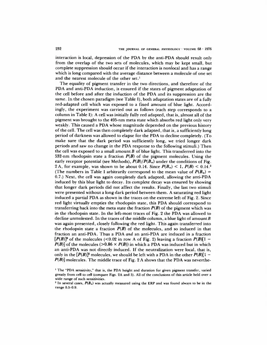
interact ion is local, depress ion o f the PDA by the ant i-PDA should result only f rom the over lap o f the two sets o f molecules, which may be kept small, but comple te suppress ion should occur if the interact ion is nonlocal and has a range which is long c o m p a r e d with the average distance between a molecule o f one set and the neares t molecule of the o the r se t?
T h e equality o f p igmen t t ransfer in the two directions, and the re fo re of the PDA and ant i-PDA induct ion, is ensu red if the states o f p igmen t adapta t ion o f the cell before and af ter the induct ion o f the PDA and its suppress ion are the same. In the chosen pa r ad i gm (see Tab le I), both adapta t ion states are of a fully r ed -adap ted cell which was exposed to a fixed a m o u n t o f blue light. Accord- i n # y , the e x p e r i m e n t was carr ied out as follows (each step cor responds to a co lumn in Tab le I): A cell was initially fully red adap ted , that is, almost all o f the p igmen t was b r o u g h t to the 495-nm meta state which absorbs red light only very weakly. This caused a PDA whose magn i tude d e p e n d e d on the previous history o f the cell. T h e cell was then comple te ly da rk adap ted , that is, a sufficiently long per iod o f darkness was allowed to elapse for the PDA to decline completely. (To make sure that the da rk per iod was sufficiently long, we tried longer da rk per iods and saw no change in the PDA response to the following stimuli.) T h e n the cell was exposed to a small a m o u n t B of blue light. This t r ans fe r red into the 532-nm rhodops in state a fract ion P(B) of the p igmen t molecules. Using the early recep tor potential (see Methods) , P(B)/P(B®) u n d e r the condit ions o f Fig. 2A , for example , was shown to be abou t 0.14. Since P(Boo) < 1, P(B) < 0.14. 2 (The num ber s in Tab le I a rb i t r a r i ly / :o r re spond to the mean value of P(B®) = 0.7.) Next , the cell was again complete ly da rk adap ted , allowing the anti-PDA induced by this blue light to decay. Its comple te decay was ensured by showing that longer dark per iods did not affect the results. Finally, the last two stimuli were p resen ted without a long da rk per iod between them. A saturat ing red light induced a partial PDA as shown in the traces on the ex t r eme left o f Fig. 2. Since red light virtually empt ies the rhodops in state, this PDA should co r re spond to t r ans fe r r ing back into the meta state the fract ion P(B) of the p igmen t which was in the rhodops in state. In the lef t-most traces of Fig. 2 the PDA was allowed to decline unmoles ted . In the traces o f the middle co lumn, a blue light o f a m o u n t B was again p resen ted , closely following the red light. This again t r ans fe r red into the rhodops in state a fraction P(B) of the molecules, and so induced in that fract ion an ant i -PDA. T h u s a PDA and an ant i-PDA are induced in a fract ion [P(B)] 2 o f the molecules (<0.02 in row A o f Fig. 2) leaving a fract ion P(B)[1 - P(B)] o f the molecules (>0.86 x P(B)) in which a PDA was induced but in which an ant i-PDA was not directly induced. I f the neutral izat ion were local, that is, only in the [P(B)] 2 molecules, we should be left with a PDA in the o ther P(B)[1 - P(B)] molecules. T h e middle trace o f Fig. 2 A shows that the PDA was never the-
The "PDA sensitivity," that is, the PDA height and duration for given pigment transfer, varied greatly from cell to cell (compare Figs. 2A and 5). All of the conclusions of this article held over a wide range of such sensitivities. 2 In several cases, P(B®) was actually measured using the ERP and was found always to be in the range 0.5-0.9.

HILl.MAN ET A~.. Nonlocal Photoreceptor Transduction Process 233
¢,,.1 ,.1
Z
Z ©
<
Z
0
Z
©
©
v
i
~1~ ~
"=~ ,,% .-.; I % - ;
<
"~ 0a
l i " E o
"2.
ea " ~ 0
qj ~ cu
e.,
"=- 0 ,g

234 T H E J O U R N A L OF G E N E R A L P H Y S I O L O G Y • V O L U M E 6 8 • 1976
less a p p a r e n t l y c o m p l e t e l y s u p p r e s s e d by the a p p r o p r i a t e b lue l igh t , i n d i c a t i n g a n o n l o c a l i n t e r a c t i o n b e t w e e n the e x c i t o r a n d i n h i b i t o r p rocesses .
As a c o n t r o l , we d e m o n s t r a t e d tha t i n d u c t i o n o f a P D A d i r ec t l y in to a f r a c t i o n less t h a n P(B)[1 - P ( B ) ] o f the m o l e c u l e s r e s u l t e d in a m u c h l a r g e r P D A t h a n t ha t
TEST CONTROL PtB) P(B)[I-P(BI]of 0 P(B/2)
FIGURE 2. The interaction between the excitor and the inhibitor processes. Late receptor potentials (LRP's). The traces of the center column show that a PDA (prolonged depolar izing afterpotential) whose unmolested course appears in the left trace is completely suppressed by an equal anti-PDA, when the two are induced in largely nonover lapping sets of molecules. The right traces show that the degree o f overlap of the sets is insufficient to explain this suppression: Each PDA in the right traces arises from a smaller number of pigment molecules than are in the par t of the PDA populat ion in which anti-PDA is not directly induced, in the center traces; and yet the PDA's in the right traces are larger than those in the center traces (see text). The cells in the center and left traces were p repared by saturating ("full") red i l lumination followed by an amount B of blue light and then 5 min of darkness. The responses shown are to 10 s red light~ followed, in the center traces, by a fur ther br ief blue exposure of amount B, (indicated by dots after light bars). In the right traces the preparat ion was full red/0.5 B blue/5 min darkness, and the stimuli again were 10-s red. The calibration bar represents 20 mV for rows A and C, 5 mV for row B. Row A is for a cell with strong pump postil lumination hyperpolar izat ion (PIH) (B. eburneus); row B is for a cell in which the PIH was weak (B. amphitrite); and rows C are sequential responses 10-20 rain after application of ouabain in a cell with a s trong PIH originally (B. eburneus). For each row the p rocedure was Sat. RIB blue/3 min dark/10 s R-B blue (center response)/3 min dark/10 s R (left response)/ 0.5 B blue/3 min dark/10 s R (right response).
o f the m i d d l e t r ace o f Fig . 2 A (if any) . S ince the P D A a m p l i t u d e is a m o n o t o n i c f u n c t i o n o f p i g m e n t t r a n s f e r r e d , this shows t ha t t he P D A o f the m i d d l e t r ace ( if any) c o r r e s p o n d s to a n u m b e r o f P D A m o l e c u l e s s m a l l e r t h a n the n u m b e r o f t hose no t d i r e c t l y c a n c e l l e d by the a n t i - P D A ; tha t is, at leas t p a r t o f the P D A

HILLMAN ET AL. Nonlocal Photoreceptor Transduction Process 235
molecules must be cancelled by remote anti-PDA's. T h e control expe r imen t consisted o f the same parad igm as the main expe r imen t but with ha/f as much blue light and without the second blue exposure (see Table I, lower half). Th u s a PDA was induced in a fraction P(B/2) o f the molecules. We used the early recep tor potential to demons t ra te that , for this cell and these exper imenta l parameters ,
P(B/2)/P(B®) ~ 0.07,
and
But
s o
o r
[P(B)/P(B®)] × [1 - {P(B)/P(B®)}] ~ 0,12.
[P(B)/P(B®)] × [1 - P(B)] > [P(B)/P(B®)] × [1 - P(B)/P(B®)],
P(B/2)/P(B®) < [P(B)/P(B.)] × [1 - P(B)],
P(B/2) < P(B)[1 - P(B)],
and the control condi t ion is fulfilled. T h e right trace o f Fig. 2 A shows that the result was a PDA clearly larger than the residual PDA in the test exper iment . T h e expe r imen t and control were repea ted for several values o f B and in several cells, with similar results.
T h e posti l lumination hyperpolar izat ion (PIH) due to the electrogenic p u m p in this p repara t ion should not be a substantial factor in the test-control compari - son o f this expe r imen t , since the p u m p apparent ly depends on the response o f the cell (Koike et al., 1971) which is very similar in the three cases (Fig. 2 A).
As an added precaut ion we p e r f o r m e d this set o f exper iments also u n d e r condit ions where . the p u m p potential could not affect the result. T h e second row of traces (Fig. 2 B) shows responses for a cell where the P IH was ext remely weak, presumably because o f metabolic decay (See Hanani and Hil lman, 1976). This is appa ren t in the middle trace, as it was for all conditions o f stimulation in this cell. T h e stimulus-coincident response and the PDA, u n d e r these condit ions, though somewhat r educed in ampl i tude, were in every o the r respect identical to the responses r eco rded when the PIH was strong. Compar ison o f the PDA in the middle trace with those in the o ther two traces o f Fig. 2 B clearly shows that there is less PDA for the "test" middle trace than for e i ther "control ." In this case, by ERP test, we found
and
P(B)/P(B®) = 0.070,
P(B/2)/P(B®) = 0.035,
P(B)/P(B.)[1 - P(B)/P(B®)] = 0.065,
so that
P(B/2)/P(B®) ~ P(B)/P(B®) [1 - P(B)/P(B®)] < P(B) /P(B.)[1 - P(B)],

2 3 6 T H E J O U R N A L OF G E N E R A L P H Y S I O L O G Y • V O L U M E 6 8 • 1 9 7 6
and P(B/2) ~ P(B)[1 - P(B)].
T h e PDA on the middle trace o f row B is nevertheless clearly close to zero, and certainly smaller than that in the r ight t race.
T h e same e x p e r i m e n t was also p e r f o r m e d af ter the applicat ion of 10 -5 M ouaba in to a cell with a s t rong p u m p hyperpolar iza t ion . T h e final two rows o f Fig. 2 show responses recorded sequentially beginning 10 min af ter per fus ion with ouabain . For each row the o rde r was center , left, r ight trace, with 3-min da rk between each two stimuli. Despite the gradual , cont inuous decline in response ampl i tude (Koike et al., 1971), it is clear that the PDA, if any, on the middle traces is smaller than those on the r ight traces. He re , by ERP measure - ment , we found
P(B)/P(B®) = 0.48,
P(B/2)/P(B®) = 0.28,
and
so that
P(B)/P(B.)[1 - P(B)/P(B®)] = 0.25,
P(B/2) <. P(B)[1 - P(B)].
T h u s , locality would predict that the middle traces have PDA's app rox ima te ly equal to or g rea te r than those of the r ight traces. Rather , the PDA's, if any, are smaller.
A lower limit on the range (distance) o f the interact ion can be der ived f rom the u p p e r limit on the value o f P(B). T h e n u m b e r o f molecules which must lie within the range mus t he at least o f the o rde r o f UP(B), if substantial excitor- inhibi tor cancellation is to occur. T h e smallest P(B) for which the m e a s u r e m e n t could be carr ied out on any cell was about 0.02, so P(B)/P(B®) = 0.032 as measured with the ERP, and no sign o f a d e p a r t u r e f rom comple te suppress ion was seen. At least the 50 neighbors nea r each molecule must the re fo re be within its interact ion range.
(B) Interaction of the Excitor Process with the Nonparticipating Molecules
Is the PDA, induced by t ransfe r r ing a fixed n u m b e r o f p igmen t molecules f r o m the rhodops in state to the meta state, inf luenced by the state ( rhodops in or meta) o f those molecules which are not t r ans fe r r ed ei ther way (and so are not directly responsible for any PDA or anti-PDA)? This was investigated as follows: T race 1 o f Fig. 3 A shows a partial PDA induced by a certain a m o u n t R o f red light in a fully b lue-adapted cell. When this PDA had decayed in the dark , a fu r the r , saturat ing, red light (R=) was p resen ted , inducing a fu r the r PDA: trace 2. In each case there was a per iod of darkness between each two stimuli and we made sure this per iod was "sa tura t ing" by seeing that longer per iods had no fu r t he r effects.
T h e initial a m o u n t R of red light had been adjusted so that the PDA's resul t ing f rom the two red exposures would be nearly identical. An initial 0.8 R red light

HILLMAN ET AL. Nonlocal Photoreceptor Transduction Process 237
r e s u l t e d in a P D A ve ry n e a r to t h a t o f t r ace 2 (Fig . 3 A) a n d the fo l l owing R~ in a P D A close to t ha t o f t r ace 1 (Fig. 3 A) , i . e . wi th a r e v e r s e d d i f f e r e n c e b e t w e e n the two P D A ' s so t ha t t he a m o u n t o f in i t ia l r e d l igh t R h f o r which the two P D A ' s w e r e e q u a l was a b o u t 0.9 R . (A s q u a r e d e p e n d e n c e o f P D A o n l igh t a m o u n t , f o r
= 4
B i °o" ~J
FIGURE 3 The interaction between the excitor process and the state of those pigment molecules not part icipating directly in the PDA induction. B. amphitr i te . (A) LRP's. Trace 1 was preceded by full blue adaptat ion and 5 min of darkness. Trace 2 was preceded by trace 1 and 3 min of dark . In trace 1 the stimulus was a limited amount R of red light, and in trace 2, a full red light. For an amount 0.8 R of red light, the ratio of the heights o f these two PDA's was approximately reversed. Tha t is, about 0.O R of red light is needed to t ransfer from the 532 rhodopsin to the 405 meta state such a fraction of the p igment populat ion that t ransferr ing the remainder induces a similar PDA. Traces 3 and 4 are like 1 and 2, respectively, except that the stimulus in 3 was a full red light, so that trace 3 shows a full PDA and trace 4 a "zero," or no-pigment change, PDA, for comparison. Dashed lines are added to guide the eye where the trace was too weak to appear in the pho tograph . The light dura t ion bars relate, f rom top to bot tom, to traces 3, 1, 2, and 4, respectively. (B) Traces show the ERP responses to a white test stimulus in the same cell. The central trace is for the cell af ter 0.9R red light, and is not halfway between the responses for the fully blue-adapted state (bottom trace) and the fully red- adapted state (top trace), indicating that 0.9 R transfers more than half (about 0.63) of the transferable pigment. The simplest explanat ion is that nonpar t ic ipat ing molecules affect the PDA. In trace 1 they are largely in the 532 rhodopsin state and in trace 2 in the 405 meta state. The calibration bars represent 20 mV and 10 s for the traces in Fig. 3 A and 1 mV and 40 ms for the traces in Fig. 3B.
e x a m p l e , w o u l d m a k e this 0.906 R . ) I f t he s ta te o f t he r e m a i n i n g m o l e c u l e s is i r r e l e v a n t , e ach o f t he two P D A ' s s h o u l d r e p r e s e n t t he t r a n s f e r in to t he m e t a s ta te o f h a l f o f t he in i t ia l ly t r a n s f e r a b l e p i g m e n t . T h e n o n p a r t i c i p a t i n g m o l e - cu les a r e m a i n l y in t h e r h o d o p s i n s ta te d u r i n g the f i rs t P D A a n d a l m o s t e n t i r e l y

238 THE JOURNAL OF GENERAL PHYSIOLOGY ' VOLUME 68 • 1976
in the meta state during the second. (Note that all the molecules have reached these states in this preparation within seconds after any stimulus [Minke et al., 1974] .)
To check whether the two equal PDA's correspond to equal pigment transfers, in which case the nonparticipating molecules have no effect, we exploit the linear dependence of the early receptor potential (ERP) on pigment change, as we did above (see Methods). The traces of Fig. 3 B show, from bottom to top, the ERP responses to white test flashes of the cell: (a) in the fully blue-adapted state (fraction of pigment in rhodopsin state = F(Ro) ~ F(Bo) + P(B®)), (b) afterRh red light (rhodopsin fraction = F(Ro) + P(Rh)), and (c) in the fully red-adapted state (rhodopsin fraction = F(Ro) + P(R~) ~ F(Bo)). (Note that P(Rn) and P(R®) are negative numbers here as pigment is transferred from rhodopsin to meta.) IfRh really transfers to the meta state half the pigment transferable by saturating red light, P(Rn) = 0.5, and the middle response should be exactly halfway between the top and bottom responses at all times. That is, presenting the response amplitudes at any time t by a, b, and c, (b-a)/(c-a) = P(Rh)/P(R=) should be 0.5. (This equality can be formally derived in the same way as that used in the Methods section.)
The measured value of (b-a)/(c-a) in this cell is 0.63; values in three other cells ranged from 0.59 to 0.67. Thus there appears to be a small influence of the nonparticipating molecules on the PDA in the direction that the PDA is facili- tated by interaction with the nonparticipating molecules in the meta state and/or inhibited by those in the rhodopsin state.
(C) The Excitor-Excitor Interaction
Two demonstrations of the existence of a facilitatory interaction among the PDA's excited in different pigment molecules appear in Figs. 4 and 5. Fig. 4 shows the response to two successive equal red pulses (latter part of each trace) in a cell which initially was fully blue adapted (early part of each trace). The PDA after the second of the two equal red pulses is grossly higher than that after the first, even though the potential apparently returns, between the two, to the same level as before the first (a hyperpolarized level, see below). Since the successive equal pulses of course transfer successively smaller amounts of pigment into the 495 meta state, the successive PDA's would be reduced if there were no facilita- tion; in fact they are considerably enhanced. From ERP observations, as above, we determined that the first red pulse in the top and bottom traces transferred P(R)/P(R~) = 0.14 and 0.22 of the transferable pigment into the meta state, respectively.
This facilitatory effect thus clearly outlasts the response. Its actual duration was examined by increasing the spacing between the successive red pulses. The degree of facilitation was found to decline with time constants which varied from cell to cell in the range of seconds to minutes. The duration corresponded approximately, in each cell, with that of the full PDA in that cell.
One must consider the possibility that the observed voltage facilitation could arise from an increase in the cell resistance after the red stimuli, and not from a conductance facilitation. Bridge measurements indeed sometimes showed such

HILLMAN E T AL. Nonlocal Photoreceptor Transduction Process 239
an increase, but never more than by a factor o f 2, and generally by much less. T h e observed facilitation was by a factor far larger than this (Fig. 4). Fur ther - more , C. Shaw has done a few voltage clamp exper iments at o u r request and has observed large conductance facilitations. These facilitations could not be due to residual cell resistance changes.
In o rde r to conf i rm that the large PIH's observed in this prepara t ion do not substantially affect the results, the expe r imen t was p e r f o r m e d as follows (Fig. 4): T h e blue adaptat ion was presented immediately af ter a full red stimulus which induced a PDA. In this way the blue stimulus caused the complete suppression o f the PDA, left no anti-PDA effect , but the red-blue succession induced a maximal p u m p PIH. Thus , the following red stimuli, p resented before the P IH
red blue
red blue
r
L_
? r 1__
FIGURE 4. The interaction between excitor processes or PDA's induced in differ- ent pigment molecules. B. eburneus. Initial red/blue adaptation (to inducemaximal pump PIH), was followed by twin red stimuli, the first of which transferred into the meta state 14% (top trace) or 22% (bottom trace, in same ceil) of the transferable pigment, as measured by the ERP. Despite the fact that the first PDA's had declined to near the steady PIH level, the following PDA's are greatly enhanced. Such a "facilitation" could arise from a nonlinear dependence of the PDA on pigment transfer. The calibration bars represent 10 s and 40 mV, respectively.
had declined more than minimally, could not increase the P IH substantially and the facilitation is not due to p u m p saturat ion.
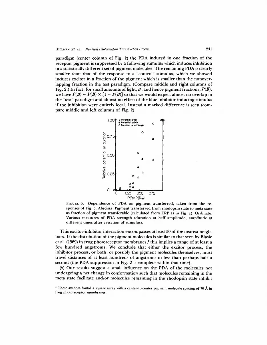
I f this facilitation has a rapid onset, it should also manifest itself in a nonl inear d e p e n d e n c e o f PDA ampl i tude on stimulus amount . We examined this depend- ence using stimulus durat ions which were short compared with that o f the full PDA in the same cell. Some sample recordings, in which the nonlineari ty o f the PDA dependence on p igment t ransfer is clearly visible, are shown in Fig. 5. Extraction o f the quantitative dependence f rom such observations, however , is complicated by the fact that not only the PDA ampli tude changes, but also its shape, so that d i f fe ren t possible measures o f the s trength o f the PDA (ampli tude at various times, width at various heights, step decrease u p o n cessation o f stimulus) give d i f fe ren t dependences . These differences are seen in Fig. fi, where various measures o f the PDA in one cell are plotted against net p igment

240 THE JOURNAL OF GENERAL PHYSIOLOGY' VOLUME 6 8 ' 1976
t ransferred, as a fraction o f the total transferable, that is, P(R) /P(R=) , which was derived f rom ERP measurements as above. One sees that, while they do differ quantitatively, all the measures of the PDA exhibit an initial nonlinear, concave- up dependence 3 Conversion of the membrane potential to conductance would have the effect o f raising the higher points on the graph, and so emphasizing fur ther the nonlinearity. The upward concavity indicates that there is a facilita- tory interaction among the PDA's or the excitor processes responsible for them,
Pigment Intensdy ffgcHon (-log) lransferred
I 45 __/ /~ 0.09
09~ _ _ J ' / ~ - ~ 0.30
0.80 ~ 037
o64_~ __ 048
o 54 L o.5~
i
0?_6 ~ 080
0 1.00
FIGURE 5. The nonlinear dependence of the PDA on pigment transfer. B. ebur- neus. The traces are the LRP responses of a blue-adapted cell to red light transfer- ring from one state to the other the indicated proportions of the transferable pigment as measured by the ERP. By any measure the PDA dependence on pigment transferred appears to be nonlinear. The calibration bar represents 20 mV., and the light durations were 10 s.
induced in different pigment molecules. No quantitative analysis is a t tempted here.
D I S C U S S I O N
(a) We have shown that the previously demonst ra ted interaction between the excitor and inhibitor components o f the transduction process in the barnacle photorecep tor is nonlocal: the excitor and inhibitor neutralize each other even when induced in different pigment molecules. In every trial of the "test"
3 The contrary thesis (linearity) was tentatively accepted by Hochstein et al., 1973, based on a wide scatter of data consistent with both a linear and a square law.
HILLMAN ET AL. Nonlocal Photoreceptor Transduction Process 241
parad igm (center co lumn of Fig. 2) the PDA induced in one fraction o f the receptor p igment is suppressed by a following stimulus which induces inhibition in a statistically d i f fe ren t set o f p igment molecules. T h e remain ing PDA is clearly smaller than that o f the response to a "control" stimulus, which we showed induces excitor in a fraction of the p igment which is smaller than the nonover- lapping fract ion in the test paradigm. (Compare middle and r ight columns o f Fig. 2.) In fact, for small amounts o f light, B, and hence p igment fractions, P(B), we have P(B) -~ P(B) x [1 - P(B)] so that we would expect almost no overlap in the "test" parad igm and almost no effect of the blue inhibi tor- inducing stimulus if the inhibition were entirely local. Instead a marked d i f ference is seen (com- pare middle and left columns o f Fig. 2).
1 0 0 !
t - ,o
075
0.50
025 n~
0 0
O Potlmtiol Qt 155 CII ¢I Potential gt 50s 0 & Duration to half height
0
• A
A
0 A
0 A
A l l
025 0,50 075 P(R)/P(R®)
FIGURE 6. Dependence of PDA on pigment transferred, taken from the re- sponses of Fig. 5. Abscissa: Pigment transferred from rhodopsin state to meta state as fraction of pigment transferable (calculated from ERP as in Fig. 1). Ordinate: Various measures of PDA strength (duration at half amplitude, amplitude at different times after cessation of stimulus).
This exci tor- inhibi tor interaction encompasses at least 50 o f the nearest neigh- bors. I f the distribution o f the p igment molecules is similar to that seen by Blasie et al. (1969) in f rog pho to recep to r membranes , ' this implies a range o f at least a few h u n d r e d angstroms. We conclude that e i ther the excitor process, the inhibitor process, or both, or possibly the pigment molecules themselves, must travel distances o f at least hundreds of angstroms in less than perhaps half a second (the PDA suppression in Fig. 2 is complete within that time).
(b) O u r results suggest a small inf luence on the PDA o f the molecules not unde rgo ing a net change in conformat ion such that molecules remaining in the meta state facilitate and /o r molecules remaining in the rhodops in state inhibit
4 T h e s e a u t h o r s f o u n d a square a r ray with a center - to-center p i g m e n t molecule spac ing o f 70 A in f rog pho to recep to r m e m b r a n e s .

242 T H E J O U R N A L o r G E N E R A L P H Y S I O L O G Y " V O L U M E 6 8 ' 1 9 7 6
the PDA. In attempting to decide whether this effect is significant or not, one must consider alternate sources of distortion of the PDA-ERP correlation. These include: (i) possible residual effects of preceding PDA's or anti-PDA's. However, additional dark times of several minutes before or between the red stimuli had no effect on the results. In every case, both the potential and the conductance of the cell had returned to their resting values long before a fur ther stimulus was presented; (ii) the effects of the electrogenic ion pump in this preparation (Koike et al., 1071). However, the activity of this pump is usually considered to depend on the change in the ionic composition of the intracellular medium due to the preceding stimulus, which change should be nearly the same for the nearly equal PDA's in this experiment; (iii) the effect of any systematic variation through the cell of the ratio of the contributions of the pigment molecules to the ERP and to the PDA. If the visual pigment were sufficiently absorbing that the initial and saturating red stimuli effectively activated different regions of the cell, an artifactual distortion would appear. However, the cell is so transparent that, even if an inhomogeneity existed (for which there is no evidence), the effect would be very small.
One should note that the state of the nonparticipating molecules is not the only difference between the experiments of Fig: 3A, traces 1 and 2. The stimulus in the second experiment was of larger amount than in the first and so resulted in a much larger number of pigment molecules making the "round trip," that is, returning to their initial state. These round trips, or the stimulus- coincident response resulting from them, might influence the PDA. However, such round trips do not induce PDA's or anti-PDA's (these are not observed after neutral stimuli; Hochstein et al., 1073) so we consider the effect unlikely.
We thus conclude that the most probable explanation for the observed result is, in fact, the presence of a small influence of the nonparticipating molecules on the PDA. The interaction (if it exists) is by definition nonlocal, but present data do not require a range beyond nearest-neighbors, and this is in a time of several seconds. (This is the duration of the saturating red stimulus [Fig. 3 A, trace 2] after which the PDA's of traces 1 and 2 have nearly the same time courses.)
(c) We have found a strong facilitatory excitor-excitor interaction manifested in the PDA. This facilitatory effect outlasts the response (Fig. 4) but has a rapid onset leading to a nonlinear dependence of the PDA amplitude on fraction of pigment transferred (Figs. 5-6). A similar effect has been seen by Hanani and Hillman, 1076, in the stimulus-coincident late receptor potential. This latter effect is seen in the first traces of our Fig. 5 but not in the remainder of our responses because of voltage saturation. With respect to the range and velocity of the excitor-excitor interaction, the argument is similar to that for the excitor- inhibitor interaction. The fact that the nonlinearity in Fig. 6 appears clearly by about 0.3 pigment transfer, however, does not require an interaction beyond nearest-neighbors. Again this interaction must take place within a few seconds.
About the functional dependence of the various interactions we can say the following: (a) The fact that a neutral stimulus induces no PDA or anti-PDA suggests that the excitor and inhibitor neutralize each other in the same ratio in which they are produced by equal (and opposite) pigment transfers, but tells us nothing directly about their rates of production. By far the simplest hypothesis

HILLMAN ET AL. Nonlocal Photoreceptor Transduction Process 243
would be that their production is linear with amount of light and that they neutralize each other in fixed ratio, independent of concentration. (b) Since the dependence of the excitor process on the nonparticipating molecules is so weak, we have not attempted to determine the functional dependence on nonpartici- pating population distribution. (c) The nonlinear dependence of the PDA on pigment transfer could arise either from a nonlinear dependence of excitor strength on pigment transfer or from an excitor-excitor interaction. The linear production hypothesis mentioned above is only compatible with the latter.
Possible mechanisms which may be suggested to explain the observations of nonlocality presented here are: electrical spread, translation of the pigment molecules themselves, or some internal transmitter related to either the excitor or the inhibitor process discussed above. Of these, electrical spread cannot explain the PDA facilitation (excitor-excitor interaction) as the effect occurs under voltage clamp. Translation of transmitter related to the inhibitor process cannot explain the excitor-excitor interaction nor the interaction of the excitor process with the nonparticipating molecules as inhibitor is involved in neither of these. Translation of pigment molecules or of an excitor-related transmitter remain as possibilities.
Cone, 1073, cited evidence for the existence of an internal transmitter in photoreceptors. In invertebrates, he noted that the maximum current response per photon absorbed in the ventral photoreceptors of Limulus appears to be too large to arise from a single-ion channel, or even a single microvillus. This could arise from either of the mechanisms found acceptable above. However, this observation requires a diffusion speed (t < 100 ms) and distance (> 1 ~m) greater than those arising from our own results and less compatible with the diffusion of a large pigment molecule in membrane. Furthermore according to Wehner and Goldsmith (1975) there appears to be no pigment diffusion in the photoreceptors of another invertebrate (crayfish).
The substantial reduction of sensitivity observed in many photoreceptors after absorption of relatively few photons (Borsellino and Fuortes, 1968; Behbehani and Srebro, 1974; Dowling, 1963) implies a spread of adaptation. Hamdorf , 1070, directly demonstrated this spread in the fly. However, none of the above mechanisms is excluded by these observations, although pigment diffusion seems implausible for the times and distances involved.
In the vertebrate photoreceptor light reduces cell conductance, so an "excitor" cannot be directly involved. Cone, 1073, noted that a spreading process is needed to explain the facts that (a) in some cases absorption of one photon can reduce the cell conductance by as much as 1%; and (b) the sacs in vertebrate rods which contain the bulk of the visual pigment are isolated from the plasma membrane. Translation of pigment or excitor molecules cannot be responsible for these phenomena, so an alternate or additional process must be present in vertebrates.
C O N C L U S I O N
We have demonstrated some nonlocalities in the barnacle photoreceptor trans- duction process. We suggest that the most likely explanation involves translation of an internal "excitor."

244 T H E J O U R N A L OF G E N E R A L P H Y S I O L O G Y • V O L U M E 6 8 " 1976
Critical readings of the manuscript by Professor I. Parnas and especially Professor R. Werman were most useful. C. Shaw kindly did some control experiments for us. Lluba Kaminsky provided technical assistance. We thank Professor William L. Pak for kindly making available to us research facilities at the Department of Biological Sciences, Purdue University, for part of the work. The work was supported in part by grants of the Central Research Fund of the Hebrew University, of Israel Commission for Basic Research, and of the United States-Israel Binational Science Founda- tion (BSF), Jerusalem, Israel.
Received for publication 17 June 1974.
R E F E R E N C E S
BEHBEHANI, M., and R. SREBRO. 1974. Discrete waves and phototransduct ion in voltage- clamped ventral photoreceptors . J . Gen. Physiol. 64:186.
BERSON, E. L., and E. B. GOLDSTEIN. 1970. Recovery of the human early receptor potential dur ing dark adaptat ion in heredi tary disease. Vision Res. 10:219.
BLASIE, J. K., C. R. WORTHINGTON, and M. M. DEWEY. 1969. Molecular localization of frog retinal receptor photo-pigment by electron microscopy and low-angle X-ray diffraction. J . Mol. Biol. 39:407.
BORSELLINO, A., and M. G. F. FUORTES. 1968. Responses to single photons in visual cells of Limulus. J. Physiol. (Lond.). 196:507.
CONE, R. A. 1973. The internal t ransmitter model for visual excitation: Some quantitative implications. In Biochemistry and Physiology of Visual Pigments. H. Langer, editor. Springer-Verlag, Berlin.
DOWLING, J. E. 1963. Neural and photochemical mechanisms of visual adaptat ion in the rat. J. Gen. Physiol. 46:1287.
HAMDORF, K. 1970. Correlat ion between the concentraton of visual pigment and sensitiv- ity in photoreceptors . Verh. Dtsch. Zool. Ges. 64:148.
HANANI, M., and P. HILLMAN. 1976. Adaptat ion and facilitation in the barnacle photore- ceptor. J. Gen. Physiol. 67:235.
HILLMAN, P., F. A. DODGE, S. HOCHSTEIN, B. W. KNIGHT, and B. MINKE. 1973. Rapid dark recovery of the invertebrate early receptor potential. J. Gen. Physiol. 62:77.
HILLMAN, P., S. HOCHSTEIN, and B. MINKE. 1972. A visual pigment with two physiologi- cally active stable states. Science (Wash. D. C.). 175:1486.
HOCHSTEIN, S., P. HILLMAN, and B. MINRE. 1974. Non-local photoreceptor pigment- membrane coupling mechanisms. Israel J. Med. Sci. 10:569.
HOCHSTEIN, S., B. MINKE, and P. HILLMAN. 1973. Antagonistic components of the late receptor potential arising from different stages of the pigment process.J . Gen. Physiol. 63:105.
KOIKE, H., H. M. BROWN, and S. HAGIWARA. 1971. Hyperpolar izat ion of a barnacle photoreceptor membrane following i l luminat ion.J . Gen. Physiol. 57:723.
MINKE, B., S. HOCHSTEIN, and P. HILLMAN. 1973a. Early receptor potential evidence for the existence of two thermally stable states in the barnacle visual pigment. J. Gen. Physiol. 62:87.
MINKE, B., S. HOCHSTE1N, and P. HILLMAN. 1973b. Antagonistic process as source of visible-light suppression of after-potential in Limulus UV photoreceptors . J. Gen. Physiol. 62:787.
MINKE, B., S. HOCHSTEIN, and P. HILLMAN. 1974a. Derivation of a quantitative kinetic model for a visual pigment from observations o f the early receptor potential. Biophys. J. 14:490.

HILLMAN ET AL. Nonlocal Photoreceptor Transduction Process 245
MINKZ, B., S. HOCHSTEIN, and P. HILLMAN. 1974b. A photoreceptor sensitivity paradox. Biol. Bull. (Woods Hole). 147:491.
SHAW, S. 1972. Decremental conduction of the visual signal in barnacle lateral eye. J. Physiol. (Lond. ). 220:145.
WZHN~a, R., and T. H. GOLDSMITH. 1975. Restrictions on translational diffusion of metarhodopsin in the membranes of a rhabdomeric photoreceptor. Biol. Bull. (Woods Hole). 149:450.
YOSHIKAMI, S., and W. A. HAGINS. 1973. Control of dark current in vertebrate rods and cones. In Biochemistry and Physiology of Visual Pigments. H. Langer, editor. Sprin- ger-Verlag, Berlin.


