Embed Size (px)
Citation preview
Pinch Forces and Instrument Tip ForcesDuring Periodontal ScalingHui Dong,* Peter Loomer,† Alfredo Villanueva,‡ and David Rempel§
Background: The prevalence of upper-extremity musculo-skeletal disorders, such as tendinitis, is elevated among dentalpractitioners. An important risk factor for these disorders isforceful pinching; however, the pinch forces and instrumentforces during scaling are unknown.
Methods: Six dentists and six senior-year dental studentswere recruited to use an instrumented periodontal scaler toperform their usual dental scaling work on patients. Thumbpinch force was measured by a pressure sensor, whereasthe forces developed at the instrument tip were measured bya six-axis load cell.
Results: Dental students applied greater mean peak pinchforce (35.7 – 3.8 N) compared to dentists (24.5 – 4.1 N) (P =0.001). On the other hand, the peak forces generated at the in-strument tip, which were directly related to the productivity ofthe dental scaling task, were higher among the dentists. Theapplication of pinch force by dentists was related to the re-quired scaling forces, whereas students applied excessivepinch force to the tools.
Conclusions: Increased experience in periodontal scalingleads to the application of less pinch force to accomplish scal-ing. Nonetheless, the applied peak pinch forces in both groupsare high and may pose a risk for the development of musculo-skeletal disorders of the distal upper extremity. J Periodontol2007;78:97-103.
KEY WORDS
Dental scaling; ergonomics; tendinitis.
Dentistry is a specialized field inhealth care that requires repeti-tive motions of the fingers and
wrists, forceful pinching, and prolongedawkward postures. Dental practitionershave a high prevalence of work-relatedmusculoskeletal disorders in the neck,shoulders, upper extremities, and lowerback.1,2 A 1997 survey by the AmericanDental Association3 reported that 9.2%of dentists had been diagnosed by aphysician as having an upper-extremitymusculoskeletal disorder; of this group,;20% required surgery and >40% re-duced their work hours.
Upper-extremity musculoskeletal dis-orders are the most common and debili-tating occupational disorders associatedwith the profession of dentistry and den-tal hygiene.4-11 Rice et al.12 comparedthree categories of dental workers (den-tists, dental hygienists, and dental assis-tants) and found that dental hygienistswere at the greatest risk for developingupper-extremity musculoskeletal disor-ders due to the long hours of dentalscaling and root planing work. Upper-extremity musculoskeletal disorders arecommon among dental hygienists; theprevalence of carpal tunnel syndromeranges from 6% to 8.5%.13-18 The Bureauof Labor Statistics reported in 1998 thatdental hygiene ranked the first amongall occupations in the United States inthe number of carpal tunnel syndromecases per 1,000 employees.19 Akessonet al.11 assessed musculoskeletal symp-toms among female dental personnel in a5-year follow-up study and concluded
* School of Public Health, University of California, Berkeley, Berkeley, CA.† School of Dentistry, University of California, San Francisco, San Francisco, CA.‡ Bioengineering Program, University of California, Berkeley.§ Department of Medicine, University of California, San Francisco.
doi: 10.1902/jop.2007.060171
J Periodontol • January 2007
97
that painful and persistent symptoms related to carpaltunnel syndrome and other work-related musculo-skeletal disorders would lead dental practitioners toselect a different occupation.
Carpal tunnel syndrome and wrist tendinitis are as-sociated with personal and occupational risk factors.Work-related risk factors include repetitive forcefulpinching or gripping, sustained non-neutral wrist po-sitions, and use of vibrating tools.20-23 Periodontalscaling and root planing poses an elevated risk for de-veloping musculoskeletal disorders. The work re-quires repeated high pinch forces. According to astudy by Bramson et al.,24 the average pinch forcesexerted during dental scaling were 11% to 20% per-cent of the maximum pinch strength, as estimatedby electromyography. Another study by Zappaet al.25 reported that the forces exerted during scalingranged from 1.01 to 10.35 N with a mean of 5.70 Namong 10 dentists. Among 10 dental hygienists, thecorresponding values ranged from 1.5 to 15.73 Nwith a mean of 5.38 N. However, no one has simulta-neously collected pinch force and instrument tipforces during periodontal scaling on patients.
The purpose of this study was to compare the peakpinch force patterns during scaling between experi-enced dentists and dental students and determinewhether peak thumb pinch force was related to theforces generated at the instrument tip. The hypothesiswas that those with greater experience performingscaling apply lower peak pinch forces than those withless experience.
MATERIALS AND METHODS
Twelve dental provider-patient pairs participated inthe study from March through June 2005. The dentalproviders (dentists and senior-year dental students)were recruited from two community clinics inOakland, California. All of the dentists had ‡2 yearsof experience in dental scaling, whereas the dentalstudents had <2 years of experience. None of the pro-viders reported having had injuries or previous surger-ies in the hand and wrist area. Patients were recruitedfrom the same clinics. All patients had moderate toheavy calculus based on radiographic and clinical ex-aminations; patients were evenly distributed betweendentists and students by level of disease. Informedconsent from both the providers and the patientswas obtained prior to data collection. The study wasapproved by the Committee on Human Research atthe University of California, San Francisco.
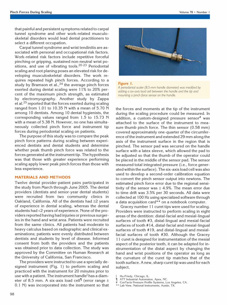
The providers were instructed to use a specially de-signed instrument (Fig. 1) to perform scaling andpracticed with the instrument for 20 minutes prior touse with a patient. The instrument handlei has a diam-eter of 8.5 mm. A six-axis load cell¶ (error range –0.1 N) was incorporated into the instrument so that
the forces and moments at the tip of the instrumentduring the scaling procedure could be measured. Inaddition, a custom-designed pressure sensor# wasattached to the surface of the instrument to mea-sure thumb pinch force. The thin sensor (0.58 mm)covered approximately one-quarter of the circumfer-ence of the instrument and extended 29 mm along theaxis of the instrument surface in the region that ispinched. The sensor pad was secured on the handlesurface with a latex sleeve, which allowed the pad tobe adjusted so that the thumb of the operator couldbe placed in the middle of the sensor pad. The sensormeasured total integrated pressure (i.e., force gener-ated within the surface). The six-axis load cell was alsoused to develop a second-order calibration equationto convert the pinch sensor output into newtons. Theestimated pinch force error due to the regional sensi-tivity of the sensor was – 4.9%. The mean error dueto time drift was 3.5% per 30 seconds. All data werecollected at 100 Hz using specialized software througha data acquisition card** on a notebook computer.
Gracey number 11 curet tips were used for scaling.Providers were instructed to perform scaling in eightareas of the dentition: distal-facial and mesial-lingualsurfaces of tooth #3, distal-lingual and mesial-facialsurfaces of tooth #14, distal-facial and mesial-lingualsurfaces of tooth #19, and distal-lingual and mesial-facial surfaces of tooth #30. Although the Gracey11 curet is designed for instrumentation of the mesialaspect of the posterior teeth, it can be adapted for in-strumentation of the distal aspect by changing thehand and wrist positions of the operator as long asthe curvature of the curet tip matches that of thetooth surface. A new, sharp curet tip was used for eachsubject.
Figure 1.A periodontal scaler (8.5-mm handle diameter) was modified byadding a six-axis load cell between the handle and the tip andmounting a pinch force sensor on the handle.
i Hu-Friedy, Chicago, IL.¶ ATT Industrial Automaton, Apex, NC.# ConTacts Pressure Profile Systems, Los Angeles, CA.** Lab View, National Instruments, Austin, TX.
Pinch Forces During Scaling Volume 78 • Number 1
98
During scaling, force data were recorded sepa-rately for each of the eight areas of the dentition. Be-fore scaling in each area, the provider was allowed toassume her or his most typical and comfortable posi-tion to hold the instrument. The sensor pad was thenadjusted to allow the thumb to be placed in the middleof the sensor pad. Baseline force measures were re-corded before scaling, while no force was applied onthe sensor pad. The provider was then instructed tobegin scaling, and ;1 minute of the scaling processwas recorded. When scaling was completed, the subjectwas instructed again to hold the instrument withoutapplying any force, while the baseline force measureswere recorded again.
The load cell and pressure sensor data were con-verted to force. The pinch force was represented byFp. The load cell output along the long axis of the in-strument, Fz, represented the push (-) and pull (+)forces applied at the tip. The absolute value of Fz
was used for subsequent analyses. The force at thetip, which was perpendicular to the long axis ofthe tool, Ft, was calculated as the geometric sum ofthe two moment outputs about the two axes per-pendicular to the long axis of the tool divided by thedistance between the load cell and the tip of the instru-ment. Because the long axis of the instrument wasroughly parallel to the tooth surface during scaling,Ft estimated the force applied perpendicular to thetooth surface during scaling.
Summary measures of Fp, Fz, and Ft were calcu-lated from a randomly selected 10-second periodof the force history for each tooth, using amplitudeprobability distribution functions (APDFs). APDF val-ues at the 50% and 90% levels represent summarymeasures that estimate the median and peak forcevalues.26
Statistical analysis was performed with a statisticalprogram.†† Analysis of variance with repeated mea-sures (RMANOVA) was used to evaluate the effectof gender, experience, and tooth area on the summaryforce measures. Significant findings were followed-upwith pair-wise comparisons using the Tukey methodto adjust for multiple comparisons. The relationshipsbetween thumb pinch force (Fp) and the forces atthe instrument tip (Fz and Ft) were analyzed usingthe linear regression methods.
RESULTS
Twelve dental providers (six dentists and six senior-year dental students) along with 12 patients partici-pated in the study. Among the participating dentists,there were five females and one male, whereas amongthe students, there were three males and three females.The average age of the dentists was 40.5 – 7.7 years,and the average age of the students was 29.8 – 3.3years. All of the dentists had ‡2 years of experience
in dental scaling, whereas the dental students had<2 years of experience.
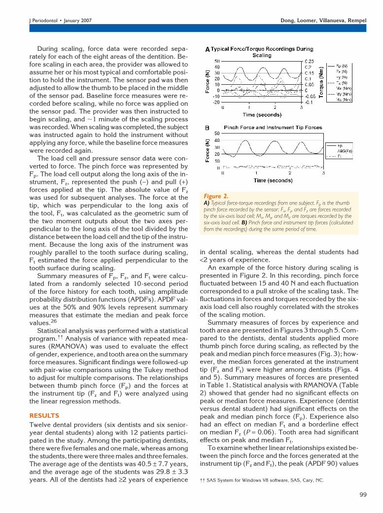
An example of the force history during scaling ispresented in Figure 2. In this recording, pinch forcefluctuated between 15 and 40 N and each fluctuationcorresponded to a pull stroke of the scaling task. Thefluctuations in forces and torques recorded by the six-axis load cell also roughly correlated with the strokesof the scaling motion.
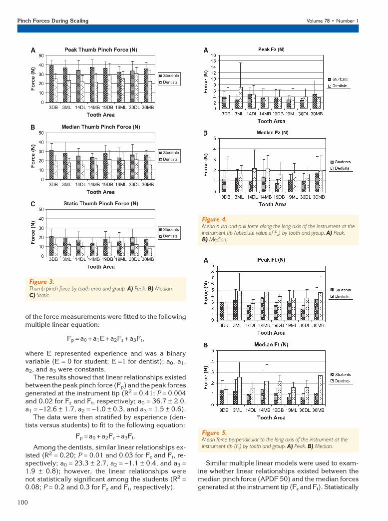
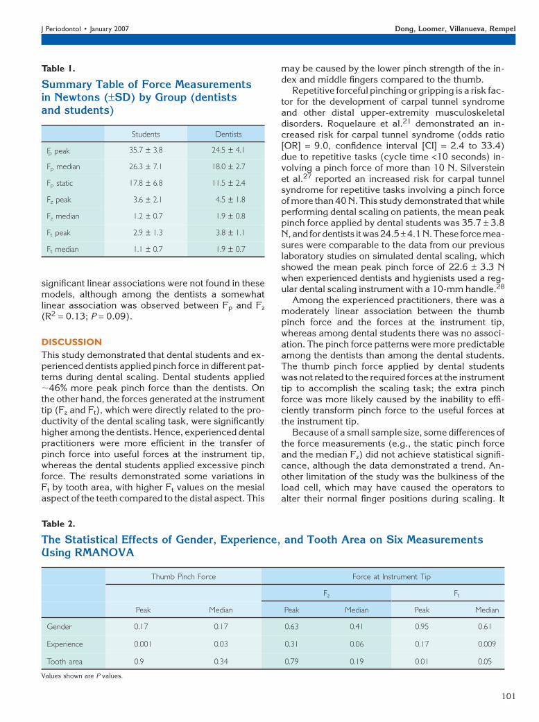
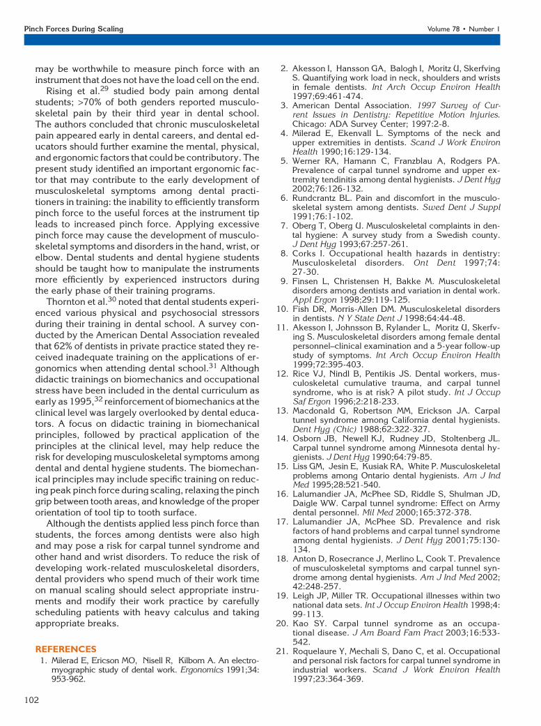
Summary measures of forces by experience andtooth area are presented in Figures 3 through 5. Com-pared to the dentists, dental students applied morethumb pinch force during scaling, as reflected by thepeak and median pinch force measures (Fig. 3); how-ever, the median forces generated at the instrumenttip (Fz and Ft) were higher among dentists (Figs. 4and 5). Summary measures of forces are presentedin Table 1. Statistical analysis with RMANOVA (Table2) showed that gender had no significant effects onpeak or median force measures. Experience (dentistversus dental student) had significant effects on thepeak and median pinch force (Fp). Experience alsohad an effect on median Ft and a borderline effecton median Fz (P = 0.06). Tooth area had significanteffects on peak and median Ft.
To examine whether linear relationships existed be-tween the pinch force and the forces generated at theinstrument tip (Fz and Ft), the peak (APDF 90) values
Figure 2.A) Typical force-torque recordings from one subject. Fp is the thumbpinch force recorded by the sensor; Fx, Fy, and Fz are forces recordedby the six-axis load cell; Mx, My, and Mz are torques recorded by thesix-axis load cell. B) Pinch force and instrument tip forces (calculatedfrom the recordings) during the same period of time.
†† SAS System for Windows V8 software, SAS, Cary, NC.
J Periodontol • January 2007 Dong, Loomer, Villanueva, Rempel
99
of the force measurements were fitted to the followingmultiple linear equation:
Fp = a0 + a1E + a2Fz + a3Ft;
where E represented experience and was a binaryvariable (E = 0 for student; E =1 for dentist); a0, a1,a2, and a3 were constants.
The results showed that linear relationships existedbetween the peak pinch force (Fp) and the peak forcesgenerated at the instrument tip (R2 = 0.41; P = 0.004and 0.02 for Fz and Ft, respectively; a0 = 36.7 – 2.0,a1 = -12.6 – 1.7, a2 = -1.0 – 0.3, and a3 = 1.5 – 0.6).
The data were then stratified by experience (den-tists versus students) to fit to the following equation:
Fp = a0 + a2Fz + a3Ft:
Among the dentists, similar linear relationships ex-isted (R2 = 0.20; P = 0.01 and 0.03 for Fz and Ft, re-spectively; a0 = 23.3 – 2.7, a2 = -1.1 – 0.4, and a3 =1.9 – 0.8); however, the linear relationships werenot statistically significant among the students (R2 =0.08; P = 0.2 and 0.3 for Fz and Ft, respectively).
Similar multiple linear models were used to exam-ine whether linear relationships existed between themedian pinch force (APDF 50) and the median forcesgenerated at the instrument tip (Fz and Ft). Statistically
Figure 3.Thumb pinch force by tooth area and group. A) Peak. B) Median.C) Static.
Figure 4.Mean push and pull force along the long axis of the instrument at theinstrument tip (absolute value of Fz) by tooth and group. A) Peak.B) Median.
Figure 5.Mean force perpendicular to the long axis of the instrument at theinstrument tip (Ft) by tooth and group. A) Peak. B) Median.
Pinch Forces During Scaling Volume 78 • Number 1
100
significant linear associations were not found in thesemodels, although among the dentists a somewhatlinear association was observed between Fp and Fz
(R2 = 0.13; P = 0.09).
DISCUSSION
This study demonstrated that dental students and ex-perienced dentists applied pinch force in different pat-terns during dental scaling. Dental students applied;46% more peak pinch force than the dentists. Onthe other hand, the forces generated at the instrumenttip (Fz and Ft), which were directly related to the pro-ductivity of the dental scaling task, were significantlyhigher among the dentists. Hence, experienced dentalpractitioners were more efficient in the transfer ofpinch force into useful forces at the instrument tip,whereas the dental students applied excessive pinchforce. The results demonstrated some variations inFt by tooth area, with higher Ft values on the mesialaspect of the teeth compared to the distal aspect. This
may be caused by the lower pinch strength of the in-dex and middle fingers compared to the thumb.
Repetitive forceful pinching or gripping is a risk fac-tor for the development of carpal tunnel syndromeand other distal upper-extremity musculoskeletaldisorders. Roquelaure et al.21 demonstrated an in-creased risk for carpal tunnel syndrome (odds ratio[OR] = 9.0, confidence interval [CI] = 2.4 to 33.4)due to repetitive tasks (cycle time <10 seconds) in-volving a pinch force of more than 10 N. Silversteinet al.27 reported an increased risk for carpal tunnelsyndrome for repetitive tasks involving a pinch forceof more than 40 N. This study demonstrated that whileperforming dental scaling on patients, the mean peakpinch force applied by dental students was 35.7 – 3.8N,and fordentists itwas 24.5 – 4.1N.These force mea-sures were comparable to the data from our previouslaboratory studies on simulated dental scaling, whichshowed the mean peak pinch force of 22.6 – 3.3 Nwhen experienced dentists and hygienists used a reg-ular dental scaling instrument with a 10-mm handle.28
Among the experienced practitioners, there was amoderately linear association between the thumbpinch force and the forces at the instrument tip,whereas among dental students there was no associ-ation. The pinch force patterns were more predictableamong the dentists than among the dental students.The thumb pinch force applied by dental studentswas not related to the required forces at the instrumenttip to accomplish the scaling task; the extra pinchforce was more likely caused by the inability to effi-ciently transform pinch force to the useful forces atthe instrument tip.
Because of a small sample size, some differences ofthe force measurements (e.g., the static pinch forceand the median Fz) did not achieve statistical signifi-cance, although the data demonstrated a trend. An-other limitation of the study was the bulkiness of theload cell, which may have caused the operators toalter their normal finger positions during scaling. It
Table 1.
Summary Table of Force Measurementsin Newtons (–SD) by Group (dentistsand students)
Students Dentists
Fp peak 35.7 – 3.8 24.5 – 4.1
Fp median 26.3 – 7.1 18.0 – 2.7
Fp static 17.8 – 6.8 11.5 – 2.4
Fz peak 3.6 – 2.1 4.5 – 1.8
Fz median 1.2 – 0.7 1.9 – 0.8
Ft peak 2.9 – 1.3 3.8 – 1.1
Ft median 1.1 – 0.7 1.9 – 0.7
Table 2.
The Statistical Effects of Gender, Experience, and Tooth Area on Six MeasurementsUsing RMANOVA
Thumb Pinch Force Force at Instrument Tip
Fz Ft
Peak Median Peak Median Peak Median
Gender 0.17 0.17 0.63 0.41 0.95 0.61
Experience 0.001 0.03 0.31 0.06 0.17 0.009
Tooth area 0.9 0.34 0.79 0.19 0.01 0.05
Values shown are P values.
J Periodontol • January 2007 Dong, Loomer, Villanueva, Rempel
101
may be worthwhile to measure pinch force with aninstrument that does not have the load cell on the end.
Rising et al.29 studied body pain among dentalstudents; >70% of both genders reported musculo-skeletal pain by their third year in dental school.The authors concluded that chronic musculoskeletalpain appeared early in dental careers, and dental ed-ucators should further examine the mental, physical,and ergonomic factors that could be contributory. Thepresent study identified an important ergonomic fac-tor that may contribute to the early development ofmusculoskeletal symptoms among dental practi-tioners in training: the inability to efficiently transformpinch force to the useful forces at the instrument tipleads to increased pinch force. Applying excessivepinch force may cause the development of musculo-skeletal symptoms and disorders in the hand, wrist, orelbow. Dental students and dental hygiene studentsshould be taught how to manipulate the instrumentsmore efficiently by experienced instructors duringthe early phase of their training programs.
Thornton et al.30 noted that dental students experi-enced various physical and psychosocial stressorsduring their training in dental school. A survey con-ducted by the American Dental Association revealedthat 62% of dentists in private practice stated they re-ceived inadequate training on the applications of er-gonomics when attending dental school.31 Althoughdidactic trainings on biomechanics and occupationalstress have been included in the dental curriculum asearly as 1995,32 reinforcement of biomechanics at theclinical level was largely overlooked by dental educa-tors. A focus on didactic training in biomechanicalprinciples, followed by practical application of theprinciples at the clinical level, may help reduce therisk for developing musculoskeletal symptoms amongdental and dental hygiene students. The biomechan-ical principles may include specific training on reduc-ing peak pinch force during scaling, relaxing the pinchgrip between tooth areas, and knowledge of the properorientation of tool tip to tooth surface.
Although the dentists applied less pinch force thanstudents, the forces among dentists were also highand may pose a risk for carpal tunnel syndrome andother hand and wrist disorders. To reduce the risk ofdeveloping work-related musculoskeletal disorders,dental providers who spend much of their work timeon manual scaling should select appropriate instru-ments and modify their work practice by carefullyscheduling patients with heavy calculus and takingappropriate breaks.
REFERENCES1. Milerad E, Ericson MO, Nisell R, Kilbom A. An electro-
myographic study of dental work. Ergonomics 1991;34:953-962.
2. Akesson I, Hansson GA, Balogh I, Moritz U, SkerfvingS. Quantifying work load in neck, shoulders and wristsin female dentists. Int Arch Occup Environ Health1997;69:461-474.
3. American Dental Association. 1997 Survey of Cur-rent Issues in Dentistry: Repetitive Motion Injuries.Chicago: ADA Survey Center; 1997:2-8.
4. Milerad E, Ekenvall L. Symptoms of the neck andupper extremities in dentists. Scand J Work EnvironHealth 1990;16:129-134.
5. Werner RA, Hamann C, Franzblau A, Rodgers PA.Prevalence of carpal tunnel syndrome and upper ex-tremity tendinitis among dental hygienists. J Dent Hyg2002;76:126-132.
6. Rundcrantz BL. Pain and discomfort in the musculo-skeletal system among dentists. Swed Dent J Suppl1991;76:1-102.
7. Oberg T, Oberg U. Musculoskeletal complaints in den-tal hygiene: A survey study from a Swedish county.J Dent Hyg 1993;67:257-261.
8. Corks I. Occupational health hazards in dentistry:Musculoskeletal disorders. Ont Dent 1997;74:27-30.
9. Finsen L, Christensen H, Bakke M. Musculoskeletaldisorders among dentists and variation in dental work.Appl Ergon 1998;29:119-125.
10. Fish DR, Morris-Allen DM. Musculoskeletal disordersin dentists. N Y State Dent J 1998;64:44-48.
11. Akesson I, Johnsson B, Rylander L, Moritz U, Skerfv-ing S. Musculoskeletal disorders among female dentalpersonnel–clinical examination and a 5-year follow-upstudy of symptoms. Int Arch Occup Environ Health1999;72:395-403.
12. Rice VJ, Nindl B, Pentikis JS. Dental workers, mus-culoskeletal cumulative trauma, and carpal tunnelsyndrome, who is at risk? A pilot study. Int J OccupSaf Ergon 1996;2:218-233.
13. Macdonald G, Robertson MM, Erickson JA. Carpaltunnel syndrome among California dental hygienists.Dent Hyg (Chic) 1988;62:322-327.
14. Osborn JB, Newell KJ, Rudney JD, Stoltenberg JL.Carpal tunnel syndrome among Minnesota dental hy-gienists. J Dent Hyg 1990;64:79-85.
15. Liss GM, Jesin E, Kusiak RA, White P. Musculoskeletalproblems among Ontario dental hygienists. Am J IndMed 1995;28:521-540.
16. Lalumandier JA, McPhee SD, Riddle S, Shulman JD,Daigle WW. Carpal tunnel syndrome: Effect on Armydental personnel. Mil Med 2000;165:372-378.
17. Lalumandier JA, McPhee SD. Prevalence and riskfactors of hand problems and carpal tunnel syndromeamong dental hygienists. J Dent Hyg 2001;75:130-134.
18. Anton D, Rosecrance J, Merlino L, Cook T. Prevalenceof musculoskeletal symptoms and carpal tunnel syn-drome among dental hygienists. Am J Ind Med 2002;42:248-257.
19. Leigh JP, Miller TR. Occupational illnesses within twonational data sets. Int J Occup Environ Health 1998;4:99-113.
20. Kao SY. Carpal tunnel syndrome as an occupa-tional disease. J Am Board Fam Pract 2003;16:533-542.
21. Roquelaure Y, Mechali S, Dano C, et al. Occupationaland personal risk factors for carpal tunnel syndrome inindustrial workers. Scand J Work Environ Health1997;23:364-369.
Pinch Forces During Scaling Volume 78 • Number 1
102
22. Bernard BP. Musculoskeletal Disorders and WorkplaceFactors. Cincinnati, OH: National Institute for Occu-pational Safety and Health; 1997.
23. Nathan PA, Meadows KD, Istvan JA. Predictors ofcarpal tunnel syndrome: An 11-year study of industrialworkers. J Hand Surg [Am] 2002;27:644-651.
24. Bramson JB, Smith S, Romagnoli G. Evaluating dentaloffice ergonomic. Risk factors and hazards. J Am DentAssoc 1998;129:174-183.
25. Zappa U, Cadosch J, Simona C, Graf H, Case D. Invivo scaling and root planing forces. J Periodontol1991;62:335-340.
26. Jonsson B. Measurement and evaluation of localmuscular strain in the shoulder during constrainedwork. J Hum Ergol (Tokyo) 1982;11:73-88.
27. Silverstein BA, Fine LJ, Armstrong TJ. Occupationalfactors and carpal tunnel syndrome. Am J Ind Med1987;11:343-358.
28. Dong H, Barr A, Loomer P, Laroche C, Young E,Rempel D. The effects of periodontal instrument han-dle design on hand muscle load and pinch force. J AmDent Assoc 2006;137:1123-1130.
29. Rising DW, Bennett BC, Hursh K, Plesh O. Reports ofbody pain in a dental student population. J Am DentAssoc 2005;136:81-86.
30. Thornton LJ, Stuart-Buttle C, Wyszynski TC, WilsonER. Physical and psychosocial stress exposures in USdental schools: The need for expanded ergonomicstraining. Appl Ergon 2004;35:153-157.
31. Murphy DC, Guay AH. The American Dental Associa-tion and dental ergonomics: Research, observationsand activities. In: Murphy DC, ed. Ergonomics and theDental Care Worker. Washington, DC: United BookPress: 1998.
32. Sturdevant CM, et al. Preliminary Considerations forOperative Dentistry, the Art and Science of OperativeDentistry. St. Louis: Mosby; 1995.
Correspondence: Dr. David Rempel, Department of Med-icine, University of California, San Francisco, 1301 South46th Street, Building 163, Richmond, CA 94804. Fax: 510/665-3423; e-mail: [email protected].
Accepted for publication August 23, 2006.
J Periodontol • January 2007 Dong, Loomer, Villanueva, Rempel
103