Embed Size (px)
Citation preview

Frederiksen et al. Environmental Health 2010, 9:32http://www.ehjournal.net/content/9/1/32
Open AccessR E S E A R C H
ResearchPlacental transfer of the polybrominated diphenyl ethers BDE-47, BDE-99 and BDE-209 in a human placenta perfusion system: an experimental studyMarie Frederiksen†1,2,3, Katrin Vorkamp†2, Line Mathiesen1, Tina Mose1 and Lisbeth E Knudsen*1
AbstractBackground: Polybrominated diphenyl ethers (PBDEs) have been widely used as flame retardants in consumer products. PBDEs may affect thyroid hormone homeostasis, which can result in irreversible damage of cognitive performance, motor skills and altered behaviour. Thus, in utero exposure is of very high concern due to critical windows in fetal development.
Methods: A human ex vivo placenta perfusion system was used to study the kinetics and extent of the placental transfer of BDE-47, BDE-99 and BDE-209 during four-hour perfusions. The PBDEs were added to the maternal circulation and monitored in the maternal and fetal compartments. In addition, the perfused cotyledon, the surrounding placental tissue as well as pre-perfusion placental tissue and umbilical cord plasma were also analysed. The PBDE analysis included Soxhlet extraction, clean-up by adsorption chromatography and GC-MS analysis.
Results and Discussion: Placental transfer of BDE-47 was faster and more extensive than for BDE-99. The fetal-maternal ratios (FM-ratio) after four hours of perfusion were 0.47 and 0.25 for BDE-47 and BDE-99, respectively, while the indicative permeability coefficient (IPC) measured after 60 minutes of perfusion was 0.26 h-1 and 0.10 h-1, respectively. The transport of BDE-209 seemed to be limited. These differences between the congeners may be related to the degree of bromination. Significant accumulation was observed for all congeners in the perfused cotyledon as well as in the surrounding placental tissue.
Conclusion: The transport of BDE-47 and BDE-99 indicates in utero exposure to these congeners. Although the transport of BDE-209 was limited, however, possible metabolic debromination may lead to products which are both more toxic and transportable. Our study demonstrates fetal exposure to PBDEs, which should be included in risk assessment of PBDE exposure of women of child-bearing age.
BackgroundPolybrominated diphenyl ethers (PBDEs) have beenwidely used as flame retardant additives in a variety ofproducts of everyday use, e.g. electric equipment, textilesand furniture upholstery. As they are not chemicallybound to the polymers, they can be emitted during theproduct's life cycle and accumulate in the environment.With logKow values of 6-7 (for tetra and penta-BDEs),they accumulate in lipid-rich tissue and biomagnify in thefood chain [1]. The congeners BDE-47 and BDE-99 are
among the most prevalent in the environment [1], eventhough their production and use was banned in largeparts of the world, including the EU in 2004 [2]. Due totheir persistency and toxicity, the tetra- to hepta-BDEshave also been added to Annex A of the Stockholm Con-vention, which aims to protect human health and theenvironment by eliminating toxic persistent organic pol-lutants (POPs) [3]. However, exposure to these com-pounds is likely to continue, due to the use of existingPBDE-containing products and the occurrence of BDE-47 and BDE-99 in the environment. The fully brominatedcongener, BDE-209, has different chemical propertiesthan BDE-47 and BDE-99 and is less persistent in theenvironment despite its extreme hydrophobicity (logKow
* Correspondence: [email protected] Department of Environment & Health, Institute of Public Health, University of Copenhagen. Oester Farimagsgade 5, DK-1014 Copenhagen K, Denmark† Contributed equallyFull list of author information is available at the end of the article
© 2010 Frederiksen et al; licensee BioMed Central Ltd. This is an Open Access article distributed under the terms of the Creative Com-mons Attribution License (http://creativecommons.org/licenses/by/2.0), which permits unrestricted use, distribution, and reproduc-tion in any medium, provided the original work is properly cited.

Frederiksen et al. Environmental Health 2010, 9:32http://www.ehjournal.net/content/9/1/32
Page 2 of 10
≥ 10) [1]. The debromination of BDE-209 can occur byphotolysis to form octa- and nonaBDEs as initial degra-dation products [4,5]. Furthermore, biotransformation ofBDE-209 in fish and rodents results in lower brominatedcongeners, for example hexaBDEs [6,7] with furtherdebromination possible. Generally, the lower brominatedcongeners are more toxic and bioaccumulative than BDE-209 [8,9]. In spite of its apparently lower persistence,BDE-209 has been found at very high concentrations inindoor environments, e.g., dust [10,11], as well as inhumans and wildlife [12,13]. Few restrictions have beenplaced on the use of BDE-209, but since June 2008 the useof BDE-209 in electronic products has been banned inthe EU [2]. The two US producers of BDE-209 and thelargest US importer recently announced to phase outBDE-209 by the end of 2013 [14].
While PBDEs are beneficial for fire safety and save lives,they are also endocrine disrupters that interfere with thy-roid hormone homeostasis [15]. The thyroid hormonesare particularly important for normal brain developmentand impairment can result in irreversible deficits in cog-nitive performance and motor skills as well as alteredbehaviour [16,17]. In animal studies, PBDEs have beenfound to alter the levels of thyroid hormones in offspringafter low doses of maternal exposure to PBDEs in bothrodents [18] and sheep [19]. In human studies, PBDEs inhouse dust have been related to changes in hormone lev-els in men [20] and elevated levels of PBDEs have beenfound in breast milk of mothers to newborn boys withcryptorchidism [21].
PBDEs have been found in various human tissues,including umbilical cord blood [12,22]. The presence ofPBDEs in cord blood indicates placental passage ofPBDEs. In the present study, an ex vivo human placentaperfusion system was applied to investigate the transferof the congeners BDE-47, BDE-99 and BDE-209 acrossthe placenta. The extent of the transfer, kinetics and accu-mulation in the placental tissue have been compared forthe three congeners. The advantage of using the humanplacenta perfusion model is the controlled environmentin which the kinetics of the transfer can be studied andextrapolation from animals to humans is avoided.
MethodsPlacenta perfusionsThe study was approved by the regional Ethics Commit-tee (KF 01-145/03 + KF(11) 260063) and the Data Protec-tion Agency. All mothers gave informed written consent.Term placentas (n = 37) were collected and successfulperfusions were performed on five placentas from fourvaginal births and one Caesarean section directly afterdelivery. The placentas were injected with a Krebs Ringerbuffer containing heparin (5000 IU/ml, 5 ml/l medium) toprevent any remaining blood in fetal vessels from coagu-
lating. The perfusions were performed in a dual perfusionsystem as previously described [23,24]. Of the 37 placen-tas collected 22 were suitable for cannulation (checkpointone in placental perfusion as described in [23]) in a chori-onic artery vein pair within 30 minutes of delivery (oneplacenta was cannulated within 58 min, Table 1). Next,the lobule containing the perfused cotyledon was cutfrom the placenta (9 cm diameter) and placed in the per-fusion chamber. The maternal circulation was connectedby blunt cannulation of the intervillous space surround-ing the perfused cotyledon. The perfusion media (100 mLeach in fetal and maternal reservoirs) were circulated byperistaltic pumps and consisted of Krebs Ringer Buffercontaining heparin (5000 IU/mL, Copenhagen UniversityHospital Pharmacy, 5 mL/L medium), glucose (0.6 g/100mL), Pen-Strep (1% Substrate department standard solu-tion, Panum Institute Copenhagen) and physiological lev-els of Human Serum Albumin (20% solution, CSLBehring Gmbh, dialysed in Krebs Ringer Buffer, maternalreservoir: 30 g/L; fetal reservoir: 40 g/L) and gassed with95% O2/5% CO2 and 95% N2/5% CO2 in the maternal andfetal reservoirs, respectively. Of the 22 cannulated pla-centas only five fulfilled the criteria set for further prog-ress in the experiments. After at least 30 minutes of pre-perfusion to stabilise temperature and oxygen content,BDE-47, BDE-99 and BDE-209 (Cambridge Isotopes Lab-oratories, 1 μg/mL in ethanol, 100 μL added; final con-centration of 1 ng/mL) and the positive control substanceantipyrine dissolved in H2O (Aldrich-Chemie, Germany;final concentration of 100 μg/mL) were added to thematernal reservoir. The applied concentrations of PBDEswere a compromise between achieving final levels rela-tively close to in vivo levels and having a detection fre-quency close to 100%. Setting the BDE-209 level wasfurther complicated by the extremely low water solubilityand generally higher blank levels than for the other con-geners. The perfusions were run for four hours, and 6 mLwere sampled at t = 0 from the maternal circulation andfrom both reservoirs before addition of the compoundsand at 2, 30, 60, 130, 190 and 240 minutes after additionof the compounds. The sampled volume was notreplaced.
The pH (7.2-7.4), glucose and lactate concentrations(glucose > 6 mM), and O2 tension in the maternal perfu-sion medium (30-35 kPa), in the fetal perfusion medium(10-15 kPa), and in the fetal venous outflow (15-20 kPa)were measured every 30 minutes using an ABL5 bloodgas analyser (Radiometer, Denmark). Adjustments weremade using 1 M HCl or 1 M NaOH to adjust pH, glucosesolution (2,61 g/L) and increasing or decreasing the rateof 95% N2/5% CO2 gassing in the fetal perfusate or 95%O2/5% CO2 gassing in the maternal perfusate to adjustpO2 after each measurement [23].

Frederiksen et al. Environmental Health 2010, 9:32http://www.ehjournal.net/content/9/1/32
Page 3 of 10
In addition to the perfusion samples, the three PBDEcongeners were analysed in umbilical cord blood and inthe placental tissue before and after the perfusion. Sam-ples of umbilical cord blood were taken if possible andprocessed to collect plasma. Subsamples of the placentaltissue were taken before perfusion as well as from the cot-yledon and the surrounding tissue after perfusion. Priorto placenta perfusions, adsorption tests had been per-formed: The chambers and tubes were perfused withoutplacental tissue to investigate loss of substance during theperfusion, for instance through adsorption to the perfu-sion system. These tests showed that 25-35% of BDE-47and BDE-99 could adsorb within the perfusion system,while no loss of BDE-209 was observed.
PBDE and antipyrine analysesThe analysis of the positive control substance antipyrinein the perfusate samples was carried out on a LaChromHPLC system equipped with a C-18 column and a Securi-tyGuard precolumn eluted with methanol:water (55:45)as previously described [25].
The analysis of the PBDEs in the perfusion medium,plasma and placental tissue followed accredited methodsfor analysis of PBDEs in biota as previously described[13,26,27]. In brief, 5 ml of perfusate/plasma or up to 24 ghomogenised placental tissue was dried with diatoma-ceous earth and extracted by Soxhlet using 350 mL and500 mL hexane:acetone (4:1), respectively. The extractswere purified by multi-layer adsorption chromatographyand reduced to 500 μL. Instrumental analysis was per-
Table 1: Perfusion variables.
Parameter\Perfusion no. 1 2 3 4 5
Volume loss, maternal (ml/min) 0.016 0.025 n.d. 0.012 0.016
Volume loss, fetal (ml/min) 0.008 0.005 0.045a 0.003 0.025
Flow, fetal (ml/min) 3.1 3.2 3.1a 2.9 3.1
Time, birth to lab (min) n.d. 29 58 24 n.d.
Pre-perfusion (min) 48 43 56 43 48
Antipyrene, FM-ratio (at 240 min) 0.95 1.21 0.92 0.74(1.04 at 190 min)
1.02
Maternal age (yr) 26 24 30 37 31
Placenta weight (g) 1077 675 550 620 1077
Cotyledon weight (g) 47 51 16 20 47
Total perfused (g) 113 157 86 112 113
Caesarean section N N N Y N
Gestation age (weeks + days) n.d. 41+3 41+0 39+1 39+4
Smoker N N N N N
Medicine N N Penicillin Innohep &locoid one week
Citalopram
Details of donors and perfusion of five individual ex vivo placenta perfusions of PBDEs. The perfusions lasted four hours, except no. 3 for which leakage was detected after two hours.n.d.: no data; afor the first two hours only.

Frederiksen et al. Environmental Health 2010, 9:32http://www.ehjournal.net/content/9/1/32
Page 4 of 10
formed by GC-MS using electron capture negative ioni-sation (ECNI) on 60 m DB-5 and 15 m DB-1 columns.
The samples were analysed in batches of 24, whichincluded procedural blanks corresponding to perfusate/plasma and placenta analytical methods, respectively, aduplicate analysis of one of the placenta samples andduplicate analyses of reference samples from the AMAPring test of persistent organic pollutants in human serum.The limit of quantification (LOQ) was defined as the low-est standard of the calibration curve deviating ≤ 20% fromthe actual value if the compound was not detected in theblank samples. Otherwise the LOQ was set to one cali-bration level above the highest blank level. Thus LOQ isdependent on instrumental sensitivity at the time of theanalysis and the blank level in each batch; typical LOQswere 0.005 and 0.17 ng/ml perfusate or plasma for BDE-47/-99 and BDE-209, respectively, or 0.001 and 0.06 ng/gww for placental tissue. However, BDE-209 concentra-tions in the blank samples were not constant, but variedbetween batches.
Total lipid content in placental tissue was determinedaccording to Smedes (1999) [28], while enzymatic lipiddetermination of triglyceride and cholesterol was per-formed on the plasma samples and total lipid was calcu-lated as described by Covaci et al (2006) [29].
Statistical methodsStudent's paired t-test was used for comparison of matri-ces and compounds. The statistical analyses were carriedout using GraphPad Prism 5.0 (GraphPad Software Inc,La Jolla, CA, USA).
Results and discussionPerfusion experimentsIn total, 89 samples of maternal and fetal perfusate, pla-cental tissue and umbilical cord plasma from the five per-fusions were analysed for PBDEs. The details of the fiveplacenta perfusions are given in Table 1. The cotyledon ofperfusion no. 3 was leaking towards the end of the perfu-sion, therefore only data from the first 60 min of this per-fusion have been used. All experiments meet the successcriteria on minimal leakage of fetal media (< 0.05 mL/min) and a fetal/maternal ratio (FM-ratio) for antipyrinetransfer of at least 0.75 (Table 1). The oxygen transfer,glucose consumption and lactate production were moni-tored during the perfusion, and pH was kept in the physi-ological range (data not shown) [23]. It is realised that thefinal sampled volume of 42 ml removed from each reser-voir may give a slightly different picture of transportwhen looking at exact values as in figures 1 and 2. As theexact same amount was removed from the fetal andmaternal reservoir at the same time-points and theresulting concentration is unchanged by removal; thelarge sample-volume demanded by the analysis protocolfor PBDE was allowed in these perfusions.
PBDE levelsThe levels of BDE-47 and BDE-99 in the maternal per-fusate declined rapidly in the beginning of the perfusion;simultaneously, a significant increase in concentrationwas observed in the fetal perfusate (pt(2-240), BDE47 = 0.0003and pt(2-240), BDE99 = 0.0032) (Figure 1a and 1b). The initialconcentration (at t = 0) of the PBDEs in the maternal per-fusate was above the expected level of 1 ng/mL that wasadded, which indicates insufficient initial mixing of the
Figure 1 Placenta perfusion of BDE-47 and BDE-99. Concentration (ng/ml) and standard deviation of four hour placenta perfusions of a) BDE-47 and b) BDE-99 after addition of 1 ng/mL of each congener to the maternal compartment at t = 0. (0-60 min: n = 5; 130-240 min: n = 4).
Placenta perfusion, BDE-47
0 60 120 180 2400.0
0.5
1.0
Maternal, BDE-47
Fetal, BDE-47
Time (min)
Co
nce
ntr
ati
on
in
pe
rfu
sio
n m
ed
ium
(n
g/m
l)
OBr
Br
Br
Br
Placenta perfusion, BDE-99
0 60 120 180 2400.0
0.5
1.0
Maternal, BDE-99
Fetal, BDE-99
Time (min)
Co
nce
ntr
ati
on
in
pe
rfu
sio
n m
ed
ium
(n
g/m
l)
OBr
Br
Br
Br
Br
b)a)

Frederiksen et al. Environmental Health 2010, 9:32http://www.ehjournal.net/content/9/1/32
Page 5 of 10
medium. After 2 minutes the levels were below theexpected initial level. Neither BDE-47 nor BDE-99 wasdetected above LOQ in any of the fetal or maternal per-fusate samples taken prior to the experiment.
At the end of the perfusion the average maternal con-centrations of BDE-47 and BDE-99 were 0.15 and 0.21ng/mL, respectively. This is somewhat higher than themedian levels of BDE-47 found in recent studies ofmaternal plasma and serum in Europe (0.003-0.02 ng/mL), but within the range of recent adult median serumlevels in the United States (0.09-0.4 ng/mL), assuming alipid content of 0.8% [30]. This shows that the concentra-tions used in this study are within the relevant range ofenvironmental exposure. The steady state concentrationsin the fetal circulation were 0.071 and 0.052 ng/mL forBDE-47 and BDE-99, respectively, which was approxi-mately 10 times higher than in the umbilical cord plasmafrom this study (Table 2), but similar to median BDE-47levels observed in US fetal serum of 0.03-0.08 ng/mLwhen assuming a lipid content of 0.3% [31,32].
The mean concentrations of BDE-47 and BDE-99 foundin the non-perfused placental tissue were 0.84 and 0.38ng/g lipid weight (lw), respectively (Table 2), and therebyin line with those previously found in Denmark and Spain[12,26]. The levels of BDE-47 were higher than those ofBDE-99, which is the general trend in mammalian sam-ples. Compared with the non-perfused tissue the concen-trations were significantly higher in the tissuesurrounding the cotyledon (p47 = 0.007; p99 = 0.013), andthe concentrations in the actually perfused cotyledonswere also significantly higher than in the surroundingarea (p47 = 0.007; p99 = 0.013) (Table 2). This shows that asignificant part of the amount of BDE-47 and BDE-99added to the system accumulates in the placental tissue.After the perfusion BDE-99 was present in higher con-centration in the placental tissue than BDE-47, i.e. BDE-99 accumulated in the tissue to a greater extent thanBDE-47.
Contamination during analysis and thereby fluctuatingblank levels are generally an obstacle in trace analysis of
Figure 2 Placenta perfusion of BDE-209. Concentration (ng/ml) and standard deviation of four hour placenta perfusions of BDE-209 after addition of 1 ng/mL to the maternal compartment at t = 0. (0-60 min: n = 5; 130-240 min: n = 4). The grey area indicates the concentration of the maximum pre-perfusion background level in the fetal circulation.
Placenta perfusion, BDE-209
0 60 120 180 2400.0
0.5
1.0Maternal, BDE-209
Fetal, BDE-209
� max pre-perfusion level
Time (min)
Co
nce
ntr
ati
on
in
pe
rfu
sio
n m
ed
ium
(n
g/m
l)
Frederiksen et al. Environmental Health 2010, 9:32http://www.ehjournal.net/content/9/1/32
Page 6 of 10
BDE-209 [e.g. [33]]. Blank levels are caused by the omni-presence of BDE-209, including computers and labora-tory equipment, which can lead to high levels of BDE-209in dust and indoor air, and contamination of samples andequipment in spite of careful precautions. The measuredlevels of BDE-209 in the non-perfused placental tissueand fetal perfusate were not significantly different fromthe relatively high blank levels (pplc = 0.3; pfet = 0.4), thuswe chose not to report BDE-209 concentrations for thesematrices (Figure 2). This critical and conservativeapproach is in agreement with conclusions of interna-tional PBDE intercalibration exercises [34] and the labo-ratory's accreditation scheme. In future perfusion studies,the use of isotope labelled BDE-209 could be consideredin order to eliminate the uncertainties resulting from lab-oratory contamination.
In the maternal perfusate, a general decrease in BDE-209 concentration could be observed (Figure 2), to a levelof 0.50 ng/mL after 4 hours of perfusion. This is roughlyone order of magnitude higher than actual observedblood levels in recent studies from Denmark (~0.014 ng/ml) and elsewhere (~0.009-0.15 ng/ml, assuming 0.8%lipid content) [30,35]. In the present study, BDE-209could not be determined in non-perfused placental tis-sue, however, in a previous study of placental tissue col-lected from the same hospital, a median level of 1.14 ng/g
lw was found [26]. If a similar level is assumed in the pres-ent pre-perfusion samples, this will mean that during theperfusion BDE-209 accumulates in both the perfused cot-yledon and the surrounding tissue with concentrations20- and 100-times the background, respectively. Thisaccumulation in placental tissue may explain why anincrease in fetal circulation cannot be observed.
Transport and kinetics of PBDEsThe ratio between the concentrations in the fetal andmaternal circulation (FM-ratio) during the perfusion wascalculated to evaluate the extent and kinetics of the trans-fer (Figure 3). Steady-state was reached after approxi-mately 190 min for both BDE-47 and BDE-99. For BDE-47 and BDE-99 the FM-ratios were 0.47 and 0.25 at steadystate, respectively, thus indicating that the transfer ofthese PBDEs was limited. The higher FM-ratio of BDE-47showed that it was transported across the placenta to agreater extent than BDE-99 (p = 0.0006). These resultsare in line with previous findings of decreasing placentaltransfer with increasing degree of bromination in pairedmaternal and umbilical cord blood [35,36]. Increasingaffinity with tissue with increasing logKOW is a likelyexplanation for the decreasing placental transfer. Othercompound characteristics that could affect the transportare molecular size and resulting steric hindrance [35,37]
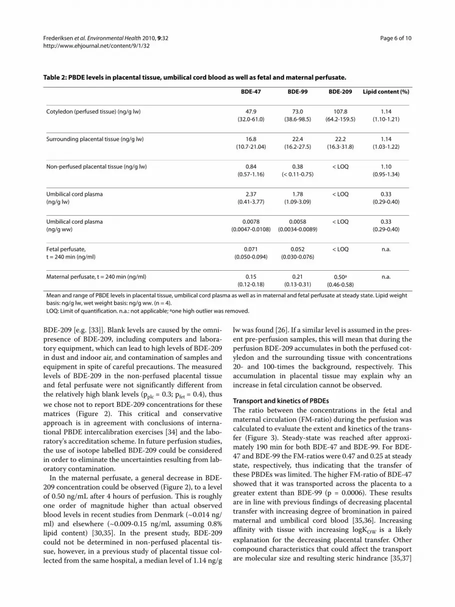
Table 2: PBDE levels in placental tissue, umbilical cord blood as well as fetal and maternal perfusate.
BDE-47 BDE-99 BDE-209 Lipid content (%)
Cotyledon (perfused tissue) (ng/g lw) 47.9(32.0-61.0)
73.0(38.6-98.5)
107.8(64.2-159.5)
1.14(1.10-1.21)
Surrounding placental tissue (ng/g lw) 16.8(10.7-21.04)
22.4(16.2-27.5)
22.2(16.3-31.8)
1.14(1.03-1.22)
Non-perfused placental tissue (ng/g lw) 0.84(0.57-1.16)
0.38(< 0.11-0.75)
< LOQ 1.10(0.95-1.34)
Umbilical cord plasma(ng/g lw)
2.37(0.41-3.77)
1.78(1.09-3.09)
< LOQ 0.33(0.29-0.40)
Umbilical cord plasma(ng/g ww)
0.0078(0.0047-0.0108)
0.0058(0.0034-0.0089)
< LOQ 0.33(0.29-0.40)
Fetal perfusate,t = 240 min (ng/ml)
0.071(0.050-0.094)
0.052(0.030-0.076)
< LOQ n.a.
Maternal perfusate, t = 240 min (ng/ml) 0.15(0.12-0.18)
0.21(0.13-0.31)
0.50a
(0.46-0.58)n.a.
Mean and range of PBDE levels in placental tissue, umbilical cord plasma as well as in maternal and fetal perfusate at steady state. Lipid weight basis: ng/g lw, wet weight basis: ng/g ww. (n = 4).LOQ: Limit of quantification. n.a.: not applicable; aone high outlier was removed.

Frederiksen et al. Environmental Health 2010, 9:32http://www.ehjournal.net/content/9/1/32
Page 7 of 10
or possibly differences in affinity for carrier proteins ofe.g. albumin which may control the transport of the largercongeners [38].
The rapid decrease in maternal levels indicates thatBDE-209 is either transferred across the placenta or accu-mulated in the tissue. While BDE-209 has been detectedquite frequently in adult blood samples [39-41] there areonly few studies on BDE-209 in cord blood. Of these,some have found BDE-209 above the LOQ [12,22,42],mostly at low detection frequencies, while others havenot [31,35,43]. Thus, in spite of the many samples belowLOQ in several studies, results from the literature indi-cate that some transport of BDE-209 across the placentaoccurs. In addition, it may be relevant to consider debro-mination products, which are both more toxic and trans-portable than the parent compound itself, whenaddressing in utero toxicity of BDE-209. However, themetabolic pathway of BDE-209 in humans is poorlyunderstood. Furthermore, BDE-209 can be degraded tolower brominated compounds, leading to a potentially
continued exposure to these more bioaccumulative andtoxic compounds.
The rate of the transfer can be studied by the indicativepermeability coefficient (IPC), which gives a quantitativeindication of the permeability of the placenta for a givencompound. IPC can be estimated from the slope of theinitial linear section of the FM-ratio graph [44]. For BDE-47 and BDE-99, the initial linear slope covered the periodfrom 0 to 60 minutes in Figure 3; from this, the IPC wascalculated to be 0.26 h-1 and 0.10 h-1 for BDE-47 andBDE-99, respectively. This indicates a higher transfer ratefor BDE-47 compared to BDE-99 (p = 0.014). For com-parison, the IPC for the control substance antipyrine, thepesticide glyphosate, the alkaloid caffeine and the preser-vative benzoic acid were 0.94 h-1, 0.11 h-1, 1.03 h-1 and 0.6h-1, respectively, when perfused in the same human pla-centa model [44], and the IPC of the lipophilic substancebenzo(a)pyrene was 0.08 h-1 [45]. This places BDE-99close to glyphosate and BDE-47 between glyphosate andbenzoic acid with regards to the rate of the placental
Figure 3 Fetal-maternal ratios of BDE-47 and BDE-99. Mean and standard deviation of fetal/maternal concentration ratios (FM-ratios) for BDE-47 and BDE-99 during four hour placenta perfusions. (0-60 min: n = 5; 130-240 min: n = 4).
FM-ratios
0 60 120 180 2400.0
0.1
0.2
0.3
0.4
0.5FM-ratio (BDE-47)
FM-ratio (BDE-99)
Time (min)
FM
-ra
tio
(Cfe
tal/C
mat
)

Frederiksen et al. Environmental Health 2010, 9:32http://www.ehjournal.net/content/9/1/32
Page 8 of 10
transport in spite of the very different properties, includ-ing, for example, aqueous solubility.
Mass balancesA mass balance was calculated for each congener to eval-uate the partitioning between the compartments atsteady state using the weight or volume and the concen-trations in the different compartments. The recoveries ofthe added compounds in the different compartments areshown in Figure 4. For BDE-47 and BDE-99, only 7.8%and 10.9%, respectively, were still in the maternal circula-tion at the end of the perfusion. Apart from the quantitywhich was removed by sampling or adsorbed to the sys-tem, the largest fraction was found in the perfused cotyle-don of the placenta for both BDE-47 and BDE-99, thoughthe amount in the surrounding tissue was almost equalbut with a lower absolute concentration (Table 2). Thus,the majority of BDE-47 and BDE-99 was accumulatedand recovered in the placental tissue. For BDE-47, 35% ofthe added amount was unaccounted for, which is inagreement with the initial system adsorption test andcould reflect, for example, binding to blood cells that
were removed by centrifugation prior to sample analysis.For BDE-99, 23% of the added amount was unaccountedfor by tissue and media content, which is also very closeto the loss observed in the system adsorption test.
No mass loss of BDE-209 was observed in the systemadsorption test; however, this could be masked by thebackground contamination with BDE-209. The BDE-209fractions reported (Figure 4) are indicative values wherehigh outliers have been removed; the system adsorptionwas negative. This might indicate that the system hascontributed slightly to the total amount of BDE-209 inthe system, but could also be a result of the higher mea-surement uncertainty for BDE-209. As for BDE-47 andBDE-99, the majority of the BDE-209 added to the perfu-sion system accumulated in the placental tissue. However,a higher percentage than those of BDE-47 and BDE-99remained in the maternal circulation after four hours ofperfusion.
The mass balances do not include metabolites of thePBDEs such as hydroxyl- and methoxy-metabolites,though it is possible that differences in the metabolisationrates may account for some of the observed differences
Figure 4 Mass balance of BDE-47, BDE-99 and BDE-209 in the placenta perfusion system. Fractions of originally added PBDE amounts in per-fusate, placental tissue and removed by sampling. The amount unaccounted for is given as system adsorption. The numbers given for BDE-209 are indicative values (marked with *) due to possible contributions from the surroundings and higher measurement uncertainty.
System adsorption
BDE-47: 34.7% BDE-99: 23.0% BDE-209*: -8%
Surrounding placental tissue
BDE-47: 16.6% BDE-99: 19.8% BDE-209*: 19%
Maternal circulation
BDE-47: 7.8% BDE-99: 10.9% BDE-209*: 27%
Fetal circulation
BDE-47: 4.3% BDE-99: 3.0% BDE-209*: 0%
Cotyledon
BDE-47: 17.5% BDE-99: 20.3% BDE-209*: 24%
Sampling
BDE-47: 19.1% BDE-99: 23.0% BDE-209*: 38%

Frederiksen et al. Environmental Health 2010, 9:32http://www.ehjournal.net/content/9/1/32
Page 9 of 10
between the compounds. However, given the short timeperiod of the study and the fact that the levels of PBDEmetabolites generally are less than 10% of the parentcompound in human plasma/serum [46,47] the metabo-lites would probably be difficult to detect with the currentstudy setup.
Benefits and limitations of the human placenta perfusion systemPlacental passage studied in a human ex vivo placentaperfusion system provides human data non-invasivelyand with no major ethical concerns as the tissue is usuallydiscarded after the birth. The setup is controlled and sev-eral compounds can be studied at the same time and caninclude kinetics, which is not possible to study in vivo.However, the method also has some limitations, forexample, the necessity to exchange the blood with buffersolutions and a time limit of about six hours available forthe transfer study [23]. To increase the solubility of thePBDEs in the buffer solutions, we added human serumalbumin at physiological levels, and even though thestudy period is relatively short, the current results indi-cate that steady-state was obtained for BDE-47 and BDE-99 during the perfusion. Placental thickness and numberof cell layers to pass from the maternal to the fetal circu-lation decrease towards the end of the pregnancy. Thus,the term placenta is considered more sensitive to xenobi-otics than the placenta at earlier stages of pregnancy [48].
ConclusionTo our knowledge this is the first placenta perfusionstudy of PBDEs. The observed transport of BDE-47 andBDE-99 across the placenta shows in utero exposure toPBDEs. The transport of BDE-47 occurred faster andmore extensively than for BDE-99. In conclusions, ourstudy clearly demonstrates fetal exposure to PBDEs, andthis must be considered in risk assessments of PBDEexposure of women of child-bearing age.
AbbreviationsAMAP: Arctic Monitoring and Assessment Programme; BDE-47: 2,2',4,4'-tetra-bromo diphenyl ether; BDE-99: 2,2',4,4',5-tetrabromo diphenyl ether; BDE-209:decabromo diphenyl ether; ECNI: Electron capture negative ionisation; FM-ratio: Fetal/maternal ratio; GC-MS: Gas chromatography mass spectrometry;HPLC: High performance liquid chromatography; IPC: Indicative permeabilitycoefficient; LOQ: Limit of quantification; PBDE: Polybrominated diphenyl ether;POP: Persistent organic pollutant.
Competing interestsThe authors declare that they have no competing interests.
Authors' contributionsMF participated in the data analysis and drafted the manuscript. KV wasresponsible for the PBDE analyses and contributed to experimental planning,data analysis and reviewing of the manuscript. LM performed the perfusionexperiments, antipyrine analyses and contributed to data analysis and draftingof the manuscript. TM contributed to initial planning of the perfusions and thereview of the manuscript. LK was responsible for the overall study including
ethical issues and contributed to experimental planning, data analysis andreviewing of the manuscript. All authors read and approved the manuscript.
AcknowledgementsThe study was financed by the Danish Ministry of the Interior and Health, Research Centre for Environmental Health's Fund (0-302-02-18/4). The authors gratefully acknowledge the donors and the collaboration with the Maternity Unit at Copenhagen University Hospital. Finally, the skilful PBDE analysis per-formed by Birgit Groth at NERI is much appreciated.
Author Details1Department of Environment & Health, Institute of Public Health, University of Copenhagen. Oester Farimagsgade 5, DK-1014 Copenhagen K, Denmark, 2Department of Environmental Chemistry & Microbiology, National Environmental Research Institute (NERI), Aarhus University, Frederiksborgvej 399, 4000 Roskilde, Denmark and 3Danish Building Research Institute, Aalborg University, Dr. Neergaards Vej 15, DK-2970 Hørsholm, Denmark
References1. de Wit CA: An overview of brominated flame retardants in the
environment. Chemosphere 2002, 46:583-624.2. BSEF, Bromine Science and Environmental Forum 2010 [http://
www.BSEF.com].3. The Stockholm Convention 2010 [http://chm.pops.int/].4. Soderstrom G, Sellstrom U, de Wit C, Tysklind M: Photolytic
debromination of the flame retardant Decabromodiphenyl ether (BDE-209). Environ Sci Technol 2004, 38:127-132.
5. Stapleton HM, Dodder NG: Photodegradation of decabromodiphenyl ether in house dust by natural sunlight. Environ Sci Technol 2008, 27:306-312.
6. Huwe JK, Smith DJ: Accumulation, Whole-Body Depletion, and Debromination of Decabromodiphenyl Ether in Male Sprague-Dawley Rats Following Dietary Exposure. Environ Sci Technol 2007, 41:2371-2377.
7. Stapleton HM, Brazil B, Holbrook RD, Mitchelmore CL, Benedict R, Konstantinov A, Potter D: In vivo and in vitro debromination of decabromodiphenyl ether (BDE 209) by juvenile rainbow trout and common carp. Environ Sci Technol 2006, 40:4653-4658.
8. Darnerud PO, Eriksen GS, Jóhannesson T, Larsen PB, Viluksela M: Polybrominated diphenyl ethers: Occurence, dietary exposure, and toxicology. Environ Health Perspect 2001, 109(supl 1):49-68.
9. Shaw SD, Berger ML, Brenner D, Kannan K, Lohmann N, Päpke O: Bioaccumulation of polybrominated diphenyl ethers and hexabromocyclododecane in the northwest Atlantic marine food web. Sci Tot Environ 2009, 407:3323-3329.
10. Harrad S, Ibarra C, Abdallah MA-E, Boon R, Neels H, Covaci A: Concentrations of brominated flame retardants in dust from United Kingdom cars, homes, and offices: Causes of variability and implications for human exposure. Environ Int 2008, 34:1170-1175.
11. Vorkamp K, Thomsen M, Frederiksen M, Pedersen M, Knudsen LE: Polybrominated diphenyl ethers (PBDEs) in the indoor environment and associations with prenatal exposure. Environ Int in press.
12. Gomara B, Herrero L, Ramos JJ, Mateo JR, Fernandez MA, Garcia JF, Gonzalez MJ: Distribution of Polybrominated Diphenyl Ethers in Human Umbilical Cord Serum, Paternal Serum, Maternal Serum, Placentas, and Breast Milk from Madrid Population, Spain. Environ Sci Technol 2007, 41:6961-6968.
13. Vorkamp K, Thomsen M, Falk K, Leslie H, Moller S, Sorensen PB: Temporal Development of Brominated Flame Retardants in Peregrine Falcon (Falco peregrinus) Eggs from South Greenland (1986-2003). Environ Sci Technol 2005, 39:8199-8206.
14. USEPA: DecaBDE phase-out intitative. 2010 [http://www.epa.gov/oppt/existingchemicals/pubs/actionplans/deccadbe.html].
15. Darnerud PO: Brominated flame retardants as possible endocrine disrupters. Int J Androl 2008, 31:152-160.
16. Darras V: Endocrine disrupting polyhalogenated organic pollutants interfere with thyroid hormone signalling in the developing brain. The Cerebellum 2008, 7:26-37.
Received: 1 March 2010 Accepted: 5 July 2010 Published: 5 July 2010This article is available from: http://www.ehjournal.net/content/9/1/32© 2010 Frederiksen et al; licensee BioMed Central Ltd. This is an Open Access article distributed under the terms of the Creative Commons Attribution License (http://creativecommons.org/licenses/by/2.0), which permits unrestricted use, distribution, and reproduction in any medium, provided the original work is properly cited.Environmental Health 2010, 9:32

Frederiksen et al. Environmental Health 2010, 9:32http://www.ehjournal.net/content/9/1/32
Page 10 of 10
17. Porterfield SP: Thyroidal dysfunction and environmental chemicals--potential impact on brain development. Environ Health Perspect 2000, 108:433-438.
18. Kuriyama SN, Wanner A, Fidalgo-Neto AA, Talsness CE, Koerner W, Chahoud I: Developmental exposure to low-dose PBDE-99: Tissue distribution and thyroid hormone levels. Toxicology 2007, 242:80-90.
19. Abdelouahab N, Suvorov A, Pasquier JC, Langlois MF, Praud JP, Takser L: Thyroid Disruption by Low-Dose BDE-47 in Prenatally Exposed Lambs. Neonatology 2009, 96:120-124.
20. Meeker JD, Johnson PI, Camann D, Hauser R: Polybrominated diphenyl ether (PBDE) concentrations in house dust are related to hormone levels in men. Sci Tot Environ 2009, 407:3425-3429.
21. Main KM, Kiviranta H, Virtanen HE, Sundquist E, Tuomisto JT, Toumisto J, Vartiainen T, Skakkebæk NE, Toppari J: Flame Retardants in Placenta and Breast Milk and Cryptorchidism in Newborn Boys. Environ Health Perspect 2007, 115:1519-1526.
22. Antignac JP, Cariou R, Zalko D, Berrebi A, Cravedi JP, Maume D, Marchand P, Monteau F, Riu A, Andre F, Le Bizec B: Exposure assessment of French women and their newborn to brominated flame retardants: Determination of tri- to deca- polybromodiphenylethers (PBDE) in maternal adipose tissue, serum, breast milk and cord serum. Environ Pollut 2009, 157:164-173.
23. Mathiesen L, Mose T, Mørck TJ, Nielsen JK, Nielsen LK, Maroun LL, Dziegel MH, Larsen LG, Knudsen LE: Quality assessment of a placental perfusion protocol. Reprod Toxico 2010 in press.
24. Mose T, Knudsen LE: Placenta perfusion -A human alternative. ALTEX 2006, 23(Suppl):358-363.
25. Mose T, Mortensen GK, Hedegaard M, Knudsen LE: Phthalate monoesters in perfusate from a dual placenta perfusion system, the placenta tissue and umbilical cord blood. Reprod Toxicol 2007, 23:83-91.
26. Frederiksen M, Thomsen M, Vorkamp K, Knudsen LE: Patterns and concentration levels of polybrominated diphenyl ethers (PBDEs) in placental tissue of women in Denmark. Chemosphere 2009, 76:1464-1469.
27. Vorkamp K, Christensen JH, Glasius M, Riget FF: Persistent halogenated compounds in black guillemots (Cepphus grylle) from Greenland--levels, compound patterns and spatial trends. Mar Pollut Bull 2004, 48:111-121.
28. Smedes F: Determination of total lipid using non-chlorinated solvents. The Analyst 1999, 124:1711-1718.
29. Covaci A, Voorspoels S, Thomsen C, van Bavel B, Neels H: Evaluation of total lipids using enzymatic methods for the normalization of persistent organic pollutant levels in serum. Sci Tot Environ 2006, 366:361-366.
30. Frederiksen M, Vorkamp K, Thomsen M, Knudsen LE: Human internal and external exposure to PBDEs - A review of levels and sources. Int J Hyg Environ Health 2009, 212:109-134.
31. Herbstman J, Sjodin A, Patterson DG, Apelberg BJ, Witter FR, Halden RU, Heidler J, Needham LL, Goldman LR: Determinants of prenatal exposure to polychlorinated biphenyls (PCBs) and polybrominated diphenyl ethers (PBDEs) in an urban population. Environ Health Perspect 2007, 115:1794-1800.
32. Mazdai A, Dodder NG, Abernathy MP, Hites RA, Bigsby RM: Polybrominated Diphenyl Ethers in Maternal and Fetal Blood Samples. Environ Health Perspect 2003, 111:1249-1252.
33. Thomas GO, Wilkinson M, Hodson S, Jones KC: Organohalogen chemicals in human blood from the United Kingdom. Environ Pollut 2006, 141:30-41.
34. de Boer J, Wells DE: Pitfalls in the analysis of brominated flame retardants in environmental, human, and food samples - including results of three international interlaboratory studies. Trends Anal Chem 2006, 25:364-372.
35. Frederiksen M, Thomsen C, Frøshaug M, Vorkamp K, Thomsen M, Becher G, Knudsen LE: Polybrominated diphenyl ethers in paired samples of maternal and umbilical cord blood plasma and associations with house dust in a Danish cohort. Int J Hyg Environ Health 2010, 213:233-242.
36. Meijer L, Weiss J, van Velzen M, Brouwer A, Bergman A, Sauer PJJ: Serum Concentrations of Neutral and Phenolic Organohalogens in Pregnant Women and Some of Their Infants in The Netherlands. Environ Sci Technol 2008, 42:3428-3433.
37. Guvenius DM, Aronsson A, Ekman-Ordeberg G, Bergman A, Norén K: Human Prenatal and Postnatal Exposure to Polybrominated Diphenyl Ethers, Polychlorinated Biphenyls, Polychlorobiphenylols, and Pentachlorophenol. Environ Health Perspect 2003, 111:1235-1241.
38. Syme MR, Paxton JW, Keelan JA: Drug transfer and metabolism by the human placenta. Clin Pharmacokinet 2004, 43:487-514.
39. Fischer D, Hooper K, Athanasiadou M, Athanassiadis I, Bergman A: Children Show Highest Levels of Polybrominated Diphenyl Ethers in a California Family of Four: A Case Study. Environ Health Perspect 2006, 114:1581-1584.
40. Thomsen C, Liane VH, Becher G: Automated solid-phase extraction for the determination of polybrominated diphenyl ethers and polychlorinated biphenyls in serum--application on archived Norwegian samples from 1977 to 2003. J Chromatogr B 2007, 846:252-263.
41. Zhu L, Ma B, Hites RA: Brominated Flame Retardants in Serum from the General Population in Northern China. Environ Sci Technol 2009, 43:6963-6968.
42. Wu K, Xu X, Liu J, Guo Y, Li Y, Huo X: Polybrominated Diphenyl Ethers in Umbilical Cord Blood and Relevant Factors in Neonates from Guiyu, China. Environ Sci Technol 2010, 44:813-819.
43. Kawashiro Y, Fukata H, Omori-Inoue M, Kubonoya K, Jotaki T, Takigam H, Sakai SI, Mori C: Perinatal Exposure to Brominated Flame Retardants and Polychlorinated Biphenyls in Japan. Endocr J 2008, 55:1071-1084.
44. Mose T, Kjaerstad MB, Mathiesen L, Nielsen JB, Edelfors S, Knudsen LE: Placental Passage of Benzoic acid, Caffeine, and Glyphosate in an Ex Vivo Human Perfusion System. J Toxicol Environ Health A 2008, 71:984-991.
45. Mathiesen L, Rytting E, Mose T, Knudsen LE: Transport of Benzo[a]pyrene in the Dually Perfused Human Placenta Perfusion Model: Effect of Albumin in the perfusion medium. Basic Clinl Pharmacol Toxicol 2009, 105:181-187.
46. Athanasiadou M, Cuadra SN, Marsh G, Bergman A, Jakobsson K: Polybrominated Diphenyl Ethers (PBDEs) and Bioaccumulative Hydroxylated PBDE Metabolites in Young Humans from Managua, Nicaragua. Environl Health Perspect 2008, 116:400-408.
47. S and Ikonomou: Occurrence and congener specific profiles of polybrominated diphenyl ethers and their hydroxylated and methoxylated derivatives in breast milk from Catalonia. Chemosphere 2009, 74:412-420.
48. Vahakangas K, Myllynen P: Experimental methods to study human transplacental exposure to genotoxic agents. Mutat Res 2006, 608:129-135.
doi: 10.1186/1476-069X-9-32Cite this article as: Frederiksen et al., Placental transfer of the polybromi-nated diphenyl ethers BDE-47, BDE-99 and BDE-209 in a human placenta perfusion system: an experimental study Environmental Health 2010, 9:32


