Embed Size (px)
Citation preview
Farida Farid et al.
235
Plasma F2-Isoprostane: A Biomarker of Lipid Peroxidation:
Correlation with Cerebral Haemodynamics in Children
with Chronic Renal Failure
Farida Farid1, Gihan Mostafa
1, Hala El-Khawas
2, Eman El-Hadidi
3, Tamer Sameeh
1
Departments of Pediatrics1, Neurology
2, Clinical Pathology
3, Ain Shams University
ABSTRACT
Background: Increased lipid peroxidation (LPO), the central feature of oxidative stress, is attributable
mainly to inflammation in chronic renal failure (CRF). Measurement of plasma F2-isoprostane may provide a
quantitative index of LPO and thus, oxidative stress in those patients . LPO is an important risk factor for
premature atherosclerosis which is the major contributing factor to the high prevalence of cerebrovascular
mortalities and morbidities in uremic patients. Cerebral atherosclerosis and other flow abnormalities in CRF
patients could be detected by Transcranial Doppler Ultrasonography (TCD) . Vitamin E is the most frequent
antioxidant strategy in CRF Objective: This study aimed to investigate LPO (indicated by plasma F2-isoprostane
levels), tissue inflammation (indicated by CRP levels) in patients with CRF in relation to cerebral
atherosclerosis, estimated by TCD. In addition, the effect of oral vitamin E supplementation, as an antioxidant,
on suppression of the enhanced LPO, measured by plasma F2-isoprostane, was also investigated. Methods:
Thirty-four patients with CRF (22 on regular hemodialysis "HD" and 12 on conservative management were
studied in comparison to 34 healthy children as controls. Beside full neurological evaluation, measurements of
plasma F2-isoprostane (ELISA) and CRP and estimation of cerebral circulation by TCD were performed, with
re-estimation of plasma F2 isoprostane after two months of Vitamin E supplementation for patients with high
plasma F2-isoprostane. Results: CRF patients, either complied in one group or subdivided into patients on
regular HD and patients on conservative management, had significantly higher plasma F2 -isoprostane and CRP
levels than healthy controls. Plasma F2-isoprostane levels were elevated in 61.8%, 72.7% and 41.7% of all CRF
patients, those patients on regular HD and patients on conservative management, respectively. There was a
significant positive correlation between plasma F2-isoprostane and CRP levels. TCD abnormalities were found
in 70.6% of CRF patients. There was significant positive correlation between TCD abnormalities and both
plasma F2-isoprostane and CRP levels. Patients with clinical neurological manifestations (41.2%) had
significantly higher levels of plasma F2- isoprostane and CRP and frequency of TCD abnormalities than those
without such manifestations. Vitamin E supplementation resulted in a significant decrease of plasma F2 -
isoprostane. Conclusion: LPO, measured by plasma F2-isoprostane, may be enhanced in uremic patients. This
may be attributable to the inflammatory process, indicated by elevated CRP levels. Both LPO and inflammation
may be important factors contributing to premature atherosclerosis in these patients. This assumption was
suggested by the significant positive association between TCD abnormalities (70.6%), which point out to the
presence of cerebral atherosclerosis, and both elevated plasma F2-isoprostane levels (61.8%) and CRP positivity
(58.8%). In addition, vitamin E supplementation, as an antioxidant therapy, may result in suppression of the
enhanced LPO in uremic patients indicated by a significant decrease of plasma levels of F2 -isoprostane levels.
But further studies on a large scale, using different regimens (doses and durations of therapy) of vitamin E, to
choose the best regimen of this antioxidant therapeutic agent in CRF patients are warranted. Also, the effect of
these agents on lowering plasma F2-isoprostane levels should be studied as this LPO marker may serve as an
Correspondence to Hala El-Khawas, e-mail: [email protected]
Egypt J. Neurol. Psychiat. Neurosurg. Vol. 46 (1) - Jan
2009
236
indicator for the effectiveness of antioxidant strategies in patients with CRF. (Egypt J. Neurol. Psychiat. Neurosurg.,
2009, 46(1): 235-246)
INTRODUCTION
Atherosclerotic vascular disease is a major
cause of morbidity and mortality in patients with
chronic renal failure. Oxidative stress, which occurs
when there is excessive free radical production or
low antioxidant levels, has emerged as an important
cofactor for the development of endothelial
dysfunction and hence, atherogenesis1.
Increased lipid peroxidation (LPO), the
central feature of oxidative stress, is attributable
mainly to both the inflammatory process of the renal
disease itself2 and hemodialysis (HD) membrane
bio-incompatibility in patients with chronic renal
failure (CRF)(3)
. Both conditions result in excessive
production of free radicals which attack the
polyunsaturated fatty acids constitutive of cellular
membranes resulting in formation of end products of
LPO4. Increased LPO, is a major risk factor for
premature atherosclerosis in CRF5,6
.
Discovery of the F2-isoprostanes, a group of
prostaglandin F2-like compounds biosynthesized
from arachidonic acid nonenzymatically, has
uncovered a new and novel facet of free radical
biology. F2-isoprostanes has been shown to be a
reliable biomarker of lipid peroxidation7. CRP is one
of the important inflammatory markers currently
considered as a cardiovascular risk factor and it is a
significant predictor of mortality in CRF8.
F2-isoprostane is toxic to endothelium
resulting in endothelial dysfunction which is the key
initial event in the development of atherosclerosis.
This results from the decreased production of the
naturally occurring vasodilator nitric oxide, leading
to vasoconstriction9. Uremic patients show a high
prevalence of cerebrovascular disease which is also
a major cause of their death10
. Besides
atherosclerosis, the underlying process of renal
disease (anemia and hypertension) and treatment
specific changes in HD (hemoconcentration and
alteration of hemostasis) could result in
cerebrovascular insufficiency and increasing the
incidence of stroke in these patients11,12
.
Transcranial Doppler (TCD) is a non-invasive
technique that measures blood flow velocities in
large intracranial arteries12
. TCD is a useful method
of assessment of cerebral circulation in uremic
patients13
.
In CRF the activity of the antioxidant defense
(e.g., vitamin E, A and C) is reduced due to low
dietary intake and/or removal by dialysis14
. Vitamin
E is the most frequent anti-oxidant strategy in CRF
patients15
. There is no doubt that the correction of
the oxidant / antioxidant imbalance is an important
approach for the reduction of the risk of
cardiovascular and cerebrovascular diseases
secondary to atherosclerosis in CRF patients16,17
.
Aim of the work
This study aimed at investigation of lipid
peroxidation (indicated by plasma F2-isoprostane
levels), tissue inflammation (indicated by CRP
levels) in patients with CRF in relation to cerebral
atherosclerosis, estimated by TCD. In addition, the
effect of oral vitamin E supplementation, as an
antioxidant, on suppression of the enhanced LPO,
measured by plasma F2- isoprostane, was also
investigated.
SUBJECTS AND METHODS
Study population
This case-control, follow-up study was
conducted in the Pediatric Dialysis Unit and
Neurology department of Ain Shams University
Hospitals. It included 34 patients with CRF. An
informed written consent for participation in the
study was signed by the parents or the legal
guardians. Patients with CRF were categorized into
2 groups as follows:
- Group I (Patients on regular hemodialysis)
It included 22 patients with end stage renal
disease (ESRD) undergoing regular
hemodialysis (HD). HD was performed with
carbonate dialysate for 2 to 4 hours in each
session. All were on thrice HD regimen per
week. All patients were on polysulfone type of
dialyzer membrane.
- Group II (Patients on conservative
management)
It included 12 patients with CRF on
conservative therapy.
Inclusion criteria:
Farida Farid et al.
237
Stable, maintenance and regular HD patients
with duration of dialysis therapy for at least 2 years
and a prognosis to survive for the duration of the
study.
Exclusion criteria:
Medical instability, therapy with agents that have
been associated with elevated plasma lipids (e.g. beta-
adrenergic blocking agents and androgens) and lipid
lowering drugs, associated condition known to increase
oxidative stress (e.g. diabetes mellitus) and presence of
clinical evidence of infection.
Clinical evaluation of the patients was done
based on clinical history from the caregivers,
reviewing follow up sheets and clinical examination.
Special emphasis was layed on, manifestations
suggesting nervous system involvement (as
convulsions, weakness, paraesthesia, transient
ischemic attacks "TIAs").
Group III (Control group)
It included 34 age-and sex-matched healthy
children with no history of renal or other medical
problems.
Study measurements
The following was done for all subjects:
a. Detailed history taking and thorough clinical
examination
b. Laboratory investigations including:
1. Blood urea, using enzymatic
rate method18
, serum creatinine,
using a modified rate Jaffe
method18,19
(Synchron CX5
system from BECKMAN,
USA).
2. C-reactive protein (CRP) by
latex agglutination.
3. Plasma F2-isoprostane by
competitive enzyme linked
immunoassay (ELISA)20
using
Bioxytech 8-isoprostane
immunoassay from Oxis
international, USA.
c. Transcranial Doppler ultrasonography “TCD”
d. Drug therapy: twenty one patients in whom
plasma F2- isoprostane levels were elevated
were given 400 IU of vitamin E in the form of
E-viton capsules 100 mg (from Cairo
Pharmaceutical Company) as one capsule per
day for 2 months.
After completion of vitamin E-
antioxidant therapy, re-estimation of plasma
F2-isoprostane levels was done.
Sample collection
Three millimeters of venous blood were
collected from each subject (after the dialysis
session in patients on regular HD). Prompt
separation of serum was carried out and used for
direct assay of urea, creatinine and CRP. CRP was
normal if less than 10 mg/ Liter1. Part of serum was
stored at -20 C until assay of F2-isoprostane.
Re-assay of F2-isoprostane was done after two
months of vitamin E therapy for CRF patients with
high plasma F2-isoprostane levels.
Determination of plasma F2-isoprostane by ELISA
Principle of F2-isoprostane assay:
This assay is a competitive enzyme-linked
immunoassay (ELISA) for determining levels of F2-
isoprostane in biological samples. Briefly, 8-epi-
prostaglandin F2α (8-EPI) or (F2-isoprostane) in the
samples or standards competes for the binding (to
the antibody coated on the plate) with F2-
isoprostane conjugated to horseradish peroxidase
(HRP). The peroxidase activity results in colour
development in the substrate when added. The
intensity of the color is proportional to the amount
of 8-EPI-HRP bound and inversely proportionate to
the amount of 8-EPI in the samples or standards21
.
Patients with CRF were considered to have
elevated serum F2-isoprostane if their levels were
above 90.5 pg/ml which was the 95th percentile of
the values of healthy controls as data distribution
was non-parametric.
Transcranial Doppler
TCD was performed, after the dialysis session
in patients on regular HD, using a pulsed Doppler
device (DWL Electronische system, GmbH,
Germany), operating at 2 MHz. The highest peak
systolic and mean blood flow velocities in 2 mm
increments in the middle, anterior and posterior
cerebral arteries, internal carotid artery, vertebral
and basilar arteries were recorded for each patient.
Co2 wash out by hyperventilation for 30 seconds
was performed while insonating MCA vessel.
Patients were considered to have positive TCD
Egypt J. Neurol. Psychiat. Neurosurg. Vol. 46 (1) - Jan
2009
238
findings of large cerebral vasculature if they have
maximum flow velocity greater than 200 cm/s,
maximum velocity in the posterior cerebral,
vertebral or basilar arteries greater than the
maximum velocity in the middle cerebral artery,
mean blood flow velocity of more than 120 cm/sec.,
turbulence, decreased flow velocity in the segment
distal to the stenotic lesion, inter-side difference of
more than 25%, spectral broadening and arterial wall
co-vibration or generalized diminished mean flow
velocities22
. Also, a drop in flow velocities less than
35% with hyperventilation denoted impaired
vasoreactivity (VR) of arterioles23
.
Statistical Analysis
The results were analyzed by commercially
available software package (Stat View, Abacus
Concepts, Inc, Berkley, CA, USA). The data were
presented as mean and standard deviation (SD). Mann
Whitney test was used for comparison between 2
groups as data distribution was non-parametric.
Spearman’s correlation coefficient “r” was used to
determine the relationship between different
quantitative variables. For all tests, a probability (p) of
less than 0.05 was considered significant.
RESULTS
The study included 34 patients with CRF. They
were 21 males and 13 females. Their ages ranged
between 6 and 16 years [mean±SD = 14.2±2.1
years]. Patients were categorized as either patients
on regular hemodialysis (group I, 22 patients; 14
males and 8 females with ages ranged between 11
and 16 years [mean±SD = 14.6±1.6 years]) and
patients on conservative managements (group II, 12
patients; 7 males and 5 females with ages ranged
between 7 and 16 years [mean±SD = 13.6±2.8
years]). Patients were compared to 34 age and sex
matched healthy children in respect to F2-
isoprostane, CRP and TCD findings.
Lipid peroxidation (measured by plasma F2-
isoprostane) and tissue inflammation (measured
by CRP) in CRF:
The mean values of plasma F2 isoprostane in all
studied patients with CRF, patients on regular HD and
patients on conservative management was 116.7566.7,
130.173.6 and 9244.3 pg/mL respectively and are
significantly higher than that of healthy controls
(49.418.7 pg/mL), (p<0.00001) (Table 1). Although,
patients on regular HD had higher plasma F2-
isoprostane levels than those on conservative
management, these differences did not reach statistical
significance (p>0.05)). Interestingly, 61.8% (21/34
patients), 72.7% (16/22 patients) and 41.7% (5/12
patients) of all CRF patients, those on regular HD and
patients on conservative management had elevated
plasma F2-isoprostane levels. CRP positivity was found
in 58.8% (20 patients), 68.2% (15 patients) and 41.7%
(5 patients) of all CRF patients, group I and group II,
respectively. The mean values of CRP were 364.87,
55.145.6 and 28.218.5mg/L in all CRF patients,
group I and group II respectively, significantly higher
than control group (1.81.6 mg/L) (p<0.001, p<0.001
and p<0.01 respectively) (Table 1). Yet, there is no
statistical difference regarding CRP between group I
and group II (p>0.05).
Relationship between plasma F2-isoprostane, as
an index of lipid peroxidation and CRP, as an
index of tissue inflammation in CRF:
There was a significant positive association
between plasma F2 isoprostane and CRP. Eighteen
out of the total 21 patients with elevated plasma F2-
isoprostane (85.7%) had positive CRP as well. On
the other hand, 11 out of the 13 patients with normal
F2-isoprostane (84.6%) had also negative results for
CRP (p<0.001).
Interestingly, CRF patients with elevated F2-
isoprostane levels had significantly higher CRP
values [meanSD = 55.853.5 mg/L] than patients
with normal plasma F2-isoprostane levels
[meanSD = 4.53.6 mg/L, p<0.001]. In addition,
CRF patients with positive CRP had significantly
higher plasma F2-isoprostane levels [mean
SD=70.919 pg/mL] than those with negative CRP
[meanSD = 457.01 pg/mL, p<0.001].
TCD results
TCD abnormalities were found in 24 out of the
studied CRF patients (70.6%). All of them had
impaired VR, 7 had also generalized decreased mean
blood flow velocities i.e. atherosclerosis and one
patient with left MCA stenosis and one patient with
right ACA stenosis.
Farida Farid et al.
239
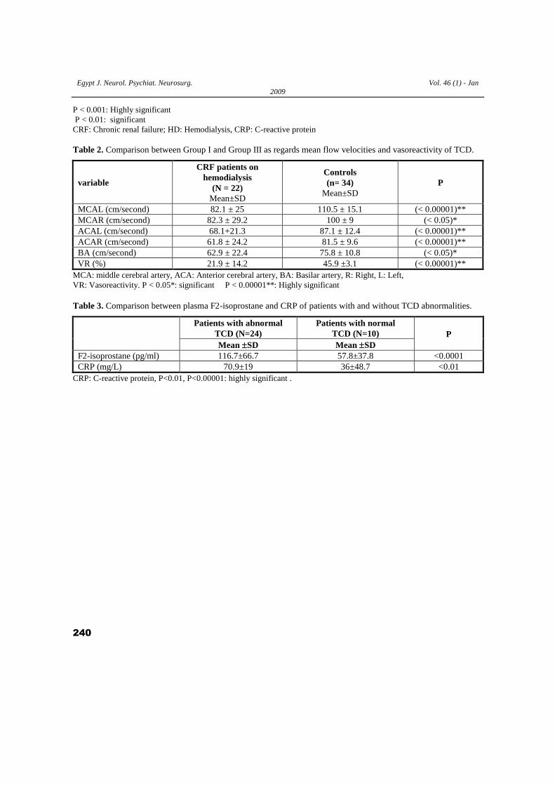
Patients on regular HD had significantly lower
MFV of the studied large cerebral vessels and VR
than healthy controls (Table 2). On the other hand
VR of patients in group II [meanSD = 29.819.2
%] was significantly lower than controls [meanSD
= 45.93.1%, p<0.05]. In contrast, MFV of large
cerebral vessels of both groups (group I &II) were
comparable (p>0.05).
Although TCD abnormalities (flow abnormalities
and/or impaired VR) were higher in patients on regular
HD (18/22 : 81.85%) than patients on conservative
management (6/12 : 50%), this difference did not reach
statistical significance (p>0.05).
Relationship between TCD abnormalities and
both oxidative stress, measured by plasma F2-
isoprostane, and tissue inflammation measured
by CRP in CRF:
Patients with abnormal TCD (24 patients,
70.6%) had significantly higher serum F2-
isoprostane and CRP than those with normal TCD
(Table 3).
There was significant positive correlation
between TCD abnormalities and both elevated
plasma F2- isoprostane (X2= 22.9, p < 0.00001) and
CRP (X2 = 10.46, p<0.01) as 21 and 19 out of the 24
patients with abnormal TCD (85.5% and 79.2%,
respectively) had also elevated plasma F2-
isoprostane and positive CRP, respectively. In
addition, all and 8 out of the 10 patients with normal
TCD had normal plasma F2 isoprostane and
negative CRP, respectively as well (Table 3).
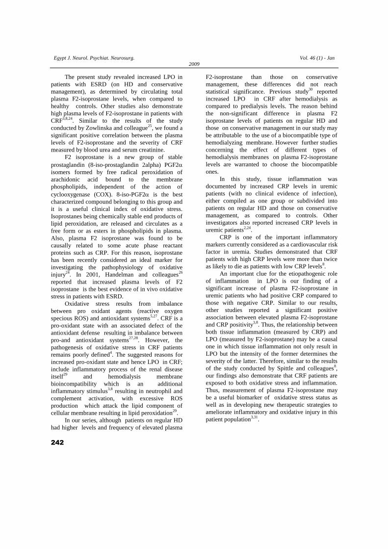
Relationship between clinical neurological
manifestations and Plasma F2 isoprostane, CRP
and TCD in CRF patients:
Clinical neurological manifestations (as TIAs,
convulsions, pyramidal weakness and peripheral
neuritis) were found in 41.2%, 50% and 25% of CRF
patients, group I, and group II, respectively. CRF
patients with neurological manifestations had
significantly higher values of plasma F2 isoprostane and
CRP and lower TCD VR than those without such
manifestations (Table 4). Interestingly all patients with
neurological manifestations had TCD abnormalities.
This frequency was significantly higher than that of
patients without such findings (50%) (p<0.05).
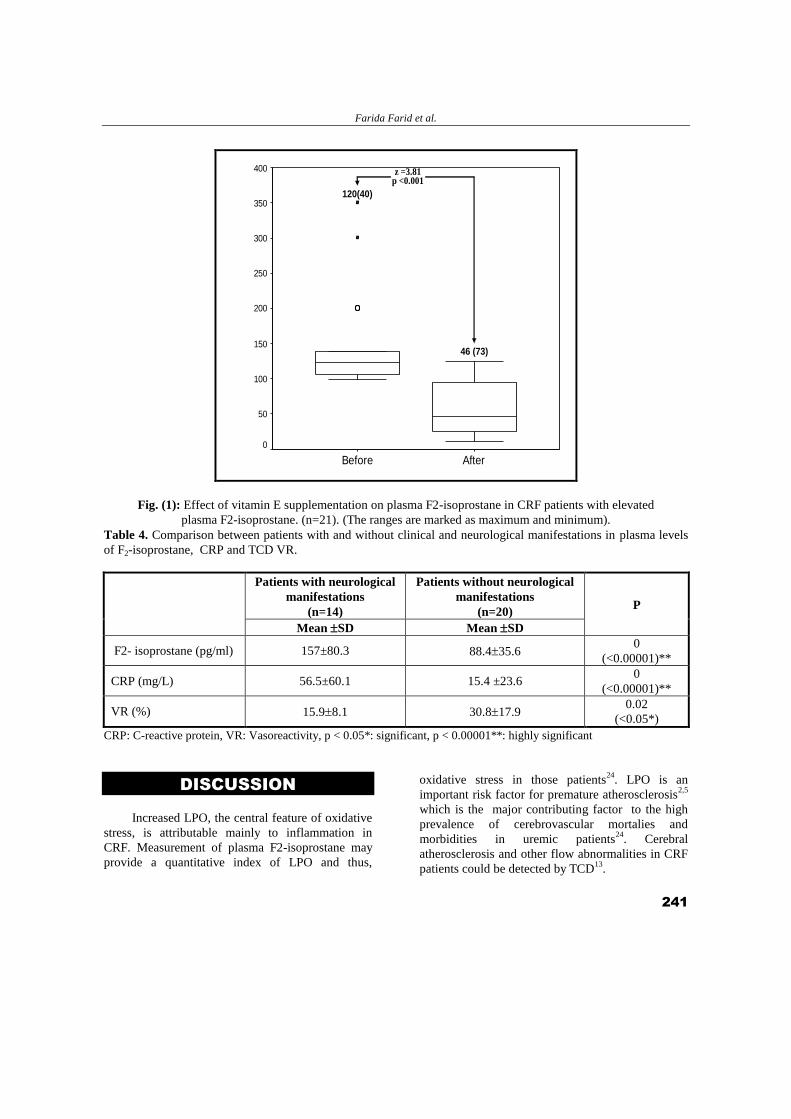
Effect of vitamin E supplementation on plasma
F2- isoprostane :
Vitamin E supplementation (400 IU daily for
2 months) resulted in a significant decrease of
plasma F2- isoprostane (Fig. 1).
Vitamin E supplementation resulted in
normalization of F2-isoprostane in 16 out of the 21
patients with elevated plasma F2-isoprostane (76.2%).
In contrast, this antioxidant therapy had no significant
effect in the remaining 5 patients (23.8%).
Correlation between plasma F2-isoprostane and
the studied clinical, laboratory and cerebral
hemodynamics (TCD) data of CRF patients:
Plasma F2-isoprostane had significant positive
correlation with CRP (r = 0.9, p<0.001) and serum
creatinine (r = 0.9, p<0.001) among patients with
CRF and significant negative correlation with TCD
VR (r = -0.57, p<0.05).
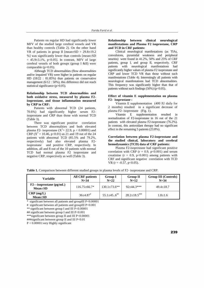
Table 1. Comparison between different studied groups in plasma levels of F2- isoprostane and CRP.
Variable All CRF patients
N=34
Group I
N=22
Group II
N=12
Group III (Controls)
N=34
F2—isoprostane (pg/mL)
Mean±SD 116.75±66.7* 130.1±73.6** 92±44.3*** 49.4±18.7
CRP (mg/L)
Mean±SD 36±4.87
# 55.1±45..6
## 28.2±18.5
### 1.8±1.6
* significant between all patients and groupIII P<0.00001
# significant between all patients and groupIII P<0.001
** significant between group I and III P<0.00001
## significant between group I and III P<0.001
***significant between group II and III P<0.00001
###significant between group II and III P<0.01
P < 0.00001:very Highly significant
Egypt J. Neurol. Psychiat. Neurosurg. Vol. 46 (1) - Jan
2009
240
P < 0.001: Highly significant
P < 0.01: significant
CRF: Chronic renal failure; HD: Hemodialysis, CRP: C-reactive protein
Table 2. Comparison between Group I and Group III as regards mean flow velocities and vasoreactivity of TCD.
variable
CRF patients on
hemodialysis
(N = 22)
Mean±SD
Controls
(n= 34)
Mean±SD
P
MCAL (cm/second) 82.1 ± 25 110.5 ± 15.1 (< 0.00001)**
MCAR (cm/second) 82.3 ± 29.2 100 ± 9 (< 0.05)*
ACAL (cm/second) 68.1+21.3 87.1 ± 12.4 (< 0.00001)**
ACAR (cm/second) 61.8 ± 24.2 81.5 ± 9.6 (< 0.00001)**
BA (cm/second) 62.9 ± 22.4 75.8 ± 10.8 (< 0.05)*
VR (%) 21.9 ± 14.2 45.9 ±3.1 (< 0.00001)**
MCA: middle cerebral artery, ACA: Anterior cerebral artery, BA: Basilar artery, R: Right, L: Left,
VR: Vasoreactivity. P < 0.05*: significant P < 0.00001**: Highly significant
Table 3. Comparison between plasma F2-isoprostane and CRP of patients with and without TCD abnormalities.
Patients with abnormal
TCD (N=24)
Patients with normal
TCD (N=10) P
Mean SD Mean SD
F2-isoprostane (pg/ml) 116.7±66.7 57.8±37.8 <0.0001
CRP (mg/L) 70.9±19 36±48.7 <0.01
CRP: C-reactive protein, P<0.01, P<0.00001: highly significant .
Farida Farid et al.
241
AfterBefore
Pla
sm
a F
2-isop
rosta
ne
(pg/m
l)
400
350
300
250
200
150
100
50
0
z =3.81p <0.001
120(40)
46 (73)
AfterBefore
Pla
sm
a F
2-isop
rosta
ne
(pg/m
l)
400
350
300
250
200
150
100
50
0
z =3.81p <0.001
120(40)
46 (73)
Fig. (1): Effect of vitamin E supplementation on plasma F2-isoprostane in CRF patients with elevated
plasma F2-isoprostane. (n=21). (The ranges are marked as maximum and minimum).
Table 4. Comparison between patients with and without clinical and neurological manifestations in plasma levels
of F2-isoprostane, CRP and TCD VR.
Patients with neurological
manifestations
(n=14)
Patients without neurological
manifestations
(n=20) P
Mean SD Mean SD
F2- isoprostane (pg/ml) 157±80.3 88.435.6 0
(<0.00001)**
CRP (mg/L) 56.5±60.1 15.4 ±23.6 0
(<0.00001)**
VR (%) 15.98.1 30.817.9 0.02
(<0.05*)
CRP: C-reactive protein, VR: Vasoreactivity, p < 0.05*: significant, p < 0.00001**: highly significant
DISCUSSION
Increased LPO, the central feature of oxidative
stress, is attributable mainly to inflammation in
CRF. Measurement of plasma F2-isoprostane may
provide a quantitative index of LPO and thus,
oxidative stress in those patients24
. LPO is an
important risk factor for premature atherosclerosis2,5
which is the major contributing factor to the high
prevalence of cerebrovascular mortalies and
morbidities in uremic patients24
. Cerebral
atherosclerosis and other flow abnormalities in CRF
patients could be detected by TCD13
.
Egypt J. Neurol. Psychiat. Neurosurg. Vol. 46 (1) - Jan
2009
242
The present study revealed increased LPO in
patients with ESRD (on HD and conservative
management), as determined by circulating total
plasma F2-isoprostane levels, when compared to
healthy controls. Other studies also demonstrate
high plasma levels of F2-isoprostane in patients with
CRF3,8,24
. Similar to the results of the study
conducted by Zowlinska and colleague25
, we found a
significant positive correlation between the plasma
levels of F2-isoprostane and the severity of CRF
measured by blood urea and serum creatinine.
F2 isoprostane is a new group of stable
prostaglandin (8-iso-prostaglandin 2alpha) PGF2
isomers formed by free radical peroxidation of
arachidonic acid bound to the membrane
phospholipids, independent of the action of
cyclooxygenase (COX). 8-iso-PGF2 is the best
characterized compound belonging to this group and
it is a useful clinical index of oxidative stress.
Isoprostanes being chemically stable end products of
lipid peroxidation, are released and circulates as a
free form or as esters in phospholipids in plasma.
Also, plasma F2 isoprostane was found to be
causally related to some acute phase reactant
proteins such as CRP. For this reason, isoprostane
has been recently considered an ideal marker for
investigating the pathophysiology of oxidative
injury24
. In 2001, Handelman and colleagues26
reported that increased plasma levels of F2
isoprostane is the best evidence of in vivo oxidative
stress in patients with ESRD.
Oxidative stress results from imbalance
between pro oxidant agents (reactive oxygen
specious ROS) and antioxidant systems5,27
. CRF is a
pro-oxidant state with an associated defect of the
antioxidant defense resulting in imbalance between
pro-and antioxidant systems27,28
. However, the
pathogenesis of oxidative stress in CRF patients
remains poorly defined4. The suggested reasons for
increased pro-oxidant state and hence LPO in CRF;
include inflammatory process of the renal disease
itself29
and hemodialysis membrane
bioincompatibility which is an additional
inflammatory stimulus3,8
resulting in neutrophil and
complement activation, with excessive ROS
production which attack the lipid component of
cellular membrane resulting in lipid peroxidation29
.
In our series, although patients on regular HD
had higher levels and frequency of elevated plasma
F2-isoprostane than those on conservative
management, these differences did not reach
statistical significance. Previous study30
reported
increased LPO in CRF after hemodialysis as
compared to predialysis levels. The reason behind
the non-significant difference in plasma F2
isoprostane levels of patients on regular HD and
those on conservative management in our study may
be attributable to the use of a biocompatible type of
hemodialyzing membrane. However further studies
concerning the effect of different types of
hemodialysis membranes on plasma F2-isoprostane
levels are warranted to choose the biocompatible
ones.
In this study, tissue inflammation was
documented by increased CRP levels in uremic
patients (with no clinical evidence of infection),
either compiled as one group or subdivided into
patients on regular HD and those on conservative
management, as compared to controls. Other
investigators also reported increased CRP levels in
uremic patients2,24
.
CRP is one of the important inflammatory
markers currently considered as a cardiovascular risk
factor in uremia. Studies demonstrated that CRF
patients with high CRP levels were more than twice
as likely to die as patients with low CRP levels8.
An important clue for the etiopathogenic role
of inflammation in LPO is our finding of a
significant increase of plasma F2-isoprostane in
uremic patients who had positive CRP compared to
those with negative CRP. Similar to our results,
other studies reported a significant positive
association between elevated plasma F2-isoprostane
and CRP positivity3,8
. Thus, the relationship between
both tissue inflammation (measured by CRP) and
LPO (measured by F2-isoprostane) may be a causal
one in which tissue inflammation not only result in
LPO but the intensity of the former determines the
severity of the latter. Therefore, similar to the results
of the study conducted by Spittle and colleagues8,
our findings also demonstrate that CRF patients are
exposed to both oxidative stress and inflammation.
Thus, measurement of plasma F2-isoprostane may
be a useful biomarker of oxidative stress status as
well as in developing new therapeutic strategies to
ameliorate inflammatory and oxidative injury in this
patient population3,31
.
Farida Farid et al.
243
In the present work, CRF patients with clinical
neurological manifestations had significantly higher
plasma F2-isorpsotane and CRP levels than those
without such manifestations. This could be
explained by the fact that increased LPO, due to
tissue inflammation is a major risk factor for
atherosclerosis in uremia5,6
. F2-isorpsotane is toxic
to endothelium resulting in endothelial dysfunction
which is the key initial event in the development of
atherosclerosis due to the reduced production of the
vasodilator nitric oxide (NO) leading to
vasoconstriction. For this reason, uremic patients
have a high prevalence of cerebrovascular disease
and strokes which are also major causes of their
death10
.
Thus, the increased levels of both plasma F2-
isoprostane and CRP in patients with than those
without clinical neurological manifestations could
be attributable to the increased frequency of
atherosclerosis in the former than the latter group.
This premature atherosclerosis (due to increased
inflammation and LPO) may be responsible for the
cerebrovascular morbidity of uremic patients.
Cerebral blood flow studies using TCD is a
non- invasive technique that measures blood flow
velocity in large intracranial arteries. It is relatively
cheap, available and can be performed with portable
machines.
In the present study, mean flow velocities of
large cerebral arteries after dialysis management of
patients on regular HD were significantly lower than
that of healthy controls. Similarly, 2 previous studies
demonstrated significantly lower MFV of patients
under regular HD than those on conservative
management13,32
.
Besides atherosclerosis, the underlying process
of the renal disease (anaemia and hypertension) in
uremic could result in cerebrovascular insufficiency
leading to brain damage and structural lesion11
.
Also, treatment specific changes in HD including
hemoconcentration (due to fluid removal),
alteration of hemostasis due to changes of plasma
fibrinogen levels and endothelial activation could
potentially interfere with cerebral blood flow12
.
Our series revealed significant decrease of VR
of uremic patients, either compiled as one group or
subdivided into patients on regular HD and patients
on conservative management, compared to healthy
controls. Impaired VR, which may be attributable to
atherosclerosis, occurred in 70.6%, 81.8% and 50%
of all CRF patients, those on regular HD and those
on conservative management, respectively. We
could not trace data in literature regarding VR of
cerebral circulation estimated by TCD in uremic
patients to be compared with our results.
The possible variable role of TCD in diagnosis
of cerebral vascular abnormalities in CRF may be
supported by our finding of a significant increase of
TCD abnormalities in CRF patients with clinical
neurological manifestations (100%) than those
without such manifestations (50%). The presence of
TCD abnormalities in half of our patients without
evident clinical neurological manifestations may
highlight the importance of TCD evaluation of
cerebral circulation in all uremic patients, even in
absence of overt clinical neurological
manifestations, for early interference before
development of a debilitating neurological disease.
In our series, patients with abnormal TCD had
significantly higher plasma F2-isorpsotane and CRP
than those with normal TCD. Furthermore, there was
significant negative correlation between VR and
both plasma F2-isorpsotane and CRP levels.
Moreover, we found a significant positive
association between TCD abnormalities and both
elevation of plasma F2-isoprostane and CRP
positivity. All the previous findings may point out to
the significant positive relationship between TCD
abnormalities and both tissue inflammation and LPO
in CRF.
The previous findings could be explained by
enhancement of LPO secondary to the inflammatory
process of uremia, both of which result in premature
atherosclerosis with abnormalities of MFV and VR
of the cerebral circulation detected by TCD. Thus,
F2-isoprostane may be a biochemical link between,
inflammation, LPO and accelerated atherosclerosis
in uremic patients3,33,34
.
Studies demonstrated low levels of vitamin E,
A and C in CRF patients due to either low dietary
intake and/or removal by dialysis14,27,31
. Vitamin E is
the most frequent antioxidant strategy in CRF
patients (15)
. In the present study vitamin E (-
tocopherol) supplementation (400 IU/day for 2
months) resulted in a significant decrease of the
elevated plasma F2- isoprostane.
The benefit of antioxidant therapy in CRF may
result from the interruption of several
Egypt J. Neurol. Psychiat. Neurosurg. Vol. 46 (1) - Jan
2009
244
pathophysiological mechanisms. Vitamin E restores
glomerular basement membrane integrity, prevents
neutrophil chemotaxis and inhibits platelets
aggregation35
.
In our series, vitamin E supplementation
resulted in normalization of the elevated plasma F2-
isoprostane in 16 out of the 21 patients with elevated
plasma F2-isoprostane levels (76.2%). On the other
hand, this antixodiant therapy had no significant
effect in the remaining 5 patients (23.8%) and
plasma F2-isoprostane levels remained high. Thus,
further studies using different regimens of vitamin E
and other antioxidants supplementation in CRF
patients with increased oxidative stress are
mandatory to determine the best regimens (including
dose and duration) of these therapeutic agents that
result in complete suppression of oxidative stress in
uremia.
Today there is no doubt that the correction of the
oxidant / antioxidant imbalance in patients with CRF
is an important approach for the reduction of the risk
of those patients to develop cardiovascular
disorders28
.
Synthetic biocompatible membranes, including
vitamin E coated membranes, had antioxidant
effects and preservation of plasma vitamin E levels
in hemodialysis patients35
. Thus, the use of vitamin
E coated membranes is recommended in CRF
patient on regular HD to decrease the oxidative
stress and hence, the enhanced atherosclerosis
responsible for the cardiovascular and
cerebrovascular mortalities in those patients.
In conclusion, both LPO, measured by plasma
F2 isoprostane, and CRP may be enhanced in
uremic patients. Both LPO and inflammation may
be important factors contributing to premature
atherosclerosis in these patients. The latter
assumption was suggested by the significant positive
association between TCD abnormalities (70.6%),
which point out to the presence of cerebral
atherosclerosis, and both elevated plasma F2
isoprostane levels (61.8%) and CRP positivity
(58.8%).
In addition, vitamin E supplementation, as an
antioxidant therapy, may result in suppression of the
enhanced LPO in uremic patients indicated by a
significant decrease of plasma levels of F2
isoprostane levels. But further studies on a large
scale, using different regimens (doses and durations
of therapy) of vitamin E, to choose the best
regimen of this antioxidant therapeutic agent in
CRF patients are warranted. Also, the effect of
these agents on lowering plasma F2-isoprostane
levels should be studied as this LPO marker may
serve as an indicator for the effectiveness of
antioxidant strategies in patients with CRF. In
addition, F2 isoprostane, CRP and TCD could
represent useful approach not only to monitor
antioxidant treatment and new dialysis therapies, but
also to monitor for occurrence of cerebral
atherosclerosis. Studies concerning anti F2-
isoprostane measures as receptor antagonists or
synthesis inhibition are needed. As F2-isoprostane
could be a target for a new therapy which decrease
LPO and hence, cerebrovascular mortalities from
atherosclerosis in CRF in future.
REFERENCES
1. Stenvinkel P, Heimburger O, Paultre F,
Diczfalusy U, Wang T, Berglund L, Jogestrand T.
Strong association between malnutrition,
inflammation and atherosclerosis in chronic
renal failure. Kid Int 1999; 55: 1899-1911.
2. Oberg BP, Mc Menamin E, Lucas FL, Mc
Monagle E, Morrow J, Ikizler TA. Increased
prevalence of oxidant stress and inflammation in
patients with moderate to severe chronic kidney
disease. Kid Int 2004; 65: 1009-1016.
3. Ikizler TA, Morrow JU, Roberts LJ, Evanson JA,
Becker V, Hakim RM,Shyr Y, Himmelfarb J.
Plasma F2 isoprostane levels are elevated in
chronic hemodialysis. Clin Nephrol 2002; 50:
190-197.
4. Massy ZA, Nyuyen-Khoa T. Oxidative stress and
chronic renal failure: markers and management. J
Nephrol 2002;15: 336-341.
5. Caimi G, Carollo C, Lo Presti R. Chronic renal
failure: oxidative stress, endothelial dysfunction
and wine. Clinical nephrology 2004; 62: 331-
335.
6. Diepeveen SHA, Verhoeven GHWE, Palen JVD,
Dvkkescher BLD, Tits LJV, Kolsters G.
Oxidative stress in patients with end-stage renal
disease prior to the start at renal replacement
therapy. Nephron Clin Pract 2004; 98: 3-7.
7. Basu S. Factors regulating isoprostane formation
in vivo. Antioxidants and redox signaling 2005;
7: 221-235.
Farida Farid et al.
245
8. Splittle MA, Hoenich NA, Handelman GJ,
Adhikarla R, Homel Levin NW. Oxidative stress
and inflammation in helodialysis pts. Am J
kidney Dis 2001; 38: 1408-1413.
9. Morris ST, Jardine AG. The vascular
endothelium in chronic renal failure. J Nephrol
2000; 13: 96-105.
10. Savazzi GM, Cusmano F, Musini S. Cerebral
imaging changes in patients with chronic renal
failure treated conservatively or on hemodialysis.
Nephron 2005; 90: 510 .
11. Skinner H, Mackaness C, Bedforth N, Mahajan
R. Cerebral hemodynamics in patients with
chronic renal failure: effects of hemodialysis.
British J of Anasthesia 2006; 442: 203-205.
12. Stefanidia I, Boch R, Mertens PR, Liakopoulos
V, Liapi G, Munn H. Influence of hemodialysis
on the mean flow velocity in the middle cerebral
artery. Clinics Nephrol 2005; 64: 29-37.
13. Kweicinski J, Pierzchala K, Szczepanska M,
Szprynger K. Doppler examination of cerebral
arteries in uremic children. Pediatric Nephrology
1998; 12: 785-787.
14. Zwolinska D, Grzeszczac W, Szczepaiska M,
Pstrusinska KK, Szprynger K. Vitamin A, E and
C as non-enzymatic antioxidants and their
relation to lipid peroxidation in children with
chronic renal failure. Nephron Clin Pract 2006;
103: 12-18.
15. Meagher EA, Barry OP, Lawson A, Rokach J,
Fitzgerald GA. Effects of Vitamin E on lipid
peroxidation in healthy persons. Am J Nephrol
2001; 285: 1178- 1182.
16. Neuzil J, Weber C, Kontush A. The role of
vitamin E in atherogenesis: linking the chemical,
biological and clinical aspects of the disease.
Atherosclerosis 2001; 157: 257-283.
17. Siems W, Quast L, Carluccia F, Wisedel I, Hirsch
D, Augustin W. Oxidative stress in chronic renal
Failure as a cardiovascular risk factor. Clin
Nephrol 2002; 58: 512-519.
18. Teitz N. Specimen collection and processing;
sources of biological variations. In: Textbook of
Clinical Chemistry, 2nd edition. W.B. Saunders.
Philadelphia 1994: p. 286.
19. Vasiliades J. Determination of serum creatinine
by a modified rate Jaffe method. Clin Chem Acta
1976; 22:1664.
20. Doumas B, Peters T. Serum and urine albumin: a
progress report on their measurement and clinical
significance. Clin Chem Acta 1997;258: 3-20.
21. Morrow JD, Harris TM, Roberts SJ. 8-
Isoprostane assay. Analyt Biochem 1990; 14: 1-
10.
22. Siebler M, Kleinschmid A. Stizer M, Steinmetz
H, Freund JH. Cerebral micro embolism in
symptomatic high grade internal carotid artery
stenosis. Neurology 1994; 44: 615-618.
23. Yokata C, Hasegawa Y, Minematsu K,
Yamaguchi T. Effect of acetazolamide reactivity
and long term outcome in patients with major
cerebral artery occlusive diseases. Stroke 1998;
29: 640-644.
24. Lim PS, Chang YM, Thien LM, Wang NP, Yang
CC, Chen TT, Hsu WM. 8-iso-prostaglandin F2
as a useful clinical biomarker of oxidative
stress in ESRD patients. Blood Purif 2002; 20:
537-542.
25. Zwolinska D, Grzeszczak W, Kilispstrusinska K,
Trusin- Ska K, Szprynger K, Szczepansk M.
Lipid peroxidation and antioxidant enzymes in
children with chronic renal faluire. Pediatr
Nephrol 2004; 19: 888- 892.
26. Handelman GJ, Walter MF, Adhikaria R, Gross J,
Dalial GE, Levin NW, Blumberg JB. Elevated
plasma F2-isoprostames in patients on long term
haemodialysis. Kidney International 2001; 59:
1960-1966.
27. Mishra OP, Pooniya V, Ali Z, Upadhyay RS,
Prasad R. Antioxidant status of children with
acute renal failure. Pediatr Nephrol 2008; 23:
2047-2051.
28. Locatelli F, Canaud B, Eckardt KV, Stenvintel P,
Wanner C, Zoccalic C. Oxidative stress in end-
stage renal disease: an emerging threat to patient
outcome. Nephrol Dial Transplant 2003; 18:
1272-1280.
29. Cengiz N, Baskin E, Agras PI, Sezgin N, Saatci
U. Relationship between chronic inflammation
and cardiovascular risk factors in children on
maintenance hemodialysis. Transplant Proc 2005;
37: 2915-2917.
30. Marjani A. Effect of hemodialysis on plasma lipid
peroxidation and erythrocyte intracellular
enzymes in Gorgan (Souch east of Caspian sea).
Int sephrol (abstract) 2005; 2:1105-1120.
31. Ferretti G, Bacchetti T, Masciangelo S, Pallotta
G. Lipid peroxidation in hemodialysis patients:
effect of vitamin C supplementation. Clin
Biochem 2008; 41: 381-386.
32. Szprynger K, Kwiecinski J, Sz cepanska M, Pierz
Chala K. Evaluation of cerebrovascular reactivity
Egypt J. Neurol. Psychiat. Neurosurg. Vol. 46 (1) - Jan
2009
246
with chronic renal failure in children. Pediatr
Nephrol 2000; 14 :993-996.
33. Pilacik B, Nofer TW, Wasowicz W. F2-
isoprostanes biomarkers of lipid peroxidation:
their utility in evaluation of oxidative stress
induced by toxic agents. Int J Occuo Med
Environ Health 2002; 15: 19-27.
34. Milne GL, Musiek ES, Morrow JD. F2-
isoprostane as markers of oxidative stress in vivo:
an overview. Biomarkers 2005; 10 Suppl 1:S10-
S23.
35. Tamg DO, Haung TP, Liu TY, Chen HW, Sung
YJ, Wei HY. Effect of vitamin E-bounded
membrane on the 8-hydroxy 2-deoxyguanosine
levels in leukocyte DNA of hemodialysis
patients. Kid Int 2000; 58: 90-99.

Egypt J. Neurol. Psychiat. Neurosurg. Vol. 46 (1) - Jan
2009
246
انمهخص انعربي
انمخيةةاألوعية اندموً أيزوبروستيه كدالنة عهي تأكسد اندهون وعالقته بديناميكية 2-استخداو إف
في مرضي انفشم انكهوى األطفال
2
2
234
2
2


