Embed Size (px)
Citation preview
�����������������
Citation: Bumbea, H.; Vladareanu,
A.M.; Dumitru, I.; Popov, V.M.; Ciufu,
C.; Nicolescu, A.; Onisai, M.;
Marinescu, C.; Cisleanu, D.; Voican,
I.; et al. Platelet Defects in Acute
Myeloid Leukemia—Potential for
Hemorrhagic Events. J. Clin. Med.
2022, 11, 118. https://doi.org/
10.3390/jcm11010118
Academic Editors: Ciprian
Tomuleasa, Paola Quarello and
Emmanuel Andrès
Received: 29 October 2021
Accepted: 21 December 2021
Published: 26 December 2021
Publisher’s Note: MDPI stays neutral
with regard to jurisdictional claims in
published maps and institutional affil-
iations.
Copyright: © 2021 by the authors.
Licensee MDPI, Basel, Switzerland.
This article is an open access article
distributed under the terms and
conditions of the Creative Commons
Attribution (CC BY) license (https://
creativecommons.org/licenses/by/
4.0/).
Journal of
Clinical Medicine
Article
Platelet Defects in Acute Myeloid Leukemia—Potential forHemorrhagic Events
Horia Bumbea 1,2 , Ana Maria Vladareanu 1,2, Ion Dumitru 1, Viola Maria Popov 3,* , Cristina Ciufu 1,2,Anca Nicolescu 1, Minodora Onisai 1,2 , Cristina Marinescu 1,2, Diana Cisleanu 1,2, Irina Voican 1
and Sinziana Sarghi 4
1 Department of Hematology, Emergency University Hospital, 050098 Bucharest, Romania;[email protected] (H.B.); [email protected] (A.M.V.); [email protected] (I.D.);[email protected] (C.C.); [email protected] (A.N.); [email protected] (M.O.);[email protected] (C.M.); [email protected] (D.C.); [email protected] (I.V.)
2 Department of Hematology, Carol Davila University of Medicine and Pharmacy, 020021 Bucharest, Romania3 Department of Hematology, Colentina Clinical Hospital, 020125 Bucharest, Romania4 (VP) Centre, Hospitalier René Dubos, 6 Avenue de l’île de France, 95300 Pontoise, France; [email protected]* Correspondence: [email protected]
Abstract: Background and objectives: In acute myeloid leukemia (AML), extensive bleeding isone of the most frequent causes of death. Impaired activation and aggregation processes wereidentified in previous studies on platelet behaviour associated with this disease. This study’s aimwas to examine platelet function in correlation with other haemorrhage risk factors (fever, sepsis,recent bleeding, uraemia, leucocytosis, haematocrit value, treatment). Design and methods: Theanalysis of platelet surface proteins (Glycoprotein Ib-IX (CD42b, CD42a), Glycoprotein IIb-IIIa (CD41,CD61), p-selectin (CD62P), granulophysin (CD63)) was conducted by flowcytometry from samples ofwhole blood in patients with acute myeloid leukaemia in different stages of diagnosis and therapy(n = 22) in comparison with healthy human controls (n = 10). Results and interpretations: Ourresults show a significant decrease in fluorescence level associated with platelet activation markers(CD63 (14.11% vs. 40.78 % p < 0.05); CD62P (15.26% vs. 28.23% p < 0.05)); adhesion markers (CD42b(69.08% vs. 84.41% p < 0.05)) and aggregation markers (CD61 (83.79% vs. 98.62% p < 0.001)) inpatients compared to controls. The levels of CD41 (80.62% vs. 86.31%, p = 0.290) and CD42a (77.98%vs. 94.15%, p = 0.99) demonstrate no significant differences in the two groups. Conclusion: The AMLpatients present changes in adhesion receptors and activation markers, suggesting a functional defector denatured intracellular signalling in platelets. The exposed data indicate that flow cytometry caneffectively identify multiple functional platelet impairments in AML pathogenesis.
Keywords: platelet adhesion; platelet activation; acute myeloid leukaemia; flow cytometry
1. Introduction
Acute myeloid leukaemia (AML) is a clonal haematopoietic stem cell disease [1,2] char-acterized by bone marrow infiltration with leukemic myeloid blast cells and low counts ofother hematopoietic lineages, producing leucopenia, anemia and thrombocytopenia. AMLis frequently associated with a life-threatening haemorrhage. Haemorrhagic complicationsare a very frequent part of the clinical picture of acute myeloid leukemia. The main causesof this complication are thrombocytopenia and/or defective platelet function, abnormalitiesof coagulation or fibrinolysis process. It was proven that the high expression of activatedGPIIbIIIa, especially p selectin and CD 63, are present in AML patients with a bleedinghistory. Abnormalities of platelet aggregation were also associated with haemorrhagic com-plications in the past. Thrombocytopenia is not correlated with a high incidence of bleedingin AML patients [3]. Abnormalities of coagulation and fibrinolysis are present especially inAPL but also, in rare cases, in other subtypes of AML such as AML [4,5]. The frequency ofdisseminated coagulation (DIC) in hematologic malignancies is not low (12.7%), and one
J. Clin. Med. 2022, 11, 118. https://doi.org/10.3390/jcm11010118 https://www.mdpi.com/journal/jcm
J. Clin. Med. 2022, 11, 118 2 of 14
half of these patients had an unfavourable evolution, haemorrhage being the main cause ofmortality in these patients [4]. Fibrinolysis is one of the most important factors involved inhaemorrhagic complication in AML. Both mechanisms, secondary and primary, of DIC,such as high levels of urokinase-type plasminogen activator (u-PA), annexin-II, tissue-typeplasminogen activator (tPA), reduced the levels of plasminogen and α2-antiplasmin, andwere present in APL (acute promyelocytic leukemia). Annexin–II, the receptor for plas-minogen and tPA, is highly expressed in endothelial cells of cerebral microvasculature,and, likely due to this expression, the incidence of intracerebral haemorrhage is morefrequent. Elastase and chymotrypsin from myeloid blasts are involved in the proteolysis ofproteins of the coagulation cascade (clotting factors and fibrinogen). Cytokines deliveredby leukemic promyelocytes induce APL coagulopathy. These are represented by IL-1β andTNFα and secondary the loss of the thrombomodulin–anticoagulant cofactor [5].
Mucosal bleeding, petechiae, ecchymosis, fundal or cerebral hemorrhages may occuras a result of thrombocytopenia or thrombocytopathia. Previous research on plateletrole in bleeding phenomenona of acute leukaemia patients identified defects in plateletproduction [6,7] and impaired platelet function [8–12]. These studies were particularlyfocused on aggregation defects [13–15] and bleeding at diagnosis [16]. Even if the risk ofbleeding was correlated with platelet count in AML, it was shown that platelet aggregationand activation tests were more important in the evaluation of haemorrhagic complicationthan platelet count alone [3]. On the other hand, aggregation studies on platelets arevery difficult to perform in severe thrombocytopenia, where this method has technicallimitations [17].
From analysing the literature, we found that platelet function analysis using whole-blood flow cytometry was performed in a relative small number of studies [18–21] and in arigorously selected group of patients [19,22].
The occurrence of haemorrhages in AML patients was thought to be due to additionalrisk factors, including fever, sepsis [20,23], anticoagulant therapy and drugs [20,24,25],coagulation anomalies, hypoalbuminemia [26], uraemia [27,28], recent bone marrow trans-plants, low haematocrit [29,30], leukopenia or leucocytosis [20], and vascular integrity [20].Data regarding the independent predictive role of these factors in acute leukaemia patients,and for the estimation of severity of the bleeding, are very poor.
Consequently, the goal of our research is to extend the flow cytometry examinationof platelet activation (CD63, CD62p,) adhesion (CD42a, CD42b) and aggregation (CD41,CD61) markers to the particular clinical context of the patient with AML.
2. Materials and Methods
The analysed group was composed of 22 patients diagnosed with AML at the BucharestEmergency University Hospital and Colentina Clinical Hospital; all patients undergoingtreatment specific for the stage of the disease. The patients’ diagnosis was establishedby bone marrow examination in accordance with World Health Organization classifica-tion [31], and the treatment was conducted according to international protocols. The AMLclassification was conducted using French-American-British (FAB) system [32,33] becausecytogenetic and molecular analysis was not available during the period in which the pa-tients were enrolled in the study, and usually the therapeutic approach was conducted inrelation to FAB classification during the study period. Ten healthy volunteers recruitedfrom the hospital staff members were enrolled as controls.
2.1. Monoclonal Antibodies
The following six monoclonal antibodies were used for the identification of plateletdefects: CD61 clone SZ21 IM1758 (FITC conjugated) [34], CD41 clone P2IM1416 (PE con-jugated) [33], CD42a clone SZ1IM1757 (FITC conjugated), CD42b clone SZ2IM1417 (PEconjugated), CD62P clone CLBThromb/6IM1164 (FITC conjugated) and CD63 clone CLB-Gran/12IM1914 (PE conjugated)—all were produced by Coulter-Immunotech [31].
J. Clin. Med. 2022, 11, 118 3 of 14
Anti CD61 antibody is directed against the Glycoprotein [Gp] IIIa (β3), sub-unit of theGp IIb-IIIa, the most frequent platelet surface integrin. The CD41 antigen (αIIb chain fromintegrin) is always non-covalently associated with the CD61. CD41 is expressed by platelets,megakaryocytes and a small subset of CD34+ cells, suggesting that CD41/CD61 is one ofthe earliest markers of the megakaryocytic lineage. The P2 antibody reacts with CD41a(GpIIbα) in the intact complex with GpIIIa but not with the GpIIb or GpIIIa separately.
Anti CD42b antibody is targeted against Gp Ib-α chain, the moiety of the von Wille-brand receptor, GpIb. GpIbα, GpIbβ, GpIX (CD42a), and GpV represent a multifunctionalreceptor (the glycoprotein Ib-V-IX). The CD42a/CD42b complex is the receptor for vonWillebrand’s factor and is known as von Willebrand factor-dependent adhesion receptor.CD42a expression is restricted to platelets and megakaryocytes. The SZ1 monoclonalantibody reacts with the complex CD42a/CD42b but does not recognize GpIb or GpIXindividually [35].
Anti CD 62P antibody is specific for p selectin, a protein present in megakaryocytes,in Weibel–Palade bodies of endothelial cells and in alpha granules of platelets. It is translo-cated to the platelet surface membrane upon platelet activation [36]. The p-selectin rolein vivo is still under study but there is strong evidence that it interferes in leukocyterecruitment and potentially supports platelet rolling [37–39].
Anti CD63 antibody is directed against Gp53 (granulophysin), a four transmembraneprotein member of the tetraspanin (TM4SF) family present on the membranes of densegranules and lysosomes b. It relocates to the plasma membrane following platelet activationand degranulation. On the membrane surface it associates with the platelet integrin αIIbβ3-CD9 complex and with the actin cytoskeleton [40].
2.2. Preparation of Whole Blood Samples for Flow Cytometry
Blood samples (3 mL peripheral blood) from patients (n = 22) and volunteers (n = 10)were used for analyses. The samples were collected by venous punction from the antecubitalvein with a minimum of stasis in Beckton Dickinson vacutainers with anticoagulant sodiumcitrate. The analysis was performed within 4–6 h from the venous punction with a FACSCalibur BD four channels flowcytometer, using the CellQuest Software.
The protocol for the platelet immunophenotyping is described below:
1. The blood samples were centrifuged at 20 ◦C and 200× g for 10 min, and the platelet-rich plasma obtained was removed and centrifuged at 20 ◦C and 800× g for 10 min toprepare the platelet pellet. The platelet pellet was washed three times with phosphate-buffered saline (PBS) containing EDTA (0.009 mol/L Na2 EDTA; 0.01 mol/L Na2HPO4; 0.0018 mol/L KH2PO4; 0.17 mol/L NaCl and 0.0033 mol/L KCl), and thenfixed by incubation for 10 min at room temperature with 2% paraformaldehyde inPBS-EDTA. The fixed platelets were washed twice with PBS-EDTA and adjustedto a concentration of 5 × 10⁹/L. Aliquots (200 µL) of the platelet suspension wereadded to 12 mm × 75 mm polystyrene tubes previously coated with 30 µL of 5%bovine albumin.
2. The platelets were incubated for 15 min with the above-described monoclonal anti-bodies conjugated with fluorocromes for surface receptors
2.3. Flow Cytometry Analysis of Platelets in Whole Blood
The flow cytometry settings were optimized for the acquisition of platelets by loga-rithmic signal amplification in all detectors. Electronic compensation was used to controlfor spectral overlap and the flow cytometer was aligned daily with CaliBriteTM beads(BDIS). A threshold on FL-1 was set to ensure that only platelet CD61-positive events werecollected. A minimum of 5000 CD61-positive platelets was collected from each sample.
For analysis, an electronic gate enclosing platelets was set, as defined by forwardscatter (FSC) and 90◦ side scatter (SSC) characteristics and CD61/CD41 platelet posi-tivity markers, excluding platelet-leukocytes aggregates as well as platelet-erythrocyteand platelet-leukocytes doublets [18]. Further analysis of platelet activation markers
J. Clin. Med. 2022, 11, 118 4 of 14
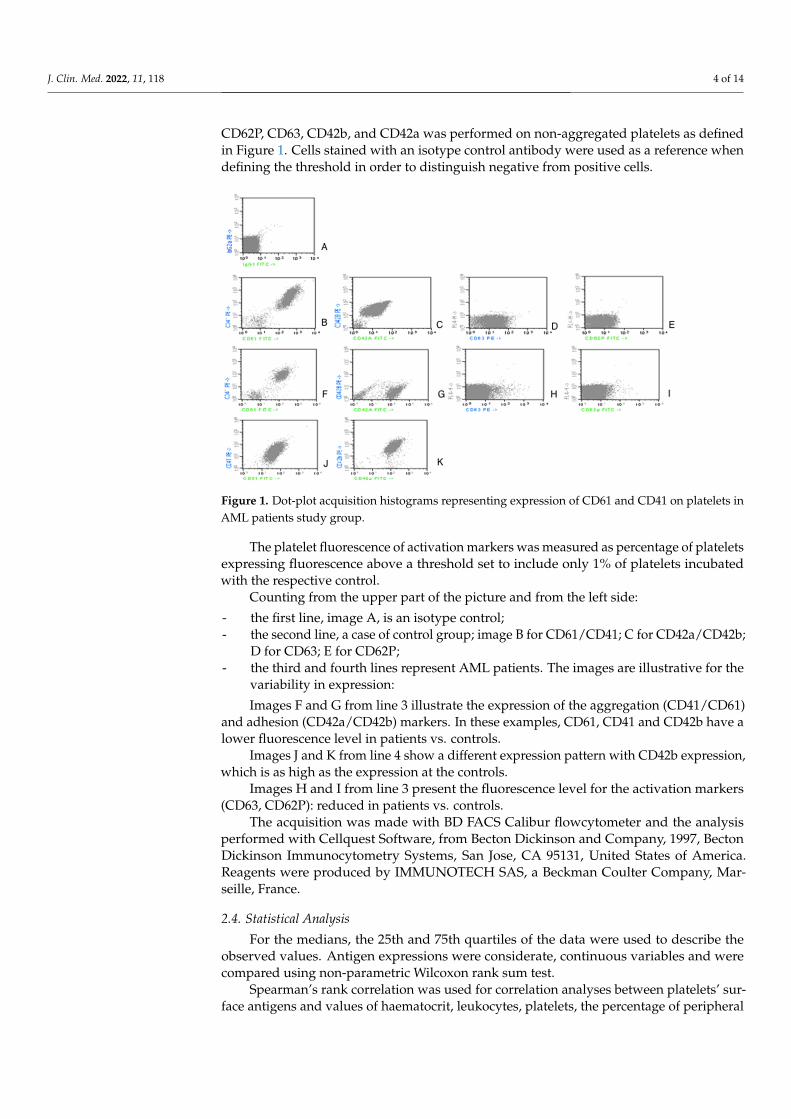
CD62P, CD63, CD42b, and CD42a was performed on non-aggregated platelets as definedin Figure 1. Cells stained with an isotype control antibody were used as a reference whendefining the threshold in order to distinguish negative from positive cells.
J. Clin. Med. 2022, 11, x FOR PEER REVIEW 4 of 15
2.3. Flow Cytometry Analysis of Platelets in Whole Blood The flow cytometry settings were optimized for the acquisition of platelets by loga-
rithmic signal amplification in all detectors. Electronic compensation was used to control for spectral overlap and the flow cytometer was aligned daily with CaliBriteTM beads (BDIS). A threshold on FL-1 was set to ensure that only platelet CD61-positive events were collected. A minimum of 5000 CD61-positive platelets was collected from each sample.
For analysis, an electronic gate enclosing platelets was set, as defined by forward scatter (FSC) and 90° side scatter (SSC) characteristics and CD61/CD41 platelet positivity markers, excluding platelet-leukocytes aggregates as well as platelet-erythrocyte and platelet-leukocytes doublets [18]. Further analysis of platelet activation markers CD62P, CD63, CD42b, and CD42a was performed on non-aggregated platelets as defined in Figure 1. Cells stained with an isotype control antibody were used as a reference when defining the threshold in order to distinguish negative from positive cells.
The platelet fluorescence of activation markers was measured as percentage of platelets expressing fluorescence above a threshold set to include only 1% of platelets incubated with the respective control.
Figure 1. Dot-plot acquisition histograms representing expression of CD61 and CD41 on platelets in AML patients study group.
Counting from the upper part of the picture and from the left side: - the first line, image A, is an isotype control; - the second line, a case of control group; image B for CD61/CD41; C for
CD42a/CD42b; D for CD63; E for CD62P; - the third and fourth lines represent AML patients. The images are illustrative for the
variability in expression: Images F and G from line 3 illustrate the expression of the aggregation (CD41/CD61)
and adhesion (CD42a/CD42b) markers. In these examples, CD61, CD41 and CD42b have a lower fluorescence level in patients vs. controls.
Images J and K from line 4 show a different expression pattern with CD42b expres-sion, which is as high as the expression at the controls.
Images H and I from line 3 present the fluorescence level for the activation markers (CD63, CD62P): reduced in patients vs. controls.
Figure 1. Dot-plot acquisition histograms representing expression of CD61 and CD41 on platelets inAML patients study group.
The platelet fluorescence of activation markers was measured as percentage of plateletsexpressing fluorescence above a threshold set to include only 1% of platelets incubatedwith the respective control.
Counting from the upper part of the picture and from the left side:
- the first line, image A, is an isotype control;- the second line, a case of control group; image B for CD61/CD41; C for CD42a/CD42b;
D for CD63; E for CD62P;- the third and fourth lines represent AML patients. The images are illustrative for the
variability in expression:
Images F and G from line 3 illustrate the expression of the aggregation (CD41/CD61)and adhesion (CD42a/CD42b) markers. In these examples, CD61, CD41 and CD42b have alower fluorescence level in patients vs. controls.
Images J and K from line 4 show a different expression pattern with CD42b expression,which is as high as the expression at the controls.
Images H and I from line 3 present the fluorescence level for the activation markers(CD63, CD62P): reduced in patients vs. controls.
The acquisition was made with BD FACS Calibur flowcytometer and the analysisperformed with Cellquest Software, from Becton Dickinson and Company, 1997, BectonDickinson Immunocytometry Systems, San Jose, CA 95131, United States of America.Reagents were produced by IMMUNOTECH SAS, a Beckman Coulter Company, Mar-seille, France.
2.4. Statistical Analysis
For the medians, the 25th and 75th quartiles of the data were used to describe theobserved values. Antigen expressions were considerate, continuous variables and werecompared using non-parametric Wilcoxon rank sum test.
Spearman’s rank correlation was used for correlation analyses between platelets’ sur-face antigens and values of haematocrit, leukocytes, platelets, the percentage of peripheral
J. Clin. Med. 2022, 11, 118 5 of 14
blasts, level of uric acid, creatinine, BUN, and body temperature, all considered continuousvariables. A general two-tailed significance level of 5% was applied.
Other clinical determinants, such as the presence of infection (localized or generalized)or cutaneous haemorrhagic syndrome, were analysed using crosstabs procedure.
All analyses were performed using SPSS statistical software, version 12.0 for Windows.
3. Results
The present study examines a group of 22 patients (13 females and 9 males) with agesranging between 28 and 83 years. At the time of the analysis, they were already diagnosedwith acute myeloid leukaemia and in different stages of disease.
Each patient was investigated by clinical examination, full blood count and peripheralblood smear with a detailed lymphocyte count. In every case, biochemistry samples (uricacid, creatinine, BUN) were taken and analysed.
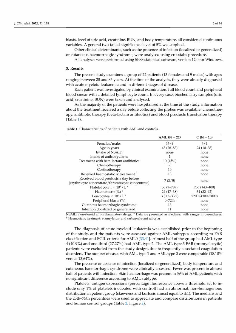
As the majority of the patients were hospitalised at the time of the study, informationabout the treatment received a day before collecting the probes was available: chemother-apy, antibiotic therapy (beta-lactam antibiotics) and blood products transfusion therapy(Table 1).
Table 1. Characteristics of patients with AML and controls.
AML (N = 22) C (N = 10)
Females/males 13/9 6/4Age in years 48 (28–83) 24 (10–38)
Intake of NSAID none noneIntake of anticoagulants 1 none
Treatment with beta-lactam antibiotics 10 (45%) noneChemotherapy 2 noneCorticotherapy 10 none
Received haemostatic iv treatment b 13 noneReceived blood products a day before
(erythrocyte concentrate/thrombocyte concentrate) 7 (2/5) none
Platelet count × 109/L a 50 (2–782) 256 (143–400)Haematocrit (%) a 24 (17–38) 34 (32–42)
Leucocytes × 109/L a 3 (0.5–33.7) 5200 (4300–7000)Peripheral blasts (%) 0–72% none
Cutaneus haemorrhagic syndrome 13 noneInfection (localized or generalized) 11 none
NSAID, non-steroid anti-inflammatory drugs; a Data are presented as medians, with ranges in parentheses;b Haemostatic treatment: etamsylatum and carbazochromi salicylas.
The diagnosis of acute myeloid leukaemia was established prior to the beginningof the study, and the patients were assessed against AML subtypes according to FABclassification and EGIL criteria for AML0 [33,41]. Almost half of the group had AML type4 (40.9%) and one-third (27.27%) had AML type 2. The AML type 3 FAB (promyelocytic)patients were excluded from the study design, due to frequently associated coagulationdisorders. The number of cases with AML type 1 and AML type 0 were comparable (18.18%versus 13.64%).
The presence or absence of infection (localized or generalized), body temperature andcutaneous haemorrhagic syndrome were clinically assessed. Fever was present in almosthalf of patients with infection. Skin haemorrhage was present in 59% of AML patients withno significant difference according to AML subtype.
Platelets’ antigen expressions (percentage fluorescence above a threshold set to in-clude only 1% of platelets incubated with control) had an abnormal, non-homogenousdistribution in patient group (skewness and kurtosis almost equal to ±1). The medians andthe 25th–75th percentiles were used to appreciate and compare distributions in patientsand human control groups (Table 2, Figure 2).
J. Clin. Med. 2022, 11, 118 6 of 14
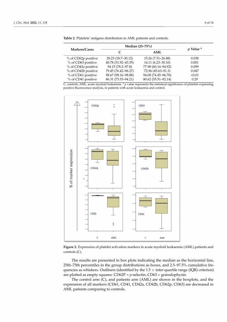
Table 2. Platelets’ antigens distribution in AML patients and controls.
Markers/CasesMedian (25–75%)
p Value *C AML
% of CD62p positive 28.23 (18.7–30.12) 15.26 (7.51–26.88) 0.038% of CD63 positive 40.78 (31.92–43.35) 14.11 (6.23–30.10) 0.001% of CD42a positive 94.15 (78.2–97.8) 77.98 (60.14–94.92) 0.099% of CD42b positive 79.45 (76.42–96.27) 72.96 (45.63–91.3) 0.047% of CD61 positive 98.67 (98.16–98.88) 94.08 (74.45–96.78) <0.01% of CD41 positive 86.31 (73.53–94.21) 80.62 (55.51–92.14) 0.29
C, controls; AML, acute myeloid leukaemia. * p value represents the statistical significance of platelets expressingpositive fluorescence analysis, in patients with acute leukaemia and control.
J. Clin. Med. 2022, 11, x FOR PEER REVIEW 6 of 15
The diagnosis of acute myeloid leukaemia was established prior to the beginning of the study, and the patients were assessed against AML subtypes according to FAB clas-sification and EGIL criteria for AML0 [33,41]. Almost half of the group had AML type 4 (40.9%) and one-third (27.27%) had AML type 2. The AML type 3 FAB (promyelocytic) patients were excluded from the study design, due to frequently associated coagulation disorders. The number of cases with AML type 1 and AML type 0 were comparable (18.18% versus 13.64%).
The presence or absence of infection (localized or generalized), body temperature and cutaneous haemorrhagic syndrome were clinically assessed. Fever was present in almost half of patients with infection. Skin haemorrhage was present in 59% of AML pa-tients with no significant difference according to AML subtype.
Platelets’ antigen expressions (percentage fluorescence above a threshold set to in-clude only 1% of platelets incubated with control) had an abnormal, non-homogenous distribution in patient group (skewness and kurtosis almost equal to ±1). The medians and the 25th–75th percentiles were used to appreciate and compare distributions in pa-tients and human control groups (Table 2, Figure 2).
Figure 2. Expression of platelet activation markers in acute myeloid leukaemia (AML) patients and controls (C).
The results are presented in box plots indicating the median as the horizontal line, 25th–75th percentiles in the group distributions as boxes, and 2.5–97.5% cumulative fre-
Figure 2. Expression of platelet activation markers in acute myeloid leukaemia (AML) patients andcontrols (C).
The results are presented in box plots indicating the median as the horizontal line,25th–75th percentiles in the group distributions as boxes, and 2.5–97.5% cumulative fre-quencies as whiskers. Outliners (identified by the 1.5 × inter-quartile range (IQR) criterion)are plotted as empty squares: CD62P = p-selectin; CD63 = granulophysin.
The control arm (C), and patients arm (AML) are shown in the boxplots, and theexpression of all markers (CD61, CD41, CD42a, CD42b, CD62p, CD63) are decreased inAML patients comparing to controls.
J. Clin. Med. 2022, 11, 118 7 of 14
3.1. Activation of Platelets in AML
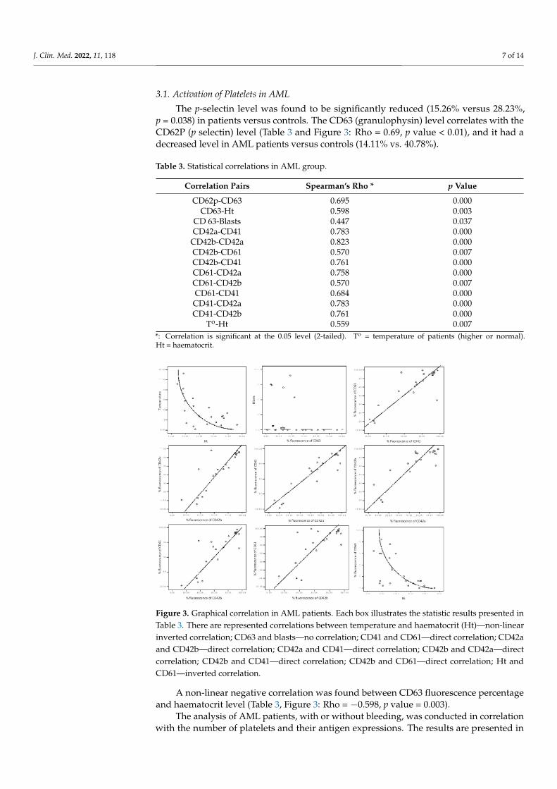
The p-selectin level was found to be significantly reduced (15.26% versus 28.23%,p = 0.038) in patients versus controls. The CD63 (granulophysin) level correlates with theCD62P (p selectin) level (Table 3 and Figure 3: Rho = 0.69, p value < 0.01), and it had adecreased level in AML patients versus controls (14.11% vs. 40.78%).
Table 3. Statistical correlations in AML group.
Correlation Pairs Spearman’s Rho * p Value
CD62p-CD63 0.695 0.000CD63-Ht 0.598 0.003
CD 63-Blasts 0.447 0.037CD42a-CD41 0.783 0.000
CD42b-CD42a 0.823 0.000CD42b-CD61 0.570 0.007CD42b-CD41 0.761 0.000CD61-CD42a 0.758 0.000CD61-CD42b 0.570 0.007CD61-CD41 0.684 0.000
CD41-CD42a 0.783 0.000CD41-CD42b 0.761 0.000
To-Ht 0.559 0.007*: Correlation is significant at the 0.05 level (2-tailed). To = temperature of patients (higher or normal).Ht = haematocrit.
J. Clin. Med. 2022, 11, x FOR PEER REVIEW 8 of 15
Figure 3. Graphical correlation in AML patients. Each box illustrates the statistic results presented in Table 3. There are represented correlations between temperature and haematocrit (Ht)—non-linear inverted correlation; CD63 and blasts—no correlation; CD41 and CD61—direct correlation; CD42a and CD42b—direct correlation; CD42a and CD41—direct correlation; CD42b and CD42a—direct correlation; CD42b and CD41—direct correlation; CD42b and CD61—direct correlation; Ht and CD61—inverted correlation.
The analysis of AML patients, with or without bleeding, was conducted in correla-tion with the number of platelets and their antigen expressions. The results are presented in Table 4. The medians and 25th–75th percentiles were used to appreciate and compare distributions in both groups (Figure 4). The median age was closely similar in both groups: 46 and 43 years old.
Table 4. Platelets’ antigens distribution in AML patients with or without bleeding.
Markers Cases and the Pres-ence of Hemorrhage
Median (25–75%)
p Value
% of CD62p positive AML NO AML YES
16.46 (7.38–47.32) 14.29 (0.86–65.18)
0.038
% of CD63 positive AML NO AML YES
16.78 (5.12–45.76) 13.44 (0.69–56.15)
0.001
% of CD42a positive AML NO AML YES
76.59 (1.44–95.81) 83.09 (35.49–98.28)
0.099
% of CD42b positive AML NO AML YES
76.35 (12.94–95.31) 63.53 (34.85–95.05
0.047
% of CD61 positive AML NO AML YES
87.51 (40.35–98.10) 94.25 (37.99–99.63)
<0.01
% of CD41 positive AML NO AML YES
73.19 (21.42–93.06) 77.33 (27.34–97.27)
0.029
Figure 3. Graphical correlation in AML patients. Each box illustrates the statistic results presented inTable 3. There are represented correlations between temperature and haematocrit (Ht)—non-linearinverted correlation; CD63 and blasts—no correlation; CD41 and CD61—direct correlation; CD42aand CD42b—direct correlation; CD42a and CD41—direct correlation; CD42b and CD42a—directcorrelation; CD42b and CD41—direct correlation; CD42b and CD61—direct correlation; Ht andCD61—inverted correlation.
A non-linear negative correlation was found between CD63 fluorescence percentageand haematocrit level (Table 3, Figure 3: Rho = −0.598, p value = 0.003).
The analysis of AML patients, with or without bleeding, was conducted in correlationwith the number of platelets and their antigen expressions. The results are presented in
J. Clin. Med. 2022, 11, 118 8 of 14
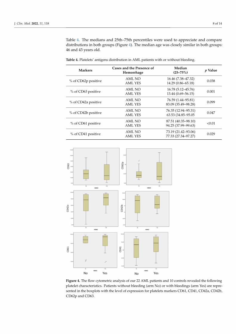
Table 4. The medians and 25th–75th percentiles were used to appreciate and comparedistributions in both groups (Figure 4). The median age was closely similar in both groups:46 and 43 years old.
Table 4. Platelets’ antigens distribution in AML patients with or without bleeding.
Markers Cases and the Presence ofHemorrhage
Median(25–75%) p Value
% of CD62p positive AML NOAML YES
16.46 (7.38–47.32)14.29 (0.86–65.18) 0.038
% of CD63 positive AML NOAML YES
16.78 (5.12–45.76)13.44 (0.69–56.15) 0.001
% of CD42a positive AML NOAML YES
76.59 (1.44–95.81)83.09 (35.49–98.28) 0.099
% of CD42b positive AML NOAML YES
76.35 (12.94–95.31)63.53 (34.85–95.05 0.047
% of CD61 positive AML NOAML YES
87.51 (40.35–98.10)94.25 (37.99–99.63) <0.01
% of CD41 positive AML NOAML YES
73.19 (21.42–93.06)77.33 (27.34–97.27) 0.029
J. Clin. Med. 2022, 11, x FOR PEER REVIEW 9 of 15
Figure 4. The flow cytometric analysis of our 22 AML patients and 10 controls revealed the fol-lowing platelet characteristics. Patients without bleeding (arm No) or with bleedings (arm Yes) are represented in the boxplots with the level of expression for platelets markers CD61, CD41, CD42a, CD42b, CD62p and CD63.
3.2. Von Willebrand Receptor in AML The CD42b expression decreases in patients versus controls (p = 0.047), while the
anti-CD42a antigen antibody, which reacts with the whole CD42a/CD42b complex, pre-sents no particular difference in the study group versus the control group.
3.3. Fibrinogen Receptor Gp IIb-IIIa, β3 integrin (CD61), the most frequent platelet surface antigen, presents a
significant decrease in fluorescence percentage levels (94.08% vs. 98.67%, p value < 0.01) in patients versus controls (Table 3).
An interesting relation between Gp IIb-IIIa and Gp Ib-IX occurred (Table 3). Even if the connection between the two protein complexes had a variable strength (Rho: 0.570–0.783), the p value < 0.01 indicated that, in patients with AML, the expression of these two platelet determinants was related, likely as a global defect of signal transduction.
3.4. Comparison between Patients with Bleeding and without Bleeding An important difference was identified in patients with bleeding, in comparison to
patients without bleeding, in the form of a decrease in Gp Ib-IX expression (CD42a/CD42b) and the activation of less platelets (CD62P and CD63), as shown in Fig-ure 4. This suggested a more severe acquired Bernard–Soulier syndrome and the block-ing of platelet activation, independent of patient age or the number of platelets. The ex-pression of fibrinogen receptor (CD61/CD41) was found with a higher level of expres-sion, even if a lower level of expression in this receptor was found in all AML patients
Figure 4. The flow cytometric analysis of our 22 AML patients and 10 controls revealed the followingplatelet characteristics. Patients without bleeding (arm No) or with bleedings (arm Yes) are repre-sented in the boxplots with the level of expression for platelets markers CD61, CD41, CD42a, CD42b,CD62p and CD63.
J. Clin. Med. 2022, 11, 118 9 of 14
3.2. Von Willebrand Receptor in AML
The CD42b expression decreases in patients versus controls (p = 0.047), while the anti-CD42a antigen antibody, which reacts with the whole CD42a/CD42b complex, presents noparticular difference in the study group versus the control group.
3.3. Fibrinogen Receptor
Gp IIb-IIIa, β3 integrin (CD61), the most frequent platelet surface antigen, presents asignificant decrease in fluorescence percentage levels (94.08% vs. 98.67%, p value < 0.01) inpatients versus controls (Table 3).
An interesting relation between Gp IIb-IIIa and Gp Ib-IX occurred (Table 3). Even if theconnection between the two protein complexes had a variable strength (Rho: 0.570–0.783),the p value < 0.01 indicated that, in patients with AML, the expression of these two plateletdeterminants was related, likely as a global defect of signal transduction.
3.4. Comparison between Patients with Bleeding and without Bleeding
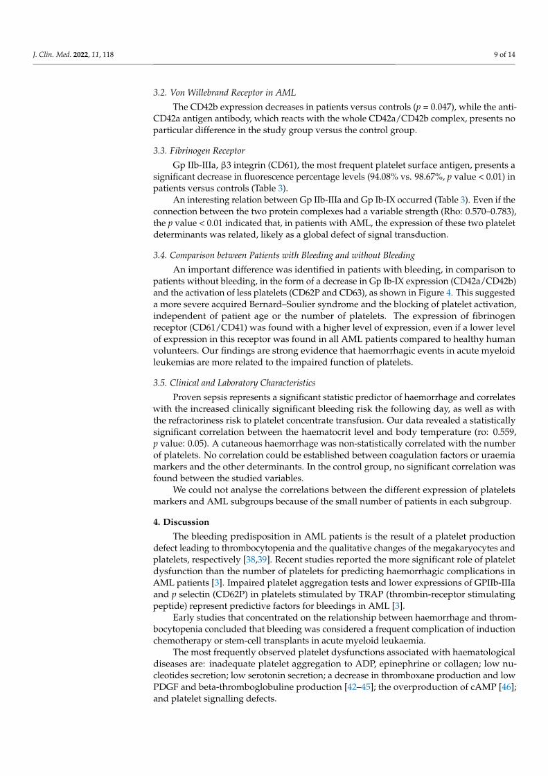
An important difference was identified in patients with bleeding, in comparison topatients without bleeding, in the form of a decrease in Gp Ib-IX expression (CD42a/CD42b)and the activation of less platelets (CD62P and CD63), as shown in Figure 4. This suggesteda more severe acquired Bernard–Soulier syndrome and the blocking of platelet activation,independent of patient age or the number of platelets. The expression of fibrinogenreceptor (CD61/CD41) was found with a higher level of expression, even if a lower levelof expression in this receptor was found in all AML patients compared to healthy humanvolunteers. Our findings are strong evidence that haemorrhagic events in acute myeloidleukemias are more related to the impaired function of platelets.
3.5. Clinical and Laboratory Characteristics
Proven sepsis represents a significant statistic predictor of haemorrhage and correlateswith the increased clinically significant bleeding risk the following day, as well as withthe refractoriness risk to platelet concentrate transfusion. Our data revealed a statisticallysignificant correlation between the haematocrit level and body temperature (ro: 0.559,p value: 0.05). A cutaneous haemorrhage was non-statistically correlated with the numberof platelets. No correlation could be established between coagulation factors or uraemiamarkers and the other determinants. In the control group, no significant correlation wasfound between the studied variables.
We could not analyse the correlations between the different expression of plateletsmarkers and AML subgroups because of the small number of patients in each subgroup.
4. Discussion
The bleeding predisposition in AML patients is the result of a platelet productiondefect leading to thrombocytopenia and the qualitative changes of the megakaryocytes andplatelets, respectively [38,39]. Recent studies reported the more significant role of plateletdysfunction than the number of platelets for predicting haemorrhagic complications inAML patients [3]. Impaired platelet aggregation tests and lower expressions of GPIIb-IIIaand p selectin (CD62P) in platelets stimulated by TRAP (thrombin-receptor stimulatingpeptide) represent predictive factors for bleedings in AML [3].
Early studies that concentrated on the relationship between haemorrhage and throm-bocytopenia concluded that bleeding was considered a frequent complication of inductionchemotherapy or stem-cell transplants in acute myeloid leukaemia.
The most frequently observed platelet dysfunctions associated with haematologicaldiseases are: inadequate platelet aggregation to ADP, epinephrine or collagen; low nu-cleotides secretion; low serotonin secretion; a decrease in thromboxane production and lowPDGF and beta-thromboglobuline production [42–45]; the overproduction of cAMP [46];and platelet signalling defects.
J. Clin. Med. 2022, 11, 118 10 of 14
The reduced level of CD63 and CD62P in our patients indicated two possible events:(1) no activation process took place or (2) there was a brief activation followed by rapid pselectin secretion.
In addition to the above-mentioned studies, which were focused on functional platelets’characteristics, our research performed on additional patient platelets that were not furtherstimulated, revealed the same low level of activation markers mentioned before. Theseresults raise the hypothesis of the lack of platelets activation in vivo, a comparable statewith the one found in vitro by the other experiments. Furthermore, the activation ofplatelets represents a vital first step in the clotting cascade.
The haematocrit role in platelets activation was previously investigated using in vitrostimulations [47]. Eugster M and Reinhart W found an inverse correlation between haemat-ocrit and a necessary amount of time for the formation of the platelet plug. Our results showan inversely proportional relation between the haematocrit and granulophysin expressionon platelets, which may be a false statistical result with no biological signification or afuture research perspective.
Specific heterozygous mutations affecting the hematopoietic transcription factorCBFA2 were previously reported to be associated with a familial platelet disorder pre-disposing to acute myeloid leukaemia [48]. Our study correlated the percentage of theperipheral blast cells with platelet-specific markers. Although peripheral blasts seemedstatistically related to CD63, this conclusion could not be graphically validated (Figure 3).This fact imposes a larger number of cases and, potentially, a different analytic strategy.
4.1. Von Willebrand Receptor in AML
The receptor for the von Willebrand Factor (CD42b) is one of the most major adhesivereceptors expressed on the surface of circulating platelets, able to interact with severalligands, including the adhesive protein von Willebrand factor, the coagulation factors(thrombin, factors XI and XII), and the membrane glycoproteins (p-selectin and Mac-1) [46].The reduced expression of CD42b in our patients might be interpreted as a particular reduc-tion in the α chain of GpIb due to the downregulation during platelet activation [49–51].Interestingly, Leinoe et al. [21] investigated if this decrease in CD42b in platelets underTRAP activation may occur because a downregulation of GpIb due to platelet activationin vivo. What we can say is that the same findings in vivo suggest that a broken activationmechanism could be involved in the expression of both p-selectin and granulophysin andthe decreasing of GpIb, considered to be a regulatory mechanism, which may witness thiseffort to activate a defective platelet.
4.2. Fibrinogen Receptor
The resting form of the CD41/CD61 complex binds to immobilized fibrinogen, and,upon platelet activation, the complex becomes a receptor for soluble fibrinogen, fibronectin,the von Willebrand Factor, vitronectin and thrombospondin [52]. This receptor is alsoinvolved in platelet aggregation [53,54]. The anti CD41 antigen antibody reacts with GpIIbin the intact GpIIb-IIIa complex, blocking fibrinogen and platelet aggregation inducedby thrombin, collagen and ADP. The reduction in the CD61 fluorescence level may beinterpreted as a selective decrease in the β3 subunit of GpIIIa, which may be a cause ofthe aggregation defect. A defective β3 integrin phosphorylation, associated with alteredoutside-in signalling, was observed [55]. This may lead to an increase in the haemorrhagicsyndrome of AML patients.
In AML, the low expression of GP1b on platelets could be explained by action ofelastase released from myeloid blasts [56]. The interaction between platelets and AMLblasts was proven by one study, and this interaction could produce functional changesand explain the presence of the dysfunctional platelet in AML patients [19]. A correlationbetween the low expression of CD62P, CD63 and the fibrinogen receptor and the high riskfor haemorrhage in the next 7 days of follow up for AML patients was reported recentlyin platelets stimulated by TRAP. In AML patients defective signalling pathways were
J. Clin. Med. 2022, 11, 118 11 of 14
found that could explain the low activation of GPIIbIIIa, an abnormal conformation ofthis activated receptor that influences its function [3,11]. In other studies, a correlationwas observed between the high expression of p selectin and GP IIbIIIa and haemorrhagichistory [20]. In AML patients, the MPL expression on blasts was correlated with theseverity of neutropenia and thrombocytopenia, especially in AML patients with t(8;21)genetic aberrancy [57].
4.3. Clinical and Laboratory Characteristics
The documented presence of the infection for the day before the study and/or thefever is associated with different types of bleeding, which can be clinically assessed asmild, significant or major. Recently, Vinholt et al. did not obtain any significant correlationbetween the infection and haemorrhagic risk of AML patients [3]. Gaydos et al. [58] foundcorrelations between platelet numbers and all types of bleedings (mild, clinically significantand major) in acute leukemia patients. Lawrence et al. [59] obtained no correlation betweenbleeding and the number of platelets, or the minimal number of the platelets. Otherstudies, which focused on the same issue, analysed this in association with the clinicalissue of prophylactic transfusion with platelet concentrate and stressed that the risk of animportant haemorrhagic complication still exists [60,61]. The absence of any correlationbetween cutaneous haemorrhage and the number of platelets, in addition to the above-described aberrant expression of phenotypic surface platelets markers, emphasizes the roleof functional and structural platelet defects in the haemorrhagic burst.
5. Conclusions
In our group of patients, we found a significant decrease in activation markers suchas p-selectin and granulophysin, associated with the decrease in GpIb and β3 integrin,demonstrating that, in AML, there could be multiple defects of signalling transduction,leading to defects of adhesion, aggregation and the secretion of platelets, consequentlyleading to haemorrhagic syndrome independent of the number of platelets in the peripheralblood. These data are complementary to other already published results in demonstratingthe multiple platelet defects of AML patients, suggesting that the risk of bleeding is directlycorrelated to the level of platelet defect.
In conclusion, the platelet behaviour in our study group offers important clues forpostulating that flow cytometry analysis could be a very important method to identifythose patients that are potential candidates to haemorrhagic disorders.
A limitation of our study is the small number of patients in the study group and thelack of genetic aberrancies data in AML patients, which could be of clinical interest infurther research. Additionally, the study was not designed as a longitudinal study, and afollow up study could be of interest for more information regarding haemorrhagic risk.
Author Contributions: H.B.: conception and design, flow cytometry data acquisition and writing of“Design and methods”, and Section 4, critical and responsible revision of the draft, guarantee of theintellectual content and final approval; A.M.V.: conception and design; S.S., V.M.P.: design, statisticalanalysis, writing of the Sections 1 and 3, drafting the Section 4, preparing the article for submission;I.D.: flow cytometry data acquisition, critical and responsible revision of the draft; D.C., I.V., C.C.,C.M., A.N. and M.O.: documentation process, gathering the clinical data, contribution to drafting theSections 1 and 4. All authors have read and agreed to the published version of the manuscript.
Funding: The current article was written based on the data collected during a National ResearchGrants in the Program “Excellence in Research” (CEEX)—MUL-TRO nr. 62/2005 and PARTE MPNProject nr. 141PED/2017-2018—funded by the Romanian Ministry of Research and Development.The study was carried out at the Departments of Haematology, Emergency University Hospital,“Carol Davila” University of Medicine and Pharmacy, Bucharest, Romania, and Colentina ClinicalHospital, Bucharest, Romania.
J. Clin. Med. 2022, 11, 118 12 of 14
Institutional Review Board Statement: The study was conducted in accordance with the Declarationof Helsinki, and approved by the Institutional Review Board no 29342/11 October 2005 and no 532/20January 2017 of Romanian Ministry of Research and Development.
Informed Consent Statement: The study was reviewed and approved by the Ethics Committee ofthe University Emergency Hospital in Bucharest, Romania. The approval codes are 29342/11 October2005 for first study. The second study approved for checking and improvement of methods has532/20 January 2017. Data were accessed anonymously. All methods were carried out in accordancewith the relevant guidelines and regulations.
Data Availability Statement: All data are available, either analysed as figures and tables presentedin the current manuscript; or as raw data upon request from any external collaborator or reviewer.
Conflicts of Interest: The authors declare no conflict of interest.
References1. Haase, D.; Feuring-Buske, M.; Konemann, S.; Fonatsch, C.; Troff, C.; Verbeek, W.; Pekrun, A.; Hiddemann, W.; Wormann, B.
Evidence for malignant transformation in acute myeloid leukemia at the level of early hematopoietic stem cells by cytogeneticanalysis of CD34+ subpopulations. Blood 1995, 86, 2906–2912. [CrossRef] [PubMed]
2. Grimwade, D.; Enver, T. Acute promyelocytic leukemia: Where does it stem from? Leukemia 2004, 18, 375–384. [CrossRef][PubMed]
3. Just Vinholt, P.; Højrup Knudsen, G.; Sperling, S.; Frederiksen, H.; Nielsen, C. Platelet function tests predict bleeding in patientswith acute myeloid leukemia and thrombocytopenia. Am. J. Hematol. 2019, 94, 891–901. [CrossRef]
4. Hatzl, S.; Uhl, B.; Hinterramskogler, M.; Leber, S.; Eisner, F.; Haring, M.; Jud, P. Acute myeloid leukemia with severe coagulationdisorder and concomitant central nervous system bleeding—A clinical diagnostic case report. Int. Fed. Clin. Chem. Lab. Med. 2018,29, 146–151.
5. Choudhry, A.; DeLoughery, T.G. Bleeding and thrombosis in acute promyelocytic leukemia. Am. J. Hematol. 2012, 87, 596–603.[CrossRef] [PubMed]
6. Matolcsy, A.; Kálmán, E.; Pajor, L.; Kónya, T.; Weber, E. Morphologic and flow cytometric analysis of circulating megakaryoblastsin chronic myeloid leukaemia. Leuk. Res. 1991, 15, 887–897. [CrossRef]
7. Wong, K.F.; Chan, J.K.C. Are ‘dysplastic’ and hypogranular megakaryocytes specific markers for myelodysplastic syndrome? Br.J. Haematol. 1991, 77, 509–514. [CrossRef] [PubMed]
8. Friedman, I.A.; Schwartz, S.O.; Leithold, S.L. Platelet Function Defects With Bleeding: Early Manifestation of Acute Leukemia.Arch. Intern. Med. 1964, 113, 177–185. [CrossRef]
9. Cowan, D.H.; Graham, R.C., Jr. Structural-functional relationships in platelets in acute leukemia and related disorders. Ser.Haematol. 1975, 8, 68–100.
10. Slichter, S.J. Relationship between platelet count and bleeding risk in thrombocytopenic patients. Transfus. Med. Rev. 2004, 18,153–167. [CrossRef]
11. Leinoe, E.B.; Hoffmann, M.H.; Kjaersgaard, E.; Nielsen, J.D.; Bergmann, O.J.; Klausen, T.W.; Johnsen, H.E. Prediction ofhaemorrhage in the early stage of acute myeloid leukaemia by flow cytometric analysis of platelet function. Br. J. Haematol. 2005,128, 526–532. [CrossRef]
12. Qian, X.; Wen-jun, L. Platelet changes in acute leukemia. Cell Biochem. Biophys. 2013, 67, 1473–1479. [CrossRef] [PubMed]13. Weyden, M.B.V.D.; Clancy, R.L.; Howard, M.A.; Firkin, B.G. Qualitative Platelet Defects with Reduced Life-Span in Acute
Leukaemia. Aust. N. Z. J. Med. 1972, 2, 339–345. [CrossRef]14. Maldonado, J.E.; Pierre, R.V. The platelets in preleukemia and myelomonocytic leukemia. Ultrastructural cytochemistry and
cytogenetics. Mayo Clin. Proc. 1975, 50, 573–587. [PubMed]15. Ganguly, P.; Sutherland, S.B.; Bradford, H.R. Defective binding of thrombin to platelets in myeloid leukaemia. Br. J. Haematol.
1978, 39, 599–605. [CrossRef] [PubMed]16. Woodcock, B.E.; Cooper, P.C.; Brown, P.R.; Pickering, C.; Winfield, D.A.; Preston, F.E. The platelet defect in acute myeloid
leukaemia. J. Clin. Pathol. 1984, 37, 1339–1342. [CrossRef] [PubMed]17. Vinholt, P.J. The role of platelets in bleeding in patients with thrombocytopenia and hematological disease. Clin. Chem. Lab. Med.
2019, 57, 1808–1817. [CrossRef]18. Matzdorff, A.C.; Kühnel, G.; Kemkes-Matthes, B.; Pralle, H. Quantitative assessment of platelets, platelet microparticles, and
platelet aggregates with flow cytometry. J. Lab. Clin. Med. 1998, 131, 507–517. [CrossRef]19. Foss, B.; Ulvestad, E.; Hervig, T.; Bruserud, Ø. Effects of cytarabine and various anthracyclins on platelet activation: Character-
ization of in vitro effects and their possible clinical relevance in acute myelogenous leukemia. Int. J. Cancer 2002, 97, 106–114.[CrossRef]
20. Psaila, B.; Bussel, J.B.; Frelinger, A.L.; Babula, B.; Linden, M.D.; Li, Y.; Barnard, M.R.; Tate, C.; Feldman, E.J.; Michelson, A.D. Dif-ferences in platelet function in patients with acute myeloid leukemia and myelodysplasia compared to equally thrombocytopenicpatients with immune thrombocytopenia. J. Thromb. Haemost. 2011, 9, 2302–2310. [CrossRef]
J. Clin. Med. 2022, 11, 118 13 of 14
21. Leinoe, E.B.; Hoffmann, M.H.; Kjaersgaard, E.; Johnsen, H.E. Multiple platelet defects identified by flow cytometry at diagnosisin acute myeloid leukaemia. Br. J. Haematol. 2004, 127, 76–84. [CrossRef]
22. Sandes, A.F.; Yamamoto, M.; Matarraz, S.; Chauffaille Mde, L.; Quijano, S.; López, A.; Oguro, T.; Kimura, E.Y.; Orfao, A.Altered immunophenotypic features of peripheral blood platelets in myelodysplastic syndromes. Haematologica 2012, 97, 895–902.[CrossRef]
23. Webert, K.; Cook, R.J.; Sigouin, C.S.; Rebulla, P.; Heddle, N.M. The risk of bleeding in thrombocytopenic patients with acutemyeloid leukemia. Haematologica 2006, 91, 1530–1537. [PubMed]
24. Böck, M.; Muggenthaler, K.H.; Schmidt, U.; Heim, M.U. Influence of antibiotics on posttransfusion platelet increment. Transfusion1996, 36, 952–954. [CrossRef] [PubMed]
25. Slichter, S.J.; Davis, K.; Enright, H.; Braine, H.; Gernsheimer, T.; Kao, K.J.; Kickler, T.; Lee, E.; McFarland, J.; McCullough, J.; et al.Factors affecting posttransfusion platelet increments, platelet refractoriness, and platelet transfusion intervals in thrombocytopenicpatients. Blood 2005, 105, 4106–4114. [CrossRef] [PubMed]
26. Friedmann, A.M.; Sengul, H.; Lehmann, H.; Schwartz, C.; Goodman, S. Do basic laboratory tests or clinical observations predictbleeding in thrombocytopenic oncology patients? A reevaluation of prophylactic platelet transfusions. Transfus. Med. Rev. 2002,16, 34–45. [CrossRef]
27. Livio, M.; Gotti, E.; Marchesi, D.; Mecca, G.; Remuzzi, G.; de Gaetano, G. Uraemic bleeding: Role of anaemia and beneficial effectof red cell transfusions. Lancet 1982, 2, 1013–1015. [CrossRef]
28. Fernandez, F.; Goudable, C.; Sie, P.; Ton-That, H.; Durand, D.; Suc, J.M.; Boneu, B. Low haematocrit and prolonged bleeding timein uraemic patients: Effect of red cell transfusions. Br. J. Haematol. 1985, 59, 139–148. [CrossRef]
29. Blajchman, M.A.; Bordin, J.O.; Bardossy, L.; Heddle, N.M. The contribution of the haematocrit to thrombocytopenic bleeding inexperimental animals. Br. J. Haematol. 1994, 86, 347–350. [CrossRef]
30. Small, M.; Lowe, G.D.; Cameron, E.; Forbes, C.D. Contribution of the haematocrit to the bleeding time. Haemostasis 1983, 13,379–384. [CrossRef]
31. Swerdlow, S.H.; Campo, E.; Harris, N.L.; Jaffe, E.S.; Pileri, S.A.; Stein, H.; Thiele, J.; Vardiman, J.W. WHO Classification of Tumoursof Haematopoietic and Lymphoid Tissues; International Agency for Research on Cancer: Lyon, France, 2008; Volume 2.
32. Bennett, J.M.; Catovsky, D.; Daniel, M.T.; Flandrin, G.; Galton, D.A.; Gralnick, H.R.; Sultan, C. Proposals for the classification ofthe acute leukaemias. French-American-British (FAB) co-operative group. Br. J. Haematol. 1976, 33, 451–458. [CrossRef]
33. Segeren, C.M.; van‘t Veer, M.B. The FAB classification for acute myeloid leukaemia—Is it outdated? Neth. J. Med. 1996, 49, 126–131.[CrossRef]
34. CD61. Beckmann Coulter Catalog. Available online: https://www.beckman.com/reagents/coulter-flow-cytometry/antibodies-and-kits/single-color-antibodies/cd61 (accessed on 29 October 2021).
35. Schlossman, S.F. Leucocyte Typing V: White Cell Differentiation Antigens, Proceedings of the Fifth International Workshop and Conference,Boston, MA, USA, 3–7 November 1993; Oxford University Press: Oxford, NY, USA, 1995.
36. McEver, R.P. Properties of GMP-140, an inducible granule membrane protein of platelets and endothelium. Blood Cells 1990, 16,73–80.
37. Palabrica, T.; Lobb, R.; Furie, B.C.; Aronovitz, M.; Benjamin, C.; Hsu, Y.-M.; Sajer, S.A.; Furie, B. Leukocyte accumulationpromoting fibrin deposition is mediated in vivo by P-selectin on adherent platelets. Nature 1992, 359, 848–851. [CrossRef][PubMed]
38. Subramaniam, M.; Frenette, P.S.; Saffaripour, S.; Johnson, R.C.; Hynes, R.O.; Wagner, D.D. Defects in hemostasis in P-selectin-deficient mice. Blood 1996, 87, 1238–1242. [CrossRef] [PubMed]
39. Falati, S.; Liu, Q.; Gross, P.; Merrill-Skoloff, G.; Chou, J.; Vandendries, E.; Celi, A.; Croce, K.; Furie, B.C.; Furie, B. Accumulationof tissue factor into developing thrombi in vivo is dependent upon microparticle P-selectin glycoprotein ligand 1 and plateletP-selectin. J. Exp. Med. 2003, 197, 1585–1598. [CrossRef]
40. Israels, S.J.; McMillan-Ward, E.M. CD63 modulates spreading and tyrosine phosphorylation of platelets on immobilized fibrinogen.Thromb. Haemost. 2005, 93, 311–318. [CrossRef]
41. Bene, M.C.; Castoldi, G.; Knapp, W.; Ludwig, W.D.; Matutes, E.; Orfao, A.; van’t Veer, M.B. Proposals for the immunologicalclassification of acute leukemias. European Group for the Immunological Characterization of Leukemias (EGIL). Leukemia 1995, 9,1783–1786.
42. Raman, B.K.S.; Van Slyck, E.J.; Riddle, J.; Sawdyk, M.A.; Abraham, J.P.; Saeed, S.M. Platelet Function and Structure in Myelo-proliferative Disease, Myelodysplastic Syndrome, and Secondary Thrombocytosis. Am. J. Clin. Pathol. 1989, 91, 647–655.[CrossRef]
43. Russell, N.H.; Salmon, J.; Keenan, J.P.; Bellingham, A.J. Platelet adenine nucleotides and arachidonic acid metabolism in themyeloproliferative disorders. Thromb. Res. 1981, 22, 389–397. [CrossRef]
44. Cowan, D.H.; Haut, M.J. Platelet function in acute leukemia. J. Lab. Clin. Med. 1972, 79, 893–905.45. Cowan, D.H.; Graham, R.C., Jr.; Baunach, D. The platelet defect in leukemia. Platelet ultrastructure, adenine nucleotide
metabolism, and the release reaction. J. Clin. Investig. 1975, 56, 188–200. [CrossRef]46. Lecchi, A.; Femia, E.A.; La Marca, S.; Onida, F.; Artoni, A. Acquired platelet dysfunction and overproduction of platelet cyclic
AMP in two patients with myeloid malignancies. Platelets 2019, 30, 1053–1056. [CrossRef]
J. Clin. Med. 2022, 11, 118 14 of 14
47. Eugster, M.; Reinhart, W.H. The influence of the haematocrit on primary haemostasis in vitro. Thromb. Haemost. 2005, 94,1213–1218. [CrossRef] [PubMed]
48. Buijs, A.; Poddighe, P.; van Wijk, R.; van Solinge, W.; Borst, E.; Verdonck, L.; Hagenbeek, A.; Pearson, P.; Lokhorst, H. A novelCBFA2 single-nucleotide mutation in familial platelet disorder with propensity to develop myeloid malignancies. Blood 2001, 98,2856–2858. [CrossRef]
49. Canobbio, I.; Balduini, C.; Torti, M. Signalling through the platelet glycoprotein Ib-V-IX complex. Cell. Signal. 2004, 16, 1329–1344.[CrossRef] [PubMed]
50. Michelson, A.D. Thrombin-induced down-regulation of the platelet membrane glycoprotein Ib-IX complex. Semin. Thromb.Hemost. 1992, 18, 18–27. [CrossRef]
51. Cahill, M.R.; Macey, M.G.; Newland, A.C. Fixation with formaldehyde induces expression of activation dependent plateletmembrane glycoproteins, P selectin (CD62) and GP53 (CD63). Br. J. Haematol. 1993, 84, 527–529. [CrossRef]
52. Bennett, J.S. Structure and function of the platelet integrin αIIbβ3. J. Clin. Investig. 2005, 115, 3363–3369. [CrossRef] [PubMed]53. Nurden, P. Bidirectional trafficking of membrane glycoproteins following platelet activation in suspension. Thromb. Haemost.
1997, 78, 1305–1315. [CrossRef] [PubMed]54. George, J.N.; Caen, J.P.; Nurden, A.T. Glanzmann’s thrombasthenia: The spectrum of clinical disease. Blood 1990, 75, 1383–1395.
[CrossRef] [PubMed]55. Glembotsky, A.C.; Bluteau, D.; Espasandin, Y.R.; Goette, N.P.; Marta, R.F.; Marin Oyarzun, C.P.; Korin, L.; Lev, P.R.; Laguens,
R.P.; Molinas, F.C.; et al. Mechanisms underlying platelet function defect in a pedigree with familial platelet disorder with apredisposition to acute myelogenous leukemia: Potential role for candidate RUNX1 targets. J. Thromb. Haemost. 2014, 12, 761–772.[CrossRef] [PubMed]
56. Aziz, K.A. An acquired form of Bernard Soulier syndrome associated with acute myeloid leukemia. Saudi Med. J. 2005, 26,1095–1098. [PubMed]
57. Rauch, P.J.; Ellegast, J.M.; Widmer, C.C.; Fritsch, K.; Goede, J.S.; Valk, P.J.; Löwenberg, B.; Takizawa, H.; Manz, M.G. MPLexpression on AML blasts predicts peripheral blood neutropenia and thrombocytopenia. Blood 2016, 128, 2253–2257. [CrossRef][PubMed]
58. Gaydos, L.A.; Freireich, E.J.; Mantel, N. The quantitative relation between platelet count and hemorrhage in patients with acuteleukemia. N. Engl. J. Med. 1962, 266, 905–909. [CrossRef] [PubMed]
59. Lawrence, J.B.; Yomtovian, R.A.; Dillman, C.; Masarik, S.R.; Chongkolwatana, V.; Creger, R.J.; Manka, A.; Hammons, T.; Lazarus,H.M. Reliability of automated platelet counts: Comparison with manual method and utility for prediction of clinical bleeding.Am. J. Hematol. 1995, 48, 244–250. [CrossRef]
60. Rebulla, P.; Finazzi, G.; Marangoni, F.; Avvisati, G.; Gugliotta, L.; Tognoni, G.; Barbui, T.; Mandelli, F.; Sirchia, G. The thresholdfor prophylactic platelet transfusions in adults with acute myeloid leukemia. Gruppo Italiano Malattie Ematologiche Malignedell’Adulto. N. Engl. J. Med. 1997, 337, 1870–1875. [CrossRef]
61. McCullough, J.; Vesole, D.H.; Benjamin, R.J.; Slichter, S.J.; Pineda, A.; Snyder, E.; Stadtmauer, E.A.; Lopez-Plaza, I.; Coutre, S.;Strauss, R.G.; et al. Therapeutic efficacy and safety of platelets treated with a photochemical process for pathogen inactivation:The Sprint Trial. Blood 2004, 104, 1534–1541. [CrossRef]


