Embed Size (px)
Citation preview
Molecular Pathogenesis of Genetic and Inherited Diseases
Plectin Regulates the Organization of Glial FibrillaryAcidic Protein in Alexander Disease
Rujin Tian,* Martin Gregor,† Gerhard Wiche,† andJames E. Goldman‡
From the Department of Physiology and Cellular Biophysics * and
the Department of Pathology and The Center for Neurobiology
and Behavior,‡ Columbia University, New York, New York; and
the Institute of Biochemistry and Molecular Cell Biology,†
University of Vienna, Vienna, Austria
Alexander disease (AxD) is a rare but fatal neurolog-ical disorder caused by mutations in the astrocyte-specific intermediate filament protein glial fibrillaryacidic protein (GFAP). Histologically, AxD is charac-terized by cytoplasmic inclusion bodies calledRosenthal fibers (RFs), which contain GFAP, smallheat shock proteins, and other undefined compo-nents. Here, we describe the expression of the cy-toskeletal linker protein plectin in the AxD brain. RFsdisplayed positive immunostaining for plectin andGFAP, both of which were increased in the AxD brain.Co-localization, co-immunoprecipitation, and in vitrooverlay analyses demonstrated direct interaction ofplectin and GFAP. GFAP with the most common AxDmutation, R239C (RC GFAP), mainly formed abnor-mal aggregates in human primary astrocytes and mu-rine plectin-deficient fibroblasts. Transient transfec-tion of full-length plectin cDNA converted theseaggregates to thin filaments, which exhibited diffusecytoplasmic distribution. Compared to wild-typeGFAP expression, RC GFAP expression lowered plec-tin levels in astrocytoma-derived stable transfectantsand plectin-positive fibroblasts. A much higher pro-portion of total GFAP was found in the Triton X-insol-uble fraction of plectin-deficient fibroblasts than inwild-type fibroblasts. Taken together, our results sug-gest that insufficient amounts of plectin, due to RCGFAP expression, promote GFAP aggregation and RFformation in AxD. (Am J Pathol 2006, 168:888–897; DOI:10.2353/ajpath.2006.051028)
Glial fibrillary acidic protein (GFAP) is a member of thetype III intermediate filament (IF) protein family and haslong been viewed as the characteristic cytoskeletal pro-
tein of astrocytes in the central nervous system (CNS).Alexander disease (AxD) is a rare but devastating disor-der of cortical white matter that predominantly affectsinfants and children. Neurological symptoms often in-clude macrocephaly and episodes of severe seizures,leading to progressive disability or death within the firstdecade of life.1 Pathologically, AxD is a type of leukodys-trophy characterized by a loss of oligodendrocytes andthe presence in astrocytes of numerous eosinophilic cy-toplasmic aggregates called Rosenthal fibers (RFs). Ma-jor molecular components of RFs include GFAP itself andthe small heat shock proteins �B crystallin and hsp27.2
Most individuals with AxD carry missense mutations inone allele of the coding region of the GFAP gene.3,4 Ourstudy focused on the most frequent and severe type ofAxD mutation, R239C (RC) GFAP.
Plectin, a member of the plakin family of cytolinkerproteins, was originally identified as an abundant IF-associated protein in C6 glioma cells5 and has subse-quently been characterized as a ubiquitously expressedcytoskeletal cross-linker.6 It is localized at the cytoskele-ton-plasma membrane interface and binds IFs, actin mi-crofilaments, microtubules, and integrins. Plectin has thecapacity to bridge multiple cellular structures, includingsarcolemma and the Z-lines of the skeletal muscle,hemidesmosomes and focal adhesion contacts.7 Muta-tions of the human plectin gene cause autosomal reces-sive epidermolysis bullosa simplex, a skin blistering dis-order accompanied by muscular dystrophy.8 Plectinknockout mice have a similar phenotype, demonstratingan important role of plectin in maintaining the structuralintegrity of skin and muscle.9
Given that a number of studies have suggested thatplectin is important in the assembly and dynamics of thecytoskeleton networks in vascular and epithelial struc-tures and in keratin-related diseases,10 we predicted thata similar association between GFAP and plectin occurredin the formation of GFAP aggregates and RFs. We there-
Supported by the National Institutes of Health (program project for cellpathology of Alexander Disease grant NS42803).
Accepted for publication November 7, 2005.
Address reprint requests to James E. Goldman, Columbia University,Department of Pathology and The Center for Neurobiology and Behavior,630 W. 168th St., New York, NY 10032. E-mail: [email protected].
American Journal of Pathology, Vol. 168, No. 3, March 2006
Copyright © American Society for Investigative Pathology
DOI: 10.2353/ajpath.2006.051028
888
fore examined plectin expression in the CNS of AxDpatients. We found that plectin levels were significantlyelevated in the Triton X-100 (TX)-insoluble fraction of theAxD brain tissues. Immunohistochemical staining of plec-tin in brain sections showed a characteristic rim stainingof RFs. The interaction was further confirmed in astro-cytes expressing RC GFAP as well as in immortalizedfibroblasts derived from plectin (�/�) mice.
Materials and Methods
DNA Cloning
To construct N-terminally GFP- or FLAG-tagged versionsof wild-type (WT) and R239C (RC) GFAP, we used themodified pcDNA3 plasmid containing a FLAG sequence(Ron Liem, Columbia University, New York, NY) andpeGFP-N1 (Clontech, Palo Alto, CA). The full GFAP cod-ing region was amplified and subcloned into the modifiedpcDNA3 and peGFP-N1 plasmids between the EcoRIand the XhoI sites. All plasmid inserts were fully se-quenced. The c-myc-tagged full-length plectin (PBN165)was described previously.11
Antibodies
Primary antibodies included mouse anti-GFAP monoclonalantibody (mAb 3402; Chemicon, Temecula, CA), guineapig anti-human plectin C-terminal polyclonal antibody (Re-search Diagnostics, Venecia, CA), and goat anti-c-mycpolyclonal antibody (sc-789-G; Santa Cruz Biotechnology,Santa Cruz, CA). Secondary antibodies included rabbitanti-goat IgG (H�L)-horseradish peroxidase (SouthernBiotechnology, Birmingham, AL), fluorescein isothiocya-nate-conjugated anti-mIgG1 (Southern Biotechnology), tet-ramethyl-rhodamine isothiocyanate-conjugated anti-rabbit(Chemicon), tetramethyl-rhodamine isothiocyanate-conju-gated anti-guinea pig (Research Diagnostics), andhorseradish peroxidase-conjugated goat anti-guinea pig(provided by Dr. Taewan Kim, Columbia University, NewYork, NY).
Cells, Transfection, and IF Staining
Human primary astrocytes were a gift from Dr. Paul Fisherat Columbia University, New York, NY.12 Ple (�/�) and(�/�) immortalized fibroblasts were derived from plectinWT and knockout mice, respectively. Cells were culturedin 5% fetal calf serum in 50% Dulbecco’s modified Ea-gle’s medium-F12 medium supplemented with 50 �g/mleach of streptomycin and penicillin. Transient transfec-tions were performed in serum-free medium using Lipo-fectamine 2000 reagents (Invitrogen, Carlsbad, CA).
For indirect immunofluorescence microscopy, cellswere grown on 22-mm glass coverslips. Twenty-fourhours after transfection, cells were fixed in 4% parafor-maldehyde for 10 to 15 minutes and subjected to indirectfluorescence/confocal laser-scanning microscopy (LSM510 META, 100X, 1.4 N.A; Zeiss, Thornwood, NY). All
images are projections generated from confocal serialsections of fluorescently labeled cells.
Human Brain Specimen Treatment, TXExtraction, and Immunoblotting
Frozen brain specimens of two children (control 1 andcontrol 2) without any history of neurological or psychiat-ric disorder and one AxD child (R239C, RC) with a similarage were obtained at autopsy with postmortem intervalswithin 12 hours. Total tissue lysates were prepared fromwhite matter homogenates of each brain by incubation for10 minutes in sodium dodecyl sulfate-polyacrylamide gelelectrophoresis (SDS-PAGE) loading buffer (62.5 mmol/LTris-HCl, 5% SDS, 10% glycerol, 20% �-mercaptoetha-nol, pH 6.8) at 95°C (T: total tissue lysate; 15 �g per lane).In certain experiments, these tissues were extracted withice-cold TX buffer (1% TX, 50 mmol/L Tris-HCl, 100mmol/L NaCl, pH 7.4) in the presence of protease inhib-itors (Complete protease inhibitor cocktail; Roche, India-napolis, IN). Tissue lysates were centrifuged for 10 min-utes at 4°C and 15,000 � g. TX-insoluble pellets werewashed twice with phosphate-buffered saline and thendenatured in SDS-PAGE loading buffer (P: cytoskeletalfraction; 10 �g per lane). The TX-soluble proteins weredenatured in SDS-PAGE loading buffer (S: TX-solublefraction; 30 �g per lane). After electrophoresis through6% or 4 to 20% gradient Tris-bis polyacrylamide gels(Invitrogen) at 20 mA for 16 hours or 150 V for 2 hours, theseparated proteins were transferred onto nitrocellulosemembranes and immunostained with primary antibodiesand 1:10,000 dilution of horseradish peroxidase-conju-gated secondary antibodies. Reactions were visualizedwith an ECL kit (Amersham, Arlington Heights, IL).
Immunohistochemical Staining
Deparaffinized brain sections were incubated at roomtemperature in 1% H2O2 for 10 minutes and 0.5% NaBH4
for 5 minutes to reduce background. Sections were thenplaced in blocking buffer [0.3% Triton X-100, 1% bovineserum albumin (BSA), and 5% goat serum in phosphate-buffered saline] for 2 hours and incubated overnight inguinea pig polyclonal antibody to plectin (1:200) in thesame buffer. All sections were incubated with species-specific biotinylated secondary antibodies (VectorLaboratories, Burlingame, CA). The avidin-biotin im-munoperoxidase method with 3,3-diaminobenzidinetetrahydrochloride was used to visualize immunoreactivecells (ABC kits, Vector Laboratories). Sufficient washeswith phosphate-buffered saline were performed betweeneach incubation step. Control sections, immunostainedby the same procedure but with the omission of primaryantibodies, were included in each batch of labeling. Sec-tions with plectin labeling were examined using an Olym-pus BX60 microscope.
Plectin and GFAP in Alexander Disease 889AJP March 2006, Vol. 168, No. 3
Co-Immunoprecipitation
Cells lysates were prepared 24 hours after transfectionusing IP buffer (10 mmol/L Tris, pH 7.4, 1.0% TX, 0.5%Nonidet P-40, 150 mmol/L NaCl, 20 mmol/L NaF, 0.2mmol/L sodium orthovanadate, 1 mmol/L EDTA, 1mmol/L EGTA, 0.2 mmol/L phenylmethyl sulfonyl fluo-ride) and centrifuged at 4°C and 15,000 � g for 10minutes. Next, 200 �g of protein from the supernatantwere incubated with rocking for 5 hours at 4°C with 1�g of GFAP antibody crosslinked to protein A Sepha-rose-conjugated beads (Sigma Chemical Co., St.Louis, MO). The beads were washed three times withIP buffer, and antigen-antibody complexes were elutedwith SDS sample buffer and analyzed by immunoblot-ting as described above.
In Vitro Overlay Assay
Bacteria were transformed with PET-15b (Novagen, Mad-ison, WI)-based plasmids (modified to encode C-terminalHis-tag). BN205 and BN192 encode a part of rat plectin(P30427) C-terminal domain (L3850 to A4687) and a partof plectin �-helical rod domain (E2235 to Q2577), respec-tively. Corresponding proteins were expressed and puri-fied on Ni-chelating resins. Bacterially expressed WTGFAP and RC GFAP proteins were tagless and purifiedby high performance liquid chromatography. For overlayassays, 2 to 3 �g of BN205, BSA, WT GFAP, and RCGFAP proteins were separated on 10% polyacrylamidegels, transferred to nitrocellulose, and blocked overnightwith 4% BSA in Tris-buffered saline-Tween 20 (0.1%).Blots were overlaid with BN205 and BN192 plectin re-combinant proteins diluted directly from elution fractions(20 mmol/L Tris, 500 mmol/L NaCl, 6 mol/L urea, 500mmol/L imidazol) 1:1000 in Tris-buffered saline-Tween 20(0.1%), containing 1 mmol/L EGTA, 2 mmol/L MgCl2, and1 mmol/L dithiothreitol, giving the final protein concentra-tion of 0.5 �g/ml. The assay was performed for 2 hours atroom temperature. Bound proteins were detected withcell culture supernatant of myc monoclonal antibody,diluted 1:100, followed by horseradish peroxidase-con-jugated goat antibodies against mouse IgGs (Jackson)diluted 1:150,000. Signal was obtained after incubationwith ECL (Pierce, Rockford, IL) substrate and exposure toX-ray film.
Statistical Methods
The percentage of cells with characteristic GFAP pheno-types was quantified by randomly choosing visual fieldsin the fluorescence microscope and counting 150 cellsper microscope slide (�40 objective). Results are ex-pressed as means � SEM. Each experiment was per-formed three times with similar results. Statistical analysiswas performed using the Student’s t-test. P values lessthan 0.05 were considered significant.
Results
Plectin Is Associated with RFs: Evidence fromBrain Sections and Tissues of AxD Patients
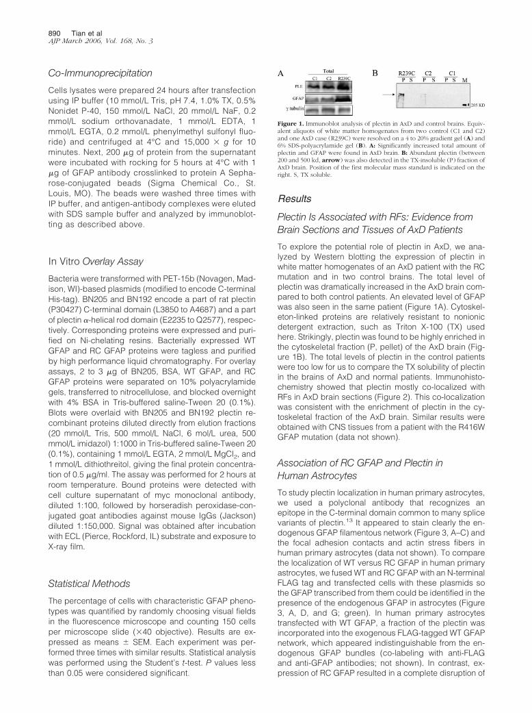
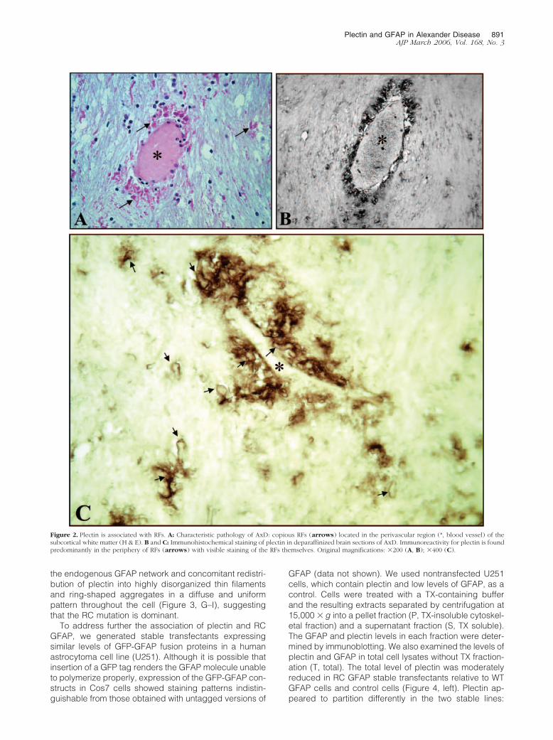
To explore the potential role of plectin in AxD, we ana-lyzed by Western blotting the expression of plectin inwhite matter homogenates of an AxD patient with the RCmutation and in two control brains. The total level ofplectin was dramatically increased in the AxD brain com-pared to both control patients. An elevated level of GFAPwas also seen in the same patient (Figure 1A). Cytoskel-eton-linked proteins are relatively resistant to nonionicdetergent extraction, such as Triton X-100 (TX) usedhere. Strikingly, plectin was found to be highly enriched inthe cytoskeletal fraction (P, pellet) of the AxD brain (Fig-ure 1B). The total levels of plectin in the control patientswere too low for us to compare the TX solubility of plectinin the brains of AxD and normal patients. Immunohisto-chemistry showed that plectin mostly co-localized withRFs in AxD brain sections (Figure 2). This co-localizationwas consistent with the enrichment of plectin in the cy-toskeletal fraction of the AxD brain. Similar results wereobtained with CNS tissues from a patient with the R416WGFAP mutation (data not shown).
Association of RC GFAP and Plectin inHuman Astrocytes
To study plectin localization in human primary astrocytes,we used a polyclonal antibody that recognizes anepitope in the C-terminal domain common to many splicevariants of plectin.13 It appeared to stain clearly the en-dogenous GFAP filamentous network (Figure 3, A–C) andthe focal adhesion contacts and actin stress fibers inhuman primary astrocytes (data not shown). To comparethe localization of WT versus RC GFAP in human primaryastrocytes, we fused WT and RC GFAP with an N-terminalFLAG tag and transfected cells with these plasmids sothe GFAP transcribed from them could be identified in thepresence of the endogenous GFAP in astrocytes (Figure3, A, D, and G; green). In human primary astrocytestransfected with WT GFAP, a fraction of the plectin wasincorporated into the exogenous FLAG-tagged WT GFAPnetwork, which appeared indistinguishable from the en-dogenous GFAP bundles (co-labeling with anti-FLAGand anti-GFAP antibodies; not shown). In contrast, ex-pression of RC GFAP resulted in a complete disruption of
Figure 1. Immunoblot analysis of plectin in AxD and control brains. Equiv-alent aliquots of white matter homogenates from two control (C1 and C2)and one AxD case (R239C) were resolved on a 4 to 20% gradient gel (A) and6% SDS-polyacrylamide gel (B). A: Significantly increased total amount ofplectin and GFAP were found in AxD brain. B: Abundant plectin (between200 and 500 kd, arrow) was also detected in the TX-insoluble (P) fraction ofAxD brain. Position of the first molecular mass standard is indicated on theright. S, TX soluble.
890 Tian et alAJP March 2006, Vol. 168, No. 3
the endogenous GFAP network and concomitant redistri-bution of plectin into highly disorganized thin filamentsand ring-shaped aggregates in a diffuse and uniformpattern throughout the cell (Figure 3, G–I), suggestingthat the RC mutation is dominant.
To address further the association of plectin and RCGFAP, we generated stable transfectants expressingsimilar levels of GFP-GFAP fusion proteins in a humanastrocytoma cell line (U251). Although it is possible thatinsertion of a GFP tag renders the GFAP molecule unableto polymerize properly, expression of the GFP-GFAP con-structs in Cos7 cells showed staining patterns indistin-guishable from those obtained with untagged versions of
GFAP (data not shown). We used nontransfected U251cells, which contain plectin and low levels of GFAP, as acontrol. Cells were treated with a TX-containing bufferand the resulting extracts separated by centrifugation at15,000 � g into a pellet fraction (P, TX-insoluble cytoskel-etal fraction) and a supernatant fraction (S, TX soluble).The GFAP and plectin levels in each fraction were deter-mined by immunoblotting. We also examined the levels ofplectin and GFAP in total cell lysates without TX fraction-ation (T, total). The total level of plectin was moderatelyreduced in RC GFAP stable transfectants relative to WTGFAP cells and control cells (Figure 4, left). Plectin ap-peared to partition differently in the two stable lines:
Figure 2. Plectin is associated with RFs. A: Characteristic pathology of AxD: copious RFs (arrows) located in the perivascular region (*, blood vessel) of thesubcortical white matter (H & E). B and C: Immunohistochemical staining of plectin in deparaffinized brain sections of AxD. Immunoreactivity for plectin is foundpredominantly in the periphery of RFs (arrows) with visible staining of the RFs themselves. Original magnifications: �200 (A, B); �400 (C).
Plectin and GFAP in Alexander Disease 891AJP March 2006, Vol. 168, No. 3
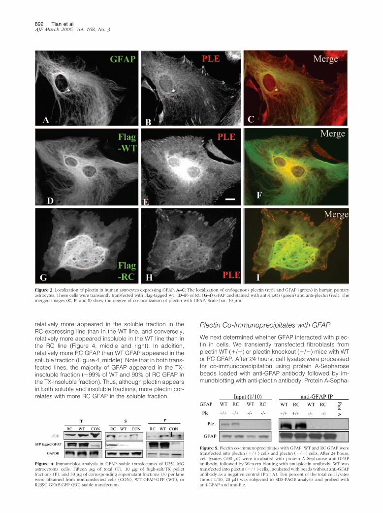
relatively more appeared in the soluble fraction in theRC-expressing line than in the WT line, and conversely,relatively more appeared insoluble in the WT line than inthe RC line (Figure 4, middle and right). In addition,relatively more RC GFAP than WT GFAP appeared in thesoluble fraction (Figure 4, middle). Note that in both trans-fected lines, the majority of GFAP appeared in the TX-insoluble fraction (�99% of WT and 90% of RC GFAP inthe TX-insoluble fraction). Thus, although plectin appearsin both soluble and insoluble fractions, more plectin cor-relates with more RC GFAP in the soluble fraction.
Plectin Co-Immunoprecipitates with GFAP
We next determined whether GFAP interacted with plec-tin in cells. We transiently transfected fibroblasts fromplectin WT (�/�) or plectin knockout (�/�) mice with WTor RC GFAP. After 24 hours, cell lysates were processedfor co-immunoprecipitation using protein A-Sepharosebeads loaded with anti-GFAP antibody followed by im-munoblotting with anti-plectin antibody. Protein A-Sepha-
Figure 5. Plectin co-immunoprecipitates with GFAP. WT and RC GFAP weretransfected into plectin (�/�) cells and plectin (�/�) cells. After 24 hours,cell lysates (200 �l) were incubated with protein A Sepharose anti-GFAPantibody, followed by Western blotting with anti-plectin antibody. WT wastransfected into plectin (�/�) cells, incubated with beads without anti-GFAPantibody as a negative control (Prot A). Ten percent of the total cell lysates(input 1/10, 20 �l) was subjected to SDS-PAGE analysis and probed withanti-GFAP and anti-Ple.
Figure 3. Localization of plectin in human astrocytes expressing GFAP. A–C: The localization of endogenous plectin (red) and GFAP (green) in human primaryastrocytes. These cells were transiently transfected with Flag-tagged WT (D–F) or RC (G–I) GFAP and stained with anti-FLAG (green) and anti-plectin (red). Themerged images (C, F, and I) show the degree of co-localization of plectin with GFAP. Scale bar, 10 �m.
Figure 4. Immunoblot analysis in GFAP stable transfectants of U251 MGastrocytoma cells. Fifteen �g of total (T), 10 �g of high-salt/TX pelletfractions (P), and 30 �g of corresponding supernatant fractions (S) per lanewere obtained from nontransfected cells (CON), WT GFAP-GFP (WT), orR239C GFAP-GFP (RC) stable transfectants.
892 Tian et alAJP March 2006, Vol. 168, No. 3
rose beads without anti-GFAP antibodies were used as abackground control (Figure 5, Prot A). As expected, plec-tin co-immunoprecipitated with both WT and RC GFAP inplectin (�/�) cells. Note that the IP buffer-insoluble GFAPwas excluded from this assay.
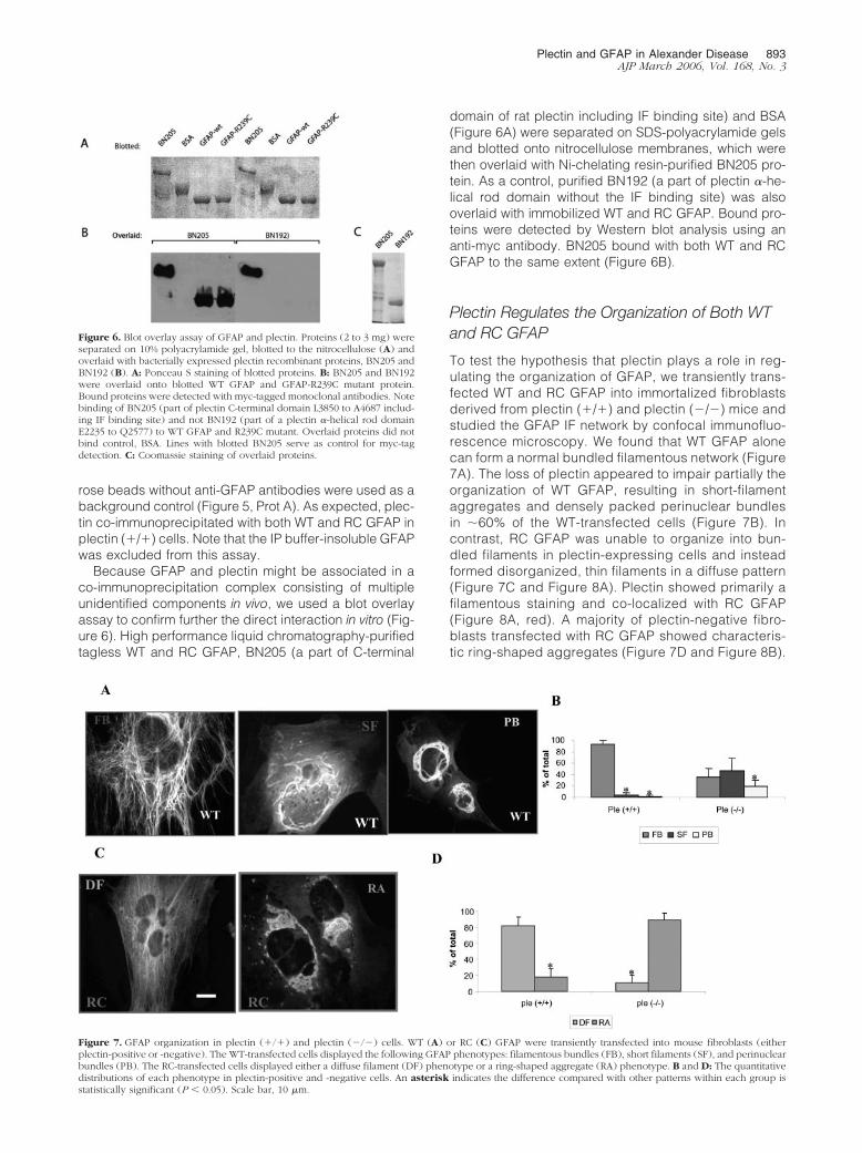
Because GFAP and plectin might be associated in aco-immunoprecipitation complex consisting of multipleunidentified components in vivo, we used a blot overlayassay to confirm further the direct interaction in vitro (Fig-ure 6). High performance liquid chromatography-purifiedtagless WT and RC GFAP, BN205 (a part of C-terminal
domain of rat plectin including IF binding site) and BSA(Figure 6A) were separated on SDS-polyacrylamide gelsand blotted onto nitrocellulose membranes, which werethen overlaid with Ni-chelating resin-purified BN205 pro-tein. As a control, purified BN192 (a part of plectin �-he-lical rod domain without the IF binding site) was alsooverlaid with immobilized WT and RC GFAP. Bound pro-teins were detected by Western blot analysis using ananti-myc antibody. BN205 bound with both WT and RCGFAP to the same extent (Figure 6B).
Plectin Regulates the Organization of Both WTand RC GFAP
To test the hypothesis that plectin plays a role in reg-ulating the organization of GFAP, we transiently trans-fected WT and RC GFAP into immortalized fibroblastsderived from plectin (�/�) and plectin (�/�) mice andstudied the GFAP IF network by confocal immunofluo-rescence microscopy. We found that WT GFAP alonecan form a normal bundled filamentous network (Figure7A). The loss of plectin appeared to impair partially theorganization of WT GFAP, resulting in short-filamentaggregates and densely packed perinuclear bundlesin �60% of the WT-transfected cells (Figure 7B). Incontrast, RC GFAP was unable to organize into bun-dled filaments in plectin-expressing cells and insteadformed disorganized, thin filaments in a diffuse pattern(Figure 7C and Figure 8A). Plectin showed primarily afilamentous staining and co-localized with RC GFAP(Figure 8A, red). A majority of plectin-negative fibro-blasts transfected with RC GFAP showed characteris-tic ring-shaped aggregates (Figure 7D and Figure 8B).
Figure 6. Blot overlay assay of GFAP and plectin. Proteins (2 to 3 mg) wereseparated on 10% polyacrylamide gel, blotted to the nitrocellulose (A) andoverlaid with bacterially expressed plectin recombinant proteins, BN205 andBN192 (B). A: Ponceau S staining of blotted proteins. B: BN205 and BN192were overlaid onto blotted WT GFAP and GFAP-R239C mutant protein.Bound proteins were detected with myc-tagged monoclonal antibodies. Notebinding of BN205 (part of plectin C-terminal domain L3850 to A4687 includ-ing IF binding site) and not BN192 (part of a plectin �-helical rod domainE2235 to Q2577) to WT GFAP and R239C mutant. Overlaid proteins did notbind control, BSA. Lines with blotted BN205 serve as control for myc-tagdetection. C: Coomassie staining of overlaid proteins.
Figure 7. GFAP organization in plectin (�/�) and plectin (�/�) cells. WT (A) or RC (C) GFAP were transiently transfected into mouse fibroblasts (eitherplectin-positive or -negative). The WT-transfected cells displayed the following GFAP phenotypes: filamentous bundles (FB), short filaments (SF), and perinuclearbundles (PB). The RC-transfected cells displayed either a diffuse filament (DF) phenotype or a ring-shaped aggregate (RA) phenotype. B and D: The quantitativedistributions of each phenotype in plectin-positive and -negative cells. An asterisk indicates the difference compared with other patterns within each group isstatistically significant (P � 0.05). Scale bar, 10 �m.
Plectin and GFAP in Alexander Disease 893AJP March 2006, Vol. 168, No. 3
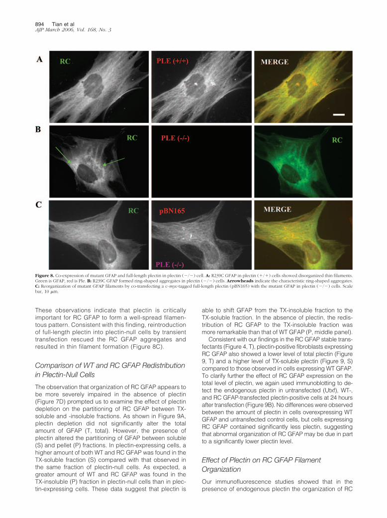
These observations indicate that plectin is criticallyimportant for RC GFAP to form a well-spread filamen-tous pattern. Consistent with this finding, reintroductionof full-length plectin into plectin-null cells by transienttransfection rescued the RC GFAP aggregates andresulted in thin filament formation (Figure 8C).
Comparison of WT and RC GFAP Redistributionin Plectin-Null Cells
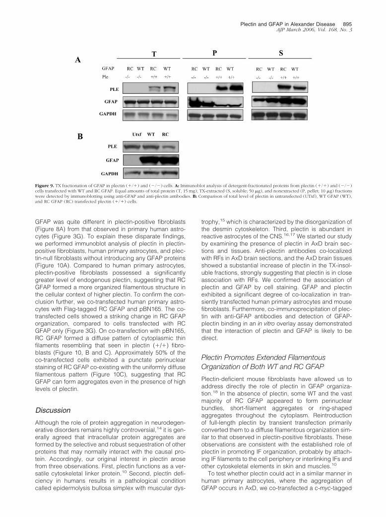
The observation that organization of RC GFAP appears tobe more severely impaired in the absence of plectin(Figure 7D) prompted us to examine the effect of plectindepletion on the partitioning of RC GFAP between TX-soluble and -insoluble fractions. As shown in Figure 9A,plectin depletion did not significantly alter the totalamount of GFAP (T, total). However, the presence ofplectin altered the partitioning of GFAP between soluble(S) and pellet (P) fractions. In plectin-expressing cells, ahigher amount of both WT and RC GFAP was found in theTX-soluble fraction (S) compared with that observed inthe same fraction of plectin-null cells. As expected, agreater amount of WT and RC GFAP was found in theTX-insoluble (P) fraction in plectin-null cells than in plec-tin-expressing cells. These data suggest that plectin is
able to shift GFAP from the TX-insoluble fraction to theTX-soluble fraction. In the absence of plectin, the redis-tribution of RC GFAP to the TX-insoluble fraction wasmore remarkable than that of WT GFAP (P, middle panel).
Consistent with our findings in the RC GFAP stable trans-fectants (Figure 4, T), plectin-positive fibroblasts expressingRC GFAP also showed a lower level of total plectin (Figure9, T) and a higher level of TX-soluble plectin (Figure 9, S)compared to those observed in cells expressing WT GFAP.To clarify further the effect of RC GFAP expression on thetotal level of plectin, we again used immunoblotting to de-tect the endogenous plectin in untransfected (Utxf), WT-,and RC GFAP-transfected plectin-positive cells at 24 hoursafter transfection (Figure 9B). No differences were observedbetween the amount of plectin in cells overexpressing WTGFAP and untransfected control cells, but cells expressingRC GFAP contained significantly less plectin, suggestingthat abnormal organization of RC GFAP may be due in partto a significantly lower plectin level.
Effect of Plectin on RC GFAP FilamentOrganization
Our immunofluorescence studies showed that in thepresence of endogenous plectin the organization of RC
Figure 8. Co-expression of mutant GFAP and full-length plectin in plectin (�/�) cell. A: R239C GFAP in plectin (�/�) cells showed disorganized thin filaments.Green is GFAP, red is Ple. B: R239C GFAP formed ring-shaped aggregates in plectin (�/�) cells. Arrowheads indicate the characteristic ring-shaped aggregates.C: Reorganization of mutant GFAP filaments by co-transfecting a c-myc-tagged full-length plectin (pBN165) with the mutant GFAP in plectin (�/�) cells. Scalebar, 10 �m.
894 Tian et alAJP March 2006, Vol. 168, No. 3
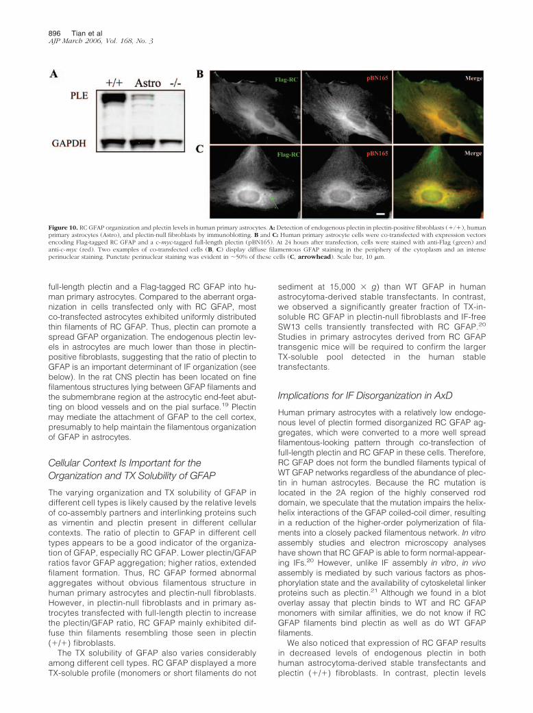
GFAP was quite different in plectin-positive fibroblasts(Figure 8A) from that observed in primary human astro-cytes (Figure 3G). To explain these disparate findings,we performed immunoblot analysis of plectin in plectin-positive fibroblasts, human primary astrocytes, and plec-tin-null fibroblasts without introducing any GFAP proteins(Figure 10A). Compared to human primary astrocytes,plectin-positive fibroblasts possessed a significantlygreater level of endogenous plectin, suggesting that RCGFAP formed a more organized filamentous structure inthe cellular context of higher plectin. To confirm the con-clusion further, we co-transfected human primary astro-cytes with Flag-tagged RC GFAP and pBN165. The co-transfected cells showed a striking change in RC GFAPorganization, compared to cells transfected with RCGFAP only (Figure 3G). On co-transfection with pBN165,RC GFAP formed a diffuse pattern of cytoplasmic thinfilaments resembling that seen in plectin (�/�) fibro-blasts (Figure 10, B and C). Approximately 50% of theco-transfected cells exhibited a punctate perinuclearstaining of RC GFAP co-existing with the uniformly diffusefilamentous pattern (Figure 10C), suggesting that RCGFAP can form aggregates even in the presence of highlevels of plectin.
Discussion
Although the role of protein aggregation in neurodegen-erative disorders remains highly controversial,14 it is gen-erally agreed that intracellular protein aggregates areformed by the selective and robust sequestration of otherproteins that may normally interact with the causal pro-tein. Accordingly, our original interest in plectin arosefrom three observations. First, plectin functions as a ver-satile cytoskeletal linker protein.10 Second, plectin defi-ciency in humans results in a pathological conditioncalled epidermolysis bullosa simplex with muscular dys-
trophy,15 which is characterized by the disorganization ofthe desmin cytoskeleton. Third, plectin is abundant inreactive astrocytes of the CNS.16,17 We started our studyby examining the presence of plectin in AxD brain sec-tions and tissues. Anti-plectin antibodies co-localizedwith RFs in AxD brain sections, and the AxD brain tissuesshowed a substantial increase of plectin in the TX-insol-uble fractions, strongly suggesting that plectin is in closeassociation with RFs. We confirmed the association ofplectin and GFAP by cell staining. GFAP and plectinexhibited a significant degree of co-localization in tran-siently transfected human primary astrocytes and mousefibroblasts. Furthermore, co-immunoprecipitation of plec-tin with anti-GFAP antibodies and detection of GFAP-plectin binding in an in vitro overlay assay demonstratedthat the interaction of plectin and GFAP is likely to bedirect.
Plectin Promotes Extended FilamentousOrganization of Both WT and RC GFAP
Plectin-deficient mouse fibroblasts have allowed us toaddress directly the role of plectin in GFAP organiza-tion.18 In the absence of plectin, some WT and the vastmajority of RC GFAP appeared to form perinuclearbundles, short-filament aggregates or ring-shapedaggregates throughout the cytoplasm. Reintroductionof full-length plectin by transient transfection primarilyconverted them to a diffuse filamentous organization sim-ilar to that observed in plectin-positive fibroblasts. Theseobservations are consistent with the established role ofplectin in promoting IF organization, probably by attach-ing IF filaments to the cell periphery or interlinking IFs andother cytoskeletal elements in skin and muscles.10
To test whether plectin could act in a similar manner inhuman primary astrocytes, where the aggregation ofGFAP occurs in AxD, we co-transfected a c-myc-tagged
Figure 9. TX fractionation of GFAP in plectin (�/�) and (�/�) cells. A: Immunoblot analysis of detergent-fractionated proteins from plectin (�/�) and (�/�)cells transfected with WT and RC GFAP. Equal amounts of total protein (T, 15 mg), TX-extracted (S, soluble; 50 �g), and nonextracted (P, pellet; 10 �g) fractionswere detected by immunoblotting using anti-GFAP and anti-plectin antibodies. B: Comparison of total level of plectin in untransfected (UTxf), WT GFAP (WT),and RC GFAP (RC) transfected plectin (�/�) cells.
Plectin and GFAP in Alexander Disease 895AJP March 2006, Vol. 168, No. 3
full-length plectin and a Flag-tagged RC GFAP into hu-man primary astrocytes. Compared to the aberrant orga-nization in cells transfected only with RC GFAP, mostco-transfected astrocytes exhibited uniformly distributedthin filaments of RC GFAP. Thus, plectin can promote aspread GFAP organization. The endogenous plectin lev-els in astrocytes are much lower than those in plectin-positive fibroblasts, suggesting that the ratio of plectin toGFAP is an important determinant of IF organization (seebelow). In the rat CNS plectin has been located on finefilamentous structures lying between GFAP filaments andthe submembrane region at the astrocytic end-feet abut-ting on blood vessels and on the pial surface.19 Plectinmay mediate the attachment of GFAP to the cell cortex,presumably to help maintain the filamentous organizationof GFAP in astrocytes.
Cellular Context Is Important for theOrganization and TX Solubility of GFAP
The varying organization and TX solubility of GFAP indifferent cell types is likely caused by the relative levelsof co-assembly partners and interlinking proteins suchas vimentin and plectin present in different cellularcontexts. The ratio of plectin to GFAP in different celltypes appears to be a good indicator of the organiza-tion of GFAP, especially RC GFAP. Lower plectin/GFAPratios favor GFAP aggregation; higher ratios, extendedfilament formation. Thus, RC GFAP formed abnormalaggregates without obvious filamentous structure inhuman primary astrocytes and plectin-null fibroblasts.However, in plectin-null fibroblasts and in primary as-trocytes transfected with full-length plectin to increasethe plectin/GFAP ratio, RC GFAP mainly exhibited dif-fuse thin filaments resembling those seen in plectin(�/�) fibroblasts.
The TX solubility of GFAP also varies considerablyamong different cell types. RC GFAP displayed a moreTX-soluble profile (monomers or short filaments do not
sediment at 15,000 � g) than WT GFAP in humanastrocytoma-derived stable transfectants. In contrast,we observed a significantly greater fraction of TX-in-soluble RC GFAP in plectin-null fibroblasts and IF-freeSW13 cells transiently transfected with RC GFAP.20
Studies in primary astrocytes derived from RC GFAPtransgenic mice will be required to confirm the largerTX-soluble pool detected in the human stabletransfectants.
Implications for IF Disorganization in AxD
Human primary astrocytes with a relatively low endoge-nous level of plectin formed disorganized RC GFAP ag-gregates, which were converted to a more well spreadfilamentous-looking pattern through co-transfection offull-length plectin and RC GFAP in these cells. Therefore,RC GFAP does not form the bundled filaments typical ofWT GFAP networks regardless of the abundance of plec-tin in human astrocytes. Because the RC mutation islocated in the 2A region of the highly conserved roddomain, we speculate that the mutation impairs the helix-helix interactions of the GFAP coiled-coil dimer, resultingin a reduction of the higher-order polymerization of fila-ments into a closely packed filamentous network. In vitroassembly studies and electron microscopy analyseshave shown that RC GFAP is able to form normal-appear-ing IFs.20 However, unlike IF assembly in vitro, in vivoassembly is mediated by such various factors as phos-phorylation state and the availability of cytoskeletal linkerproteins such as plectin.21 Although we found in a blotoverlay assay that plectin binds to WT and RC GFAPmonomers with similar affinities, we do not know if RCGFAP filaments bind plectin as well as do WT GFAPfilaments.
We also noticed that expression of RC GFAP resultsin decreased levels of endogenous plectin in bothhuman astrocytoma-derived stable transfectants andplectin (�/�) fibroblasts. In contrast, plectin levels
Figure 10. RC GFAP organization and plectin levels in human primary astrocytes. A: Detection of endogenous plectin in plectin-positive fibroblasts (�/�), humanprimary astrocytes (Astro), and plectin-null fibroblasts by immunoblotting. B and C: Human primary astrocyte cells were co-transfected with expression vectorsencoding Flag-tagged RC GFAP and a c-myc-tagged full-length plectin (pBN165). At 24 hours after transfection, cells were stained with anti-Flag (green) andanti-c-myc (red). Two examples of co-transfected cells (B, C) display diffuse filamentous GFAP staining in the periphery of the cytoplasm and an intenseperinuclear staining. Punctate perinuclear staining was evident in �50% of these cells (C, arrowhead). Scale bar, 10 �m.
896 Tian et alAJP March 2006, Vol. 168, No. 3
remained unchanged in the untransfected controls andWT transfectants. We were unable to confirm this find-ing of reduced plectin expression in frozen brain tis-sues of AxD patients. However, we do not know thenumbers of astrocytes in the tissues, the ratio of WT toRC GFAP proteins in these astrocytes, and the effectsof storage at low temperature on plectin solubility, allvariables that could alter plectin levels with respect toother proteins. Examination of AxD brain sections andtissues showed that astrocytes in the subcortical whitematter area accumulate more GFAP and GFAP assem-bly regulatory proteins such as �B crystallin and plec-tin.22 The organization of GFAP, including both WTand RC GFAP, might be highly dependent on theratio of GFAP assembly regulatory proteins to GFAP inastrocytes. A lower plectin/GFAP ratio, resulting fromthe reduced level of total plectin in response to RCGFAP expression in astrocytes, would promote theabnormal organization and aggregation of GFAP intoRFs in AxD.
Pathology in the CNS has been linked to a plectinmutation. The C-terminal domain of plectin consists of sixhighly conserved repeats, and the IF-binding domain ofplectin has been mapped to a short segment betweenrepeats 5 and 6.23 A homozygous insertion close to theIF-binding domain of plectin has been linked to epider-molysis bullosa simplex-muscular dystrophy in at leastone patient who presented with marked desmin accumu-lations in muscle tissue and severe cerebral atrophy.15
Combined with other case reports of brain atrophy asso-ciated with epidermolysis bullosa simplex,24,25 this sug-gests that perturbations of the plectin/IF interactions dueto plectin mutations or IF mutations can lead to neurolog-ical pathology.
Acknowledgments
We thank Dr. Albee Messing, Dr. Michael Brenner, andDr. Ron Liem for reviewing the manuscript and criticaldiscussions; and Dr. Tae-wan Kim, Dr. Paul Fisher, andDr. Guomei Tang for kindly providing reagents and hu-man astrocytes.
References
1. Messing A, Goldman JE, Johnson AB, Brenner M: Alexander disease:new insights from genetics. J Neuropathol Exp Neurol 2001,60:563–573
2. Goldman JE, Corbin E: Isolation of a major protein component ofRosenthal fibers. Am J Pathol 1988, 130:569–578
3. Brenner M, Johnson AB, Boespflug-Tanguy O, Rodriguez D, Gold-man JE, Messing A: Mutations in GFAP, encoding glial fibrillary acidicprotein, are associated with Alexander disease. Nat Genet 2001,27:117–120
4. Rodriguez D, Gauthier F, Bertini E, Bugiani M, Brenner M, N�Guyen S,Goizet C, Gelot A, Surtees R, Pedespan JM, Hernandorena X, Tron-coso M, Uziel G, Messing A, Ponsot G, Pham-Dinh D, Dautigny A,Boespflug-Tanguy O: Infantile Alexander disease: spectrum of GFAPmutations and genotype-phenotype correlation. Am J Hum Genet2001, 69:1134–1140
5. Wiche G, Herrmann H, Leichtfried F, Pytela R: Plectin: a high-molec-
ular-weight cytoskeletal polypeptide component that copurifies withintermediate filaments of the vimentin type. Cold Spring Harb SympQuant Biol 1982, 46:475–482
6. Herrmann H, Wiche G: Plectin and IFAP-300K are homologous pro-teins binding to microtubule-associated proteins 1 and 2 and to the240-kilodalton subunit of spectrin. J Biol Chem 1987, 262:1320–1325
7. Steinbock FA, Wiche G: Plectin: a cytolinker by design. Biol Chem1999, 380:151–158
8. Uitto J, Pulkkinen L: Molecular complexity of the cutaneous basementmembrane zone. Mol Biol Rep 1996, 23:35–46
9. Andra K, Lassmann H, Bittner R, Shorny S, Fassler R, Propst F, WicheG: Targeted inactivation of plectin reveals essential function in main-taining the integrity of skin, muscle, and heart cytoarchitecture.Genes Dev 1997, 11:3143–3156
10. Wiche G: Role of plectin in cytoskeleton organization and dynamics.J Cell Sci 1998, 111:2477–2486
11. Nikolic B, MacNulty E, Mir B, Wiche G: Basic amino acid residuecluster within nuclear targeting sequence motif is essential for cyto-plasmic plectin-vimentin network junctions. J Cell Biol 1996,134:1455–1467
12. Su ZZ, Kang DC, Chen Y, Pekarskaya O, Chao W, Volsky DJ, FisherPB: Identification of gene products suppressed by human immuno-deficiency virus type 1 infection or gp120 exposure of primary humanastrocytes by rapid subtraction hybridization. J Neurovirol 2003,9:372–389
13. Stegh AH, Herrmann H, Lampel S, Weisenberger D, Andra K, SeperM, Wiche G, Krammer PH, Peter ME: Identification of the cytolinkerplectin as a major early in vivo substrate for caspase 8 during CD95-and tumor necrosis factor receptor-mediated apoptosis. Mol Cell Biol2000, 20:5665–5679
14. Bossy-Wetzel E, Schwarzenbacher R, Lipton SA: Molecular pathwaysto neurodegeneration. Nat Med 2004, 10:S2–S9
15. Schroder R, Kunz WS, Rouan F, Pfendner E, Tolksdorf K, Kappes-Horn K, Altenschmidt-Mehring M, Knoblich R, van der Ven PF,Reimann J, Furst DO, Blumcke I, Vielhaber S, Zillikens D, Eming S,Klockgether T, Uitto J, Wiche G, Rolfs A: Disorganization of thedesmin cytoskeleton and mitochondrial dysfunction in plectin-relatedepidermolysis bullosa simplex with muscular dystrophy. J Neuro-pathol Exp Neurol 2002, 61:520–530
16. Lie AA, Schroder R, Blumcke I, Magin TM, Wiestler OD, Elger CE:Plectin in the human central nervous system: predominant expressionat pia/glia and endothelia/glia interfaces. Acta Neuropathol (Berl)1998, 96:215–221
17. Kalman M, Szabo A: Plectin immunopositivity appears in the astro-cytes in the white matter but not in the gray matter after stab wounds.Brain Res 2000, 857:291–294
18. Andra K, Nikolic B, Stocher M, Drenckhahn D, Wiche G: Not justscaffolding: plectin regulates actin dynamics in cultured cells. GenesDev 1998, 12:3442–3451
19. Nakazawa E, Ishikawa H: Ultrastructural observations of astrocyte end-feet in the rat central nervous system. J Neurocytol 1998, 27:431–440
20. Hsiao VC, Tian R, Long H, Perng MD, Brenner M, Quinlan RA,Goldman JE: Alexander-disease mutation of GFAP causes filamentdisorganization and decreased solubility of GFAP. J Cell Sci 2005,118:2057–2065
21. Portet S, Vassy J, Hogue CW, Arino J, Arino O: Intermediate filamentnetworks: in vitro and in vivo assembly models. C R Biol 2004,327:970–976
22. Head MW, Hurwitz L, Kegel K, Goldman JE: AlphaB-crystallin regu-lates intermediate filament organization in situ. Neuroreport 2000,11:361–365
23. Steinbock FA, Nikolic B, Coulombe PA, Fuchs E, Traub P, Wiche G:Dose-dependent linkage, assembly inhibition and disassembly ofvimentin and cytokeratin 5/14 filaments through plectin’s intermediatefilament-binding domain. J Cell Sci 2000, 113:483–491
24. Kletter G, Evans OB, Lee JA, Melvin B, Yates AB, Bock HG: Congen-ital muscular dystrophy and epidermolysis bullosa simplex. J Pediatr1989, 114:104–107
25. Doriguzzi C, Palmucci L, Mongini T, Bertolotto A, Maniscalco M,Chiado-Piat L, Zina AM, Bundino S: Congenital muscular dystrophyassociated with familial junctional epidermolysis bullosa letalis. EurNeurol 1993, 33:454–460
Plectin and GFAP in Alexander Disease 897AJP March 2006, Vol. 168, No. 3


