Embed Size (px)
Citation preview

Surface Physicochemical Properties at the Micro andNano Length Scales: Role on Bacterial Adhesion andXylella fastidiosa Biofilm DevelopmentGabriela S. Lorite1¤, Richard Janissen1, João H. Clerici1, Carolina M. Rodrigues2, Juarez P. Tomaz2, BorisMizaikoff3, Christine Kranz3, Alessandra A. de Souza2, Mônica A. Cotta1*
1 Departamento de Física Aplicada, Instituto de Física Gleb Wataghin, Universidade Estadual de Campinas, Campinas, São Paulo, Brazil, 2 Centro APTACitros Sylvio Moreira, Instituto Agronômico de Campinas, Cordeirópolis, São Paulo, Brazil, 3 Institute of Analytical and Bioanalytical Chemistry, University ofUlm, Ulm, Germany
Abstract
The phytopathogen Xylella fastidiosa grows as a biofilm causing vascular occlusion and consequently nutrient andwater stress in different plant hosts by adhesion on xylem vessel surfaces composed of cellulose, hemicellulose,pectin and proteins. Understanding the factors which influence bacterial adhesion and biofilm development is a keyissue in identifying mechanisms for preventing biofilm formation in infected plants. In this study, we show that X.fastidiosa biofilm development and architecture correlate well with physicochemical surface properties afterinteraction with the culture medium. Different biotic and abiotic substrates such as silicon (Si) and derivatizedcellulose films were studied. Both biofilms and substrates were characterized at the micro- and nanoscale, whichcorresponds to the actual bacterial cell and membrane/ protein length scales, respectively. Our experimental resultsclearly indicate that the presence of surfaces with different chemical composition affect X. fastidiosa behavior fromthe point of view of gene expression and adhesion functionality. Bacterial adhesion is facilitated on more hydrophilicsurfaces with higher surface potentials; XadA1 adhesin reveals different strengths of interaction on these surfaces.Nonetheless, despite different architectural biofilm geometries and rates of development, the colonization processoccurs on all investigated surfaces. Our results univocally support the hypothesis that different adhesion mechanismsare active along the biofilm life cycle representing an adaptation mechanism for variations on the specific xylemvessel composition, which the bacterium encounters within the infected plant.
Citation: Lorite GS, Janissen R, Clerici JH, Rodrigues CM, Tomaz JP, et al. (2013) Surface Physicochemical Properties at the Micro and Nano LengthScales: Role on Bacterial Adhesion and Xylella fastidiosa Biofilm Development. PLoS ONE 8(9): e75247. doi:10.1371/journal.pone.0075247
Editor: Zoya Leonenko, University of Waterloo, Canada
Received April 4, 2013; Accepted August 13, 2013; Published September 20, 2013
Copyright: © 2013 Lorite et al. This is an open-access article distributed under the terms of the Creative Commons Attribution License, which permitsunrestricted use, distribution, and reproduction in any medium, provided the original author and source are credited.
Funding: This work was financially supported by FAPESP (Fundação de Amparo à Pesquisa do Estado de São Paulo, through grant #2010/51748-7 andfellowships #2009/53021-0, 2006/06397-6), DAAD (Deutscher Akademischer Austauschdienst), DFG (Deutsche Forschungsgemeinschaft), CNPq(Conselho Nacional de Desenvolvimento Científico e Tecnológico) and CAPES (Coordenação de Aperfeiçoamento de Pessoal de Nível Superior). Thefunders had no role in study design, data collection and analysis, decision to publish, or preparation of the manuscript.
Competing interests: The authors have declared that no competing interests exist.
* E-mail: [email protected]
¤ Current address: Institute of Biomedicine, University of Oulu, Oulu, Finland
Introduction
Biofilms are complex microbial communities associated withsurfaces with important biological functions including bacterialinfection and enhanced resistance against antimicrobial agents[1]. The bacterial biofilm life cycle is known as a multi-stageprocess, which current models subdivide into five developmentstages [2]. During all these stages, biofilm development may bemediated by a number of factors including nutrientenvironment, pH, temperature and surface properties [2].Several factors may affect the initial microbial adhesion to suchsurfaces; these factors include the physicochemical propertiesof surfaces and cells, and the particular conditions within the
environment where adhesion occurs [3]. Moreover, surfaceproperties may be altered by the adsorption of (macro)molecules from the liquid environment onto the substrateforming a conditioning film, which has been considered as theinitial step of biofilm formation [3-5]. Advanced understandingthe influence of surfaces properties on biofilm formation willtherefore lead to strategies for preventing microbial adhesion,and consequently, biofilm development.
Within microbiology research, many previous studies [6-12]have focused on the question which different substrate surfaceproperties - such as roughness, topography, stiffness,hydrophobicity and charge density - play important roles duringbacterial adhesion and/or biofilm formation. The variability of
PLOS ONE | www.plosone.org 1 September 2013 | Volume 8 | Issue 9 | e75247

results found in literature, however, evidence the importance ofexperimental conditions in drawing conclusions about thesystematic behavior of bacterial cells. Furthermore, surfacesare seldom homogeneous at both micro and nanoscale; suchheterogeneities - as in micropatterned surfaces - can influencecell processes such as phagocytosis of filamentousEscherichia coli bacteria by macrophages [13]. We can thusexpect that cell adhesion will be influenced as well. This is animportant issue to the organization of larger structures such asbiofilms. Hochbaum and Aizenberg [14] have shown thatbacterial ordering and oriented attachment on the single celllevel can be induced by nanometer-scale periodic surfacefeatures. The surface patterning causes spontaneous anddistinct cell arrangement for Gram-positive and Gram-negativebacterial strains and can be modeled by maximizing thebacteria contact area with the surface. The authors suggestthat this behavior is a general phenomenon involved in naturalbiofilm organization.
In our present study, we address the role of surfaceproperties at different length scales on adhesion of the Gram-negative bacteria Xylella fastidiosa, which has recently beenincluded in the list of top 10 plant pathogenic bacteria inmolecular plant pathology [15]. This microorganism is a xylem-inhabiting bacterium responsible for diseases of economicallyimportant crops such as plum, almond, peach, coffee,grapevine and citrus [16,17]; its widely accepted mechanism ofpathogenicity is attributed to water and nutrient stress due theblockage of the xylem vessels caused by biofilm formation.Nonetheless, the precise mechanism of X. fastidiosa adhesionand biofilm formation on a molecular level is not wellunderstood to date.
Moreover, different X. fastidiosa biofilm architectures havebeen reported for the two main environments where thebacterium is found, i.e., insect cuticle or xylem vessel. Ininsects, cells adopt a polar configuration regarding theattachment point and form a mat-like structure; in plants,attachment to the xylem wall does not show any preferentialorientation [18]. The biofilm architecture is a crucial issue sinceit may related to the bacterial adhesiveness. In fact, thetransmission from host to vector relies on a balance regardingthe pathogen adhesiveness: the bacteria should be sufficientlyadherent to be acquired by the insect, but not so much as to bewithheld within the host [19]. This phenotypic plasticity, whichallows bacteria survival in multiple environments, is most likelyregulated by signals provided by the habitat.
Despite the importance of the environment in the bacteria lifecycle, in vitro studies of X. fastidiosa biofilms have been largelyperformed on abiotic surfaces which are not related to thenatural bacterial habitat [20-24]. Cellulose, hemicellulose,pectin and proteins are constituents from xylem vessels.Among the cell wall proteins, extensins and hydroxyproline/proline-rich, which are structural proteins, are known toreinforce the polysaccharide networks [25]. Recently a largenumber of other cell wall proteins in different organs werefound by proteomics approaches ( [26,27], accessible via thepublic database (www.polebio.scsv.ups-tlse.fr/WallProtDB/))revealing a great diversity of protein functions in the dynamicsof cell walls, which involves organization and rearrangements
of polysaccharides in addition to cell-to-cell communication[28]. The plant cell walls forming the xylem can be degraded byenzymes, thus facilitating the spreading of the bacteria, andmost likely, providing carbohydrates as an additional nutrientsource [20]. In plant studies of X. fastidiosa biofilm formationare very difficult to perform due to the dynamics of microbialcell movement and the simultaneous presence of differentstages of biofilms within the xylem vessels. Therefore, surfacescontaining xylem constituents, which can also have theirproperties mapped by physicochemical tools at different scales,have not been used on in vitro studies of this bacteria to date.Within this work we use a pool of techniques to study bacteria-surface interactions, as well as its role on biofilm development,at the different length scales of interest to the problem. Acomparative study of the main surface properties usuallyconsidered in microbiology research, for substrates whichsimulate the natural bacteria habitat, was thus carried out.
In order to address the role of the surface properties on theadhesion of X. fastidiosa, we have used both abiotic (silicon)and biotic (derivatized cellulose) surfaces to study biofilmdevelopment. Abiotic surfaces are considered inert materials(e.g. glass, silicon) while biotic surfaces contain substanceswhich can be used as nutrients by the microbial cells.Regarding biotic surface mimicry, we used derivatized cellulose(or cellulose functionalized by chemical modification) thin filmsto resemble more closely the plant conditions since celluloserepresents the major constituent in the natural X. fastidiosahabitat. In this context, synthetic cellulose - such as celluloseacetate (CA) and ethyl cellulose (EC) - thin polymer films wereused in order to provide supports with biotic character anddifferent chemical functional groups (acetate ester groups andethyl ether groups, respectively) resulting in electrical chargedifferences, whereas EC exhibits less negative charge as CAand is comparable to the standard cellulose. The analysis ofsurface properties was performed using micro- and nanoscaleanalytical techniques at the relevant bacterial cell andmembrane/protein length scales, respectively. Microscopy andgene expression analyses show that the presence of differenttypes of cellulose at the surface influence the adhesion,development rate and architecture of X. fastidiosa biofilms.Surface properties like roughness, hydrophobicity/hydrophilicityand surface potential were characterized considering inaddition the effect of exposure to the culture medium. Finally,force-distance curves between the transmembrane XadA1adhesion protein and the investigated surfaces were recordedfor determining potential interactions at the initial stages ofadhesion. Taken these data altogether, a scenario withrelevant parameters controlling bacterial adhesion can bedrawn.
Material and Methods
Bacteria strain and growth conditionsThe X. fastidiosa subspecies pauca strain used in this study
was 9a5c. The strain was grown in Periwinkle Wilt (PW)medium including Bovine Serum Albumin (BSA) as describedin previous work [4].
Biofilm Development Depends on Surface Properties
PLOS ONE | www.plosone.org 2 September 2013 | Volume 8 | Issue 9 | e75247

Preparation of silicon and derivatized cellulosesurfaces
Silicon surfaces (<100> oriented, cut into square shapes ofapproximately 2x2 cm2) were rinsed with acetone, 2-propanoland deionized (DI) water to remove the organic contaminationfrom the surfaces. To obtain the cellulose acetate (CA)solution, typically 0.7 g of cellulose acetate powder (Sigma-Aldrich) was dissolved in a solution containing 7.4 ml of aceticacid, 13.8 mL of acetone and 7.4 ml of deionized water. In theethyl cellulose (EC) case, 0.3 g of ethyl cellulose powder(Sigma-Aldrich) was dissolved in 30 ml ethanol. Both solutionswere deposited onto the silicon substrates via spin coating(3000 rpm, 30 s). The samples were dried overnight at roomtemperature (RT). All surfaces were sterilized via autoclavingprocedures.
Biofilm developmentX. fastidiosa cells were grown from frozen state to a liquid
PW medium for static biofilm development. The cells wereincubated simultaneously at 28°C on different autoclavedsurfaces for 7, 14, 21 days-cycles without mediumreplenishing. All samples were prepared under similarconditions and repeated from 2 to 5 times.
Real-time quantitative polymerase chain reaction (RT-qPCR)
Biofilms were scraped from the different surfaces and theRNA was extracted using RNeasy Mini Kit (Qiagen), followingthe manufacturer protocol. To RT-qPCR analysis cDNAsynthesis was performed using 1 µg total RNA usingSuperScriptII Reverse Transcriptase (Life Technologies), andRandom Primers oligonucleotide 0,1 mM (Life Technologies),1x Reaction Buffer (Life Technologies), dNTP 0,1 mM andRNAseOUT RNAse Inhibitor (100 U) (Life Technologies), in atotal volume of 20 µl. RT-qPCR was performed in duplicate inan optical 96-well plate with ABI 7500 Fast sequence detectionsystem (Life Technologies), in a volume of 25 µl, containing 2µl of cDNA, 1X SYBR Green Master Mix (Life Technologies),ROX as passive reference and 200 nM of both reverse andforward primers. The following thermal conditions were usedfor all PCR reactions: 50°C for 2 min, 95°C for 10 min and 40cycles of 95°C for 15 s and 60°C for 60 s. The mRNA levelswere quantified in relation to the endogenous control genepetC. The expression level was presented as 2-ΔΔCt, where ΔCt= Cttarget – CtpETC and ΔΔCt = ΔCttreatment - ΔCtSi. The results ofmeasurements were expressed as mean ± standard deviation(SD). Data were evaluated by one-way analysis of variance(ANOVA) with a significance level of α=0.05 followed byTukey’s post hoc test.
Optical and Electron MicroscopyThe samples were mapped by Zeiss (AXIO Scope A1)
optical microscopy to quantify the number and diameter ofindividual biofilms on the whole sample surface. We usedIMAGEJ software to estimate the biofilm diameter assumingcircular areas. Scanning electron microscopy (SEM) imageswere acquired using a dual-beam FIB/SEM system (Quanta 3D
FEG, FEI Company, Eindhoven, The Netherlands). Thesamples were gently water-rinsed (to remove the PW mediumand no adherent microbial cells) and were then dried at RTovernight
Atomic force microscopy (AFM)Topography images, surface potential and force-distance
curves were acquired using an Agilent AFM system Model5500 (Agilent Technologies, Chandler, AZ, USA). AFMtopography images were acquired in tapping mode in air usingconical Si tips with a typical tip radius of 10 nm and tip length of~20 µm (NSC14/AIBS MikroMash, Tallin, Estonia). The springconstant and resonance frequency were typically within therange 1.8-12.5 Nm-1 and 110-220 kHz, respectively. Toevaluate the roughness of the surface, the root-mean-squaredroughness (RMS) was determined over 5x5 µm2 areas for eachsample. Spectral analysis of the images was carried out bycalculating the local maxima for the Power Spectral DensityFunction. All AFM images were analyzed using freely availablesoftware (Gwyddion V. 2.29). SP images were acquiredsimultaneously with topography in tapping mode under N2 flowusing Pt-coated Si tips (NSC18/Pt, MikroMash, Tallin, Estonia).In order to observe possible surfaces changes by PW medium,all surfaces were kept in contact with PW medium for 24 h. Thesamples were then rinsed in DI water and dried at RTovernight. In the Amplitude Mode-Kelvin Probe ForceMicroscopy technique used here, we define the ‘zero’ SP valuefor the first point of the image; thus only variations of SP alongthe image can be compared quantitatively. Therefore, in orderto compare SP values from different surfaces and thus excludetip effects arising from metal coating wear [29] we have usedthe Si substrate as a common reference in the images; for thatwe have scanned edges of the cellulose film which show the Sisubstrate as well. We thus compare absolute SP* values fromthe different samples according to the following equation: SP* =SP(cellulose) – SP(Si). Each SP* value was obtained byaveraging five cross sections per image across the edgebetween homogeneous regions of cellulose film and silicon. Toevaluate SP variation along the surface, the root-mean-squared SP roughness (SP-RMS) was determined over 5x5µm2 areas for each sample similarly to the procedure carriedout for topography images. In order to assess the electricalproperties of the PW film, we have removed it to form a step onthe Si surface. The same procedure for SP* evaluation wasfollowed to the PW film - in this case SP* = SP(PW film) - SP(Si).
XadA1 immobilization on AFM tip and force-distancecurves
Conical Si tips (NSC19/AIBS MikroMash, Tallin, Estonia)with spring constant typically within the range 0.17-1.7 Nm-1
were cleaned in piranha solution (H2SO4:H2O2 = 3:1) for 5 minand rinsed with running DI water. The cantilevers weresubsequently incubated in 3-(glycidoxypropyl) trimethoxysilane(GOPTES) solution for 30 min; cantilevers were then rinsedwith ethanol and DI water and dried under N2-flow. Toimmobilize XadA1 on the tip, the cantilevers were immersed in15 µm/ml XadA1 in PBS (pH 7.4) buffer for 30 min and washed
Biofilm Development Depends on Surface Properties
PLOS ONE | www.plosone.org 3 September 2013 | Volume 8 | Issue 9 | e75247
in TBS-TT buffer (20 mM Tris/HCl, 500 mM NaCl, 0.5% (v/v)Tween 20, 0.2% (v/v) Triton-X100, pH 7.4) and TBS buffer (10mM Tris/HCl, 150 mM NaCl, pH 7.4) for 5 min each. Finally, thetips were rinsed with PBS buffer for 1 min and stored in thesame buffer solution. The successful immobilization of theXadA1 adhesion protein on the tip was subsequently confirmedby fluorescence microscopy (Figure S1) via specific polyclonalXadA1 antibody [30] and fluorescein labeled anti-rabbit IgGsecond antibody (Rheabiotech, Campinas, Brazil). Details ofthe experimental method to obtain purified XadA1 protein andantibody were published previously [51]. All force-distancecurves were obtained using liquid cell buffered with PBS (pH7.4) solution or PW medium (control experiment) at RT.Cantilevers were moved at a velocity of 200 nm/s with total z-displacement of ~700 nm. Data were acquired ~200 times ateach of five different locations on each surface. For the CA andEC surfaces, control experiments included force-distancecurves acquired on Si surfaces immediately before and afterthe measurements on the derivatized cellulose surfaces.Control experiments were also carried out with non-functionalized tips for all the investigated surfaces for both PBSand PW medium. Spring constants were experimentallyobtained after tip-functionalization using thermal noise method.All adhesion peaks in the retracting force-distance curves weredetermined in order to build force histograms for each surface.For this statistical analysis, we estimated the noise level of themeasurement using over 2500 force-distance curves. Thus,adhesion forces lower than the estimated noise (9±5 pN) orabsence of adhesion peaks were both considered as null force(e.g. 0 nN). The average adhesion forces and deviation werecalculated using Gaussian fitting to the histogram data usingthe commercial software Origin 8.0 (OriginLabs, USA).
Surface contact angle measurementsThese measurements were performed as described in a
previous work [4]. Samples exposed to PW medium fordifferent time intervals were all washed with DI water and driedat room temperature overnight. Contact angles lower than 10°were considered as complete wetting.
Results and Discussion
Experimental approachThe X. fastidiosa subspecies pauca strain 9a5c was used
within this study. The strain was grown in Periwinkle Wilt (PW)medium [31] including Bovine Serum Albumin (BSA) asdescribed in a previous work [4]. X. fastidiosa cells wereincubated at 28°C for several days on cellulose acetate (CA)and ethyl cellulose (EC) as well as bare, clean silicon surfaces,for static biofilm development.
Biofilm development was assessed from optical andscanning electron microscopy (OM and SEM, respectively)images of samples dried overnight. Real-time quantitativepolymerase chain reaction (RT-qPCR) was used to evaluatespecific gene expression changes at different biofilmdevelopment stages. In order to probe changes in thehydrophilic character of the different substrates used here,contact angle measurements were performed for all three
surface compositions. Substrate surface properties were alsomainly investigated using Atomic and Kelvin Probe ForceMicroscopy (AFM and KPFM, respectively), as well as forcespectroscopy measurements with XadA1 adhesin-functionalized silicon tips. In particular, KPFM [32,33] allowstopography and surface potential (SP) imaging of the sample,within a nanometer range spatial resolution. The contactpotential between tip and sample is defined by the differencebetween their work functions, Δϕ [34,35]. By applying externalbias voltages and compensating the contact potential, KPFMcancels this particular electrostatic interaction between tip andsample and makes it possible to measure SP = Δϕ/e, where erepresents the electron charge, simultaneously with the sampletopography. KPFM has been used extensively as a uniquemethod to characterize the nano-scale electronic/electricalproperties of metal/semiconductor surfaces, semiconductordevices and more recently, organic materials/devices andbiological materials [36]. Although an important variable toadhesion models, SP values are not usually determined inbiological processes, and the zeta potential of the surface isused instead. Zeta potential is an electrokinetic method, whichprovides the potential difference between the dispersionmedium and the stationary fluid of layer attached to adispersed particle. Such measurements on cellulosic materialshave been extensively studied in literature [37]; for CA and ECsurfaces values range in the -10 to -50mV [38,39], dependingon various parameters such as pH, polymer swelling, fiberstructure, etc. However, zeta potential measurements are notsuitable for surface mapping or relating potential andtopography inomogeneities. Therefore, we have elected adirect measurement of SP (in dry atmosphere) to comparesuch inomogeneities in our samples. However, it is important tonotice that, once immersed in liquid, the surface electrostaticproperties can be altered by the presence of ions in thesurrounding solution, due to chemical adsorption or electricaldouble layer formation. Screening of such inomogeneities canoccur in solution; this issue will be further addressed in thesubsequent sections.
X. fastidiosa biofilm architecture and development ratedependence on surface properties
Both development and architecture of X. fastidiosa biofilmswere compared at silicon (abiotic, stiff) and derivatizedcellulose (biotic, soft) surfaces. Figure 1 shows the sizedistribution and typical architecture for biofilms on Si, EC andCA. In good agreement with previous studies [4,23], thebiofilms typically present circular shapes with a different rangeof diameters along a 21-day cycle for all investigated surfaces(Figure 1 A-D, F, H); no ridges or wrinkles were observedduring the entire period [40]. Considering the same growthtime, Si surfaces present a larger number of biofilms (approx.5-fold) compared to derivatized cellulose surfaces. Despite thedispersion of diameter values, there is a clear predominance ofsmaller biofilm structures (approximately 20-80µm in diameter)on all surfaces after 7 and 21-day growth cycles (Figure 1 A-C). This expected behavior is attributed to the initial and laststages of the life cycle of X. fastidiosa biofilms, as e.g., thedetachment of bacteria from the biofilms provides nucleation
Biofilm Development Depends on Surface Properties
PLOS ONE | www.plosone.org 4 September 2013 | Volume 8 | Issue 9 | e75247

centers for new sites [41]. For 14-day growth periods, thenumber of small biofilm structures along Si and EC surfacesdecreases significantly independent of the surface material,while the biofilm diameter increases (Figure 1 A, B). Thisbehavior can be interpreted as the result of a competitionprocess, i.e., some biofilms grow in size at the expenses of thesmaller ones due to nutrient consumption [23,41] or simply dueto the spatial overlap of the biofilms. In contrast, although thetotal number of 14-day biofilms on CA also decreases, they donot present significant changes in diameter distribution untilthey are 21 days old (Figure 1 C). This analysis also revealslarger biofilm diameters on Si surfaces; for example, the largestbiofilm observed on CA surfaces after 21 days (approximately350 µm diameter) is only half the size of the largest biofilmstructure observed on Si surfaces after only 14 days(approximately 650 µm diameter). Contrarily to CA samples,biofilms on EC surfaces also reach larger diameters after only14 days (approximately 550 µm diameter). Interestingly,despite a similar overall shape, the edges of the biofilms varysignificantly according to the substrate. SEM images reveal amore extensive first layer and well-defined edges for X.fastidiosa biofilms on Si and EC surfaces (Figure 1 E, G); infact, particularly on Si, the first layer usually extends wellbeyond the more massive region of the biofilm for all growthtimes considered in the present experiments. For CA surfaces,however, the biofilm edges are not well defined; a randomdistribution of groups of cells is observed in the region aroundthe biofilms, but usually disconnected from the large matrixdeposit (Figure 1 I). In fact, even for older (21 days) samples,the regions surrounding the edges of the biofilms are not
entirely filled with cells, as in the case of Si and EC surfaces,i.e., patches of exposed substrate can be found among thebacteria surrounding the biofilms on CA surfaces. Furthermore,if Si and CA surfaces are exposed side-by-side to bacteria,biofilm formation occurs earlier on Si than on CA, as shown bycorresponding AFM experiments (Figure 2). These resultsprovide a first indication that enhanced adhesion and higherdevelopment rates for X. fastidiosa biofilm growth are prevalenton bare Si surface compared to EC and CA surfaces,respectively, and agree with previous studies which reportenhanced adhesion on stiffer substrates [10,42]. Moreover,these results imply that cell-surface interaction stronglydepends on the surface properties, which may also affect thebiofilm architecture at the microscale during later bacterial lifecycle stages.
Investigation of silicon and derivatized cellulosesurface properties
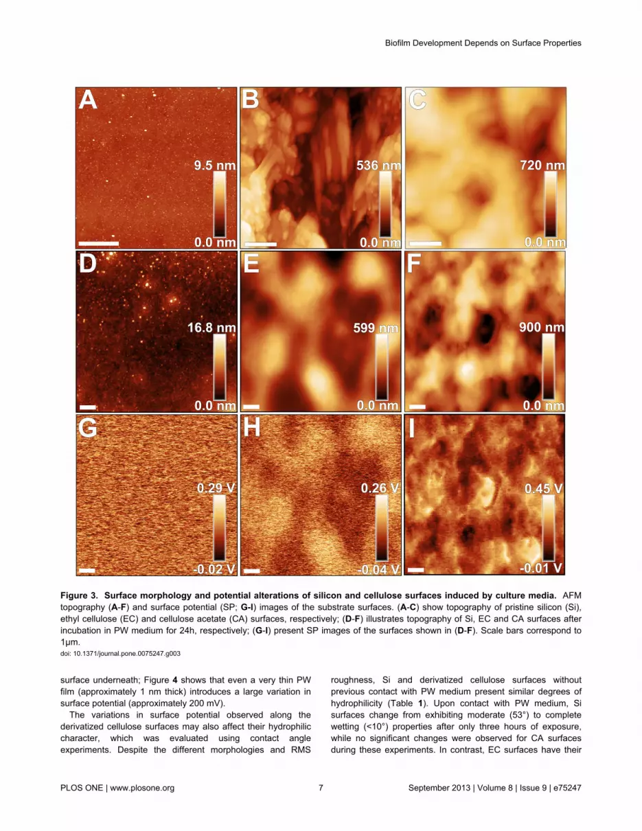
Surface properties in general can also be modified uponinteraction with the culture medium [3-5]. In order to assess therole of such effect on bacterial adhesion and biofilm formation,AFM and contact angle analysis techniques were applied tocharacterize the investigated surfaces before and after contactwith PW medium. AFM topography images reveal differentmorphologies for each pristine surface (exemplary shown inFigure 3). As expected, the Si surface presents a flatmorphology, while the derivatized cellulose films provide anirregular surface structure. CA samples present granularmorphologies homogeneously distributed along the surface(Figure 3), whereas both granular and fiber morphologies were
Figure 1. Biofilm architecture and quantitative biofilm size distribution. Size distribution histograms (A-C) and color enhancedSEM images of biofilms architecture (D-I) grown on bare silicon (Si; A, D, E), ethyl cellulose (EC; B, F, G) and cellulose acetate(CA; C, H, I) substrates in PW medium. Images E, G and I show details of the biofilm edges on each substrate at highermagnification. Scale bars correspond to 20µm for D, F, H and 5µm for E, G, I. The insets in A-C show re-scaled histograms to moreprecisely visualize the presence of larger biofilms.doi: 10.1371/journal.pone.0075247.g001
Biofilm Development Depends on Surface Properties
PLOS ONE | www.plosone.org 5 September 2013 | Volume 8 | Issue 9 | e75247

observed for EC derivatized films (Figure 3). As a consequenceof the different morphologies, these surfaces present distinctRMS roughness values (typically 0.5 nm for Si; 140±40 nm and110±6 nm for EC and CA surfaces, respectively). This scenariochanges upon exposure to PW medium, which emulates thesurface encountered by bacteria upon adhesion in ourexperiments. Despite larger RMS values (approximately 1.5nm), the Si surfaces before and after exposure remain highlyplanar (Figure 3) and homogeneous; in contrast, the PWconditioning film deposited on EC and CA cellulose samplespresents opposite characteristics. In the first case, the fiber andgrain-like morphologies are buried by the conditioning film,although many features still protrude from the smooth surfacedue to the initial height variation on the EC surface.Consequently, the RMS values (80±30 nm) are diminished oncoated EC surfaces (Figure 3). On the other hand, CA surfacespresent more prominent features than the initial morphologyand larger (by approximately 60%) RMS values (180±50 nm).Furthermore, spectral analysis of the obtained topographyimages for CA surfaces shows that the dominant spatialfrequency decreases by a factor of approximately 2, if theconditioning film is present (i.e., from 3.8 to 1.9 µm-1).
These results altogether indicate that the conditioning film isnot homogeneously deposited on CA surfaces; the depositiondoes not precisely follow the topography of the initial film, dueto the changes observed in both RMS roughness and dominantspatial frequency for the CA surface exposed to PW medium.This behavior suggests that the conditioning film does notpurely result from the physical deposition of chemical species
within the PW medium on CA surfaces. To further investigatethis aspect, we measured the surface potential (SP) on Si andderivatized cellulose surfaces. For the latter, SP variationswere observed (Figure S2) indicating an inhomogeneouscharge distribution at the micron-range length scale,particularly when the fiber morphology is present on the ECsample.
After interaction with PW medium, however, oppositebehaviors are observed for the cellulose films: while SP-RMSdiminishes for EC surfaces as the smoothening effect of PWfilm deposition takes place, SP-RMS increases for the CAsurface, and shows dominant spatial frequencies similar tothose evident in the corresponding topography images (Figure3). Despite these opposite trends, the SP-RMS values afterPW exposure are in the same range for both EC and CAsurfaces (typically 50-70 mV), although electrically charged andirregularly-shaped micron size domains are more evident onCA surfaces. Moreover, both derivatized cellulose surfacesshow smaller average SP values (Figure S3) regarding thenearby Si reference, which has also been exposed to PW; theSP inhomogeneities are still present on the surface, asindicated by the darker patches close to the cellulose film edge(Figure S3). The difference in surface potential betweenderivatized cellulose and the nearby Si surface (SP*) dependson the type of cellulose used. Average SP* values obtained forEC and CA were (-150±10) mV and (-200±40) mV,respectively. The effect of the PW film on the electrostaticproperties of the surface was evaluated after locally removingthe film to form a step structure, and thus, exposing the Si
Figure 2. Comparison of biofilm growth on pristine silicon and cellulose acetate surfaces. AFM topography images ofXylella fastidiosa biofilms grown on bare silicon (Si) and cellulose acetate (CA) surfaces side-by-side in periwinkle wilt (PW) medium(scale bar 5 µm). Images are shown with different height contrasts to illustrate individually the CA topography (A) and biofilm (B)more accurately. As a control image, please refer to Figure S2A in supplemental information.doi: 10.1371/journal.pone.0075247.g002
Biofilm Development Depends on Surface Properties
PLOS ONE | www.plosone.org 6 September 2013 | Volume 8 | Issue 9 | e75247
surface underneath; Figure 4 shows that even a very thin PWfilm (approximately 1 nm thick) introduces a large variation insurface potential (approximately 200 mV).
The variations in surface potential observed along thederivatized cellulose surfaces may also affect their hydrophiliccharacter, which was evaluated using contact angleexperiments. Despite the different morphologies and RMS
roughness, Si and derivatized cellulose surfaces withoutprevious contact with PW medium present similar degrees ofhydrophilicity (Table 1). Upon contact with PW medium, Sisurfaces change from exhibiting moderate (53°) to completewetting (<10°) properties after only three hours of exposure,while no significant changes were observed for CA surfacesduring these experiments. In contrast, EC surfaces have their
Figure 3. Surface morphology and potential alterations of silicon and cellulose surfaces induced by culture media. AFMtopography (A-F) and surface potential (SP; G-I) images of the substrate surfaces. (A-C) show topography of pristine silicon (Si),ethyl cellulose (EC) and cellulose acetate (CA) surfaces, respectively; (D-F) illustrates topography of Si, EC and CA surfaces afterincubation in PW medium for 24h, respectively; (G-I) present SP images of the surfaces shown in (D-F). Scale bars correspond to1µm.doi: 10.1371/journal.pone.0075247.g003
Biofilm Development Depends on Surface Properties
PLOS ONE | www.plosone.org 7 September 2013 | Volume 8 | Issue 9 | e75247

Figure 4. Surface potential of culture media conditioning film formed on pristine silicon. AFM topography (A) and surfacepotential (B) images showing the edge of the thin conditioning film caused by periwinkle wilt (PW) medium on silicon (Si) substrate(scale bar 2 µm). The corresponding profiles of height (1) and surface potential differences (2) are shown below.doi: 10.1371/journal.pone.0075247.g004
Biofilm Development Depends on Surface Properties
PLOS ONE | www.plosone.org 8 September 2013 | Volume 8 | Issue 9 | e75247

hydrophilic character enhanced over a longer period of 24 h.These results are better understood by taking into account thedifferent morphologies described above. Stiff and flat surfacessuch as Si contribute to more compact conditioning filmformation, as previously reported [4]. Thus, the exposedsurface for contact angle experiments in this case is mainlycomposed of this film. On the other hand, for EC and CAsurfaces PW medium may take longer to form a continuousconditioning layer; although almost as stiff as Si (as evaluatedfrom force-distance curves using a Si tip in PBS medium,Figure S4, which shows a ~15% difference in stiffness), thesesurfaces present pores and a more irregular morphology whichmay hinder or delay a continuous coverage. Furthermore,surface inhomogeneities - such as surface roughness - mayadditionally affect the contact angle, and thus, the surfacewetting properties [43], as observed for the CA surface.
Our results indicate that surface roughness may not beconsidered an independent variable in X. fastidiosa adhesionand biofilm development. In fact, how surface roughnessactually affects adhesion at a fundamental level is still underdiscussion [44,45]. Liu et al. [44] investigated theoretically andexperimentally the role of surface roughness during adhesion.In this case, the experimental adhesion studies were performedon silicon surfaces with different degrees of roughness usingAFM cantilevers. In contrast to bacterial adhesion and biofilmliterature concerning surface roughness [8,12], the authorsconclude that the adhesion force decreases as surfaceroughness increases for a determined feature size. This isindeed observed in our studies, as the largest biofilm structureshave been observed on the very flat Si surface. Despite thefact that the physical models for adhesion discussed above donot consider the role of surface chemical species orbiomolecules such as e.g., adhesins, our results show thatroughness is not a determinant feature for X. fastidiosaadhesion, as this process is not inhibited on any of the surfacesstudied herein, which show distinctly different roughnessvalues.
Regarding the hydrophobicity/hydrophilicity of the surfaces,hydrophobic termination has been reported as favorable forbacterial adhesion [6,7], but surface properties after interactionwith the culture medium are not usually considered in mostreports. Adsorption of molecules from the surrounding solutionat the substrate surface may change its properties analogouslyto the natural process of conditioning film formation. In a recent
Table 1. Contact angle variation as a function of exposuretime to PW broth for the three surfaces analyzed in thisstudy.
Contact angle [°]
Time in contact with PW browth Si surface EC surface CA surfacew/o contact 53±6 69±4 63±83 h < 10 26±1 63±16 h < 10 32±1 45±112 h < 10 30±1 70±124 h < 10 21±1 67±1
doi: 10.1371/journal.pone.0075247.t001
study [4], we reported variations in physicochemical propertiesof abiotic surfaces due to the conditioning film formed by PWmedium constituents and their effect on X. fastidiosa biofilmformation. Moreover, attached bacterial cells and biofilms areonly observed after time intervals (approximately 4 h) expectedfor changes in hydrophilicity on Si substrates associated withPW medium effects [46]. Considering these effects observedon all surfaces here, our findings suggest that hydrophobicsurfaces do not present favorable conditions for X. fastidiosabiofilm formation. Additionally, the extension of the first layerbeyond the biofilm deposit (Figure 1 E, G, I) is larger for morehydrophilic surfaces, thus suggesting that cell-surfaceinteraction is favored in this case. Furthermore, the largerdiameter of biofilms grown on CA surfaces after 21 days mayresult from a more continuous surface coverage with PW filmexpected from longer growth times [4].
In addition to roughness and hydrophobicity, electrostaticinteractions can also enhance initial bacteria adhesion tosurfaces [7,22] but only few reports have addressed the role ofthe surface potential on such processes [7]. Our experimentalresults show strong evidence of charge distribution changes atabiotic and biotic surfaces due to PW culture medium effect.The Derjaguin-Landau-Verwey-Overbeek (DLVO) theory hasbeen used to explain adhesion of microorganisms to differentinterfaces [47-49]. This theory considers that the initial celladhesion results from van der Waals attractive forces and fromthe repulsive interactions from overlap between the electricaldouble layer of cell and substrate, whose magnitudes aredependent on the surface potentials. Although ourmeasurements neglect ion shielding and other dynamical,electrical effects arising from surface contact to solution, theSP* values and SP-RMS obtained under N2 flow aresignificantly larger than expected from zeta potentialmeasurements [37-39]. Therefore, electrical gradients due tothe micron-sized electrically charged domains on the surface ofthe cellulose substrates may still be present whenever theseare immersed in solution. The interaction potential between thissurface and the bacteria membrane should depend on bothsurface potentials, according to the DLVO theory [50], andthus, the charged domains should most certainly affect themechanisms X. fastidiosa relies upon for adhesion. Thesedata, coupled with our findings of biofilm development, indicatethat higher surface potential values and electrically moreuniform surfaces provide more favorable conditions for X.fastidiosa biofilm formation. Indeed, the micron-sized electricalinhomogeneities present on the cellulose samples are roughlyin the length scale of a bacterial cell. Assuming a negativelycharged cell membrane for Gram-negative bacteria, adhesionwill be certainly affected under such circumstances.
XadA1 protein adhesion characteristics on differentsurface compositions
Several reports have shown the important role of fimbrial andafimbrial adhesins in X. fastidiosa attachment to surfaces andto each other [51,52]. In this particular case, XadA1 adhesin(ORF XF1516) was recently reported as a relevant protein atall stages of biofilm formation [52]. In order to evaluate how thisadhesion protein interacts with surfaces of different
Biofilm Development Depends on Surface Properties
PLOS ONE | www.plosone.org 9 September 2013 | Volume 8 | Issue 9 | e75247

composition and homogeneity, we have measured theadhesion forces between XadA1-functionalized AFM Si tips(Figure S1) and the substrate surfaces using AFM forcespectroscopy (Figure 5 A, E). The control experiments clearlyindicate adhesion peaks also between the surfaces and non-functionalized AFM tips when the PW medium was used(Figure S5). This result reveals that the proteins and peptideswithin the culture medium may promote non-specific adhesion,a role that has been indeed suggested for BSA [53]. Therefore,in an effort to isolate the interaction between XadA1 and thesurface from non-specific interactions, all force-distance curveswere obtained in PBS buffer (pH 7.4), which did not presentany adhesion peaks independent of the surface during controlexperiments (Figure S6).
When recording AFM tip-sample approach curves underthese conditions, no adhesion peaks due to van der Waalsattractive forces were observed likely due to shielding effectsfrom the surrounding ions within the buffer medium. For theretraction curves with XadA1-functionalized AFM tips, Si andEC surfaces present similar results with a high percentage ofadhesion events (Table 2 and Figure 5 B, C). For both cases,the adhesion forces are in the range of 50-60 pN. Multiples ofthese values (Figure 5 C, D) were also found, particularly on
the rough cellulose surface, thereby indicating multiple ruptureevents. Figure 5 (F, G) shows that the measured forces arehomogeneously distributed along the several regions of thesample probed in the experiment for both Si and EC surfaces.Contrarily, the interaction between XadA1 and CA surfaces(Figure 5 D, Table 2) occurs at a much lower percentage andalso with significantly lower adhesion forces (approximately 30pN). Furthermore, these events were localized at specificsample regions probed during these studies; some of the CAregions show basically null adhesion forces (Figure 5 H).
Table 2. Average adhesion forces and percentage of singleadhesion events from force spectroscopy experiments atthe different surfaces analyzed in this study.
Surface Adhesion force [pN] Adhesion events [%]Silicon 50±35 97Ethyl cellulose 57±22 99Cellulose acetate 27±8 20
doi: 10.1371/journal.pone.0075247.t002
Figure 5. Interaction forces of bacterial adhesin XadA1 on silicon and cellulose surfaces. Schematics of the forcespectroscopy (A) measurements at five different positions on the surface where force curves were acquired. The force histograms(B-D) for bare silicon (Si, B) ethyl cellulose (EC, C) and cellulose acetate (CA, D), show the total number of events (please noticethe different scales for the number of events); multiple rupture events were observed for EC and CA. Typical retraction force-distance curves for AFM tips coated with XadA1 adhesion protein (E) for Si (i), EC (ii) and CA (iii). The inset shows the fluorescenceimage of the functionalized AFM tip and cantilever visualized using fluorophore labeled second antibodies (scale bar = 40µm).Measured interaction forces (F-H) between XadA1 coated AFM tip and bare Si (F), EC (G) and CA (H) substrates in PBS buffershown in sequential acquisition order. The colors indicate a different region of the sample probed in each set of force curves. Thebin sizes for (B), (C) were adjusted to 22pN and (D) to 5pN to allow accurate visualization of the molecular interactioncharacteristics.doi: 10.1371/journal.pone.0075247.g005
Biofilm Development Depends on Surface Properties
PLOS ONE | www.plosone.org 10 September 2013 | Volume 8 | Issue 9 | e75247

Therefore, from the molecular point of view, the interactionwith the specific adhesin XadA1 is reduced for CA surfaces inabsence of the PW conditioning film. From the previousanalysis of the surface roughness and surface potential on CAsurfaces exposed to PW, as well as contact anglemeasurements, it is hypothesized that the PW film coverage isnot continuous on CA surfaces. This implies that the initial CAsurface is also exposed upon bacteria adhesion, thus inhibitingthe role of XadA1 at many locations of these substrates, assuggested by the spatial distribution of adhesion eventsreflected in our ensemble of force curves (Figure 5 H). Thishypothesis might also explain the difference in biofilm edgesregarding the two derivatized cellulose films (Figure 1 G, I) ifwe assume that XadA1 is implicated in the initial adhesion tothe surface. However, it is important to notice that the adhesionprocess of X. fastidiosa to CA surfaces may also occur viadifferent adhesins (not investigated in this study) or alternativemechanisms such as e.g., the production of extracellularpolymeric substances (EPS) [46] or via carbohydrate-bindingproteins [5]. Our force spectroscopy control experiments haveshown that proteins such as BSA may also enhance adhesion;similarly to the cell membrane, the surface of the non-functionalized Si AFM tip may exhibit negative charge densitiesdue to the presence of the native oxide [54]. Thus, it isemphasized that although X. fastidiosa adhesion and biofilmdevelopment did not appear enhanced on CA surfaces ascompared to Si, biofilm formation still occurs. These results arein agreement with different colonization rates observed in plant[55].
This conclusion also supports the hypothesis that differentbacterial adhesion mechanisms may be active along the biofilmlife cycle influencing the biofilm architecture, such as thoseobserved in the transmission vector (insect) and plant xylem[18]. Additionally, no shape transitions or morphogenesis, suchas those reported for Candida albicans [56], are observed forX. fastidiosa in all samples analyzed here. We cannot preclude,however, changes in adhesin density and localization at thecell membrane activated by habitat cues.
Endoglucanases gene expression dependence on thesurface properties
Our observations show different biofilm development ratesand architecture also between the two soft, electrically-inhomogeneous cellulose substrates used here. In fact, Killinyet al. [20] have shown that host structural polysaccharidesmediate gene regulation in X. fastidiosa; this regulation createsthe necessary phenotypic changes for vector transmission.Gene expression analysis also evidences the differencebetween the two types of cellulose substrates in our study. X.fastidiosa has genes encoding endoglucanases enzymeswhich may degrade cell wall constituents; the expression ofthese genes could thus be affected by the presence of differentcelluloses on the surface where the cells attach. Theexpression of three genes (Table S1) related with host cell walldegradation (XF0810, XF0818 and XF2708, http://aeg.lbi.ic.unicamp.br/xf/) was analyzed by qRT-PCR forbacteria grown on derivatized cellulose, in comparison tosilicon. Figure 6 shows the gene expression behavior for eachderivatized cellulose surface, using the biofilms grown on Si asreference. The expression of these genes was induced after 7and 21 days especially on EC as surface, and was repressedafter 14 days of biofilm formation. This gene expression patternsuggests a negative feedback mechanism arising from theenzyme level present in the immediate environment.
The expression behavior of the genes involved in host cellwall degradation reveals an up-and-down regulation behaviorduring the biofilm formation, particularly when grown on EC asbiotic surface. This behavior may result from a homeostasisstrategy to regulate the proteins/enzymes levels required forcellular maintenance [57]. Figure 6 also shows a distinctdifference between gene expressions on derivatized cellulosesurfaces. Particularly for XF0818, the expression is alwaysenhanced - at 7 and 21 days - if the bacteria are cultivated onthe EC surface as compared to CA. E. coli clones, whichexpress XF0818, also exhibited cellulose activity and efficientlydegraded cellulose [58]. If the rate of degradation varies withthe substrate - due to the different chemical structures of ECand CA - so does the carbohydrate amount available to thebacteria, which consequently affects cell metabolism, biofilm
Figure 6. Endoglucanase gene expression dependence on surface composition. Gene expression fold change for the threeendoglucanases genes analyzed in the present study for (A) 7, (B) 14 and (C) 21 days of biofilm growth on bare silicon (Si), ethylcellulose (EC) and cellulose acetate (CA) surfaces. One-way ANOVA with Tukey’s post hoc test was applied in (A), (B), (C).Asterisks denote significance level of α=0.05. Bars represent ± standard deviation.doi: 10.1371/journal.pone.0075247.g006
Biofilm Development Depends on Surface Properties
PLOS ONE | www.plosone.org 11 September 2013 | Volume 8 | Issue 9 | e75247

formation and biofilm development rate. This process shouldbe facilitated on the EC surfaces also due to the larger contactarea between the biofilm and the substrate, as compared to CAsurfaces.
These observations clearly indicate the important role ofsurface properties inside the plant or host on X. fastidiosadevelopment. Furthermore, the homogeneity of theseproperties at micro and nano length scales as well as the effectof conditioning films formed due to the necessary experimentalconditions for bacteria survival and proliferation should beconsidered as well. Particularly, from the biological point ofview, the dependence of protein expression and interaction onsurface properties suggests the existence of an adaptationmechanism for variations on the specific xylem vessel and sapcompositions, which the bacterium encounters within infectedplants. In general, our results demonstrate that the surfaceplays an important role in biofilm formation of X. fastidiosa.Moreover, surface properties can be changed by thesurrounding solution. Therefore, we suggest that themanagement strategy to control this phytopathogen shouldconsider the use of compounds such as soil nutritionaltreatments that could change xylem sap properties andconsequently xylem surface characteristics to impair thebacterial colonization.
Conclusion
In summary, our work shows that stiffer and electrically morehomogeneous surfaces with larger surface potential exhibitenhanced X. fastidiosa adhesion and proliferation, likely due toa stronger cell-surface interaction under these circumstances.Changes in biofilm architecture and development - such as thecolonies sizes and morphological edge features - are attributedto the different physicochemical properties of the biotic andabiotic surfaces studied here, particularly to theinhomogeneities at the microscale, which include the effect ofadsorbed species and the conditioning film formed due to theculture medium. The main adhesion protein used by X.fastidiosa, XadA1, presents similar interactions forces for bothSi and EC surfaces; for CA, however, we observed inhibitedXadA1 interaction and a spatial dependence on the regionsprobed by force spectroscopy. Changes in the gene expressionfor endoglucanases are also observed depending on the typeof surface where adhesion takes place; these changes suggestthe influence of the habitat on the bacterial cell metabolism aswell. Moreover, habitat modification can interfere on thebacterial colonization.
Supporting InformationAdditional supporting figures and tables are provided in the
online available supplemental information.
Supporting Information
Figure S1. Epifluorescence images for non-functionalizedsilicon AFM tip (A) and XadA1 coated AFM tip (B). For theXadA1 immobilization proof, the coated AFM tips wereincubated to a specific anti-rabbit IgG antibody for XadA1 and
visualized using a fluorescein labeled anti-rabbit IgG secondantibody. Images were acquired with an inverted Nikon EclipseTE2000U microscope and a photon-counting EMCCD camera(IXON3, Andor, Ireland).(TIF)
Figure S2. AFM topography (A, B) and surface potential(C, D) images of cellulose acetate (A, C) and ethyl cellulose(B, D) thin films (scale bar 1 µm).(TIF)
Figure S3. AFM topography (A, B) and surface potential(C, D) images of cellulose acetate (CA; A, C) and ethylcellulose (EC; B, D) thin film step edges on silicon (Si)substrates after incubation in periwinkle wilt (PW) medium(scale bar 2 µm).(TIF)
Figure S4. Force-distance curves acquired on the threesubstrates studied, using Si tips measured in PBSmedium. The stiffness values of the plotted linear fits (dashedlines) are shown in the corresponding colors.(TIF)
Figure S5. AFM force histogram of a non-functionalizedAFM probe on bare silicon (Si) in periwinkle wilt (PW)medium. The inset shows a zoom-in to illustrate the forcedistribution in more detail including a Gaussian fitting (blackcurve).(TIF)
Figure S6. Typical approach (red) and retraction (blue)force-distance curves of non-functionalized AFM probeson bare silicon (Si; i), ethyl cellulose (EC; ii) and celluloseacetate (CA; iii) in PBS buffer.(TIF)
Table S1. Xylella fastidiosa gene sequences encodingenzymes that may degrade cell wall components, such ascellulose.(DOCX)
Acknowledgements
We thank Prof. José Roberto Ribeiro Bortoleto, UNESP, for hisassistance with contact angle measurements and Alberto L.D.Moreau and Duber M. Murillo Munar, IFGW, UNICAMP, fortheir assistance in SPM image acquisition. We alsoacknowledge the Focused Ion Beam Center UUlm forassistance with SEM imaging.
Author Contributions
Conceived and designed the experiments: GSL RJ BM CKAAS MAC. Performed the experiments: GSL RJ JHC CMRJPT. Analyzed the data: GSL RJ JHC JPT AAS MAC.Contributed reagents/materials/analysis tools: GSL RJ JHCCMR JPT BM AAS MAC. Wrote the manuscript: GSL RJ MAC.
Biofilm Development Depends on Surface Properties
PLOS ONE | www.plosone.org 12 September 2013 | Volume 8 | Issue 9 | e75247

References
1. Muranaka LS, Takita MA, Olivato JC, Kishi LT, de Souza AA (2012)Global Expression Profile of Biofilm Resistance to AntimicrobialCompounds in the Plant-Pathogenic Bacterium Xylella fastidiosaReveals Evidence of Persister Cells. J Bacteriol 194: 4561-4569. doi:10.1128/JB.00436-12. PubMed: 22730126.
2. Sauer K (2003) The genomics and proteomics of biofilm formation.Genome Biol 4: 219. doi:10.1186/gb-2003-4-6-219. PubMed:12801407.
3. Donlan RM (2002) Biofilms: Microbial Life on Surfaces. Emerg InfectDis 8: 881-890. doi:10.3201/eid0809.020063. PubMed: 12194761.
4. Lorite GS, Rodrigues CM, de Souza AA, Kranz C, Mizaikoff B et al.(2011) The role of conditioning film formation and surface chemicalchanges on Xylella fastidiosa adhesion and biofilm evolution. J ColloidInterface Sci 359: 289-298. doi:10.1016/j.jcis.2011.03.066. PubMed:21486669.
5. Killiny N, Almeida RPP (2009) Xylella fastidiosa afimbrial adhesinsmediate cell transmission to plants by leafhopper vectors. Appl EnvironMicrobiol 75: 521-528. doi:10.1128/AEM.01921-08. PubMed:19011051.
6. Teixeira P, Oliveira R (1999) Influence of surface characteristics on theadhesion of Alcaligenes denitrificans to polymeric substrates. J AdhesSci Technol 13: 1287-1294. doi:10.1163/156856199X00190.
7. Sheng X, Ting YP, Pehkonen SO (2008) The influence of ionicstrength, nutrients and pH on bacterial adhesion to metal. J ColloidInterface Sci 321: 256-264. doi:10.1016/j.jcis.2008.02.038. PubMed:18343395.
8. Oh YJ, Lee NR, Jô W, Jung WK, Lim JS (2009) Effects of subtrates onbiofilm formation observed by atomic force microscopy.Ultramicroscopy 109: 874-880.
9. Gubner R, Beech IB (2000) The effect of extracellular polymericsubstances on the attachment of Pseudomonas NCIMB 2021 to AISI304 and 316 stainless steel. Biofouling 15: 25-36. doi:10.1080/08927010009386295. PubMed: 22115289.
10. Lichter JA, Thompson MT, Delgadillo M, Nishikawa T, Rubner MF et al.(2008) Substrata Mechanical Stiffness Can Regulate Adhesion ofViable Bacteria. Biomacromolecules 9: 1571-1578. doi:10.1021/bm701430y. PubMed: 18452330.
11. Saha N, Monge C, Dulong V, Picart C, Glinel K (2013) Influence ofPolyelectrolyte Film Stiffness on Bacterial Growth. Biomacromolecules14: 520-258. doi:10.1021/bm301774a. PubMed: 23289403.
12. Rizzello L, Sorce B, Sabella S, Vecchio G, Galeone A et al. (2011)Impact of nanoscale topography on genomics and proteomics ofadherent bacteria. ACS Nano 5: 1865-1876. doi:10.1021/nn102692m.PubMed: 21344880.
13. Möller J, Luehmann T, Hall H, Vogel V (2012) The Race to the Pole:How High-Aspect Ratio Shape and Heterogeneous Environments limitPhagocytosis of Filamentous Escherichia coli Bacteria byMacrophages. Nano Lett 12: 2901-2905. doi:10.1021/nl3004896.PubMed: 22591454.
14. Hochbaum AI, Aizenberg J (2010) Bacteria Pattern Spontaneously onPeriodic Nanostructure Arrays. Nano Lett 10: 3717-3721. doi:10.1021/nl102290k. PubMed: 20687595.
15. Mansfield J, Genin S, Magori S, Citovsky V, Sriariyanum M et al. (2012)Top 10 plant pathogenic bacteria in molecular plant pathology. MolPlant Pathol 13: 614-629. doi:10.1111/j.1364-3703.2012.00804.x.PubMed: 22672649.
16. de Lima JEO, Miranda VS, Hartung JS, Brlansky RH, Coutinho A et al.(1998) Coffee leaf scorch bacterium: axenic culture, pathogenicity, andcomparison with Xylella fastidiosa of citrus. Plant Dis 82: 94-97. doi:10.1094/PDIS.1998.82.1.94.
17. Purcell AH, Hopkins DL (1996) Fastidious xylem-limited bacterial plantpathogens. Annu Rev Phytopathol 34: 131-151. doi:10.1146/annurev.phyto.34.1.131. PubMed: 15012538.
18. Newman KL, Almeida RP, Purcell AH, Lindow SE (2004) Cell-cellsignaling controls Xylella fastidiosa interactions with both insects andplants. Proc Natl Acad Sci U S A 101: 1737-1742. doi:10.1073/pnas.0308399100. PubMed: 14755059.
19. Chatterjee S, Wistrom C, Lindow SE (2008) A cell–cell signaling sensoris required for virulence and insect transmission of Xylella fastidiosa.Proc Natl Acad Sci U S A 105: 2670-2675. doi:10.1073/pnas.0712236105. PubMed: 18268318.
20. Killiny N, Almeida RP (2009) Host structural carbohydrate inducesvector transmission of a bacterial plant pathogen. Proc Natl Acad Sci US A 106: 22416-22420. doi:10.1073/pnas.0908562106. PubMed:20018775.
21. Killiny N, Prado SS, Almeida RP (2010) Chitin utilization by the insect-transmitted bacterium Xylella fastidiosa. Appl Environ Microbiol 76:6134-6140. doi:10.1128/AEM.01036-10. PubMed: 20656858.
22. Osiro D, Colnago LA, Otoboni AM, Lemos EG, de Souza AA et al.(2004) A kinect model for Xylella fastidiosa adhesion, biofilm formationand virulence. FEMS Microbiol Lett 236: 313-318. doi:10.1111/j.1574-6968.2004.tb09663.x. PubMed: 15251213.
23. Moreau ALD, Lorite GS, Rodrigues CM, de Souza AA, Cotta MA (2009)Fractal analysis of Xylella fastidiosa biofilm formation. J Appl Phys 106:024702. doi:10.1063/1.3173172.
24. Cobine PA, Cruz LF, Navarrete F, Duncan D, Tygart M et al. (2013)Xylella fastidiosa Differentially Accumulates Mineral Elements in Biofilmand Planktonic Cells. PLOS ONE 8(1): e54936. doi:10.1371/journal.pone.0054936. PubMed: 23349991.
25. Showalter AM (1993) Structure and function of plant cell wall proteins.Plant Cell. 5: 9-23. doi:10.1105/tpc.5.1.9. PubMed: 8439747.
26. Jamet E, Albenne C, Boudart G, Irshad M, Canut H et al. (2008)Recent advances in plant cell wall proteomics. Proteomics 8: 893-908.doi:10.1002/pmic.200700938. PubMed: 18210371.
27. Albenne C, Canut H, Boudart G, Zhang Y, San Clemente H et al.(2009) Plant cell wall proteomics: mass spectrometry data, a trove forresearch on protein structure/function relationships. Mol Plants 2:977-989.
28. Albenne C, Canut H, Jamet E (2013) Plant cell wall proteomics: theleadership of Arabidopsis thaliana. Front. Plant Sci 4: 111.
29. Narváez AC, Chiaramonte T, Vicaro KO, Clerici JH, Cotta MA (2009)Evidence of space charge regions within semiconductor nanowiresfrom Kelvin probe force microscopy. Nanotechnology 20: 465705. doi:10.1088/0957-4484/20/46/465705. PubMed: 19843990.
30. Meng Y, Li Y, Galvani CD, Hao G, Turner JN et al. (2005) Upstreammigration of Xylella fastidiosa via pilus-driven twitch- ing motility. JBacteriol 187: 5560-5567. doi:10.1128/JB.187.16.5560-5567.2005.PubMed: 16077100.
31. Davis MJ, French WJ, Schaad NW (1981) Axenic culture of thebacteria associated with phony disease of peach and plum leaf scald.Curr Microbiol 6: 309-314. doi:10.1007/BF01566883.
32. Weaver JMR, Abraham DW (1991) High resolution atomic forcemicroscopy potentiometry. J Vac Sci Technol B 9: 1559. doi:10.1116/1.585423.
33. Jacobs HO, Leuchtmann P, Homan OJ, Stemmer AJ (1998) Resolutionand contrast in Kelvin probe force microscopy. J Appl Phys 84: 1168.
34. Shusterman S, Raizman A, Sher A, Paltiel Y, Schwarzman A et al.(2007) Nanoscale mapping of strain and composition in quantum dotsusing kelvin probe force microscopy. Nano Lett 7: 2089-2093. doi:10.1021/nl071031w.
35. Enevoldsen GH, Glatzel T, Christensen MC, Lauritsen JV,Besenbacher F (2008) Atomic scale Kelvin probe force microscopystudies of the surface potential variations on the TiO2(110) surface.Phys Rev Lett 100: 236104. doi:10.1103/PhysRevLett.100.236104.PubMed: 18643521.
36. Melitz W, Shena J, Kummel AC, Lee S (2011) Kelvin probe forcemicroscopy and its application. Surf Sci Rep 66: 1-27. doi:10.1016/j.surfrep.2010.10.001.
37. Hubbe MA (2006) Sensing the electrokinetic potential of cellulosic fibersurfaces. Bioresources 1: 116-149.
38. Jin H, Zhou W, Cao J, Stoyanov SD, Blijdenstein TBJ et al. (2012)Super stable foams stabilized by colloidal ethyl cellulose particles. SoftMatter 8: 2194-2205. doi:10.1039/c1sm06518a.
39. Elimelech M, Chen WH, Waypa JJ (1994) Measuring the Zeta(Electrokinetic) Potential of Reverse Osmosis Membranes by aStreaming Potential Analyzer. Desalination 95: 269-286. doi:10.1016/0011-9164(94)00064-6.
40. Asally M, Kittisopikul M, Rué P, Du Y, Hu Z et al. (2012) Localized celldeath focuses mechanical forces during 3D patterning in a biofilm. ProcNatl Acad Sci U S A 109: 18891-18896. doi:10.1073/pnas.1212429109.PubMed: 23012477.
41. Hunt SM, Werner EM, Huang B, Hamilton MA, Stewart PS (2004)Hypothesis for the Role of Nutrient Starvation in Biofilm Detachment.Appl Environ Microbiol 70: 7418-7425. doi:10.1128/AEM.70.12.7418-7425.2004. PubMed: 15574944.
42. Bakker DP, Huijs FM, de Vries J, Klijnstra JW, Busscher HJ et al.(2003) Bacterial Deposition to Fluoridated and Non-FluoridatedPolyurethane Coatings with Different Elastic Modulus and SurfaceTension in a Parallel Plate and a Stagnation Point Flow Chamber.Colloids Surf B Biointerfaces 32: 179-190. doi:10.1016/S0927-7765(03)00159-0.
Biofilm Development Depends on Surface Properties
PLOS ONE | www.plosone.org 13 September 2013 | Volume 8 | Issue 9 | e75247

43. de Gennes PG (1985) Wetting: statics and dynamics. Rev Mod Phys57: 827-863. doi:10.2466/pr0.1985.57.3.827.
44. Liu DL, Martin J, Burnham NA (2007) Optimal roughness for minimaladhesion. Appl Phys Lett 91: 043107.
45. Liu DL, Martin J, Burnham NA (2010) Which Fractal ParameterContributes Most to Adhesion? J Adhes Sci Technol 24: 2383-2396.doi:10.1163/016942410X508280.
46. Lorite GS, de Souza AA, Neubauer D, Mizaikoff B, Kranz C et al.(2013) On the role of extracellular polymeric substances during earlystages of Xylella fastidiosa biofilm formation. Colloids Surf BBiointerfaces 102: 519-525. doi:10.1016/j.colsurfb.2012.08.027.PubMed: 23164974.
47. Azeredo J, Visser J, Oliveira R (1999) Exopolymers in bacterialadhesion: interpretation in terms of DLVO and XDLVO theories.Colloids Surf B Biointerfaces 14: 141-148.
48. Hermansson M (1999) The DLVO theory in microbial adhesion.Colloids Surf B Biointerfaces 14: 105-119. doi:10.1016/S0927-7765(99)00029-6.
49. Dorobantu LS, Bhattacharjee S, Foght JM, Gray MR (2009) Analysis offorce interactions between AFM tips and hydrophobic bacteria usingDLVO theory. Langmuir 25: 6968-6976. doi:10.1021/la9001237.PubMed: 19334745.
50. Hogg R, Healy TW, Fuerstka DW (1966) Dispersions, translator, MutualCoagulation of Colloidal. Faraday Society 66: 1638-1651
51. Feil H, Feil WS, Lindow SE (2007) Contribution of fimbrial and afimbrialadhesins of Xylella fastidiosa to attachment to surfaces and virulence togrape. Phytopathology 97: 318-324. PubMed: 18943651.
52. Caserta R, Takita MA, Targon ML, Rosselli-Murai LK, de Souza AP etal. (2010) Expression of Xylella fastidiosa fimbrial and afimbrial proteinsduring biofilm formation. Appl Environ Microbiol 76: 4250-4259. doi:10.1128/AEM.02114-09. PubMed: 20472735.
53. Roach P, Farrar D, Perry CC (2005) Interpretation of proteinadsorption: surface-induced conformational changes. J Am Chem Soc127: 8168-8173. PubMed: 15926845.
54. Ishii M, Hamilton B (2004) Electron trapping at the Si (111) atomic stepedge. Appl Phys Lett 85: 1610-1612. doi:10.1063/1.1787162.
55. Chatterjee S, Almeida RPP, Lindow SE (2008) Linving in two worlds:the plant and insect lifestyles of Xylella fastidiosa. Annu. RevPhytophatol 46: 243-271.
56. Beaussart A, Alsteens D, El-Kirat-Chatel S, Lipke PN, Kucharíková S etal. (2012) Single-molecule imaging and functional analysis of Alsadhesins and mannans during Candida albicans morphogenesis. ACSNano 6: 10950-10964. PubMed: 23145462.
57. Robledo M, Rivera L, Jiménez-Zurdo JI, Rivas R, Dazzo F et al. (2012)Role of Rhizobium endoglucanase CelC2 in cellulose biosynthesis andbiofilm formation on plant roots and abiotic surfaces. Microb CellFactories 11: 125. PubMed: 22970813.
58. Wulff NA, Carrer H, Pascholati SF (2006) Expression and purification ofcellulase Xf818 from Xylella fastidiosa in Escherichia coli. CurrMicrobiol 53: 198-203. doi:10.1007/s00284-005-0475-2. PubMed:16874548.
Biofilm Development Depends on Surface Properties
PLOS ONE | www.plosone.org 14 September 2013 | Volume 8 | Issue 9 | e75247


