Embed Size (px)
Citation preview
Polystyrene nanoparticle trafficking across MDCK-II
Farnoosh Fazlollahi, MS1,8,*, Susanne Angelow, PhD2,5, Nazanin R. Yacobi, PhD1,8, RonaldMarchelletta, PhD6, Alan S.L. Yu, PhD2,5, Sarah F. Hamm-Alvarez, PhD6, Zea Borok,MD1,2,3,†, Kwang-Jin Kim, PhD1,2,5,6,7, and Edward D. Crandall, PhD, MD1,2,4,8,‡
1 Will Rogers Institute Pulmonary Research Center, University of Southern California, LosAngeles, CA, USA2 Department of Medicine, University of Southern California, Los Angeles, CA, USA3 Department of Biochemistry and Molecular Biology, University of Southern California, LosAngeles, CA, USA4 Department of Pathology, University of Southern California, Los Angeles, CA, USA5 Department of Physiology and Biophysics, University of Southern California, Los Angeles, CA,USA6 Department of Pharmacology and Pharmaceutical Sciences, University of Southern California,Los Angeles, CA, USA7 Department of Biomedical Engineering, University of Southern California, Los Angeles, CA,USA8 Mork Family Department of Chemical Engineering and Materials Science, University ofSouthern California, Los Angeles, CA, USA
AbstractPolystyrene nanoparticles (PNP) cross rat alveolar epithelial cell monolayers via non-endocytictranscellular pathways. To evaluate epithelial cell type-specificity of PNP trafficking, we studiedPNP flux across Madin Darby canine kidney cell II monolayers (MDCK-II). Effects of calciumchelation (EGTA), energy depletion (sodium azide (NaN3) or decreased temperature), andendocytosis inhibitors methyl-β-cyclodextrin (MBC), monodansylcadaverine and dynasore weredetermined. Amidine-modified PNP cross MDCK-II 500 times faster than carboxylate-modifiedPNP. PNP flux did not increase in the presence of EGTA. PNP flux at 4°C and after treatmentwith NaN3 decreased 75% and 80%, respectively. MBC exposure did not decrease PNP flux,whereas dansylcadaverine- or dynasore-treated MDCK-II exhibited ~80% decreases in PNP flux.Confocal laser scanning microscopy revealed intracellular colocalization of PNP with clathrinheavy chain. These data indicate that PNP translocation across MDCK-II (1) occurs via clathrin-mediated endocytosis and (2) is dependent upon PNP physicochemical properties. We concludethat uptake/trafficking of nanoparticles into/across epithelia is dependent both on properties of thenanoparticles and the specific epithelial cell type.
*To whom correspondence should be addressed: Farnoosh Fazlollahi, M.S., Department of Medicine, University of SouthernCalifornia, HMR 914, 2011 Zonal Avenue, Los Angeles, CA 90033, USA, Office: 323 442-1217, Fax: 323 226-2899,[email protected].†Z. Borok is Ralph Edgington Chair in Medicine.‡E. D. Crandall is Hastings Professor and Kenneth T. Norris Jr. Chair of Medicine.Publisher's Disclaimer: This is a PDF file of an unedited manuscript that has been accepted for publication. As a service to ourcustomers we are providing this early version of the manuscript. The manuscript will undergo copyediting, typesetting, and review ofthe resulting proof before it is published in its final citable form. Please note that during the production process errors may bediscovered which could affect the content, and all legal disclaimers that apply to the journal pertain.
NIH Public AccessAuthor ManuscriptNanomedicine. Author manuscript; available in PMC 2012 October 1.
Published in final edited form as:Nanomedicine. 2011 October ; 7(5): 588–594. doi:10.1016/j.nano.2011.01.008.
NIH
-PA Author Manuscript
NIH
-PA Author Manuscript
NIH
-PA Author Manuscript
Keywordsepithelial transport; endocytosis; clathrin; dynamin; surface charge
BackgroundNanoparticles (NP) are defined as particles with at least one dimension <100 nm. Small size,large surface area, high surface reactivity and composition-dependent characteristics of NPare uniquely suited for various nanoscale operations [1,2,3]. Engineered NP are promisingcandidates for therapeutic and diagnostic applications in biomedicine (e.g., highly sensitiveand stable probes to detect changes in intracellular molecules [4], early detection anddiagnosis [5], and targeted delivery of therapeutics to specific cell types [4,5]), althoughsome NP exhibit cellular toxicity [3,6]. Internalization of NP into cells can take place viaendocytic and/or non-endocytic pathways [7,8,9,10,11]. It has been reported that uptake ofNP into cells is influenced by surface charge and size of NP [8,9,12,13,14]. Understandingmechanisms of NP interactions with cells of interest will help lead to improved design of NPas efficient and safe reporters/carriers for biomedical investigations and interventions [8].
Our laboratory recently reported the trafficking profile of polystyrene NP (PNP) withvariable size and surface charge across primary cultured rat alveolar epithelial cellmonolayers (RAECM) [13,14]. Trafficking rates of PNP across RAECM are dependent onboth surface charge density and size, in that we found (a) ~20–40 times greater flux forpositively (vs negatively) charged PNP and (b) larger PNP (100 or 120 nm) cross RAECM~3–4 times slower than smaller PNP (20 nm) [14]. PNP appear to be translocated acrossRAECM via non-endocytic transcellular pathways, possibly involving diffusion across thelipid bilayer of cell plasma membranes [13].
In this study, we utilized Madin Darby canine kidney cell line monolayers (MDCK-II) as analternative epithelial model with which to study characteristics of PNP uptake/trafficking.MDCK-II are thought to represent the epithelial barrier of the renal tubule [15,16]. Effectsof size and surface charge density of PNP, and effects of EGTA and inhibitors ofendocytosis, on PNP trafficking rates across MDCK-II were investigated. We found thatprocesses likely involved in translocation of PNP across MDCK-II are energy- and surfacecharge-dependent and, in contrast to PNP trafficking across RAECM, take place viaclathrin-mediated endocytosis. These data indicate that characteristics of NP uptake/trafficking across epithelial barriers are both cell type-specific and dependent on NPphysicochemical properties.
MethodsNanoparticles
Fluorescently labeled polystyrene nanoparticles (PNP) were purchased from Thermo FisherScientific (Waltham, MA). Carboxylate-modified (COO-, 20 and 100 nm diameter with−304.3 and −320 μEq/g surface charge, respectively) and amidine-modified (HNC-NH2+,20 and 120 nm with 80.2 and 39.7 μEq/g surface charge, respectively) PNP were used.Excitation/emission wavelengths for carboxylate- and amidine-modified PNP are 580/605and 490/515 nm, respectively.
MDCK-IIMDCK-II cells were plated on day 0 onto tissue culture-treated polycarbonate filters(Transwell, 12 mm diameter, 0.4 μm diameter pores, Corning-Costar, Cambridge, MA) at
Fazlollahi et al. Page 2
Nanomedicine. Author manuscript; available in PMC 2012 October 1.
NIH
-PA Author Manuscript
NIH
-PA Author Manuscript
NIH
-PA Author Manuscript
1.5 ×10⁵ cells/cm2 and maintained in Dulbecco’s modified Eagle’s (DME) medium (ThermoFisher Scientific) with 5% fetal bovine serum (Thermo Fisher Scientific), 200 U/mLpenicillin (Sigma, St. Louis, MO), 200 μg/mL streptomycin (Sigma) and 2% L-glutamine(Sigma) at 37°C in a humidified atmosphere of 5% CO2 + 95% air. Cells were fed everyother day starting on day 3 in culture when they form confluent monolayers. A rapidscreening device (Millicell-ERS; Millipore, Bedford, MA) was used to checktransmonolayer electrical resistance (Rt). Confluent MDCK-II were studied on days 5–7 (Rt~150 Ωcm2).
Apical-to-basolateral PNP fluxTo measure rates of PNP trafficking across MDCK-II, apical fluid was replaced at t = 0 withfresh culture medium containing various PNP at 176 μg/mL. Apical-to-basolateral (a-b) fluxat 37°C was estimated from PNP appearing in basolateral fluid over 24 h. PNPconcentrations in downstream fluid were estimated spectrofluorometrically usingSpectraMax M2 (Molecular Devices, Sunnyvale, CA). Apical [PNP] was determined at t = 0and 24 h. [PNP] was calculated using standard curves generated with known concentrationsof PNP suspended in culture medium. Flux was calculated as J = (CV)/S/Δt, where J is flux(a-b) of PNP, C is [PNP] in basolateral fluid at Δt, V is basolateral fluid volume, S isnominal surface area of monolayer (1.13 cm2) and Δt is length of time for flux measurement.
To determine if decreased tight junctional resistance leads to increased trafficking rates, fluxof various PNP was measured in the presence of 2 mM ethylene glycol-bis-(2-aminoethyl)-N,N,N′,N′-tetraacetic acid (EGTA, Sigma) in both apical and basolateral fluids. Monolayerswere pretreated with EGTA for 30 min, after which apical fluid was replaced at t = 0 withfresh culture medium containing various PNP at 176 μg/mL and 2 mM EGTA.
To determine effects of metabolic inhibition/energy depletion, flux across MDCK-II wasmeasured at 4°C or in the presence of 10 mM sodium azide (Sigma) [17]. In the latterexperiments, MDCK-II were bathed on both sides with culture medium containing sodiumazide for 30 min prior to and during apical exposure to 176 μg/mL PNP.
To explore mechanisms of PNP translocation, effects of inhibition of lipid raft-mediatedendocytosis (including caveolin-mediated endocytosis) were evaluated using methyl-β-cyclodextrin [18,19], monodansylcadaverine was employed for inhibition of clathrin-mediated endocytosis [20,21,22], and dynasore was used to inhibit dynamin-dependentendocytosis (including both clathrin- and caveolin-mediated endocytosis) [23]. Monolayerswere bathed on both sides with culture medium containing 10–200 μM methyl-β-cyclodextrin (Sigma), 200 μM monodansylcadaverine (Sigma) or 80 μM dynasore (Sigma)for 30 min prior to replacing apical fluid at t = 0 with fresh culture medium containing both176 μg/mL of various PNP and specific endocytosis inhibitors.
Immunofluorescence and confocal laser scanning microscopyMDCK-II were exposed apically to 176 μg/mL amidine-modified (120 nm) or carboxylate-modified (100 nm) PNP at 37°C for 24 h. Exposed monolayers were washed 3 times withice-cold phosphate buffered saline (PBS, pH 7.2) and fixed in 3.7% formaldehyde (J.T.Baker, Phillipsburg, NJ) at room temperature for 15 min. Monolayers were then treated with0.5% Triton X-100 (TX-100, Bio-Rad, Hercules, CA) at room temperature for 15 min,followed by rinsing with PBS and blocking with PBS containing both 5% BSA and 0.2%TX-100 for 1 h at room temperature. Mouse antibody against human zonula occludens-1(ZO-1, Zymed Laboratories, San Francisco, CA) diluted 1:100 using 1% BSA in PBS wasincubated with fixed monolayers at 37°C for 1 h, followed by washing with PBS and furtherincubating monolayers with secondary antibody (goat anti-mouse antibody conjugated to
Fazlollahi et al. Page 3
Nanomedicine. Author manuscript; available in PMC 2012 October 1.
NIH
-PA Author Manuscript
NIH
-PA Author Manuscript
NIH
-PA Author Manuscript
Alexa 594 or Alexa 488, Invitrogen) diluted 1:100 using 1% BSA in PBS at 37°C for 1 h.Monolayers were then rinsed with PBS and mounted on microscope slides with mountingmedium (containing the nuclear staining dye 4′,6-diamidino-2-phenylindole (DAPI) (Vector,Burlingame, CA)).
For labeling clathrin heavy chain, MDCK-II were fixed and permeablized with ice-coldethanol at −20°C for 5 min and blocked with PBS containing both 5% BSA and 0.2%TX-100 at room temperature for 1 h. Mouse anti-human clathrin heavy chain antibody (BDBiosciences, San Jose, CA) diluted 1:100 in 1% BSA in PBS was incubated with fixedmonolayers for 1 h at 37°C. After washing monolayers 3 times with PBS, secondaryantibody (goat anti-mouse antibody labeled with Alexa 488, Invitrogen) in 1% BSA in PBSwas incubated with monolayers at 37°C for 1 h. Monolayers were then rinsed with PBS andmounted on microscope slides with mounting medium containing DAPI. Negative controlsinclude: i) monolayers incubated with PNP in the presence of 200 μM dansylcadaverine for1 h, ii) monolayers not exposed to PNP, and iii) monolayers incubated with a primary orsecondary antibody alone. These controls were similarly processed as above. Images wereacquired with a Zeiss confocal laser scanning microscope (510 Meta NLO CLSM imagingsystem, Jena, Germany) equipped with argon and red/green HeNe lasers mounted on avibration-free table and attached to an incubation chamber.
Data analysisData are presented as mean ± standard deviation (n = total number of monolayers). Student’st-tests were performed for comparisons of two group means. One-way analysis of variancefollowed by post-hoc tests based on modified Newman-Keuls procedures was performed forcomparisons of more than two group means. P<0.05 was considered statistically significant.
ResultsFigure 1A shows trafficking rates of amidine-modified (positively charged 20 and 120 nm)and carboxylate-modified (negatively charged 20 and 100 nm) PNP across MDCK-II withapical [PNP] of 176 μg/mL. Trafficking of positively charged PNP is 500 times faster thanthat for comparably sized but negatively charged PNP. Trafficking rates of smaller (20 nm)PNP across MDCK-II are not significantly different from those of larger (100 and 120 nm)PNP with similar surface charge. Figure 1B shows an expanded view of carboxylate-modified (negatively charged 20 and 100 nm) PNP trafficking rates across MDCK-II.
Confocal photomicrographs (at 1 μm intervals) of MDCK-II exposed apically to 176 μg/mLPNP (amidine-modified 120 nm) for 24 h are shown in Figure 2(A-D). PNP are clearly seenintracellularly. Similar observations were noted with carboxylated-modified 100 nm PNP(data not shown).
Monolayer resistance of MDCK-II decreased >95% in the presence of 2 mM EGTA (whichchelates free Ca2+ in bathing fluids). Flux of amidine-modified PNP across MDCK-II wasnot increased in the presence of EGTA (Figure 3). It can be noted that flux of 120 nmamidine modified PNP across MDCK-II was decreased by EGTA treatment, while that of 20nm amidine-modified PNP did not change. On the other hand, flux of carboxylated (20 and100 nm) PNP in the presence of EGTA were not significantly different from those observedfor control (i.e., EGTA-free) monolayers (data not shown).
Flux of amidine-modified (20 and 120 nm) PNP decreased by ~75% across MDCK-II at 4°Ccompared to control at 37°C (Figure 3), while carboxylate-modified (20 and 100 nm) PNPflux decreased by ~60% at 4°C compared to control at 37°C (data not shown). When cellularATP was decreased by treating monolayers with 10 mM NaN3, flux of positively charged
Fazlollahi et al. Page 4
Nanomedicine. Author manuscript; available in PMC 2012 October 1.
NIH
-PA Author Manuscript
NIH
-PA Author Manuscript
NIH
-PA Author Manuscript
PNP (20 and 120 nm) decreased by ~80% compared to those of NaN3-free control (Figure3).In contrast, flux of carboxylated (20 and 100 nm) PNP measured in the presence of NaN3was not significantly different from those observed for control (i.e., in the absence of NaN3)monolayers. Rt of MDCK-II at 4°C and of NaN3-treated MDCK-II did not decreasecompared to respective controls.
Figure 4 shows flux of amidine-modified PNP (20 and 120 nm) in the presence of 10 μMMBC, 200 μM dansylcadaverine or 80 μM dynasore. Flux of these positively charged PNPdid not decrease in the presence of 10–200 μM MBC. Similarly, flux of carboxylated PNP(20 and 100 nm) measured in the presence of MBC was not significantly different fromthose observed in the absence of MBC (data not shown). Rt of MDCK-II treated with MBCdecreased by ~20%, compared to that of control monolayers.
In the presence of dansylcadaverine or dynasore, flux of amidine-modified PNP (20 and 120nm) decreased by ~85% compared to controls. On the other hand, flux of carboxylated PNP(20 and 100 nm) measured in the presence of dansylcadaverine or dynasore were notsignificantly different from respective controls (data not shown). Rt of MDCK-II treatedwith dansylcadaverine or dynasore decreased by ~50% and increased by ~50%, respectively,compared to those of control monolayers.
Figure 5 (Panels A and B) shows colocalization of intracellular amidine-modified 120 nmPNP with clathrin heavy chain after 1 h exposure to apical [PNP] of 176 μg/mL. WhenMDCK-II cells were co-incubated apically with 200 μM monodansylcadaverine and 120 nmpositively charged PNP for 1 h and processed for confocal microscopy, very few amidine-modified 120 nm PNP were found inside the cells. The few PNP found intracellularly didnot colocalize with clathrin heavy chain in these cells (Figure 5, Panel C).
DiscussionThis study demonstrates that translocation of PNP across MDCK-II occurs transcellularly ina manner significantly dependent on surface charge. Positively charged PNP traverseMDCK-II via a clathrin-mediated pathway. The mechanism of PNP trafficking acrossepithelial monolayers (i.e., MDCK-II vs RAECM) appears to be highly cell type-specific.
We recently reported that cationic PNP (amidine-modified) are translocated across RAECM20–40 times faster than anionic PNP (carboxyl-modified) [14]. By contrast, the traffickingrates of amidine-modified PNP across MDCK-II are 500 times faster than those of carboxyl-modified PNP. Negatively charged components (e.g., glycocalyx) on cell plasmamembranes of epithelia may be at least in part responsible for promoting translocation ofpositively charged (but not negatively charged) NP, although titration of the membranenegative charge on RAECM caused only a relatively small decrease in amidine-modifiedPNP flux [13]. Harush-Frenkel et al [9] reported 2-fold greater uptake of cationic(polyethylene glycol)-D,L-polylactide (PEG-PLA; ~100 nm) NP in MDCK cells than that ofcomparably sized anionic PEG-PLA NP. Des Rieux et al [24] reported that amidine-coatedpolystyrene particles (200 nm) have higher hydrophobicity and translocate faster acrossintestinal epithelial cell monolayers (Caco-2 cells) than similarly sized carboxylate-coatedpolystyrene particles. Our laboratory reported that positively charged PNP are morehydrophobic than negatively charged PNP [13], which may have contributed to more rapidtranslocation of amidine-modified PNP through the lipid bilayers of RAECM plasmamembranes.
Trafficking of small hydrophilic solutes across epithelial barriers can occur via paracellularpathways [25]. Although treatment of epithelial cells with EGTA (a calcium chelator) isreported to increase paracellular permeability by changing the distribution of a number of
Fazlollahi et al. Page 5
Nanomedicine. Author manuscript; available in PMC 2012 October 1.
NIH
-PA Author Manuscript
NIH
-PA Author Manuscript
NIH
-PA Author Manuscript
tight and adherens junctional/cytoskeletal proteins [25,26,27,28], including redistribution ofsome of these proteins into intracellular compartments by clathrin-mediated endocytosis[29,30], it is unknown if NP traverse normal and/or EGTA-disrupted tight junctions. In thisstudy, trafficking rates of PNP (small and large; positive and negative surface charge) acrossMDCK-II in the presence of 2 mM EGTA did not increase, indicating that these PNP do nottraverse MDCK-II via tight junctional pathways under normal conditions or followingEGTA treatment. The mechanism(s) responsible for the EGTA-induced decrease in flux of120 nm (but not 20 nm) amidine-modified PNP are currently unknown, although we canspeculate that the low extracellular Ca2+ might have altered formation of clathrin-coatedpits. Trafficking rates of 20 and 100 nm carboxylate-modified PNP across MDCK-II do notappear to take place via endocytosis with or without EGTA treatment. These findings, alongwith localization of PNP predominantly in cytoplasm but not at cell-cell junctions, suggeststhat PNP cross MDCK-II via transcellular pathways. Yacobi et al [13] concluded thattranslocation of PNP across RAECM also takes place predominantly via transcellularpathways.
PNP trafficking was decreased by ~75% across MDCK-II when temperature was loweredfrom 37 to 4°C. In this regard, Dausend et al [7] reported uptake into HeLa cells of PNP(amino group- and cetyltrimethylammonium chloride-modified at particle surfaces with~100 nm diameter) was decreased by ~60% at 4°C compared to that observed at 37°C.Guerriero et al [31] reported ~95% decrease in cellular ATP level after treating MDCK-IIcells with 2% (10 mM) sodium azide. In this study, 10 mM sodium azide reduced traffickingrates of positively charged PNP by ~75% across MDCK-II. These data suggest that ATP/energy-dependent mechanism(s) may be involved in translocation of positively charged PNPacross MDCK-II.
Methyl-β-cyclodextrin extracts cholesterol from plasma membranes and therefore inhibitslipid raft-mediated endocytosis (including caveolin-mediated endocytosis, CLIC/GEECendocytosis, arf6-medited endocytosis, flotillin-mediated endocytosis and macropinocytosis[19,22]). Flux of PNP observed in the presence of 10–200 μM methyl-β-cyclodextrin did notdecrease, suggesting that translocation of PNP across MDCK-II is not taking place via lipidraft-mediated endocytosis. Our laboratory has recently reported that translocation of PNPacross RAECM was also not decreased in the presence of 10–200 μM methyl-β-cyclodextrin[13].
Involvement of clathrin-mediated endocytosis in uptake/translocation of various NP in cellsand tissues (e.g., mesoporous silica nanoparticles in human mesenchymal stem cells [32],PEG-PLA NP into HeLa cells [8] and fullerenic NP into rat fibroblasts and rat hepatomacells [33]) has been reported. Clathrin is composed of three light and three heavy chains thatform a triskelion. Assembly of the triskelion leads to formation of a net-like basket (clathrin-coated pit) at the cell plasma membrane. The maximum diameter of clathrin-coated pits is~100–150 nm [7]. Monodansylcadaverine has been widely used as an inhibitor of clathrin-mediated endocytosis, although its effects on macropinocytosis and phagocytosis remaincontroversial [22]. Monodansylcadaverine inhibits activity of transglutaminase (a keyregulator of actin assembly and dynamics), as exemplified by inhibition of clathrin-mediatedendocytosis of a number of ligands such as transferrin [22]. Treatment of MDCK-II with200 μM monodansylcadaverine in our study led to ~80% decrease in trafficking rates ofamidine-modified PNP (20 and 120 nm). It is of particular note that Harush-Frenkel et al[8,9] suggested a charge-dependent clathrin-mediated mechanism, in that uptake ofpositively (but not negatively) charged PEGylated D,L-polylactide (PLA) NP into HeLa andMDCK-II cells was demonstrated. In this regard, our data on much greater trafficking ofpositively charged (compared to that of negatively charged) PNP across MDCK-II may begoverned by a similar charge-dependence of clathrin-mediated endocytosis. This
Fazlollahi et al. Page 6
Nanomedicine. Author manuscript; available in PMC 2012 October 1.
NIH
-PA Author Manuscript
NIH
-PA Author Manuscript
NIH
-PA Author Manuscript
observation of clathrin-mediated trafficking of positively charged PNP across MDCK-IIappears to be cell type-specific, since trafficking of the same PNP across RAECM does nottake place via clathrin-mediated endocytosis [13]. In this study, confocal microscopyrevealed colocalization of positively charged PNP with clathrin heavy chains in MDCK-II,while such colocalization was absent when the same experiments were performed inRAECM [13].
The large GTPase dynamin is a protein that self-assembles into a helical structure and wrapsaround the neck of newly formed cell plasma membrane invaginations to pinch them offfrom the cell plasma membrane, thus forming intracellular vesicles. Dynamin is essential forformation of caveolin- and clathrin-coated vesicles and plays a role in some lipid raft-mediated processes. Dynasore is a small cell permeable molecule that inhibits dynaminGTPase and rapidly blocks formation of these vesicles [34]. Our data showing that thepresence of 80 μM dynasore in bathing fluids decreases trafficking rates of positivelycharged PNP across MDCK-II most likely reflects decreased trafficking via clathrin-mediated pathways. In HeLa cells, 80 μM dynasore inhibited uptake of positively chargedPNP (~100 nm hydrodynamic diameter) 2-fold compared to that of negatively charged PNPof comparable size [7]. We reported recently that dynasore has no effect on trafficking ratesof all types of PNP across RAECM, indicating that PNP trafficking across RAECM does nottake place via endocytosis that requires dynamin activity [13].
In summary, we have shown that translocation of amidine-modified 20 and 120 nm PNPacross MDCK-II occurs transcellularly, requires cellular energy and involves clathrin/dynamin-dependent endocytosis. Higher trafficking rates of amidine-modified PNPcompared to those of carboxylate-modified PNP of similar size across MDCK-II may berelated to the charge-selective behavior of clathrin-mediated endocytosis. By contrast, noneof the PNP was reported to traverse RAECM via endocytic mechanisms [13,14]. Weconclude that NP interactions with particular epithelial barriers are dependent on bothphysicochemical properties of NP and specific (epithelial) cell types.
AcknowledgmentsContract grant sponsor: Hastings Foundation, Whittier Foundation and National Institutes of Health ResearchGrants DK062283, EY011386, EY017923, ES017034, ES018782, HL038621, HL038658, HL062569 andHL089445.
List of abbreviated terms
MDCK Madin Darby Canine Kidney cell monolayers
MDCK-II type II MDCK
PNP polystyrene nanoparticles
MBC methyl-β-cyclodextrin
NP nanoparticles
RAECM rat alveolar epithelial cell monolayers
DME Dulbecco’s modified Eagle’s medium
Rt transmonolayer electrical resistance
EGTA ethylene glycol-bis-(2-aminoethyl)-N,N,N′,N′-tetraacetic acid
TX-100 Triton X-100
ZO-1 zonula occludens-1
Fazlollahi et al. Page 7
Nanomedicine. Author manuscript; available in PMC 2012 October 1.
NIH
-PA Author Manuscript
NIH
-PA Author Manuscript
NIH
-PA Author Manuscript
DAPI 4′,6-diamidino-2-phenylindole
PEG-PLA polyethylene glycol-D,L-polylactide
References1. Farokhzad OC, Langer R. Impact of nanotechnology on drug delivery. ACS Nano. 2009; 3:16–20.
[PubMed: 19206243]2. Card JW, Zeldin DC, Bonner JC, Nestmann ER. Pulmonary applications and toxicity of engineered
nanoparticles. Am J Physiol Lung Cell Mol Physiol. 2008; 295:L400–L411. [PubMed: 18641236]3. Xia T, Kovochich M, Liong M, Zink JI, Nel AE. Cationic polystyrene nanosphere toxicity depends
on cell-specific endocytic and mitochondrial injury pathways. ACS Nano. 2008; 2:85–96. [PubMed:19206551]
4. Cuenca AG, Jiang H, Hochwald SN, Delano M, Cance WG, Grobmyer SR. Emerging implicationsof nanotechnology on cancer diagnostics and therapeutics. Cancer. 2006; 107:459–66. [PubMed:16795065]
5. Freitas RA Jr. Nanotechnology, nanomedicine and nanosurgery. Int J Surg. 2005; 3:243–46.[PubMed: 17462292]
6. Yacobi NR, Phuleria HC, Demaio L, Liang CH, Peng CA, Sioutas C, et al. Nanoparticle effects onrat alveolar epithelial cell monolayer barrier properties. Toxicol In Vitro. 2007; 21:1373–81.[PubMed: 17555923]
7. Dausend J, Musyanovych A, Dass M, Walther P, Schrezenmeier H, Landfester K, et al. Uptakemechanism of oppositely charged fluorescent nanoparticles in hela cells. Macromol Biosci. 2008;8:1135–43. [PubMed: 18698581]
8. Harush-Frenkel O, Debotton N, Benita S, Altschuler Y. Targeting of nanoparticles to the clathrin-mediated endocytic pathway. Biochem Biophys Res Commun. 2007; 353:26–32. [PubMed:17184736]
9. Harush-Frenkel O, Rozentur E, Benita S, Altschuler Y. Surface charge of nanoparticles determinestheir endocytic and transcytotic pathway in polarized MDCK cells. Biomacromolecules. 2008; 9 :435–43. [PubMed: 18189360]
10. Hansen CG, Nichols BJ. Molecular mechanisms of clathrin-independent endocytosis. J Cell Sci.2009; 122:1713–21. [PubMed: 19461071]
11. Geiser M, Rothen-Rutishauser B, Kapp N, Schurch S, Kreyling W, Schulz H, et al. Ultrafineparticles cross cellular membranes by nonphagocytic mechanisms in lungs and in cultured cells.Environ Health Perspect. 2005; 113:1555–60. [PubMed: 16263511]
12. Rejman J, Oberle V, Zuhorn IS, Hoekstra D. Size-dependent internalization of particles via thepathways of clathrin- and caveolae-mediated endocytosis. Biochem J. 2004; 377:159–69.[PubMed: 14505488]
13. Yacobi NR, Malmstadt N, Fazlollahi F, Demaio L, Marchelletta R, Hamm-Alvarez SF, et al.Mechanisms of alveolar epithelial translocation of a defined population of nanoparticles. Am JRespir Cell Mol Biol. 2010; 42:604–14. [PubMed: 19574531]
14. Yacobi NR, Demaio L, Xie J, Hamm-Alvarez SF, Borok Z, Kim KJ, et al. Polystyrene nanoparticletrafficking across alveolar epithelium. Nanomedicine. 2008; 4:139–45. [PubMed: 18375191]
15. Angelow S, Kim KJ, Yu AS. Claudin-8 modulates paracellular permeability to acidic and basicions in MDCK II cells. J Physiol. 2006; 571:15–26. [PubMed: 16322055]
16. Angelow S, Schneeberger EE, Yu AS. Claudin-8 expression in renal epithelial cells augments theparacellular barrier by replacing endogenous claudin-2. J Membr Biol. 2007; 215:147–59.[PubMed: 17516019]
17. Lei L, Sun H, Liu D, Liu L, Li S. Transport of val-leu-pro-val-pro in human intestinal epithelial(caco-2) cell monolayers. J Agric Food Chem. 2008; 56:3582–86. [PubMed: 18442243]
18. Parton RG, Richards AA. Lipid rafts and caveolae as portals for endocytosis: New insights andcommon mechanisms. Traffic. 2003; 4:724–738. [PubMed: 14617356]
Fazlollahi et al. Page 8
Nanomedicine. Author manuscript; available in PMC 2012 October 1.
NIH
-PA Author Manuscript
NIH
-PA Author Manuscript
NIH
-PA Author Manuscript
19. Doherty GJ, McMahon HT. Mechanisms of endocytosis. Annu Rev Biochem. 2009; 78:857–902.[PubMed: 19317650]
20. Wang LH, Rothberg KG, Anderson RG. Mis-assembly of clathrin lattices on endosomes reveals aregulatory switch for coated pit formation. J Cell Biol. 1993; 123:1107–17. [PubMed: 8245121]
21. Davies PJ, Cornwell MM, Johnson JD, Reggianni A, Myers M, Murtaugh MP. Studies on theeffects of dansylcadaverine and related compounds on receptor-mediated endocytosis in culturedcells. Diabetes Care. 1984; 7:35–41. [PubMed: 6145551]
22. Ivanov AI. Pharmacological inhibition of endocytic pathways: Is it specific enough to be useful?Methods Mol Biol. 2008; 440:15–33. [PubMed: 18369934]
23. Kirchhausen T, Macia E, Pelish HE. Use of dynasore, the small molecule inhibitor of dynamin, inthe regulation of endocytosis. Methods Enzymol. 2008; 438:77–93. [PubMed: 18413242]
24. Rieux A, Ragnarsson EG, Gullberg E, Preat V, Schneider YJ, Artursson P. Transport ofnanoparticles across an in vitro model of the human intestinal follicle associated epithelium. Eur JPharm Sci. 2005; 25:455–65. [PubMed: 15946828]
25. Collares-Buzato CB, McEwan GT, Jepson MA, Simmons NL, Hirst BH. Paracellular barrier andjunctional protein distribution depend on basolateral extracellular ca2+ in cultured epithelia.Biochim Biophys Acta. 1994; 1222:147–58. [PubMed: 8031850]
26. Mounier J, Vasselon T, Hellio R, Lesourd M, Sansonetti PJ. Shigella flexneri enters human coloniccaco-2 epithelial cells through the basolateral pole. Infect Immun. 1992; 60:237–48. [PubMed:1729185]
27. Knipp GT, Ho NF, Barsuhn CL, Borchardt RT. Paracellular diffusion in caco-2 cell monolayers:Effect of perturbation on the transport of hydrophilic compounds that vary in charge and size. JPharm Sci. 1997; 86:1105–10. [PubMed: 9344165]
28. Sergent T, Parys M, Garsou S, Pussemier L, Schneider YJ, Larondelle Y. Deoxynivalenol transportacross human intestinal caco-2 cells and its effects on cellular metabolism at realistic intestinalconcentrations. Toxicol Lett. 2006; 164:167–76. [PubMed: 16442754]
29. Giepmans BN, van Ijzendoorn SC. Epithelial cell-cell junctions and plasma membrane domains.Biochim Biophys Acta. 2009; 1788:820–31. [PubMed: 18706883]
30. Ivanov AI, Nusrat A, Parkos CA. Endocytosis of epithelial apical junctional proteins by a clathrin-mediated pathway into a unique storage compartment. Mol Biol Cell. 2004; 15:176–88. [PubMed:14528017]
31. Guerriero CJ, Weisz OA. N-wasp inhibitor wiskostatin nonselectively perturbs membrane transportby decreasing cellular ATP levels. Am J Physiol Cell Physiol. 2007; 292:1562–66.
32. Huang DM, Hung Y, Ko BS, Hsu SC, Chen WH, Chien CL, et al. Highly efficient cellular labelingof mesoporous nanoparticles in human mesenchymal stem cells: Implication for stem cell tracking.Faseb J. 2005; 19:2014–16. [PubMed: 16230334]
33. Li W, Chen C, Ye C, Wei T, Zhao Y, Lao F, et al. The translocation of fullerenic nanoparticles intolysosome via the pathway of clathrin-mediated endocytosis. Nanotechnology. 2008; 19:145102–114. [PubMed: 21817752]
34. Macia E, Ehrlich M, Massol R, Boucrot E, Brunner C, Kirchhausen T. Dynasore, a cell-permeableinhibitor of dynamin. Dev Cell. 2006; 10:839–50. [PubMed: 16740485]
Fazlollahi et al. Page 9
Nanomedicine. Author manuscript; available in PMC 2012 October 1.
NIH
-PA Author Manuscript
NIH
-PA Author Manuscript
NIH
-PA Author Manuscript
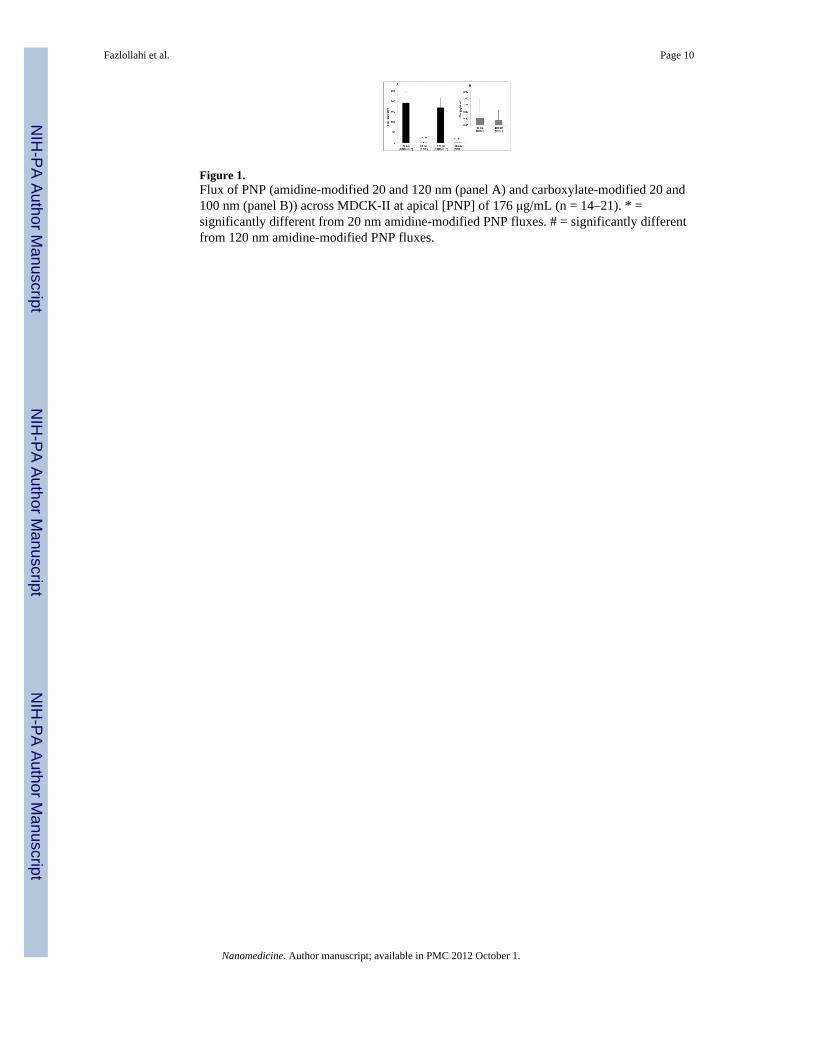
Figure 1.Flux of PNP (amidine-modified 20 and 120 nm (panel A) and carboxylate-modified 20 and100 nm (panel B)) across MDCK-II at apical [PNP] of 176 μg/mL (n = 14–21). * =significantly different from 20 nm amidine-modified PNP fluxes. # = significantly differentfrom 120 nm amidine-modified PNP fluxes.
Fazlollahi et al. Page 10
Nanomedicine. Author manuscript; available in PMC 2012 October 1.
NIH
-PA Author Manuscript
NIH
-PA Author Manuscript
NIH
-PA Author Manuscript
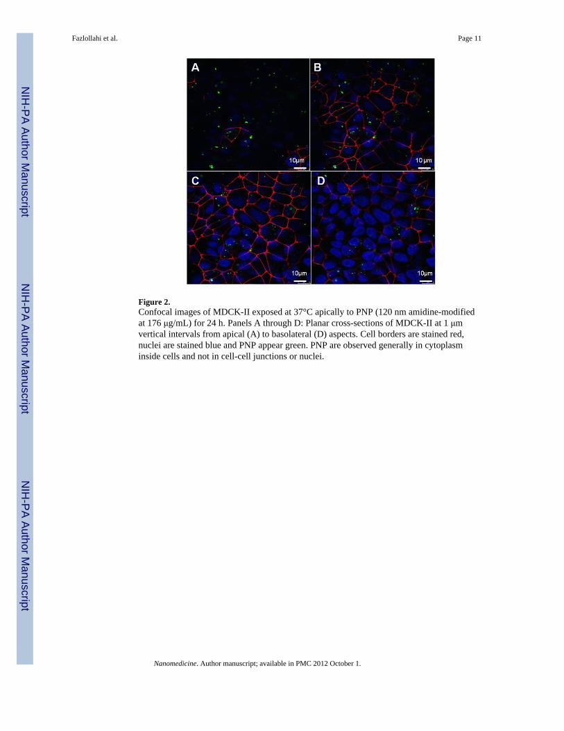
Figure 2.Confocal images of MDCK-II exposed at 37°C apically to PNP (120 nm amidine-modifiedat 176 μg/mL) for 24 h. Panels A through D: Planar cross-sections of MDCK-II at 1 μmvertical intervals from apical (A) to basolateral (D) aspects. Cell borders are stained red,nuclei are stained blue and PNP appear green. PNP are observed generally in cytoplasminside cells and not in cell-cell junctions or nuclei.
Fazlollahi et al. Page 11
Nanomedicine. Author manuscript; available in PMC 2012 October 1.
NIH
-PA Author Manuscript
NIH
-PA Author Manuscript
NIH
-PA Author Manuscript
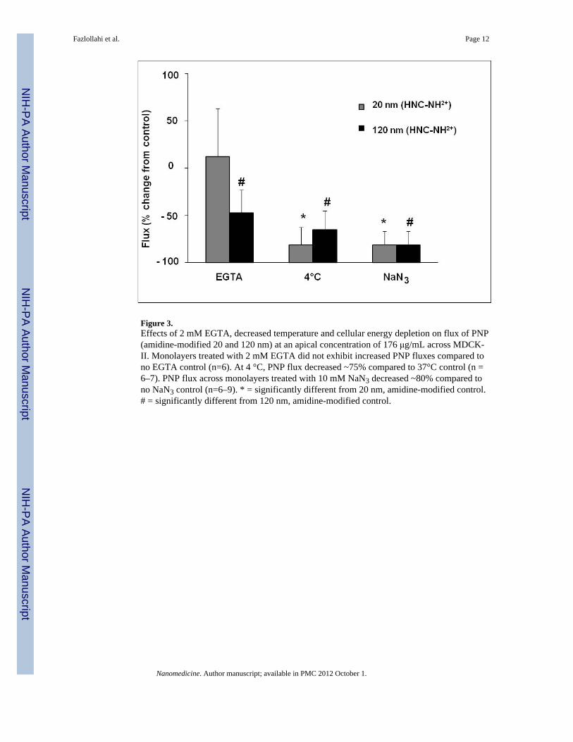
Figure 3.Effects of 2 mM EGTA, decreased temperature and cellular energy depletion on flux of PNP(amidine-modified 20 and 120 nm) at an apical concentration of 176 μg/mL across MDCK-II. Monolayers treated with 2 mM EGTA did not exhibit increased PNP fluxes compared tono EGTA control (n=6). At 4 °C, PNP flux decreased ~75% compared to 37°C control (n =6–7). PNP flux across monolayers treated with 10 mM NaN3 decreased ~80% compared tono NaN3 control (n=6–9). * = significantly different from 20 nm, amidine-modified control.# = significantly different from 120 nm, amidine-modified control.
Fazlollahi et al. Page 12
Nanomedicine. Author manuscript; available in PMC 2012 October 1.
NIH
-PA Author Manuscript
NIH
-PA Author Manuscript
NIH
-PA Author Manuscript
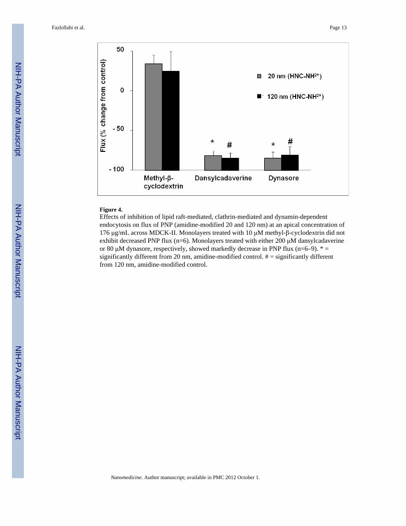
Figure 4.Effects of inhibition of lipid raft-mediated, clathrin-mediated and dynamin-dependentendocytosis on flux of PNP (amidine-modified 20 and 120 nm) at an apical concentration of176 μg/mL across MDCK-II. Monolayers treated with 10 μM methyl-β-cyclodextrin did notexhibit decreased PNP flux (n=6). Monolayers treated with either 200 μM dansylcadaverineor 80 μM dynasore, respectively, showed markedly decrease in PNP flux (n=6–9). * =significantly different from 20 nm, amidine-modified control. # = significantly differentfrom 120 nm, amidine-modified control.
Fazlollahi et al. Page 13
Nanomedicine. Author manuscript; available in PMC 2012 October 1.
NIH
-PA Author Manuscript
NIH
-PA Author Manuscript
NIH
-PA Author Manuscript
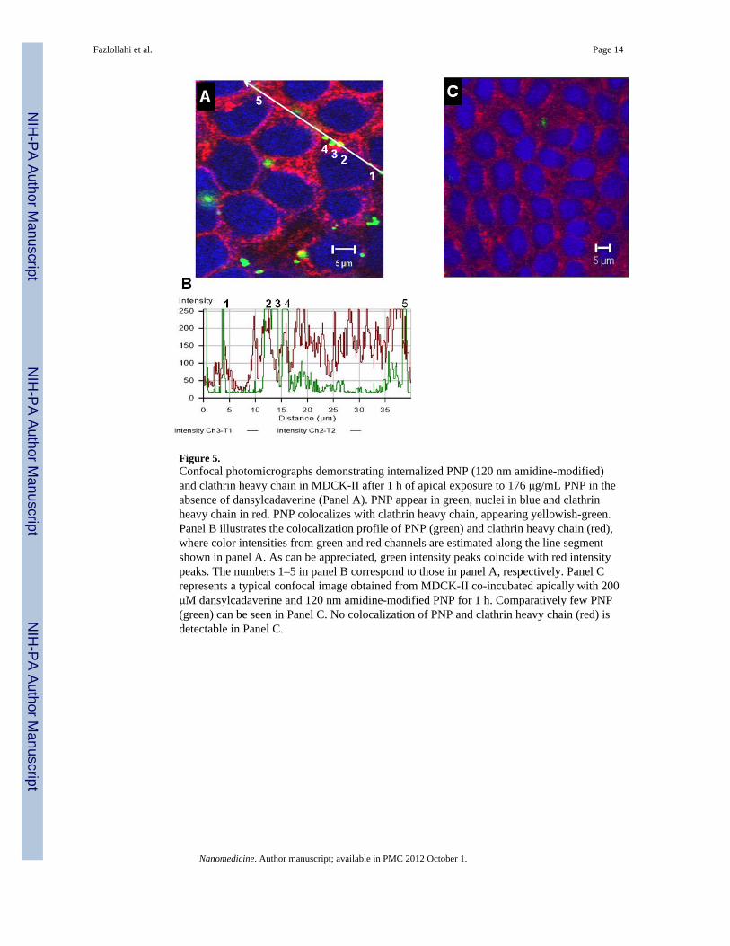
Figure 5.Confocal photomicrographs demonstrating internalized PNP (120 nm amidine-modified)and clathrin heavy chain in MDCK-II after 1 h of apical exposure to 176 μg/mL PNP in theabsence of dansylcadaverine (Panel A). PNP appear in green, nuclei in blue and clathrinheavy chain in red. PNP colocalizes with clathrin heavy chain, appearing yellowish-green.Panel B illustrates the colocalization profile of PNP (green) and clathrin heavy chain (red),where color intensities from green and red channels are estimated along the line segmentshown in panel A. As can be appreciated, green intensity peaks coincide with red intensitypeaks. The numbers 1–5 in panel B correspond to those in panel A, respectively. Panel Crepresents a typical confocal image obtained from MDCK-II co-incubated apically with 200μM dansylcadaverine and 120 nm amidine-modified PNP for 1 h. Comparatively few PNP(green) can be seen in Panel C. No colocalization of PNP and clathrin heavy chain (red) isdetectable in Panel C.
Fazlollahi et al. Page 14
Nanomedicine. Author manuscript; available in PMC 2012 October 1.
NIH
-PA Author Manuscript
NIH
-PA Author Manuscript
NIH
-PA Author Manuscript


