Embed Size (px)
Citation preview

The Role of Alveolar Epithelium in Radiation-InducedLung InjuryCeline Almeida1,3, Devipriya Nagarajan1,3, Jian Tian4, Sofia Walder Leal1,3, Kenneth Wheeler2,3,
Michael Munley1, William Blackstock1, Weiling Zhao1,3*
1 Department of Radiation Oncology, Wake Forest School of Medicine, Winston-Salem, North Carolina, United States of America, 2 Department of Radiology, Wake Forest
School of Medicine, Winston-Salem, North Carolina, United States of America, 3 Brain Tumor Center of Wake Forest University, Wake Forest School of Medicine, Winston-
Salem, North Carolina, United States of America, 4 Radiation Research Laboratories, Department of Radiation Medicine, Loma Linda University and Medical Center, Loma
Linda, California, United States of America
Abstract
Pneumonitis and fibrosis are major lung complications of irradiating thoracic malignancies. In the current study, wedetermined the effect of thoracic irradiation on the lungs of FVB/N mice. Survival data showed a dose-dependent increasein morbidity following thoracic irradiation with single (11–13 Gy) and fractionated doses (24–36 Gy) of 137Cs c-rays.Histological examination showed a thickening of vessel walls, accumulation of inflammatory cells, collagen deposition, andregional fibrosis in the lungs 14 weeks after a single 12 Gy dose and a fractionated 30 Gy dose; this damage was also seen 5months after a fractionated 24 Gy dose. After both single and fractionated doses, i] aquaporin-5 was markedly decreased, ii]E-cadherin was reduced and iii] prosurfactant Protein C (pro-SP-c), the number of pro-SP-c+ cells and vimentin expressionwere increased in the lungs. Immunofluorescence analysis revealed co-localization of pro-SP-c and a-smooth muscle actin inthe alveoli after a single dose of 12 Gy. These data suggest that, i] the FVB/N mouse strain is sensitive to thoracic radiation ii]aquaporin-5, E-cadherin, and pro-SP-c may serve as sensitive indicators of radiation-induced lung injury; and iii] theepithelial-to-mesenchymal transition may play an important role in the development of radiation-induced lung fibrosis.
Citation: Almeida C, Nagarajan D, Tian J, Leal SW, Wheeler K, et al. (2013) The Role of Alveolar Epithelium in Radiation-Induced Lung Injury. PLoS ONE 8(1):e53628. doi:10.1371/journal.pone.0053628
Editor: Philip J. Tofilon, National Cancer Institute, United States of America
Received September 25, 2012; Accepted November 30, 2012; Published January 11, 2013
Copyright: � 2013 Almeida et al. This is an open-access article distributed under the terms of the Creative Commons Attribution License, which permitsunrestricted use, distribution, and reproduction in any medium, provided the original author and source are credited.
Funding: The authors have no support or funding to report.
Competing Interests: The authors have declared that no competing interests exist.
* E-mail: [email protected]
Introduction
Thoracic radiotherapy (RT) is one of the important therapeutic
modalities for treating lung cancer, breast cancer, and various
lymphomas. Pneumonitis and lung fibrosis are the major
radiation-induced complications following thoracic RT [1,2].
Symptoms of radiation pneumonitis, such as low-grade fever,
mild cough, or dyspnea, usually occur 2–3 months after
irradiation. The risk of developing radiation-induced pneumonitis
depends on the dose, volume of lung irradiated, and dose
fractionation scheme [1,2]. Radiation can disrupt epithelial and
endothelial integrity leading to edema, recruitment of leukocytes,
angiogenesis, and a cascade of molecular events that alters the
microenvironment [3] that creates a self-sustaining cycle of
inflammation and chronic oxidative stress [3,4]. The main
histological features of radiation-induced pneumonitis include, i]
a dose-dependent leakage of proteins into the alveolar space, ii]
thickening of the alveolar septa, iii] edema of the interstitium, iv]
alteration of the capillaries, and v] changes in type II pneumocytes
and alveolar macrophages [5]. Lung fibrosis can appear 6 months
to a year after irradiation, and is characterized by excessive
fibroblast proliferation and massive deposition of extracellular
matrix [6]. The alveoli eventually collapse and are obliterated by
connective tissue. Patients with radiation-induced lung fibrosis
have severe physiologic abnormalities and chronic respiratory
failure [7]. Although inflammation and fibroblast/myofibroblast
activation have been recognized as important contributors, the
exact mechanism(s) underlying pulmonary fibrosis remain elusive
[3].
Respiratory epithelium performs an important function as a
barrier against microbial infection/exterior insults and modulation
of the airway immune response [8]. Severe injury and retarded
repair of the alveolar epithelium is sufficient to promote the
fibrotic process [9,10]. The alveolar epithelium consists of two cell
types, alveolar epithelial type I (AE1) and alveolar epithelial type II
(AE2) cells. AE1 cells are responsible for gas exchange, regulate
liquid homeostasis in the alveoli, and are highly susceptible to
oxidative stress and toxic insults. AE2 cells serve two major
functions in the lung, i] synthesizing, secreting, and regulating lung
surfactants, and ii] repopulating AE1 cells [8]. Recent studies
suggest that epithelial cells can also undergo transdifferentiation
into myofibroblasts, through a process termed ‘‘epithelial–mesen-
chymal transition’’ (EMT) [11]. Alveolar EMT has been
demonstrated in human idiopathic pulmonary and experimental
pulmonary fibrosis [12,13]. EMT is characterized by, i] a shift
from an epithelial to a bipolar morphology, ii] increased
expression of the mesenchymal markers, alpha-smooth muscle
actin (a-SMA) and vimentin, and iii] decreased expression of the
epithelial marker, E-cadherin [14]. Although a growing body of
evidence supports the involvement of EMT in the development of
fibrosis, its role in radiation-induced lung injury has not been
established in vivo.
PLOS ONE | www.plosone.org 1 January 2013 | Volume 8 | Issue 1 | e53628

The sensitivity of the mouse lung to radiation-induced
pneumonitis and fibrosis is strain-dependent [15–17]. Sharplin et
al. compared lung injury in two C3H, three C57BL and one CBA
mouse strain (dose range studied was 10–14.5 Gy). C3H and CBA
strains developed only classical pneumonitis during the early and
late phase; no visible fibrosis was observed. However, C57BL
mouse strains developed extensive fibrosis during the latent period
[18] Radiation-induced fibrosis in C57BL strain occurs following
whole-thoracic irradiation (WTI) with single doses of 12–15 Gy as
early as 24 weeks postirradiation [19]. In contrast, focal alveolar
fibrosis is not observed until 66 weeks after 30 Gy of fractionated
(f) WTI [19,20]. The translatability of the C57BL/6 mouse data to
humans has been questioned recently in an NIH sponsored
workshop on Animal Models and Medical Countermeasures
Development for Radiation-Induced Lung Damage [21]. The
primary concerns with the C57BL/6 model were that, i] overt
pneumonitis is poorly defined in the C57BL/6 mouse, ii]
decrements in lung function and ultimately the survival of these
mice after irradiation is predominantly due to an accumulation of
fluid in the pleural space (pleural effusion) which is not related to
either radiation-induced pneumonitis or fibrosis [22,23]. Studies
have been focused on development of new mouse models for
assessing radiation-induced chronic lung injury [22,23]. Recently,
FVB transgenic mice have been used to study the effect of various
genes on radiation-induced organ injury and tumor growth [24–
26]; however, the lung radiosensitivity of FVB mice has not been
studied. Here, we report the response of female FVB/N mice to
single and fractionated doses of WTI. Our data suggest that in
female FVB/N mice, i] the lungs are sensitive to radiation, ii] WTI
modulates the alveolar epithelium, and iii] WTI induces EMT in
the lung.
Materials and Methods
AnimalsSix week-old female FVB/N were purchased from The Jackson
Laboratory (Bar Harbor, Maine). The mice were housed in the
AALAC-accredited animal care facility at Wake Forest School of
Medicine (WFSM) and acclimated for 2 weeks before initiating the
studies. All animal care and experimental procedures were
performed in strict accordance with the NIH Guide for Care
and Use of Laboratory Animals. All protocols were approved by
the WFSM Institutional Animal Care and Use Committee before
experiments began.
IrradiationPrior to irradiation, 3 mice were CT scanned to determine the
size of the lungs, and a Cerrobend collimator was constructed to
restrict the beam to just the thorax. Mice were irradiated using a137Cs irradiator (Model 81A, Shephend & Associates, Glendale,
CA) at a dose rate of 3.68 Gy/min. Groups of mice ( n = 10 )
received single (11, 12, or 13 Gy) or fractionated (24, 30 or 36 Gy
given as 3, 4, 5 or 6 Gy fractions, 2 fractions/wk over 3 wk) dose
of thoracic irradiation. Animals were monitored up to 5 months
postirradiation, and the body weight and survival of animals
recorded.
Sample collectionThe surviving mice in the 12 Gy single dose group and in the
30 Gy fractionated dose group were euthanized at 14 weeks with
ketamine/xylazine (80/20 mg/kg, i.p.) due to stress symptoms.
The mice treated 24 Gy fractionated dose were euthanized at 1, 2
and 5 months postirradiation. The left lung was inflated with PBS,
dissected and fixed in 10% neutral formalin for histological and
immunohistochemical analysis. The right lungs were snap frozen
in liquid nitrogen and kept at 280uC for protein analysis.
HistopathologyLung sections (5 mm) were stained with either hemotoxylin/
eosin (H&E) to assess the histological changes or with Masson’s
trichrome to identify the sites of collagen deposition. All analyses
were performed on coded slides by a blinded pathologist (JT).
Immunohistochemical analysisLung sections (5 mm) were mounted on slides, deparaffinized,
and hydrated in xylene, 95% ethanol, 80% ethanol, 75% ethanol,
and 1X PBS, pH 7.4. Following antigen retrieval with citrate
buffer, tissue sections were preincubated with Rodent Block M for
20 min (Biocare Medical, Concord, CA) and then incubated with
pro-surfactant protein-c (pro-SP-c, 1:2,000) , CD 45 or a-SMA
(1:500) antibodies (Abcam, Cambridge, MA). Control slides were
incubated with the appropriate IgG. After washing with PBS, the
tissue sections were incubated with rabbit or mouse HRP-
polymers for 30 min (Biocare Medical, Concord, CA); diamino-
benzidine (DAB) was used for visualization. The number of
prosurfactant Protein C (pro-SP-c) positive cells was counted using
morphormetric method.
Double immunofluorescence stainingA double-color immunofluorescence analysis was performed to
identify the expression of a-SMA, a mesenchymal cell marker, in
AE2 cells in the lung. Briefly, after deparaffinization, antigen
retrieval, and incubation with Rodent Block M as described above,
the sections were incubated with a mixture of anti-a-SMA (1:500,
Abcam, Cambridge, MA) and anti-pro-SP-c (1:2,000, Abcam,
Cambridge, MA) antibodies at 4uC overnight. After washing with
PBS, the sections were incubated with FITC conjugated Alexa
Fluor 488 goat anti-mouse (Invitrogen, Carlsbad, CA) and Texas
Red-conjugated anti-rabbit secondary antibodies (Abcam) at room
temperature for 30 min. Nuclei were counterstained with 49-6-
diamidino-2-phenylindole (DAPI), and the sections analyzed using
a fluorescence microscope.
Western Blot analysisFrozen lung tissue (50 mg) was ground in a mortar with liquid
nitrogen, homogenized with 1 mL RIPA buffer with 1 mM
PMSF, 1 mg/mL aprotinin, 1 mg/mL leupeptin, 1 mM Na3VO4
and 1 mM NaF, and stored in aliquots at 270uC until assayed.
The lysate (20 mg) was mixed with an equal volume of sample
buffer, denatured by boiling, and then separated on a 10–15%
polyacrylamide mini-gel. The proteins were transferred to
nitrocellulose membranes (Amersham, Arlington Heights, IL),
blocked with 5% milk, and incubated overnight with pro-SP-c
(1:2,000, Abcam), TGF-ß1 (1:1,000, Abcam), vascular cell
adhesion molecule 1 (Vcam-1) (1:1,000 , Santa CruZ, Biotech-
nology, Santa Cruz, CA), E-cadherin (1:1,500, Abcam), vimentin
(1:1,000, Santa Cruz Biotechnology), aquaporin-5 (1:1,000, Santa
Cruz Biotechnology) or ß-actin antibodies (1:10,000, Sigma, St.
Louis, MO). The blots were then incubated with anti-mouse or
anti-rabbit IgG horseradish peroxidase conjugated antibodies (GE
healthcare, Piscataway, NJ) for 1 h at room temperature. Finally,
the signal was detected using standard chemical luminescence
methodology (ECL plus; GE healthcare). For densitometrical
analysis of bands on western blot, the densitometric signal for the
targeting protein (for example E-cadherin) was normalized to ß-
actin and expressed as the density ratio of target to non-irradiated
control.
Radiation and Lung Injury
PLOS ONE | www.plosone.org 2 January 2013 | Volume 8 | Issue 1 | e53628

Morphometric analysisThe morphometric analysis of pro-SP-C+ cell number was
conducted using stereological Investigator software as previous
described with modification [27,28]. Ten fields (250 mm6250 mm)
on the stained slide were randomly selected. Images were acquired
from each field and the number of cells with positive staining
counted in the selected fields and expressed as the number of cells/
mm2. Large airways and lung vessels were excluded.
Statistical AnalysisStatistical analysis was performed using one-sample Student’s t
tests to compare the differences between the irradiated and
unirradiated lung tissue response. A p value of #0.05 was
considered significant.
Results
Survival after single and fractionated doses of WTITo determine the sensitivity of FVB/N mice to WTI, 8–12
week-old female mice were irradiated with single (11–13 Gy) or
fractionated doses (18–36 Gy) of WTI. Single doses of WTI led to
a dose-dependent decrease in survival (Fig. 1A); ,50% of the mice
were dead at 11 and 14 weeks after irradiation with 13 and 12 Gy,
respectively. In contrast, 90% of the mice irradiated with 11 Gy
survived to 5 months postirradiation. Similarly, fWTI led to a
dose-dependent decrease in survival (Fig. 1B); ,50% of mice were
dead at 9 and 11 weeks after irradiation with total doses of 36 and
30 Gy, respectively. In contrast, all of the mice irradiated with
total doses of 24 Gy survived up to 5 months postirradiation. The
biological effective dose (BED) for an 11 Gy is about 51.33 and 56
for 24 Gy fractionated dose. The BED for 12 Gy is roughly an
equivalence of 36 Gy fractionated dose. However, our observation
on survival rates showed that biological effect of single dose
appeared more toxic than fractionated dose.
Histological changes after single and fractionated dosesof WTI
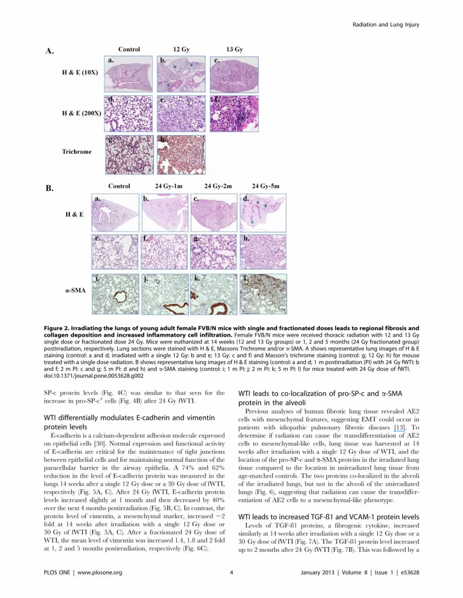
The surviving mice irradiated with a single dose of 12 Gy or a
fractionated dose of 30 Gy showed stress symptoms at 14weeks
postirradiation. Consequently, they were euthanized, the lungs
excised, and a portion of the lung tissue prepared for histological
analysis after H & E or Masson’s trichrome staining. After a single
13 Gy dose, H & E stained lungs showed markedly thickened
alveolar walls, collapsed alveoli, foam-like cells in the alveolar
space, and diffuse accumulation of inflammatory cells (Fig. 2).
Similar histological changes were noted in the lungs of mice
receiving 30 Gy fWTI (data not shown). Increased inflammatory
cell infiltration, collagen deposition, and regional fibrosis were seen
in the mouse lungs irradiated with a single dose of 12 Gy (Fig. 2A,
middle panel). No markedly chances in lung morphology were
observed at 1 month postirradiation with 24 Gy fWTI, when
compared with the non-irradiated control (Fig. 2B, second panels).
By 2 months postirradiation, enhanced inflammatory cell infiltra-
tion and alveolar wall thickness have been seen in the irradiated
lungs (Fig. 2B; third panels). At 5 months postirradiation, multiple
fibrotic lesions were presented in the irradiated lungs (Fig. 2B,
right panel). Immunohistochemical staining with a-SMA, a
marker of smooth muscle cells/myofibroblasts, showed increased
accumulation of myofibroblasts in the these lungs (Fig. 2B, lower
panels).
WTI leads to dose- and time-dependent reductions inaquaporin-5
Aquaporin-5 is an important AE1 cell-specific water channel
protein that mediates water transport across the airway epithelium
[29]. To examine the effect of radiation, the protein level of
aquaporin-5 in irradiated and non-irradiated lung tissues was
determined using Western blots. At 14 weeks postirradiation, a
similar marked reduction in aquaporin-5 was measured in lungs
irradiated with a single 12 Gy dose or a 30 Gy dose of fWTI
(Fig. 3A). A time-dependent reduction in the aquaporin-5 level was
also measured in lungs irradiated with 24 Gy fWTI (Fig. 3B).
Densitometric analysis revealed that the mean level of aquaporin-5
in the irradiated lungs was reduced by 32, 67, and 66% at 1, 2 and
5 months postirradiation when compared with unirradiated
controls (Fig. 3C).
WTI leads to an increase in pro-SP-c protein and pro-SP-c+ AE2 cells
A similar increase in the number of pro-SP-c+ AE2 cells was
measured in the lungs irradiated with a single 12 Gy dose or a
30 Gy dose of fWTI (Fig. 4A). After 24 Gy of fWTI, the number
of pro-SP-c+ cells increased up to 2 months postirradiation when
compared with unirradiated controls and remained relatively
constant over the next 3 months (Fig. 4B). The pro-SP-c protein
level was increased similarly at 14 weeks after a single 12 Gy dose
and 30 Gy fWTI; the time-dependence of the increase in the pro-
Figure 1. Irradiating the lungs of young adult female FVB/N mice with single and fractionated doses of 137Cs c-rays leads to dose-dependent increases in mortality. Groups of 8–10 week-old female FVB/N mice were irradiated with single (11–13 Gy) or fractionated (18–36 Gy)doses of c rays, and their survival recorded up to 22 weeks postirradiation.doi:10.1371/journal.pone.0053628.g001
Radiation and Lung Injury
PLOS ONE | www.plosone.org 3 January 2013 | Volume 8 | Issue 1 | e53628
SP-c protein levels (Fig. 4C) was similar to that seen for the
increase in pro-SP-c+ cells (Fig. 4B) after 24 Gy fWTI.
WTI differentially modulates E-cadherin and vimentinprotein levels
E-cadherin is a calcium-dependent adhesion molecule expressed
on epithelial cells [30]. Normal expression and functional activity
of E-cadherin are critical for the maintenance of tight junctions
between epithelial cells and for maintaining normal function of the
paracellular barrier in the airway epithelia. A 74% and 62%
reduction in the level of E-cadherin protein was measured in the
lungs 14 weeks after a single 12 Gy dose or a 30 Gy dose of fWTI,
respectively (Fig. 5A, C). After 24 Gy fWTI, E-cadherin protein
levels increased slightly at 1 month and then decreased by 40%
over the next 4 months postirradiation (Fig. 5B, C). In contrast, the
protein level of vimentin, a mesenchymal marker, increased ,2
fold at 14 weeks after irradiation with a single 12 Gy dose or
30 Gy of fWTI (Fig. 5A, C). After a fractionated 24 Gy dose of
WTI, the mean level of vimentin was increased 1.4, 1.8 and 2 fold
at 1, 2 and 5 months postirradiation, respectively (Fig. 6C).
WTI leads to co-localization of pro-SP-c and a-SMAprotein in the alveoli
Previous analyses of human fibrotic lung tissue revealed AE2
cells with mesenchymal features, suggesting EMT could occur in
patients with idiopathic pulmonary fibrotic diseases [13]. To
determine if radiation can cause the transdifferentiation of AE2
cells to mesenchymal-like cells, lung tissue was harvested at 14
weeks after irradiation with a single 12 Gy dose of WTI, and the
location of the pro-SP-c and a-SMA proteins in the irradiated lung
tissue compared to the location in unirradiated lung tissue from
age-matched controls. The two proteins co-localized in the alveoli
of the irradiated lungs, but not in the alveoli of the unirradiated
lungs (Fig. 6), suggesting that radiation can cause the transdiffer-
entiation of AE2 cells to a mesenchymal-like phenotype.
WTI leads to increased TGF-ß1 and VCAM-1 protein levelsLevels of TGF-ß1 proteins, a fibrogenic cytokine, increased
similarly at 14 weeks after irradiation with a single 12 Gy dose or a
30 Gy dose of fWTI (Fig. 7A). The TGF-ß1 protein level increased
up to 2 months after 24 Gy fWTI (Fig. 7B). This was followed by a
Figure 2. Irradiating the lungs of young adult female FVB/N mice with single and fractionated doses leads to regional fibrosis andcollagen deposition and increased inflammatory cell infiltration. Female FVB/N mice were received thoracic radiation with 12 and 13 Gysingle dose or fractionated dose 24 Gy. Mice were euthanized at 14 weeks (12 and 13 Gy groups) or 1, 2 and 5 months (24 Gy fractionated group)postirradiation, respectively. Lung sections were stained with H & E, Massons Trichrome and/or a-SMA. A shows representative lung images of H & Estaining (control: a and d; irradiated with a single 12 Gy: b and e; 13 Gy: c and f) and Masson’s trichrome staining (control: g; 12 Gy: h) for mousetreated with a single dose radiation. B shows representative lung images of H & E staining (control: a and d; 1 m postirradiation (PI) with 24 Gy fWTI: band f; 2 m PI: c and g; 5 m PI: d and h) and a-SMA staining (control: i; 1 m PI: j; 2 m PI: k; 5 m PI: l) for mice treated with 24 Gy dose of fWTI.doi:10.1371/journal.pone.0053628.g002
Radiation and Lung Injury
PLOS ONE | www.plosone.org 4 January 2013 | Volume 8 | Issue 1 | e53628

Figure 3. Irradiating the lungs of young adult female FVB/N mice with either a single 12 Gy dose or fractionated 24 or 30 Gy dosesof 137Cs c-rays leads to a marked decrease in the expression of aquaporin-5 in the lung. Lung tissues were collected at 14 weeks (12 Gyand 30 Gy) or at 1, 2 and 5 months (24 Gy) PI. Cell lysates were generated from the snap-frozen left lung from 4 mice and run on a 10–15%polyacrylamide mini-gel. Each lane contains the pooled lysate from 2 mice. ß-actin served as loading control. A: The reduction in aquaporin-5 proteinat 14 weeks after a single 12 Gy or a fractionated 30 Gy dose is similar. B: The reduction in aquaporin-5 protein occurs as early as 1 month after afractionated 24 Gy dose and remains reduced for the next 4 months. C: Quantification of the aquaporin-5 protein levels in 3B shows that thereduction in aquaporin-5 is progressive over the first 2 months postirradiation (PI) and then remains at a relatively constant low level up to 5 monthsPI. Densitometry was used to quantify the protein in each lane; all irradiation values in 2C were normalized to the unirradiated control valuesobtained on the same gel. The data in 2C are the mean 6 1 SEM.doi:10.1371/journal.pone.0053628.g003
Figure 4. Irradiating the lungs of young adult female FVB/N mice with either a single 12 Gy dose or fractionated 24 or 30 Gy dosesof 137Cs c-rays leads to increases in pro-SP-c protein and pro-SP-c positive cells in the lung. A: The increase in pro-SP-c+ cells is similar at14 weeks after a single 12 Gy dose or a fractionated 30 Gy dose. B: The increase in pro-SP-c+ cells after a fractionated 24 Gy dose is progressive overthe first 2 months postirradiation (PI) and then remains relatively constant up to 5 months PI. C: Upper gel, the increase in pro-SP-c protein at 14weeks after a single 12 Gy or a fractionated 30 Gy dose is similar. Lower gel, pro-SP-c protein is increase by 1 month PI and remains relativelyconstant up to 5 months PI. For 4A and 4B, lung tissues were collected and prepared for histochemical analysis either at 14 weeks (12 Gy and 30 Gy)or 1, 2 and 5 months (24 Gy) PI. Sections (5 mm) of lung tissue were stained with pro-SP-c antibodies, and the pro-SP-c+ cells were analyzed usingmorphometric method. Data are the mean 6 1 SEM; n = 3; * p,0.05 vs. sham control. The gels in 4C were prepared and analyzed as described inFig. 3.doi:10.1371/journal.pone.0053628.g004
Radiation and Lung Injury
PLOS ONE | www.plosone.org 5 January 2013 | Volume 8 | Issue 1 | e53628

decrease in the TGF-ß1 protein level over the next 3 months that
did not reach the level in the age-matched unirradiated controls.
VCAM-1 is a member of the immunoglobulin superfamily with a
critical role in mediating the adhesion of leukocytes to endothelial
cells in various acute and chronic inflammatory diseases [31]. The
VCAM-1 protein level was increased slightly at 14 weeks after
irradiation with a single 12 Gy dose or a 30 Gy dose of fWTI
(Fig. 7A). After 24 Gy of fWTI, the VCAM-1 protein level
increased substantially up 2 months postirradiation, but returned
to unirradiated control values by 5 m postirradiation (Fig. 7B).
Discussion
Radiation-induced pneumonitis and subsequent fibrosis are
major dose-limiting complications for patients receiving thoracic
RT. Various mouse strains have been used to study radiation-
induced normal tissue injury; their response is strain-dependent. In
the current study, we demonstrated for the first time, i] the
sensitivity of young adult female FVB/N mice to thoracic
irradiation and ii] the involvement of EMT in radiation-induced
lung injury.
In our study, irradiating the thorax of young adult female FVB/
N mice with single doses of 12 and 13 Gy led to 50% mortality at
14 and 11 weeks postirradiation, respectively (Fig. 1). In contrast,
Steel et al reported that the median survival time of C57BL mice
after doses of 12–20 Gy is between 200–300 days [32]. Yang et al
reported that ,50% of the C57BL mice died within 32 weeks after
a single 14.5 Gy dose of WTI [33]. O’Brien et al. reported that the
mean survival time of C57BL mice was 21.861.5 weeks after a
single 18 Gy dose of WTI [34]. Histological analysis of lung tissues
from FVB/N mice revealed an extensive alveolar inflammation
and regional fibrosis 14 weeks after a single 12 Gy dose of WTI
(Fig. 2). In contrast, fibrotic lesions were observed 24 and 26 weeks
Figure 5. Irradiating the lungs of young adult female FVB/N mice with either a single 12 Gy dose or fractionated 24 or 30 Gy dosesof 137Cs c-rays leads to decreased levels of E-cadherin and increased levels of vimentin protein in the lung. A, C: Although thevimentin data is variable, the decrease in E-cadherin and the increase in vimentin are similar at 14 weeks after a single 12 Gy dose or a fractionated30 Gy dose. B, D: After a fractionated 24 Gy dose, there is a slight increase in E-cadherin at 1 month postirradiation (PI) followed by a progressivedecrease up to 5 months PI. After a fractionated 24 Gy dose, there is a progressive increase in vimentin up to 2 month PI that remains relativelyconstant for the next 3 months. All methods and analyses are identical to those described in Fig. 3.doi:10.1371/journal.pone.0053628.g005
Figure 6. Pro-SP-c and a-SMA protein co-localize in the lungs ofFVB/N mice at 14 weeks postirradiation with a single dose of12 Gy. Lung section (5 mm) from an unirradiated control (A) and anirradiated mouse (B) were co-stained with pro-SP-c and a-SMAantibodies. In the representative photomicrographs the pro-SP-cprotein is red, the a-SMA protein is green, and the nuclei are blue.doi:10.1371/journal.pone.0053628.g006
Radiation and Lung Injury
PLOS ONE | www.plosone.org 6 January 2013 | Volume 8 | Issue 1 | e53628

postirradiation in fibrosis-prone C57/B6 mice after single 12 Gy
and 12.5 Gy doses of WTI, respectively [35,36]. These results
indicate that the lungs of young adult female FVB/N mice are
radiosensitive, making them an appropriate model for studying
both radiation-induced pneumonitis and fibrosis, particularly since
many transgenic mice are created on a FVB/N background.
One feature of radiation pneumonitis is excessive leakage of
fluid from the vessels into the alveolar space, which could impede
alveolar gas exchange. Aquaporin-5 has been described as playing
a critical role in maintaining water permeability across the cell
membrane and also in removal of pulmonary edema fluid from the
alveolar space [37]. Aquaporin-5 knock-out mice exhibit a 90%
decrease in airspace-capillary water permeability, suggesting an
important role for aquaporin-5 in maintaining normal lung
physiological function [38]. Decreased expression of aquaporin 5
has been reported in pathological conditions such as acute lung
injury [39,40] and lung fibrosis [41]. Moreover, a significant
reduction in aquaporin-5 mRNA and protein level has been
associated with pulmonary inflammation and edema resulting
from adenoviral infection [40]. Gabazza et al. noted decreased
expression of aquaporin-5 in alveolar type I cells in a mouse model
of bleomycin-induced lung fibrosis; aquaporin-5 knock-out mice
revealed a fibrotic phenotype [41]. Taken together, these data
indicate that down-regulation of aquaporin-5 results in abnormal
lung fluid metabolism in many diseases. In our study, a decrease in
the aquaporin-5 protein level was measured in lungs of mice after
a single 12 Gy dose or a fractionated 30 Gy dose of WTI (Fig. 3A);
a time-dependent decrease in the aquaporin-5 protein level was
measure after a fractionated 24 Gy dose of WTI (Fig. 3B, C).
These novel findings suggest that aquaporin-5 may be an
important biomarker of radiation-induced lung injury.
The lung alveolar epithelium is in direct contact with air. AE1
cells are terminally differentiated, highly susceptible to injury, and
incapable of self-renewal. In contrast, AE2 cells are highly resistant
to insults, able to self-renew, and serve as stem cells for producing
AE1cells; AE2 cells are known as the defenders of the alveoli [42].
When the alveolar epithelium is damaged, AE2 cells start
proliferating and transdifferentiate into AE1 cells to re-establish
a functional alveolar epithelium. Alveolar epithelial injury followed
by abnormal epithelial repair appears to be a key pathological
feature of lung fibrosis. Increased proliferation/hyperplasia of AE2
cells has been frequently observed in injured and irradiated lungs
[43,44]. Pro-SP-c, a surfactant protein, is expressed only by AE2
cells; thus, it has been used as a marker of type II cell
differentiation in the mammalian lung [13]. The increased
number of pro-SP-c+ cells (Fig. 4A, B) and the increased pro-SP-
c protein levels (Fig. 4C) measured after both single and
fractionated doses of WTI likely reflects proliferation of AE2 cells
in an attempt to repair the radiation-induced lung damage.
Myofibroblasts, a-SMA expressing fibroblasts, are a prominent
source of type I collagen and fibrogenic/inflammatory cytokines in
fibrotic lesions. Several origins for myofibroblasts have been
proposed; resident fibroblasts seem to be the most common one.
Bone marrow-derived circulating cells have also been suggested as
an alternative source of myofibroblasts. Epperly et al. demon-
strated that marrow-derived cells constitute 20 to 50% of the cells
in radiation-induced fibrotic areas using GFP–positive bone
marrow cells [45]. Consistent with these results, collagen-
producing lung fibroblasts derived from bone marrow progenitor
cells have been detected in fibrotic tissues in bleomycin-induced
fibrosis [46]. However, these marrow-derived fibroblasts did not
express a-SMA and were resistant to the fibroblast to myofibro-
blast conversion by TGF-ß1 [46]. Recent studies indicate that
myofibroblasts can also arise from resident epithelial cells [12,47]
that undergo EMT. Liu et al. [48] reported expression of a-SMA
and vimentin in kidney tubular epithelial cells in a rat model of
radiation nephropathy, supporting the hypothesis that radiation
leads to the transition of tubular epithelial cells to myofibroblasts.
EMT and increased cell motility has also been reported in the
irradiated human lung A549 cells [49]. Using an in vitro model, we
have demonstrated that irradiation of AE2 cells resulted a
transition of epithelial to a myofibroblast-like phenotype, which
was mediated by the ROS/ERK/GSK-3ß/Snail pathway [50].
However, EMT in the irradiated lung has not been previously
reported. In this study, a significant decrease in the protein levels
of the epithelial cell markers, E-cadherin (Fig. 5) and aquaporin-5
(Fig. 3), was measured after both single and fractionated doses of
WTI. This was accompanied by a concomitant increase in
vimentin (Fig. 5), a mesenchymal marker. Moreover, double
immunoflorescence staining showed co-localization of the pro-SP-
c and a-SMA proteins in the alveoli of irradiated lungs (Fig. 6),
implying that AE2 cells had gained a mesenchymal-like pheno-
type. Our data suggest that AE2 to mesenchymal transition occurs
in the irradiated lungs of FVB/N mice. However, it is not known if
other epithelial cells in the lung can differentiate into myofibro-
blasts. The relative contributions of epithelial cells in the irradiated
lung to the overall increase in the myofibroblast population and
the pathogenic role that EMT plays in radiation-induced lung
fibrosis remain to be investigated.
TGF-b1 plays an important role in the development of lung
fibrosis [51]. Increased TGF-b1 expression leads to recruitment of
monocytes and macrophages to the inflammatory site, enhances
the maturation and activation of fibroblasts, and stimulates EMT
Figure 7. Irradiating the lungs of young adult female FVB/Nmice with either a single 12 Gy dose or fractionated 24 or30 Gy doses of 137Cs c-rays leads to increase in VCAM-1 andTGF-ß1 protein in the lung. A: The increase in VCAM-1 and TGF-ß1 issimilar at 14 weeks after a single 12 Gy dose or a fractionated 30 Gydose. B: Both VCAM-1 and TGF-ß1 increase up to 2 months after afractionated 24 Gy dose and then decrease up to 5 monthspostirradiation.doi:10.1371/journal.pone.0053628.g007
Radiation and Lung Injury
PLOS ONE | www.plosone.org 7 January 2013 | Volume 8 | Issue 1 | e53628

[52]. Chronic radiation-induced lung injury has been reported to
increase expression and activation of TGF-b1, which leads to
parenchymal cell depletion and excess fibrosis [53]. TGF-b1
expression in the plasma of patients immediately after RT has
been used as an important marker to predict the risk for radiation-
induced lung injury [54]. In our study, both single and
fractionated doses of WTI increased the TGF-b1 protein levels
in the lung (Fig. 7). The sustained increase in the TGF-ß1 protein
level in the irradiated lungs of FVB/N mice further demonstrates
the important role of TGF-ß1 in the pathogenesis of radiation-
induced lung injury.
Recently, chromatin remodeling via posttranslational histone
modification was found to function in a genome-wide manner and
contributes to an extensive range of biological functions [55].
Histone deacetylases (HDACs) are known as modulators of gene
transcription, which is important for cell function, proliferation
and differentiation. HDAC inhibitors have been reported to
induce protein hyperacetylation, chromatin remodeling, transcrip-
tional activation and repression, cell-cycle arrest, and cell death
[56,57]. Preclinical studies indicate that HDAC inhibitors can
effectively block cardiac, skin, liver and renal fibrosis [58]. In vitro
and in vivo investigations indicate that HDAC inhibitors modulate
fibrosis by suppressing ECM production, inhibiting myofibroblast
activation, blocking EMT, and/or reducing pro-inflammatory
cytokine production [11,59–63]. Furthermore, recent studies
suggest that topical treatment of rat skin with HDAC inhibitor
4-Phenyl butyrate has been shown to promote wound healing,
reduce skin fibrosis, and decrease tumorigenesis after irradiation
[64]. In light of these findings, effect of on HDAC inhibitors on
radiation-induced lung damage in FVB/N mice is underway in
our lab.
In summary, the current findings indicate that the lungs of
female FVB/N mice are radiosensitive and represent an appro-
priate model for investigating radiation-induced lung inflamma-
tion and fibrosis. The marked changes in the levels of the alveolar
epithelial proteins, E-cadherin, aquaporin-5, and pro-SP-c,
appears to be associated with the development of radiation-
induced lung injury in FVB/N mice, suggesting that these proteins
may serve as sensitive indicators of radiation-induced lung
damage. WTI resulted in the loss of epithelial markers and a
subsequent increase in the levels of mesenchymal proteins,
indicating that EMT occurs in irradiated lung tissue. Although
the in vivo significance of EMT is unclear, radiation-induced
alterations in the alveolar epithelium phenotype implicate
impairment of alveolar epithelial function in producing fibrosis.
Future research to identify and quantify the specific mechanisms
involved in producing radiation-induced fibrosis should provide
targets for the development of interventions that prevent/
ameliorate this devastating complication of WTI.
Author Contributions
Conceived and designed the experiments: WZ KW WB MM. Performed
the experiments: CA DN WZ. Analyzed the data: STS JT WZ.
Contributed reagents/materials/analysis tools: WZ. Wrote the paper: WZ.
References
1. Movsas B, Raffin TA, Epstein AH, Link CJ, Jr. (1997) Pulmonary radiationinjury. Chest 111: 1061–1076.
2. Tsoutsou PG, Koukourakis MI (2006) Radiation pneumonitis and fibrosis:mechanisms underlying its pathogenesis and implications for future research.
Int J Radiat Oncol Biol Phys 66: 1281–1293.
3. Rubin P, Johnston CJ, Williams JP, McDonald S, Finkelstein JN (1995) A
perpetual cascade of cytokines postirradiation leads to pulmonary fibrosis.
Int J Radiat Oncol Biol Phys 33: 99–109.
4. Ghafoori P, Marks LB, Vujaskovic Z, Kelsey CR (2008) Radiation-induced lung
injury. Assessment, management, and prevention. Oncology (Williston Park) 22:37–47.
5. Travis EL (1980) The sequence of histological changes in mouse lungs aftersingle doses of x-rays. Int J Radiat Oncol Biol Phys 6: 345–347.
6. Marks LB (1994) The pulmonary effects of thoracic irradiation. Oncology(Williston Park) 8: 89–106.
7. Marks LB, Yu X, Vujaskovic Z, Small W, Jr., Folz R, Anscher MS (2003)Radiation-induced lung injury. Semin Radiat Oncol 13: 333–345.
8. Folkerts G, Nijkamp FP (1998) Airway epithelium: more than just a barrier!Trends Pharmacol Sci 19: 334–341.
9. Adamson IY, Young L, Bowden DH (1988) Relationship of alveolar epithelialinjury and repair to the induction of pulmonary fibrosis. Am J Pathol 130: 377–
383.
10. Sisson TH, Mendez M, Choi K, Subbotina N, Courey A, Cunningham A, Dave
A, Engelhardt JF, Liu X, White ES, Thannickal VJ, Moore BB, Christensen PJ,Simon RH (2010) Targeted injury of type II alveolar epithelial cells induces
pulmonary fibrosis. Am J Respir Crit Care Med 181: 254–263.
11. Radisky DC (2005) Epithelial-mesenchymal transition. J Cell Sci 118: 4325–
4326.
12. Willis BC, duBois RM, Borok Z (2006) Epithelial origin of myofibroblasts during
fibrosis in the lung. Proc Am Thorac Soc 3: 377–382.
13. Kim KK, Kugler MC, Wolters PJ, Robillard L, Galvez MG, Brumwell AN,
Sheppard D, Chapman HA (2006) Alveolar epithelial cell mesenchymal
transition develops in vivo during pulmonary fibrosis and is regulated by theextracellular matrix. Proc Natl Acad Sci U S A 103: 13180–13185.
14. Hodge S, Holmes M, Banerjee B, Musk M, Kicic A, Waterer G, Reynolds PN,Hodge G, Chambers DC (2009) Posttransplant bronchiolitis obliterans
syndrome is associated with bronchial epithelial to mesenchymal transition.Am J Transplant 9: 727–733.
15. Franko AJ, Sharplin J (1994) Development of fibrosis after lung irradiation inrelation to inflammation and lung function in a mouse strain prone to fibrosis.
Radiat Res 140: 347–355.
16. Franko AJ, Sharplin J, Ward WF, Taylor JM (1996) Evidence for two patterns of
inheritance of sensitivity to induction of lung fibrosis in mice by radiation, one of
which involves two genes. Radiat Res 146: 68–74.
17. Franko AJ, Nguyen GK, Sharplin J, Vriend R (1996) A comparison of the
ultrastructure of perfusion-deficient and functional lung parenchyma in CBAmice during the late phase after irradiation. Radiat Res 146: 586–589.
18. Sharplin J, Franko AJ (1989) A quantitative histological study of strain-
dependent differences in the effects of irradiation on mouse lung during the earlyphase. Radiat Res 119: 1–14.
19. Giri PG, Kimler BF, Giri UP, Cox GG, Reddy EK (1985) Comparison of single,
fractionated and hyperfractionated irradiation on the development of normaltissue damage in rat lung. Int J Radiat Oncol Biol Phys 11: 527–534.
20. Giri PG, Kimler BF, Giri UP, Cox GG (1986) Hyperfractionation protects
against long-term fibrosis in rat lung. Br J Cancer Suppl 7: 324–326.
21. Williams JP, Jackson IL, Shah JR, Czarniecki CW, Maidment BW, DiCarlo AL(2012) Animal models and medical countermeasures development for radiation-
induced lung damage: report from an NIAID Workshop. Radiat Res 177:e0025–e0039.
22. Jackson IL, Vujaskovic Z, Down JD (2010) Revisiting strain-related differences
in radiation sensitivity of the mouse lung: recognizing and avoiding theconfounding effects of pleural effusions. Radiat Res 173: 10–20.
23. Jackson IL, Vujaskovic Z, Down JD (2011) A further comparison of pathologies
after thoracic irradiation among different mouse strains: finding the bestpreclinical model for evaluating therapies directed against radiation-induced
lung damage. Radiat Res 175: 510–518.
24. Grundmann O, Fillinger JL, Victory KR, Burd R, Limesand KH (2010)Restoration of radiation therapy-induced salivary gland dysfunction in mice by
post therapy IGF-1 administration. BMC Cancer 10: 417.
25. Houchen CW, Stenson WF, Cohn SM (2000) Disruption of cyclooxygenase-1gene results in an impaired response to radiation injury. Am J Physiol
Gastrointest Liver Physiol 279: G858–G865.
26. Biswas S, Guix M, Rinehart C, Dugger TC, Chytil A, Moses HL, Freeman ML,Arteaga CL (2007) Inhibition of TGF-beta with neutralizing antibodies prevents
radiation-induced acceleration of metastatic cancer progression. J Clin Invest
117: 1305–1313.
27. Thore CR, Anstrom JA, Moody DM, Challa VR, Marion MC, Brown WR
(2007) Morphometric analysis of arteriolar tortuosity in human cerebral white
matter of preterm, young, and aged subjects. J Neuropathol Exp Neurol 66:337–345.
28. Huffman Reed JA, Rice WR, Zsengeller ZK, Wert SE, Dranoff G, Whitsett JA
(1997) GM-CSF enhances lung growth and causes alveolar type II epithelial cellhyperplasia in transgenic mice. Am J Physiol 273: L715–L725.
29. Cole TJ, Solomon NM, Van DR, Monk JA, Bird D, Richardson SJ, Dilley RJ,
Hooper SB (2004) Altered epithelial cell proportions in the fetal lung ofglucocorticoid receptor null mice. Am J Respir Cell Mol Biol 30: 613–619.
30. Takeichi M (1991) Cadherin cell adhesion receptors as a morphogenetic
regulator. Science 251: 1451–1455.
Radiation and Lung Injury
PLOS ONE | www.plosone.org 8 January 2013 | Volume 8 | Issue 1 | e53628

31. Molla M, Gironella M, Miquel R, Tovar V, Engel P, Biete A, Pique JM, Panes J
(2003) Relative roles of ICAM-1 and VCAM-1 in the pathogenesis of
experimental radiation-induced intestinal inflammation. Int J Radiat Oncol
Biol Phys 57: 264–273.
32. Steel GG, Adams K, Peckham MJ (1979) Lung damage in C57B1 mice
following thoracic irradiation: enhancement by chemotherapy. Br J Radiol 52:
741–747.
33. Yang X, Walton W, Cook DN, Hua X, Tilley S, Haskell CA, Horuk R,
Blackstock AW, Kirby SL (2011) The chemokine, CCL3, and its receptor,
CCR1, mediate thoracic radiation-induced pulmonary fibrosis. Am J Respir Cell
Mol Biol 45: 127–135.
34. O’Brien TJ, Letuve S, Haston CK (2005) Radiation-induced strain differences in
mouse alveolar inflammatory cell apoptosis. Can J Physiol Pharmacol 83: 117–
122.
35. Johnston CJ, Piedboeuf B, Baggs R, Rubin P, Finkelstein JN (1995) Differences
in correlation of mRNA gene expression in mice sensitive and resistant to
radiation-induced pulmonary fibrosis. Radiat Res 142: 197–203.
36. Rube CE, Uthe D, Schmid KW, Richter KD, Wessel J, Schuck A, Willich N,
Rube C (2000) Dose-dependent induction of transforming growth factor beta
(TGF-beta) in the lung tissue of fibrosis-prone mice after thoracic irradiation.
Int J Radiat Oncol Biol Phys 47: 1033–1042.
37. King LS, Yasui M, Agre P (2000) Aquaporins in health and disease. Mol Med
Today 6: 60–65.
38. Verkman AS, Yang B, Song Y, Manley GT, Ma T (2000) Role of water
channels in fluid transport studied by phenotype analysis of aquaporin knockout
mice. Exp Physiol 85 Spec No: 233S–241S.
39. Takayasu H, Nakazawa N, Montedonico S, Puri P (2007) Reduced expression of
aquaporin 5 water channel in nitrofen-induced hypoplastic lung with congenital
diaphragmatic hernia rat model. J Pediatr Surg 42: 415–419.
40. Towne JE, Harrod KS, Krane CM, Menon AG (2000) Decreased expression of
aquaporin (AQP)1 and AQP5 in mouse lung after acute viral infection.
Am J Respir Cell Mol Biol 22: 34–44.
41. Gabazza EC, Kasper M, Ohta K, Keane M, D’Alessandro-Gabazza C,
Fujimoto H, Nishii Y, Nakahara H, Takagi T, Menon AG, Adachi Y, Suzuki K,
Taguchi O (2004) Decreased expression of aquaporin-5 in bleomycin-induced
lung fibrosis in the mouse. Pathol Int 54: 774–780.
42. Fehrenbach H (2001) Alveolar epithelial type II cell: defender of the alveolus
revisited. Respir Res 2: 33–46.
43. Coggle JE (1987) Proliferation of type II pneumonocytes after X-irradiation.
Int J Radiat Biol Relat Stud Phys Chem Med 51: 393–399.
44. McCormack FX, King TE, Jr., Bucher BL, Nielsen L, Mason RJ (1995)
Surfactant protein A predicts survival in idiopathic pulmonary fibrosis.
Am J Respir Crit Care Med 152: 751–759.
45. Epperly MW, Guo H, Gretton JE, Greenberger JS (2003) Bone marrow origin of
myofibroblasts in irradiation pulmonary fibrosis. Am J Respir Cell Mol Biol 29:
213–224.
46. Hashimoto N, Jin H, Liu T, Chensue SW, Phan SH (2004) Bone marrow-
derived progenitor cells in pulmonary fibrosis. J Clin Invest 113: 243–252.
47. McAnulty RJ (2007) Fibroblasts and myofibroblasts: their source, function and
role in disease. Int J Biochem Cell Biol 39: 666–671.
48. Liu DG, Wang TM (2008) Role of connective tissue growth factor in
experimental radiation nephropathy in rats. Chin Med J (Engl ) 121: 1925–1931.49. Jung JW, Hwang SY, Hwang JS, Oh ES, Park S, Han IO (2007) Ionising
radiation induces changes associated with epithelial-mesenchymal transdiffer-
entiation and increased cell motility of A549 lung epithelial cells. Eur J Cancer43: 1214–1224.
50. Nagarajan D, Melo T, Deng Z, Almeida C, Zhao W (2012) ERK/GSK3beta/Snail signaling mediates radiation-induced alveolar epithelial-to-mesenchymal
transition. Free Radic Biol Med 52: 983–992.
51. Calveley VL, Jelveh S, Langan A, Mahmood J, Yeung IW, Van DJ, Hill RP(2010) Genistein can mitigate the effect of radiation on rat lung tissue. Radiat
Res 173: 602–611.52. Kasai H, Allen JT, Mason RM, Kamimura T, Zhang Z (2005) TGF-beta1
induces human alveolar epithelial to mesenchymal cell transition (EMT). RespirRes 6: 56.
53. Fleckenstein K, Zgonjanin L, Chen L, Rabbani Z, Jackson IL, Thrasher B,
Kirkpatrick J, Foster WM, Vujaskovic Z (2007) Temporal onset of hypoxia andoxidative stress after pulmonary irradiation. Int J Radiat Oncol Biol Phys 68:
196–204.54. Kong FM, Ao X, Wang L, Lawrence TS (2008) The use of blood biomarkers to
predict radiation lung toxicity: a potential strategy to individualize thoracic
radiation therapy. Cancer Control 15: 140–150.55. Han P, Hang CT, Yang J, Chang CP (2011) Chromatin remodeling in
cardiovascular development and physiology. Circ Res 108: 378–396.56. Hassig CA, Tong JK, Fleischer TC, Owa T, Grable PG, Ayer DE, Schreiber SL
(1998) A role for histone deacetylase activity in HDAC1-mediated transcrip-tional repression. Proc Natl Acad Sci U S A 95: 3519–3524.
57. Remiszewski SW (2002) Recent advances in the discovery of small molecule
histone deacetylase inhibitors. Curr Opin Drug Discov Devel 5: 487–499.58. Pang M, Zhuang S (2010) Histone deacetylase: a potential therapeutic target for
fibrotic disorders. J Pharmacol Exp Ther 335: 266–272.59. Niki T, Rombouts K, De BP, De SK, Rogiers V, Schuppan D, Yoshida M,
Gabbiani G, Geerts A (1999) A histone deacetylase inhibitor, trichostatin A,
suppresses myofibroblastic differentiation of rat hepatic stellate cells in primaryculture. Hepatology 29: 858–867.
60. Park JH, Jung Y, Kim TY, Kim SG, Jong HS, Lee JW, Kim DK, Lee JS, KimNK, Kim TY, Bang YJ (2004) Class I histone deacetylase-selective novel
synthetic inhibitors potently inhibit human tumor proliferation. Clin Cancer Res10: 5271–5281.
61. Yoshikawa M, Hishikawa K, Marumo T, Fujita T (2007) Inhibition of histone
deacetylase activity suppresses epithelial-to-mesenchymal transition induced byTGF-beta1 in human renal epithelial cells. J Am Soc Nephrol 18: 58–65.
62. Kaimori A, Potter JJ, Choti M, Ding Z, Mezey E, Koteish AA (2010) Histonedeacetylase inhibition suppresses the transforming growth factor beta1-induced
epithelial-to-mesenchymal transition in hepatocytes. Hepatology 52: 1033–1045.
63. Mannaerts I, Nuytten NR, Rogiers V, Vanderkerken K, van Grunsven LA,Geerts A (2010) Chronic administration of valproic acid inhibits activation of
mouse hepatic stellate cells in vitro and in vivo. Hepatology 51: 603–614.64. Chung YL, Wang AJ, Yao LF (2004) Antitumor histone deacetylase inhibitors
suppress cutaneous radiation syndrome: Implications for increasing therapeuticgain in cancer radiotherapy. Mol Cancer Ther 3: 317–325.
Radiation and Lung Injury
PLOS ONE | www.plosone.org 9 January 2013 | Volume 8 | Issue 1 | e53628


