Embed Size (px)
Citation preview
5. Portable confocal scanning optical microscopyof Australopithecus africanus enamel structure
T.G. BROMAGEHard Tissue Research UnitDep’ts of Biomaterials and Basic SciencesNew York University College of Dentistry345 East 24th Street, New YorkNY 10010-4086, [email protected]
R.S. LACRUZInstitute for Human EvolutionB.P.I. for Palaeontological ResearchUniversity of the WitwatersrandP. Bag 3 WITS 2050Johannesburg, South [email protected]
A. PEREZ-OCHOAInstituto de Postgrado y Extension UniversitariaCentro Superior de Estudios UniversitariosLA SALLE Universidad Autonoma de MadridAv. Lasalle, 10 Madrid 28003, [email protected]
A. BOYDEHard Tissue Research UnitDental BiophysicsQueen Mary University of LondonNew Road, London E1 1BB, [email protected]
Keywords: portable confocal microscope, hominid skeletal microstructure
Abstract
The study of hominid enamel microanatomical features is usually restricted to the examination of fortuitous enamelfractures by low magnification stereo-zoom microscopy or, rarely, because of its intrusive nature, by high magnifi-cation compound microscopy of ground thin sections. To contend with limitations of magnification and specimenpreparation, a Portable Confocal Scanning Optical Microscope (PCSOM) has been specifically developed
193
S.E. Bailey and J.-J. Hublin (Eds.), Dental Perspectives on Human Evolution, 193–209.© 2007 Springer.
194 Bromage et al.
for the non-contact and non-destructive imaging of early hominid hard tissue microanatomy. This uniqueinstrument can be used for high resolution imaging of both the external features of enamel, such as perikymata andmicrowear, as well as internal structures, such as cross striations and striae of Retzius, from naturally fractured orworn enamel surfaces. Because there is veritably no specimen size or shape that cannot be imaged (e.g. fracturedenamel surfaces on intact cranial remains), study samples may also be increased over what would have beenpossible before. We have applied this innovative technology to the study of enamel microanatomical featuresfrom naturally occurring occluso-cervical fractures of the South African hominid, Australopithecus africanusrepresenting different tooth types. We present for the first time detailed information regarding cross striationperiodicity for this species and, in addition, we present data on striae-EDJ angles in a large sample of teeth andcrown formation time for a molar of A. africanus. Our results characterize a pattern of enamel development forA. africanus, which is different to that reported for the genus Paranthropus, as previously observed.
Introduction
Most fossils are either translucent or, if theyare surface reflective, are not flat. In bothcases, light interacts with the sample over aconsiderable vertical range and is reflected(or the fluorescent light emanates) from athick layer. The challenge we face for thenon-destructive examination of enamel is howto obtain research-grade images of microan-tomical features in the field setting from suchsurfaces and sub-surface volumes. We havefound a solution in development of portableconfocal microscopy for the evaluation ofrare and unique early hominid fossils. Ourultimate objective is to image features, suchas cross-striations and striae of Retzius, forthe purpose of describing aspects of the hardtissue biology and the organismal life andevolutionary histories of our extinct ancestors.
The principle of the Portable ConfocalScanning Optical Microscope (PCSOM) isto eliminate the scattered, reflected, orfluorescent light from out of focus planes,allowing only light originating from the planeof focus of the objective lens to contribute toimage formation. It does this at the severalconjugate focal planes (each plane repre-senting the image of the other, that is interme-diate, eye point, and image recording device),and thus eliminates light coming from all outof focus planes. In practice, an illuminatedspot in the plane of focus is scanned across
the field of view and an image is compiled.Confocal scanning optical microscopy thusdiffers from conventional light microscopy,where light from the focus plane of theobjective lens, as well as from all out offocus planes across the entire field of view,is observed. The history and various technicalachievements in confocal microscopy aresummarized in Boyde (1995).
There is much interest in obtaining detailsof hominid enamel microanatomy fromfractured surfaces, but such surfaces are rarelygiving of all the desired detail; amongstexisting instruments, the resolving power,such as that of stereo-zoom microscopy, anddetail from below the surface, limited asin scanning electron microscopy, has beenwanting. However, the PCSOM provides Z-axis through focus imaging of topographicallycomplex surfaces at relatively high magni-fications revealing a plane view of enamelmicrostructure (e.g. striae of Retzius and crossstriations). Further, with the employ of circu-larly polarized light, the PCSOM providessome sub-surface enamel crystallite orien-tation contrast as well (Bromage et al., 2005).
Beyond a simple description of thePCSOM, we report here initial studies usingthis technology to assess Australopithecusafricanus crown formation time, cross striationperiodicity, and variation on the enamelextension rate for selected teeth. The phylo-genetic relationships of A. africanus with
Confocal Microscopy of A. AFRICANUS Enamel 195
other broadly contemporaneous hominidsremain moot (Tobias, 1980; White et al.,1981; Berger et al., 2002), thus it is our aimto employ enamel developmental parameterspotentially useful in Plio-Pleistocene hominidspecies comparisons (Grine and Martin, 1988).
Materials and Methods
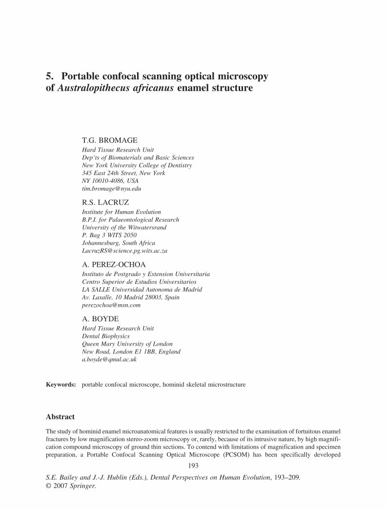
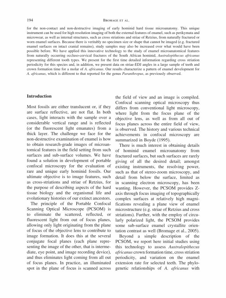
We employ a PCSOM based on the Nipkowdisk technique (Nipkow, 1884) described indetail by Petran and Hadravsky (e.g., 1966)and first commercialized in the early 1980’s.The Petran and Hadravsky design uses aso-called “two-sided” disk; the specimen isilluminated through an array of pinholes onone side of the disk whilst detected through aconjugate array of pinholes on the other (via anumber of delicately aligned mirrors). Appli-cations of this technology to bone and toothmicroanatomy were described by Boyde et al.(1983). Another Nipkow disk design (the oneused here) employs a “single-sided” disk inwhich the illumination and detection pinholeis one in the same (Kino, 1995); that is,illuminating light and its reflections from theobject pass through the same pinhole, which isimaged by the eyepiece objective or camera.This latter design is robust and able to tolerateour relatively extreme portable applications.
To date we have developed two versions ofthe PCSOM; the 1K2 (Figure 1) and the 2K2(Figure 2). Both employ a one-sided Nipkowdisk Technical Instrument Co. K2S-BIOconfocal module (Zygo Corp., Sunnyvale,CA), specifically configured for paleoanthro-pological research problems (Bromage et al.,2003). Like other confocal scanning opticalmicroscopes, the final image derives from theplane of focus, thus it eliminates the fog due tothe halo of reflected, scattered or fluorescentlight above and below the plane of focus,which otherwise confounds image content inconventional light microscopy.
An interesting feature of the single-sided diskdesign by Kino (1995) is the approach taken
Figure 1. Diagram of the 1K2 PCSOM (see textfor details).
to suppress internal, non-image-related reflec-tions that are a significant problem in thistype of system; light reflecting from internalcomponents of the microscope, having nothingto do with forming an image, degrades the
Figure 2. Diagram of the 2K2 PCSOM (see textfor details).
196 Bromage et al.
image. This method is the classical methodof illuminating with polarized light to stoplight reflections from within the optical system(e.g. from optical hardware within the bodyof the microscope), but not the useful lightreflecting from the specimen and returningthrough the objective lens. Linear polarizinglight filters and a single quarter-wave platefilter, described further below, provide themeans for eliminating the unwanted reflectedlight. A consequence of the single-sided diskdesign is that it significantly reduces thenumber of mirrors in the light path makingthe alignment of the optics less critical. Theresult is a very robustly constructed instrumentable to withstand transport and relatively roughhandling (e.g., as checked-in baggage forair travel).
The microscope configurations includeseveral other features critical to our research.Consideration was given to obtainingobjective lenses with relatively long workingdistances (i.e., ca. 20 mm) because oftenwe have little control over the geometryof broken fossil bone surfaces examinedunder remote field or museum conditions,and so we must be prepared to imagethrough long Z-height positions to avoidinterference between the fossil surface andthe objective nosepiece. Objectives choseninclude 5x and 10x lenses (34 mm and 19 mmworking distances respectively; Thales-OptemInc., Fairport, NY, USA) and Mitutoyo 20xand 50x lenses (20 mm and 13 mm workingdistances respectively; Mitutoyo Asia PacificPte Ltd, Singapore). Flexibility in magnifi-cation is achieved by both the introductionof a Thales-Optem 0.5x or 1.9x CCD adapteror by converting the fixed magnificationoptical assembly described above into a zoomsystem, which involves the introduction ofa Thales-Optem 70XL zoom module (1–7x)between the K2S-BIO module coupler and themanual coarse/fine focus module. For fullyautomated image acquisition, we motorizedthe Z focus (below).
Automation in X, Y, and Z axes has beenvariously implemented onto the PCSOM. The1K2 includes a motorized RS232 Z-steppingmotor control setup (Thales-Optem Inc.,Fairport, NY, USA) in place of the manualcoarse/fine focus module when automation isdesired. This setup includes an independentlypowered OEM (original equipment manufac-turer) computer controller board connectedto a stepping motor, which moves in smalldiscrete steps, fitted to the Z-focus moduleand the serial port of the computer. Includedsoftware permits one to drive the focus tostored set positions between the desired endsof travel, or to incrementally drive the focusby any stipulated distance until all opticalplanes within the field of view have beenimaged. Movement in X and Y-axes arecarried out on a manual microscope stage.The 2K2 includes a KP53 motorized precisionmicro-stepping X-Y stage from the SemprexCorporation (Campbell, CA, USA), and aVexta 2-phase Z-axis stepping motor (OrientalMotor USA Corp., Torrance, CA, USA).Integrated XYZ movement is performed byan Oasis 4i PCI stepper motor controllerboard for XY stage and Z focus. A three-axistrackball/mouse control of XYZ axes allowsmanual stage and focus movement to aid real-time viewing.
Portable image acquisitions are transmittedthrough the FireWire™ IEEE 1394 digitalinterface now common on notebook anddesktop computers, thus eliminating theneed for a framegrabber. The 1K2 usesa 4-pin IEEE 1394 high resolution 12bit monochrome QIMAGING Retiga 1300camera (Burnaby, BC, Canada), which has a2/3′′ monochrome progressive scan interlineCCD containing 1280 × 1024 pixels. Real-time image previewing capability facili-tates camera setup conditions, which areadjusted by software interface. Adjustmentsinclude integration time, gain, and offset.The 2K2 uses a JVC KY-F1030U 6-pinIEEE 1394 digital camera containing a 1/2′′
Confocal Microscopy of A. AFRICANUS Enamel 197
color progressive scan interline CCD and1360 × 1024 output pixels, operating at 7.5frames per second live.
The 175W (1K2) and 300W (2K2) LambdaLS Xenon Arc Lamps (Sutter InstrumentCompany, Novato, CA, USA) transmit a flatand intense beam of light via a liquid lightguide. It operates at wavelengths suitablefor both fluorescence and white light illumi-nation (320nm to 700nm output in an ozone-free bulb), is robustly constructed and pre-aligned, and is economically packaged andlightweight, housing its own power supply.
The 1K2 employs A Sony VAIO MobilePentium notebook PC computer for imagecapture. We currently use a VAIO SRX27(800MHz; 256k RAM; Windows XP). Itweighs less than 3 pounds, thus satisfying ourneed for maximum portability, and it containsa 4-pin IEEE 1394 interface. A ShuttleXPC SB52G2 computer with a Pentium4Intel processor and Windows XP Profes-sional (Shuttle Computer Group Inc., LosAngeles, CA, USA) supports fully automatedXYZ stage movement and image acquisition.A reasonably lightweight and thin standard1024 × 768 15′′ monitor (Dell Inc., Round
Rock, TX, USA) was chosen for our real-timeviewing.
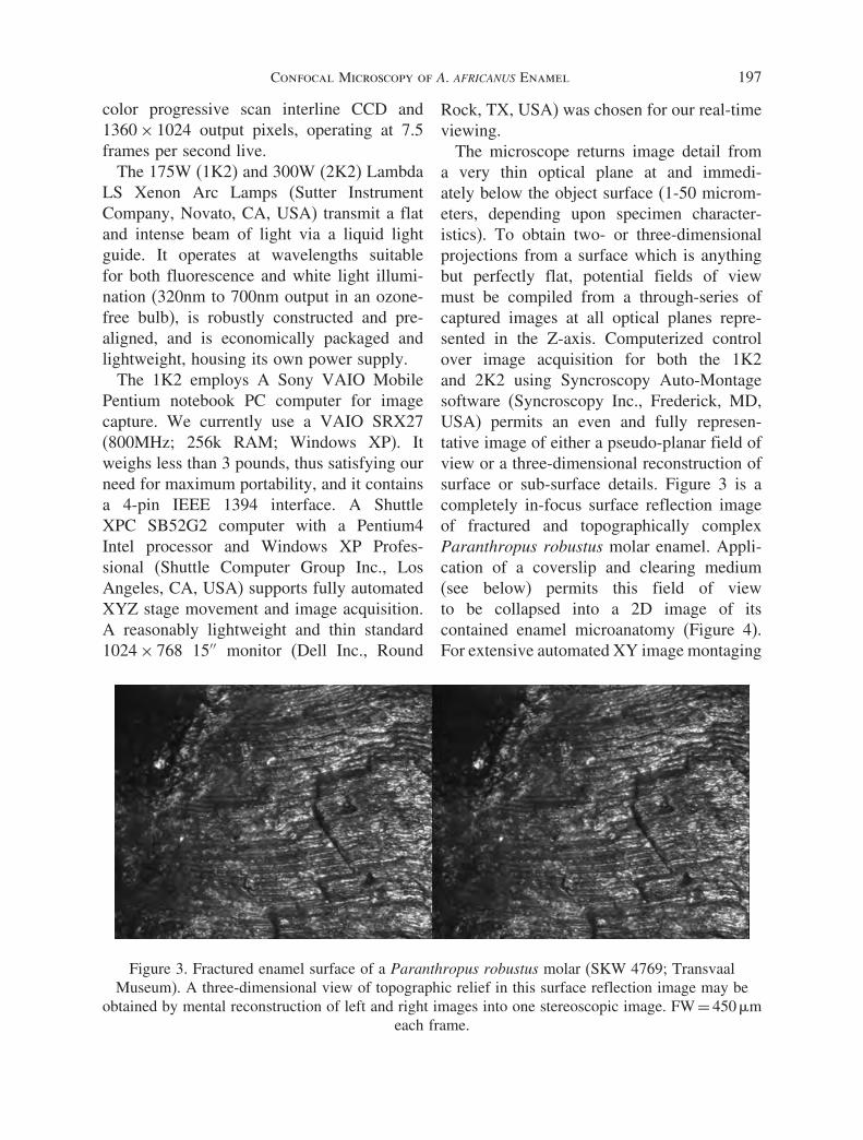
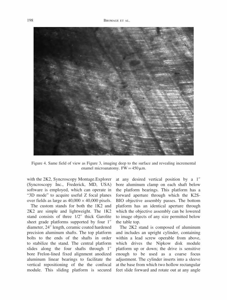
The microscope returns image detail froma very thin optical plane at and immedi-ately below the object surface (1-50 microm-eters, depending upon specimen character-istics). To obtain two- or three-dimensionalprojections from a surface which is anythingbut perfectly flat, potential fields of viewmust be compiled from a through-series ofcaptured images at all optical planes repre-sented in the Z-axis. Computerized controlover image acquisition for both the 1K2and 2K2 using Syncroscopy Auto-Montagesoftware (Syncroscopy Inc., Frederick, MD,USA) permits an even and fully represen-tative image of either a pseudo-planar field ofview or a three-dimensional reconstruction ofsurface or sub-surface details. Figure 3 is acompletely in-focus surface reflection imageof fractured and topographically complexParanthropus robustus molar enamel. Appli-cation of a coverslip and clearing medium(see below) permits this field of viewto be collapsed into a 2D image of itscontained enamel microanatomy (Figure 4).For extensive automated XY image montaging
Figure 3. Fractured enamel surface of a Paranthropus robustus molar (SKW 4769; TransvaalMuseum). A three-dimensional view of topographic relief in this surface reflection image may be
obtained by mental reconstruction of left and right images into one stereoscopic image. FW = 450 �meach frame.
198 Bromage et al.
Figure 4. Same field of view as Figure 3, imaging deep to the surface and revealing incrementalenamel microanatomy. FW = 450 �m.
with the 2K2, Syncroscopy Montage.Explorer(Syncroscopy Inc., Frederick, MD, USA)software is employed, which can operate in“3D mode” to acquire useful Z focal planesover fields as large as 40,000 × 40,000 pixels.
The custom stands for both the 1K2 and2K2 are simple and lightweight. The 1K2stand consists of three 1/2′′ thick Garolitesheet grade platforms supported by four 1′′diameter, 24′′ length, ceramic coated hardenedprecision aluminum shafts. The top platformbolts to the ends of the shafts in orderto stabilize the stand. The central platformslides along the four shafts through 1′′bore Frelon-lined fixed alignment anodizedaluminum linear bearings to facilitate thevertical repositioning of the the confocalmodule. This sliding platform is secured
at any desired vertical position by a 1′′bore aluminum clamp on each shaft belowthe platform bearings. This platform has aforward aperture through which the K2S-BIO objective assembly passes. The bottomplatform has an identical aperture throughwhich the objective assembly can be loweredto image objects of any size permitted belowthe table top.
The 2K2 stand is composed of aluminumand includes an upright cylinder, containingwithin a lead screw operable from above,which drives the Nipkow disk moduleplatform up or down; the drive is sensitiveenough to be used as a coarse focusadjustment. The cylinder inserts into a sleeveat the base from which two hollow rectangularfeet slide forward and rotate out at any angle
Confocal Microscopy of A. AFRICANUS Enamel 199
appropriate for the balance of weight andrequired workspace. The platform for holdingthe K2S-BIO attaches to a sleeve aroundthe cylinder, which rides on a bearing thatconveys the module in any rotational positionwithin the workspace.
Each microscope automatically switchesbetween 110V and 220V electrical supplies(only the Nipow disk motor requires an optional110V/220V adaptor), fits into two suitcases(Pelican Products, Inc., Torrance, CA, USA),and may be set up and tested within one hour ofarrival at museum locations.
Enamel Microstructure
Naturally fractured Australopithecus africanusteeth from Member 4 of the SterkfonteinFormation, dated to approximately 2.5 my(Vrba, 1995) were examined. The workhas only begun, and to date four naturallyfractured molars, one previously sectionedmolar and one canine have been imaged forthis preliminary study. They include: STW11 (RM3), STW 90 (RM3), STW 190 (Leftmaxillary molar fragment), STW 284 (LM2),STW 37 (LM3), and STW 267 (canine).The fractured surface of the tooth, exposingenamel in cross section, was placed approxi-mately perpendicular to the optical axis, overwhich was placed a drop of immersion oil,and over this, a glass cover slip according tostandard microscopal investigation. Becausethe fractured surfaces were not perfectly flat,images were Z-montaged in SyncroscopyAuto-Montage.
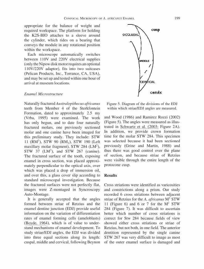
It is generally accepted that the anglesformed between striae of Retzius and theenamel dentine junction (EDJ) provide usefulinformation on the variation of differentiationrates of enamel forming cells (ameloblasts)(Boyde, 1964), which is of value to under-stand mechanisms of enamel development. Tostudy striae/EDJ angles, the EDJ was dividedinto three equal sections along its length:cuspal, middle and cervical, following Beynon
Figure 5. Diagram of the divisions of the EDJwithin which striae/EDJ angles are measured.
and Wood (1986) and Ramirez Rozzi (2002)(Figure 5). The angles were measured as illus-trated in Schwartz et al. (2003: Figure 2A).In addition, we provide crown formationtime for the molar STW 284. This specimenwas selected because it had been sectionedpreviously (Grine and Martin, 1988) andthus there was good control over the planeof section, and because striae of Retziuswere visible through the entire length of theprotocone cusp.
Results
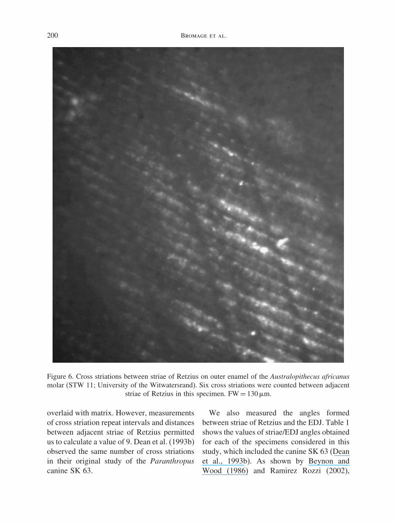
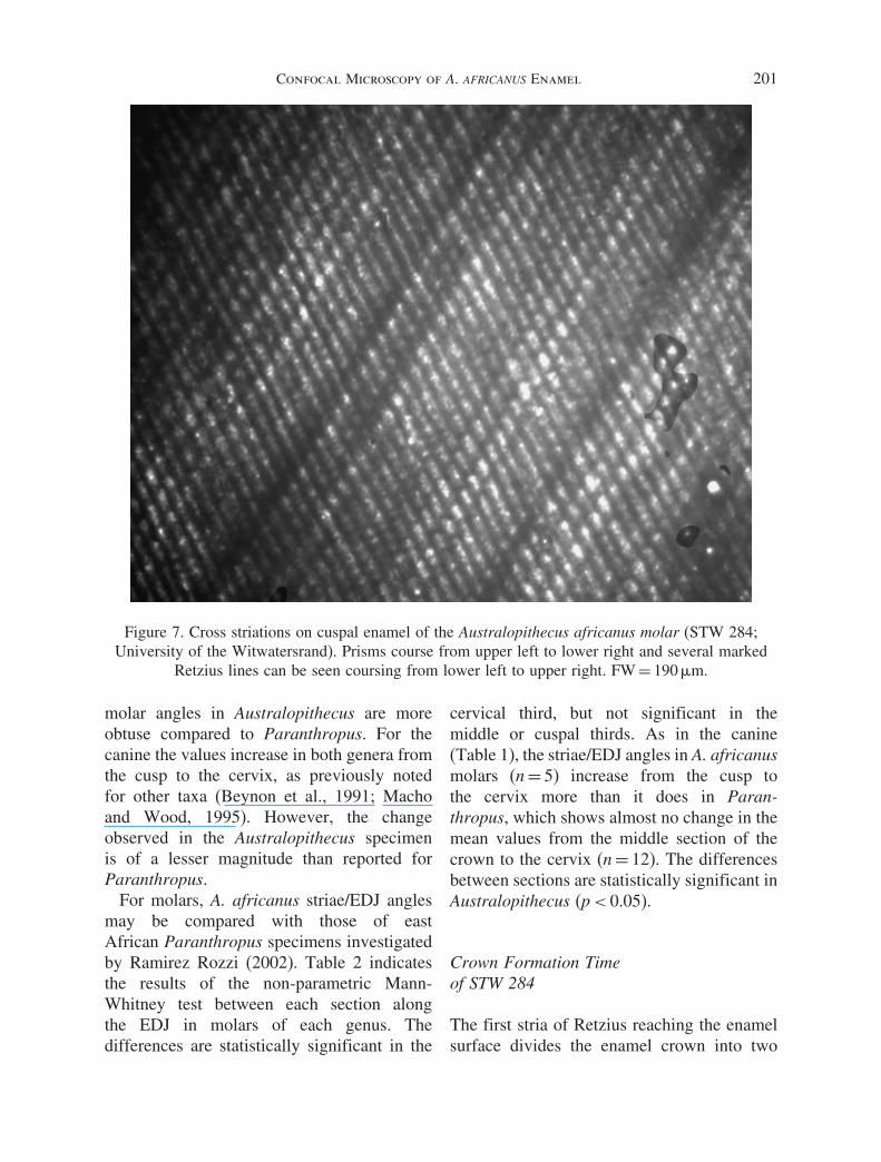
Cross striations were identified as varicositiesand constrictions along a prism. Our studyrecorded 6 cross striations between adjacentstriae of Retzius for the A. africanus M3 STW11 (Figure 6) and 6 or 7 for the M2 STW284 (Figure 7). It was difficult to ascertainbetter which number of cross striations iscorrect for Stw 284 because fields of viewshowed either cross striations or striae ofRetzius, but not both, in one field. The anteriordentition represented by the single canineSTW 267 was very difficult to image as mostof the outer enamel surface is damaged and
200 Bromage et al.
Figure 6. Cross striations between striae of Retzius on outer enamel of the Australopithecus africanusmolar (STW 11; University of the Witwatersrand). Six cross striations were counted between adjacent
striae of Retzius in this specimen. FW = 130 �m.
overlaid with matrix. However, measurementsof cross striation repeat intervals and distancesbetween adjacent striae of Retzius permittedus to calculate a value of 9. Dean et al. (1993b)observed the same number of cross striationsin their original study of the Paranthropuscanine SK 63.
We also measured the angles formedbetween striae of Retzius and the EDJ. Table 1shows the values of striae/EDJ angles obtainedfor each of the specimens considered in thisstudy, which included the canine SK 63 (Deanet al., 1993b). As shown by Beynon andWood (1986) and Ramirez Rozzi (2002),
Confocal Microscopy of A. AFRICANUS Enamel 201
Figure 7. Cross striations on cuspal enamel of the Australopithecus africanus molar (STW 284;University of the Witwatersrand). Prisms course from upper left to lower right and several marked
Retzius lines can be seen coursing from lower left to upper right. FW = 190 �m.
molar angles in Australopithecus are moreobtuse compared to Paranthropus. For thecanine the values increase in both genera fromthe cusp to the cervix, as previously notedfor other taxa (Beynon et al., 1991; Machoand Wood, 1995). However, the changeobserved in the Australopithecus specimenis of a lesser magnitude than reported forParanthropus.
For molars, A. africanus striae/EDJ anglesmay be compared with those of eastAfrican Paranthropus specimens investigatedby Ramirez Rozzi (2002). Table 2 indicatesthe results of the non-parametric Mann-Whitney test between each section alongthe EDJ in molars of each genus. Thedifferences are statistically significant in the
cervical third, but not significant in themiddle or cuspal thirds. As in the canine(Table 1), the striae/EDJ angles in A. africanusmolars (n = 5) increase from the cusp tothe cervix more than it does in Paran-thropus, which shows almost no change in themean values from the middle section of thecrown to the cervix (n = 12). The differencesbetween sections are statistically significant inAustralopithecus (p < 0.05).
Crown Formation Timeof STW 284
The first stria of Retzius reaching the enamelsurface divides the enamel crown into two
202 Bromage et al.
Table 1. Striae-EDJ angle values for eachspecimen studied at each division along the EDJ.The numbers in brackets indicate the number of
angles measured for each section
Specimen number Tooth Area Mean (n)
SK 63 UC Cuspal 14.5 (4)Middle 31.8 (9)Cervical 34.6 (8)
STW 279 UC Cuspal ?Middle 34.2 (4)Cervical 41.4 (5)
STW 284 UM2 Cuspal 20.0 (2)Middle 31.3 (6)Cervical 40.6 (5)
STW 190 frag Cuspal 14.6 (3)Middle 28.0 (5)Cervical 39.3 (4)
STW 90 Lm3 Cuspal 21.0 (2)Middle 30.7 (7)Cervical 34.7 (7)
STW 11 UM3 Cuspal 16.5 (2)Middle 28.5(7)Cervical 51.4 (7)
STW 37 UM3 Cuspal 18.0 (2)Middle 35.0 (5)Cervical 38.0 (4)
portions, which identify cuspal (or apposi-tional) and cervical (or imbricational) devel-opmental periods (Beynon and Wood, 1987).To calculate crown formation time in STW
Table 2. Results of Mann-Whitney test ofParanthropus and A. africanus for the striae-EDJ
angle values at each division along the EDJ
Sample Mean SD p value
CuspalEA Paranthropus 12 13.2 5.2A. africanus 5 18.2 2.6 N.S.MiddleEA Paranthropus 12 26.7 6.9A. africanus 5 33.9 4.5 N.S.CervicalEA Paranthropus 12 26.0 6.5A. africanus 5 42.0 6.0 p < 0.05
The values shown for E.A. Paranthropus were taken fromRamirez Rozzi (2002) and include, on the lower dentition, sixM3, a possible M2 or M3, and two M1 or M2. The upperdentition consists of one M2, one M3, and a possible M2 orM3 (Ramirez Rozzi 2002: Table 15.2)
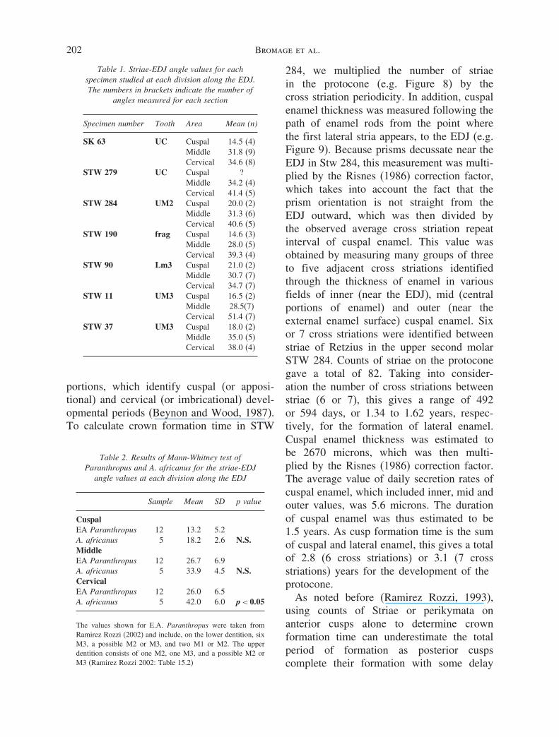
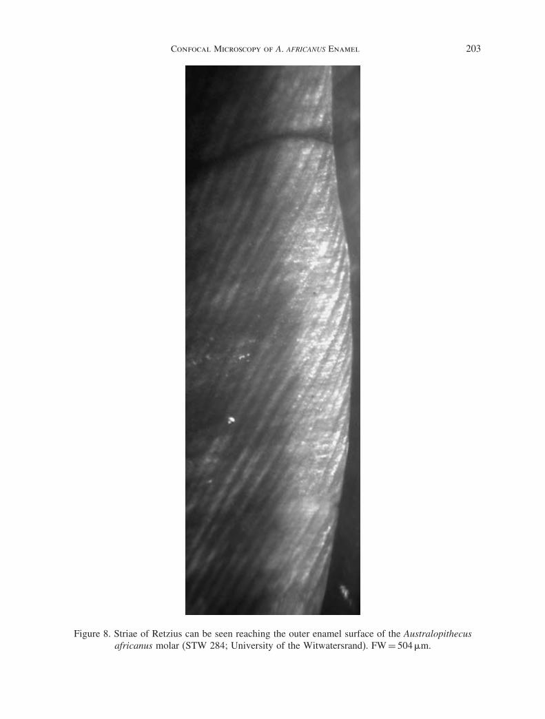
284, we multiplied the number of striaein the protocone (e.g. Figure 8) by thecross striation periodicity. In addition, cuspalenamel thickness was measured following thepath of enamel rods from the point wherethe first lateral stria appears, to the EDJ (e.g.Figure 9). Because prisms decussate near theEDJ in Stw 284, this measurement was multi-plied by the Risnes (1986) correction factor,which takes into account the fact that theprism orientation is not straight from theEDJ outward, which was then divided bythe observed average cross striation repeatinterval of cuspal enamel. This value wasobtained by measuring many groups of threeto five adjacent cross striations identifiedthrough the thickness of enamel in variousfields of inner (near the EDJ), mid (centralportions of enamel) and outer (near theexternal enamel surface) cuspal enamel. Sixor 7 cross striations were identified betweenstriae of Retzius in the upper second molarSTW 284. Counts of striae on the protoconegave a total of 82. Taking into consider-ation the number of cross striations betweenstriae (6 or 7), this gives a range of 492or 594 days, or 1.34 to 1.62 years, respec-tively, for the formation of lateral enamel.Cuspal enamel thickness was estimated tobe 2670 microns, which was then multi-plied by the Risnes (1986) correction factor.The average value of daily secretion rates ofcuspal enamel, which included inner, mid andouter values, was 5.6 microns. The durationof cuspal enamel was thus estimated to be1.5 years. As cusp formation time is the sumof cuspal and lateral enamel, this gives a totalof 2.8 (6 cross striations) or 3.1 (7 crossstriations) years for the development of theprotocone.
As noted before (Ramirez Rozzi, 1993),using counts of Striae or perikymata onanterior cusps alone to determine crownformation time can underestimate the totalperiod of formation as posterior cuspscomplete their formation with some delay
Confocal Microscopy of A. AFRICANUS Enamel 203
Figure 8. Striae of Retzius can be seen reaching the outer enamel surface of the Australopithecusafricanus molar (STW 284; University of the Witwatersrand). FW = 504 �m.
204 Bromage et al.
Figure 9. Cuspal enamel of Australopithecus africanus molar (STW 284; University of theWitwatersrand). Prisms can be identified running almost vertically towards the outer enamel surface
(top). Cross striations are seen along each prism as dark horizontal lines. FW = 1.3 mm. The boundarybetween lateral and cuspal enamel is located slightly more cervically and could not be imaged here.
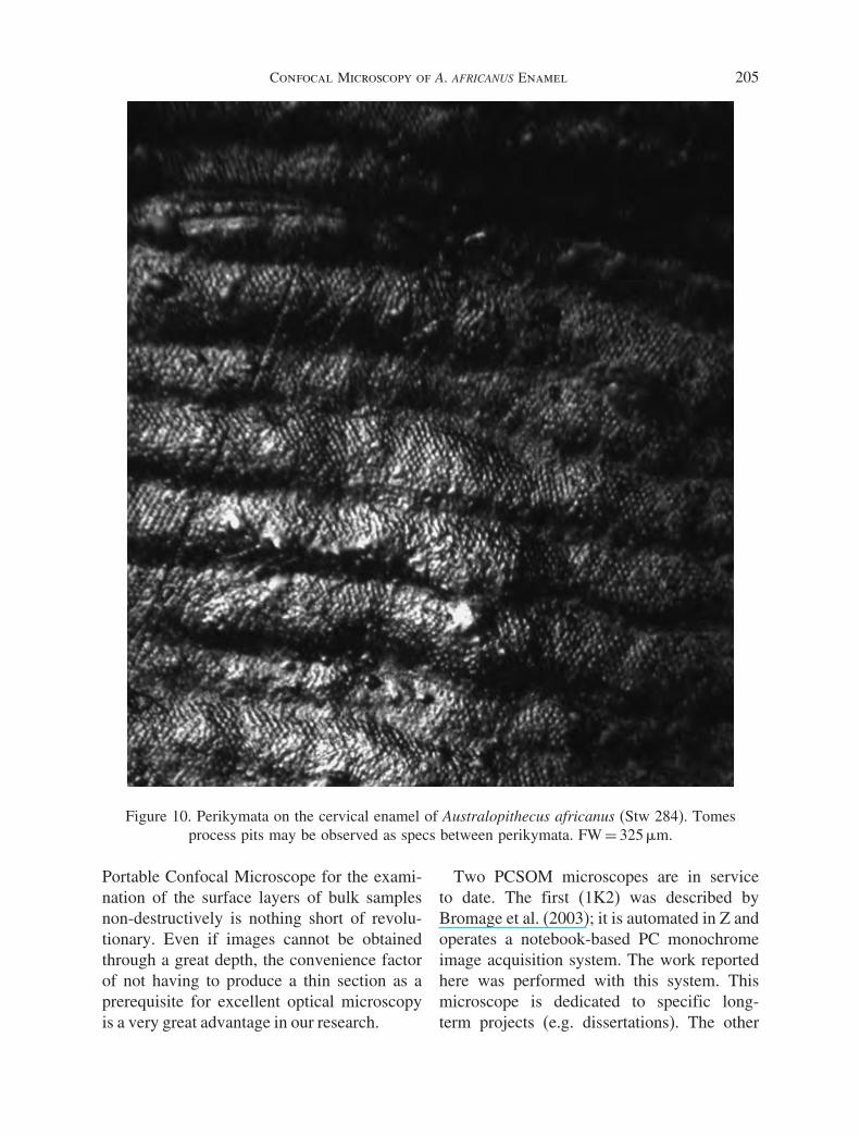
relative to the anterior cusps (e.g., Kraus andJordan, 1965). The last visible stria on theprotocone was followed to its correspondingperikyma and this was followed to thehypocone (e.g., Figure 10). The perikymatacervical to it on this cusp were counted givinga total of 12 perikymata, or an additional 0.2years of growth. This gives a total of 3.0
(6 cross striations) or 3.2 (7 cross striations)years for the crown development of STW 284.
Discussion
While the improvement over conventional lightmicroscopy in imaging thin sections may notbe substantial, the improvement made by the
Confocal Microscopy of A. AFRICANUS Enamel 205
Figure 10. Perikymata on the cervical enamel of Australopithecus africanus (Stw 284). Tomesprocess pits may be observed as specs between perikymata. FW = 325 �m.
Portable Confocal Microscope for the exami-nation of the surface layers of bulk samplesnon-destructively is nothing short of revolu-tionary. Even if images cannot be obtainedthrough a great depth, the convenience factorof not having to produce a thin section as aprerequisite for excellent optical microscopyis a very great advantage in our research.
Two PCSOM microscopes are in serviceto date. The first (1K2) was described byBromage et al. (2003); it is automated in Z andoperates a notebook-based PC monochromeimage acquisition system. The work reportedhere was performed with this system. Thismicroscope is dedicated to specific long-term projects (e.g. dissertations). The other
206 Bromage et al.
microscope (2K2) is fully automated in X,Y, and Z. With development of the PCSOMthe potential for non-destructive mineralizedtissue research on rare and unique earlyhominid remains is great.
Enamel development (amelogenesis) is afunction of the number of cells involvedin matrix secretion, secretion rates, and therate at which these cells become differ-entiated along the enamel-dentine junction(EDJ). All active cells during amelogenesisperiodically stop normal secretory activity,creating features known as striae of Retzius(cf. Boyde, 1990). Their periodicity can becalculated by recording the number of dailycell secretions or cross striations between eachstria (cf. Bromage 1991).
Originally, Boyde (1964) proposed amethod to estimate the rate of cell differen-tiation based on the angles formed betweenthe striae of Retzius and the EDJ. More acuteangles indicate a higher ameloblast differ-entiation rate. A later study (Beynon andWood, 1986) made use of this method inan analysis of isolated teeth attributed toParanthropus and Homo to assess differencesbetween these two genera. They found thatthe angles formed between the EDJ and thestriae were more acute in Paranthropus thanin Homo, with means of 23 and 31 degreesrespectively. Their measurements were takenon the occlusal third of the crown. RamirezRozzi (1993, 1998, 2002) used a larger samplederived from the Omo Shungura Formation,Ethiopia, to assess possible temporal changesin the rates of ameloblast differentiation inisolated teeth from a well stratified and datedchronological sequence. Measurements weretaken on three sections along the EDJ (cuspal,central – equivalent to our “middle” region –and cervical areas). In agreement with Beynonand Wood (1986), Ramirez Rozzi found fastrates of enamel differentiation in the genusParanthropus, though he noted differencesbetween two sets identified within the Omosample, of P. aethiopicus and P. boisei. The
only published record of angles formed bystriae of Retzius and the EDJ in A. africanus isthat of Grine and Martin (1988) and, althoughno measurements were given, they observedthat Paranthropus showed more acute anglesthan A. africanus. Thus, at present, thereis almost no data on the microanatomicalfeatures of this species.
An important aspect in studies of dentaldevelopment using microanatomy is the crossstriation periodicity. Most commonly, thisvalue is assessed from histological groundsections or by scanning electron microscopy,but both methods are intrusive and laborious.Thus only four studies to date have includedinformation regarding cross striation period-icity on hominid fossils; a P. boisei premolar(Beynon and Dean, 1987), a molar of P. boisei(Dean 1987), a P. robustus canine (Deanet al., 1993b), and a Neandertal molar (Deanet al., 2001). The periodicities recorded inthese samples range from 7 to 9 cross stria-tions. Here we report a relatively significantsample of cross striation periodicities for asingle hominid species.
Values of cross striation periodicityobserved in the small sample of teethattributed to A. africanus are highly variable.For the two molars observed the numbersranged from 6 to 7. The anterior dentition,represented here by a single canine (STW267), presented a calculated value of ninecross striations which is the same numberobserved by Dean et al. (1993b) in the Paran-thropus canine SK 63. This variation fallswithin the cross striation periodicity valuesrecorded for modern humans (6–12) (Deanand Reid, 2001) and is similar to chimpanzees(6–8) (Reid et al., 1998b; Smith, 2004) andother hominids.
Results from this preliminary study indicatethat there may be some differences ingrowth mechanisms of enamel tissue betweenA. africanus and Paranthropus. In general,rates of ameloblast differentiation inA. africanus, measured as the angles formed
Confocal Microscopy of A. AFRICANUS Enamel 207
between striae of Retzius and the EDJ,decrease as the development of the crownapproaches the more cervical aspects ofthe tooth; that is, the angles have highervalues in A. africanus than in Paranthropus(Table 1). It could be argued that some ofthese differences are the result of studyingnaturally fractured teeth where there is nocontrol of the plane of section. However, thefact that all A. africanus molars studied showthe same pattern of difference from EastAfrican Paranthropus molars, suggests thatthe plane of fracture does not significantlyaffect the results.
The crown formation time of a single molarof A. africanus was estimated to be 3.0to 3.2. years. The former value is similarto the mean of crown formation time ofmolars attributed to P. boisei and greaterthan values of P. aethiopicus (Ramirez Rozzi,1993). However, the crown formation timeof STW 284 is slightly less than reportedfor modern human second molars (Beynonand Wood, 1987; Dean et al., 1993a; Reidet al., 1998a) in spite of the fact that A.africanus molars have thicker enamel andgreater occlusal area. All of this taken togetheremphasizes differences already noted betweenextant and extinct taxa on the one hand, andbetween different hominid species on the other(Beynon and Wood, 1987; Beynon and Dean,1988; Bromage and Dean, 1985; Dean et al.,2001).
Conclusions
The Portable Confocal Scanning OpticalMicroscope was specifically developed tooffer superb analytical light microscopy ofearly hominid skeletal material. Limitationsover the handling and transport of rarefossils have motivated its development so thatspecimens may be examined by whatever thecircumstances dictate.
This study has added new information onthe growth processes of enamel identified
in the southern African hominid taxa A.africanus. Given the results obtained here, itwould be important to assess growth processesfor the South African taxon P. robustus, forwhich almost no information on molar devel-opment is available, to possibly help betterestablish relationships among early Africanhominids.
Acknowledgments
Support for this work was generouslyprovided by the L.S.B. Leakey Foundation,the Blanquer and March Foundations (Spain),the Palaeoanthropology Scientific Trust(PAST, South Africa) and Dr. D. McSherry.For the availability of hominid specimensand assistance, the Department of Palaeon-tology, Transvaal Museum, Pretoria, SouthAfrica, and the Palaeoanthropology ResearchUnit, Department of Anatomy, Universityof the Witwatersrand, Johannesburg, SouthAfrica, are gratefully acknowledged. Muchappreciation to Shara Bailey and Jean-JacquesHublin for organizing the symposium, “Dentalperspectives on human evolution: State of theart research in dental paleoanthropology” andto Shara Bailey and anonymous reviewers forcritical comments on the manuscript.
References
Berger, L., Lacruz, R.S., de Ruiter, D.J., 2002.Revised age estimates of Australopithecusbearing deposits at Sterkfontein, South Africa.American Journal of Physical Anthropology119, 192–197.
Beynon, A.D., Dean, M.C., 1987. Crown formationtime of a fossil hominid premolar tooth.Archives of Oral Biology 32, 773–780.
Beynon, A.D., Dean, M.C., 1988. Distinct dental devel-opment patterns in early fossil hominids. Nature335, 509–514.
Beynon, A.D., Dean, M.C., Reid, D.J., 1991. Ahistological study on the chronology of thedeveloping dentition of gorilla and orangutan.American Journal of Physical Anthropology 86,295–309.
208 Bromage et al.
Beynon, A.D., Wood, B., 1986. Variations inenamel thickness and structure in East Africanhominids. American Journal of PhysicalAnthropology 70, 177–193.
Beynon, A.D., Wood, B., 1987. Patterns and ratesof enamel growth on the molar teeth of earlyhominids. Nature 326, 493–496.
Boyde, A., 1964. The structure and developmentof mammalian enamel. Ph.D. Dissertation,University of London.
Boyde, A., 1990. Developmental interpretations ofdental microstructure. In: Jean de Rousseau, C.(Ed.), Primate Life History and Evolution.Wiley-Liss Publ., New York, pp. 229–267.
Boyde, A., 1995. Confocal optical microscopy. In:Wootton, R., Springall, D.R., Polak, J.M. (Eds.),Image Analysis in Histology: Conventional andConfocal Microscopy. Cambridge UniversityPress, Cambridge, UK, pp. 151–196.
Boyde, A., Petran, M., Hadravsky, M., 1983. Tandemscanning reflected light microscopy of internalfeatures in whole bone and tooth samples.Journal of Microscopy 132, 1–7.
Bromage, T.G., 1991. Enamel incremental period-icity in the pig-tailed macaque: a polychromefluorescent labelling study of dental hardtissues. American Journal of Physical Anthro-pology 86, 205–214.
Bromage, T.G., Dean, M.C., 1985. Re-evaluation ofthe age at death of immature fossil hominids.Nature 317, 525–527.
Bromage, T.G., Perez-Ochoa, A., Boyde, A., 2003. Theportable confocal microscope: scanning opticalmicroscopy anywhere. In: Méndez Vilas, A.(Ed.), Science, Technology and Education ofMicroscopy: An Overview. Formatex, Badajoz,Spain, pp. 742–752.
Bromage, T.G., Perez-Ochoa, A., Boyde, A., 2005.Portable confocal microscope reveals fossilhominid microstructure. Microscopic Analysis19, 5–7.
Dean, M.C., 1987. Growth layers and incrementalmarkings in hard tissues, a review ofthe literature and some preliminary obser-vations about enamel structure of Paran-thropus boisei. Journal of Human Evolution 16,157–172.
Dean, M.C., Beynon, A.D., Reid, D.J., Whittaker,D.K., 1993a. A longitudinal study of toothgrowth in a single individual based on long andshort period markings in dentine and enamel.International Journal of Osteoarchaeology 3,249–264.
Dean, M.C., Beynon, A.D., Thackeray, J.F., Macho,G.A., 1993b. Histological reconstruction ofdental development and age at death ofa juvenile Paranthropus robustus specimen,SK 63, from Swartkrans, South Africa.American Journal of Physical Anthropology 91,401–419.
Dean, M.C., Leakey, M., Reid, D., Schrenk, F.,Schwartz, G., Stringer, C., Walker, A., 2001.Growth processes in teeth distinguish modernhumans from Homo erectus and earlierhominins. Nature 44, 628–631.
Dean, M.C., Reid, D.J., 2001. Perikymata and distri-bution on Hominid anterior teeth. AmericanJournal of Physical Anthropology 116,209–215.
Grine, F.E., Martin, L.B., 1988. Enamel thickness anddevelopment in Australopithecus and Paran-thropus. In: Grine, F.E. (Ed.), The Evolu-tionary History of the “Robust” Australo-pithecines. Aldine de Gruyter, New York,pp. 3–42.
Kino, G.S., 1995. Intermediate optics in Nipkow diskmicroscopes. In: Pawley, J.B. (Ed.), Handbookof Biological Confocal Microscopy. PlenumPress, New York, pp. 155–165.
Kraus, B.S., Jordan, R.E., 1965. The human dentitionbefore birth. Lea & Febiger Publ., Philadelphia.
Macho, G.A., Wood, B.A., 1995. The role of timeand timing in hominid dental evolution. Evolu-tionary Anthropology 4, 17–31.
Nipkow, P., 1884. Elektrisches teleskop. Patentschrift30105 (Kaiserliches Patentamt, Berlin),patented 06.01.1884.
Petran, M., Hadravsky, M., 1966. Method andarrangement for improving the resolvingpower and contrast. United States PatentNo. 3,517,980, priority 05.12.1966, patented30.06.1970 US.
Ramirez Rozzi, F., 1993. Tooth development inEast African Paranthropus. Journal of HumanEvolution 24, 429–454.
Ramirez Rozzi, F., 1998. Can enamel microstructurebe used to establish the presence of differentspecies of Plio-Pleistocene hominids from Omo,Ethiopia? Journal of Human Evolution 35,543–576.
Ramirez Rozzi, F., 2002. Enamel microstructurein hominids: New characteristics for a newparadigm. In: Minugh-Purvis, N., McNamara,K.J. (Eds.), Human Evolution Through Devel-opmental Change. Johns Hopkins UniversityPress, Baltimore, pp. 319–348.
Confocal Microscopy of A. AFRICANUS Enamel 209
Reid, D.J., Beynon, A.D., Ramirez Rozzi, F.V.,1998a. Histological reconstruction of dentaldevelopment in four individuals from aMedieval site in Picardie, France. Journal ofHuman Evolution 35, 463–478.
Reid, D.J., Schwartz, G.T., Dean, M.C., Chandrasekera,M.S., 1998b. A histological reconstruction ofdental development in the common chimpanzee.Journal of Human Evolution 35, 427–448.
Risnes, S., 1986. Enamel apposition rate and theprism periodicity in human teeth. Scandi-navian Journal of Dental Research 94,394–404.
Schwartz, G.T., Liu, W., Zheng, L. (2003). Prelim-inary investigation of dental microstructurein the Yuanmou hominoid (Lufengpithecushudienensis), Yumn Province, China. Journal ofHuman Evolution 44, 189–202.
Smith, T.M., 2004. Incremental development ofprimate dental enamel. Ph.D. Dissertation, StateUniversity of New York, Stony Brook.
Tobias, P.V., 1980. Australopithecus afarensis andA. africanus: critique and an alternativehypothesis. Palaeontologica Africa 23, 1–17.
Vrba, E.S., 1995. The fossil record of African antelopes(Mammalia, Bovidae) in relation to humanevolution and paleoclimate. In: Vrba, E.S. (Ed.),Paleoclimate and Evolution, With Emphasis onHuman Origins. Yale University Press, NewHaven, pp. 385–424.
White, T.D., Johanson, D.C., Kimbel, W.H., 1981.Australopithecus africanus: its phyletic positionreconsidered. South African Journal of Science77, 445–471.


