Embed Size (px)
Citation preview

pubs.acs.org/Biochemistry Published on Web 10/06/2010 r 2010 American Chemical Society
9372 Biochemistry 2010, 49, 9372–9384
DOI: 10.1021/bi101156j
Positional Effects on Helical Ala-Based Peptides†
Richard P. Cheng,*,‡ Prashant Girinath,§ Yuta Suzuki,§ Hsiou-Ting Kuo,‡ Hao-Chun Hsu,‡ Wei-Ren Wang,‡
Po-An Yang,‡ Donald Gullickson,§ Cheng-Hsun Wu,‡ Marc J. Koyack,§ Hsien-Po Chiu,§ Yi-Jen Weng,‡ Pier Hart, )
Bashkim Kokona, ) Robert Fairman, ) Tzu-En Lin,‡ and Olivia Barrett§
‡Department of Chemistry, National Taiwan University, Taipei 10617, Taiwan, §Department of Chemistry, University at Buffalo,The State University of New York, Buffalo, New York 14260-3000, and )Department of Biology, Haverford College,
Haverford, Pennsylvania 19041
Received July 20, 2010
ABSTRACT:Helix-coil equilibrium studies are important for understanding helix formation in protein folding,and for helical foldamer design. The quantitative description of a helix using statistical mechanical models isbased on experimentally derived helix propensities and the assumption that helix propensity is position-independent. To investigate this assumption, we studied a series of 19-residue Ala-based peptides, to measurethe helix propensity for Leu, Phe, and Pff at positions 6, 11, and 16. Circular dichroism spectroscopy revealedthat substituting Ala with a given amino acid (Leu, Phe, or Pff) resulted in the following fraction helix trend:KXaa16>KXaa6>KXaa11. Helix propensities for Leu, Phe, and Pff at the different positions were derivedfrom the CD data. For the same amino acid, helix propensities were similar at positions 6 and 11, but muchhigher at position 16 (close to the C-terminus). A survey of protein helices revealed that Leu/Phe-Lys (i, iþ 3)sequence patterns frequently occur in two structural patterns involving the helix C-terminus; however, thesecases include a left-handed conformation residue. Furthermore, no Leu/Phe-Lys interactionwas found exceptfor the Lys-Phe cation-π interaction in two cases of Phe-Ala-Ala-Lys. The apparent high helix propensity atposition 16 may be due to helix capping, adoption of a 310-helix near the C-terminus perhaps with Xaa-Lys(i, i þ 3) interactions, or proximity to the peptide chain terminus. Accordingly, helix propensity is generallyposition-independent except in the presence of alternative structures or in the proximity of either chainterminus. These results should facilitate the design of helical peptides, proteins, and foldamers.
Helices represent one of the most common structural motifs inboth proteins (1-3) and non-natural foldamers (4-8). One thirdof all protein residues adopt an R-helical conformation (1-3),leading to various experimental studies on R-helix formationenergetics, including helix propensity (9-27), capping energetics(18, 22, 28-39), and intrahelical interactions (21, 22, 25, 40-66).Helices are also the most prevalent structure in foldamers (4-8),which are non-natural polymers and oligomers that can adoptcompact, well-defined three-dimensional conformations andstructures.Foldamers such as peptoids (67),β-peptides (4, 68, 69),oligo-phenylene ethynylenes (70), and oligo-arylamides (71, 72)have been designed to adopt helical conformations (6, 8).Furthermore, intrahelical interactions (73-77) and helix propen-sity differences (76, 78, 79) have also been explored in helicalfoldamers. Therefore, fundamental studies of the helix-coilequilibrium are important for understanding helix formation inprotein folding, and in foldamer design and applications.
The helix propensity of natural amino acids has been experimen-tally measured in random copolymers (9, 10), monomeric helicalpeptides (12, 14, 16, 21-27), coiled coil systems (13), and helices innatural proteins (18-20, 24). In particular, experimental measure-ments onmonomeric helical systems (12, 14, 16, 21, 22, 24-27) have
been coupled with statistical mechanical models based on eitherZimm-Bragg (80) or Lifson-Roig (81) theory to obtain helixformation parameters, and thus absolute (nonrelative) helix form-ing energetics. Both theories assume that every amino acid can onlyadopt either a helical or a nonhelical conformation (80, 81), effec-tively invoking a multistate equilibrium for helical peptides invol-ving multiple residues. Furthermore, the probability of adopting ahelical conformation for a given residue depends on its side chain(propensity) and the conformation of neighboring residues (co-operativity) (80, 81). Using either statistical mechanical model, helixformation for every amino acid is represented by at least twoparameters: helix initiation parameter and helix propagation para-meter (i.e., helix propensity) (80, 81).Helix capping parameters havealso been incorporated into the modified Lifson-Roig theory(12, 34, 35, 38). Further advancements in describing the helix-coilequilibrium have included intrahelical side chain-side chain inter-actions (21, 22, 25, 38, 41, 45-48, 50-54, 58-66) and interactionbetween charged amino acids and the helix dipole (38, 41, 66, 82).The influence of temperature (83-89) and trifluoroethanol(87, 88, 90) on the helix propensity parameter has also been studied.
The quantitative description of monomeric helix formationusing statistical mechanical models has been built on a foundationbased on experimentally derived helix propagation parameters(helix propensities) (12, 14, 16, 21, 22, 24-27). In particular,seminal work by Baldwin and co-workers on such systems withminimal intrahelical side chain interactions has provided the helixpropagation parameters and capping parameters for deducing thefraction helix of monomeric Ala-based peptides (12, 34, 35, 90).
†This work was supported by the NYSTAR James D. Watson Investi-gator Program (R.P.C.), Kapoor funds (R.P.C.), The State University ofNew York at Buffalo (R.P.C.), the National Science Foundation (R.P.C.,CHE0809633; R.F., MCB-0211754), National Taiwan University (R.P.C.),and theNational ScienceCouncil (R.P.C., NSC-97-2113-M-002-019-MY2).*To whom correspondence should be addressed. Phone: þ886-2-
33669789. Fax: þ886-2-23636359. E-mail: [email protected].

Article Biochemistry, Vol. 49, No. 43, 2010 9373
One basic assumption in these statistical mechanical models isthat the helix propensity for a given amino acid does not changewith position or peptide length (80, 81). Interestingly, studies byKemp showed that the helix propensity of Ala is peptide length-dependent (85, 91). Although the position independence of helixpropensity was observed in Ala-based monomeric helices (92) anda protein helix (93), the position dependence of helix propensitywas observed in another set of Ala-based monomeric helices(94-96). However, the Ala-based helices had guest positions withvarying immediate neighboring residues, thereby unable to com-pletely rule out the immediate neighboring context as the reasonfor the apparent position dependence of helix propensity (94-96).Herein, we present studies on a series of Ala-based peptides withneutral non-Ala residues [leucine, phenylalanine, and pentafluor-ophenylalanine (Pff)] at various guest positions with the sameimmediate surrounding residues ((2 residues), to study the effectof these amino acids at different positions of a helical peptide andto measure the helix propensity for these amino acids at thesedifferent positions.
MATERIALS AND METHODS
General. All of the chemical reagents except those indicatedotherwise were purchased from Aldrich. Organic and high-performance liquid chromatography (HPLC) solvents were fromEMD Science andMerck Taiwan.N-9-Fluorenylmethoxycarbo-nyl (Fmoc) amino acids, 1-hydroxybenzotriazole (HOBt), andO-1H-benzotriazol-1-yl-1,1,3,3-tetramethyluronium hexafluoro-phosphate (HBTU) were fromNovaBiochem, Fmoc-PAL-PEG-PS resin was from Applied Biosystems. Analytical reverse phase(RP) HPLC was performed on an Agilent 1100 series chroma-tography system using a Vydac C18 column (4.6 mm diameter,250 mm length). Preparative RP-HPLC was performed on aWaters Breeze chromatography system using Vydac RP C4 andC18 columns (22 mm diameter, 250 mm length). Mass spectro-metry of the peptides was performed on a matrix-assisted laserdesorption ionization time-of-flight (MALDI-TOF) spectro-meter (Bruker Daltonics Biflex IV) using R-cyano-4-hydroxycin-namic acid as the matrix. Determination of peptide concentra-tions was performed on a UV-vis spectrophotometer (Agilent8453 or Jasco V-650). Circular dichroism (CD) spectra wererecorded on a Jasco J715 or J815 spectrometer using a 1mmpathlength cell. Each reported CD value was the mean of threewavelength scans or the mean of 61 readings at 222 nm. Datawere normalized in terms of per residue molar ellipticity (degreessquare centimeters per decimole).Peptide Synthesis. Peptides were synthesized by solid phase
peptide synthesis using Fmoc-based chemistry (97). For a typicalpeptide synthesis, Fmoc-PAL-PEG-PS (50 μmol) was swollen inN,N-dimethylformamide (DMF, 5mL) for 30minbefore the firstcoupling. The resin was then washed with DMF (5 mL, 5 �1.5 min). This was followed by Fmoc deprotection with a 20%piperidine/DMF mixture (5 mL, 3 � 8 min). The resin wassubsequently washed with DMF (5 mL, 5 � 1.5 min). A mixtureof 3 equiv of the appropriately protected Fmoc amino acid,HOBt, and HBTU was dissolved in DMF (1 mL). Diisopropy-lethylamine (DIEA, 8 equiv) was then added to the solution. Thesolution was then mixed thoroughly and applied to the resin. Thevial that contained the solutionwas rinsedwithDMF (2� 1mL),and its contents were added to the reactionmixture. The couplingreaction was typically conducted for 45 min. The coupling timesvaried for different amino acids dependingupon their positions in
the sequence. The first amino acidwas coupled for 8 h. The eighthto 14th residues that were attached to the resin were coupledfor 1.5 h. For capping with acetic anhydride, a solution of Ac2O(20 equiv), DIEA (20 equiv), and DMF (3 mL) was added to theresin. The reaction mixture was shaken for 2 h. The resin wassubsequently washedwithDMF (5mL, 5� 1.5min) andCH2Cl2(5 mL, 5 � 0.5 min) and lyophilized overnight.
Peptides were deprotected and cleaved off the resin when theresin was treated with a 95:5 trifluoroacetic acid (TFA)/triiso-propylsilane mixture (10 mL) for 2 h. The reaction mixture wasthen filtered through glass wool, and the resin was washed withTFA (3� 3mL). The combined filtratewas then evaporated witha gentle stream ofN2. The resulting oil was washed with hexanes,dissolved in water, and lyophilized. The peptides (1 mg/mLaqueous solution) were analyzed using analytical RP-HPLC ona 25 cm C18 column (diameter of 4.6 mm) using a flow rate of1 mL/min, a linear gradient (rate of 1% per minute) from 100 to0% A (solvent A, 99.9% water and 0.1% TFA; solvent B, 90%acetonitrile, 10% water, and 0.1% TFA). Appropriate linearsolvent A/solvent B gradients were used for purification onRP-HPLC preparative C4 and C18 columns; all peptides werepurified to greater than 98% purity. The identity of the peptideswas confirmed by MALDI-TOF.Circular Dichroism Spectroscopy. CD data were collected
using a 1 mm path length cell. The concentration of the peptidestock solution was determined by the tyrosine absorbance in 6Mguanidinium chloride (ε276= 1455, ε278= 1395, ε280= 1285, andε282 = 1220) (98). CD measurements were reported at peptideconcentrations of 70-80 μM in 1 M NaCl, 1 mM sodium phos-phate, 1 mM sodium citrate, and 1 mM sodium borate (pH 7) at0 �C. The data were analyzed using Kaleidagraph version 3.52(Synergy Software). Each reported CD value was the mean of atleast three determinations. Data were expressed in terms of meanresiduemolar ellipticity (degrees square centimeters per decimole).Themean residuemolar ellipticity of the peptides was independentof peptide concentration (80-160 μM). The fraction helix of eachpeptide (fhelix) was calculated from the mean residue molarellipticity at 222 nm and the number of backbone amides (N)using eq 1.
fhelix ¼ ½θ�22240000 1-
2:5
N
� � ð1Þ
Helix Propensity of Amino Acids. The statistical mechan-ical parameters for the host residues Ala, Lys, Gly, Tyr, acetyl,and carboxyamide were derived numerically on the basis ofmodified Lifson-Roig theory (12, 26, 34, 38, 81) using the least-squares method by minimizing the sum of the square of thedifference between the calculated and experimental values. Forall residues, v was set to 0.048 (12). The helix propensity of theamino acid at the guest position was then numerically derivedfrom the fhelix of the corresponding peptide based on modifiedLifson-Roig theory (12, 26, 34, 38, 81). The free energy for helixformation (ΔG) was calculated as -RT ln(w). To obtain thepotential side chain-side chain interaction energetics, the statis-tical weight for each specific intrahelical side chain-side chaininteraction (p) was calculated from the experimental fhelix on thebasis of the modified nesting block method (41, 66, 99). Thesecalculations were performed using in-house computer codewritten in Cþþ. The free energy of each specific side chain-sidechain interaction (ΔG) was calculated as -RT ln(p).

9374 Biochemistry, Vol. 49, No. 43, 2010 Cheng et al.
Survey of Natural Protein Structures. The survey wasperformed on PDBselect (April 2009, 25% threshold) (100, 101),a database of nonredundant protein chains. The R-helical con-formation for each residue was defined by backbone dihedrals asdescribed by Balaram and co-workers (102, 103). The residueswere selected using in-house code written in ActivePerl 5.8.8.819,and the dihedral angles were compiled using DSSP (104).Segments of six or more R-helical residues were considered toavoid end effects. The occurrence was compiled for Leu-Lys andPhe-Lys (i, i þ 3) residue patterns, and for the specific sequencesLeu-Ala-Ala-Lys and Phe-Ala-Ala-Lys. These occurrences werecompiled using in-house code written in ActivePerl 5.8.8.819. Thepropensity of each residue pattern was calculated by dividing theoccurrence of the sequence pattern in R-helices by the expectedoccurrence for the sequence pattern basedon the structural contextfor each residue in the database. The expected occurrence and thecorresponding standard deviation were obtained by bootstrap-ping (105) the sequence pattern against the appropriate contextsacross PDBselect. The bootstrapping was performed using in-house code written in Cþþ. Dividing the difference between theoccurrence and the expected occurrence by the standard deviationgave theZ value, which was used to obtain the P value based on anormal distribution (106, 107). The structures were examined andoverlaid to generate the superimposed figures using DiscoveryStudio version 2.1 (Accelrys).
RESULTS
Peptide Design and Synthesis. A series of Ala-based pep-tides was designed with one Gly, Leu, Phe, or Pff incorporated atposition 6, 11, or 16 or at the terminus (Table 1), analogous tothose studied by Baldwin and co-workers (12, 35). The threepositions (6, 11, and 16) have the same immediate neighboring
amino acids ((2 residues) but differ in the overall position in thehelical peptide. Position 6 is close to the N-terminus; position 16is close to the C-terminus, and position 11 is close to the middleof the peptide. In addition to these Ala-based peptides with asingle Gly, Leu, Phe, or Pff substitution, Ala-based peptides withvarying numbers of Leu residues incorporated at positions 6, 11,and 16 were also investigated. These uncharged non-hydrogenbonding residues were chosen because such side chains will haveminimal interactions with the helix dipole and no hydrogenbonding with the backbone. We also synthesized the peptidewithAla at all three positions (6, 11, and 16) to serve as a control.Furthermore, 10 Ala-based peptides were investigated with Ala,Gly, Leu, Phe, or Pff at the N- or C-terminus to derive properstatistical mechanical capping parameters for Gly, Leu, Phe, andPff based on modified Lifson-Roig theory (12, 26, 34, 38, 81).The N-termini of all peptides were acetylated, and the C-terminiwere designed to be a carboxyamide so there would be no bias(created by charged termini) on the statisticalmechanical cappingparameters derived for these residues. Tyr was incorporated tofacilitate the determination of the concentration by UV-vis(98, 108), and the Gly-Gly intervening sequence was includedto minimize interference in the circular dichroism signal by theTyr chromophore (109). Multiple Lys residues were evenlydistributed within the sequence to increase solubility, minimizeaggregation, and balance the attractive and repulsive interactionwith the helixmacrodipole. All peptides were synthesized by solidphase peptide synthesis using Fmoc-based chemistry (97). Uponcleavage with concomitant side chain deprotection, the peptideswere purified by RP-HPLC to>98% purity. The concentrationof the peptideswas determined by theEdelhochmethod (98, 108).Analogous peptides have been shown to be monomeric insolution (12, 26, 27, 35, 110), and the CD spectrum of eachpeptide did not change significantly between 80 and 160 μM.
Table 1: Sequences and Experimentally Measured Fraction Helix (fhelix) Values of Ala-Based Peptides
peptide sequencea fhelix Baldwinb fhelix
KAla Ac-YGG KAAAA KAAAA KAAAA K-NH2 0.677 0.549( 0.006
KGly6 Ac-YGG KAGAA KAAAA KAAAA K-NH2 0.349( 0.010
KGly11 Ac-YGG KAAAA KAGAA KAAAA K-NH2 0.176( 0.005
KGly16 Ac-YGG KAAAA KAAAA KAGAA K-NH2 0.289( 0.004
KLeu6 Ac-YGG KALAA KAAAA KAAAA K-NH2 0.513( 0.006
KLeu11 Ac-YGG KAAAA KALAA KAAAA K-NH2 0.546 0.502( 0.006
KLeu16 Ac-YGG KAAAA KAAAA KALAA K-NH2 0.616( 0.006
KLeu611 Ac-YGG KALAA KALAA KAAAA K-NH2 0.467( 0.006
KLeu616 Ac-YGG KALAA KAAAA KALAA K-NH2 0.557( 0.006
KLeu1116 Ac-YGG KAAAA KALAA KALAA K-NH2 0.561( 0.010
KLeu61116 Ac-YGG KALAA KALAA KALAA K-NH2 0.492 0.469( 0.006
KPhe6 Ac-YGG KAFAA KAAAA KAAAA K-NH2 0.455( 0.007
KPhe11 Ac-YGG KAAAA KAFAA KAAAA K-NH2 0.426 0.426( 0.008
KPhe16 Ac-YGG KAAAA KAAAA KAFAA K-NH2 0.494( 0.006
KPff6 Ac-YGG KAZAA KAAAA KAAAA K-NH2 0.320( 0.008
KPff11 Ac-YGG KAAAA KAZAA KAAAA K-NH2 0.264( 0.006
KPff16 Ac-YGG KAAAA KAAAA KAZAA K-NH2 0.391( 0.013
NCapAla Ac-AA KAAAA KAAAA KAA GGY-NH2 0.573( 0.011
CCapAla Ac YGG AA KAAAA KAAAA KAA-NH2 0.576( 0.017
NCapGly Ac-GA KAAAA KAAAA KAA GGY-NH2 0.512( 0.009
CCapGly Ac YGG AA KAAAA KAAAA KAG-NH2 0.421( 0.012
NCapLeu Ac-LA KAAAA KAAAA KAA GGY-NH2 0.459( 0.009
CCapLeu Ac YGG AA KAAAA KAAAA KAL-NH2 0.543( 0.013
NCapPhe Ac-FA KAAAA KAAAA KAA GGY-NH2 0.502( 0.011
CCapPhe Ac YGG AA KAAAA KAAAA KAF-NH2 0.388( 0.008
NCapPff Ac-ZA KAAAA KAAAA KAA GGY-NH2 0.377( 0.011
CCapPff Ac YGG AA KAAAA KAAAA KAZ-NH2 0.402( 0.013
aThe amino acids are represented using the standard one-letter code; Z is pentafluorophenylalanine. bValues reported by Baldwin and co-workers (12).
Article Biochemistry, Vol. 49, No. 43, 2010 9375
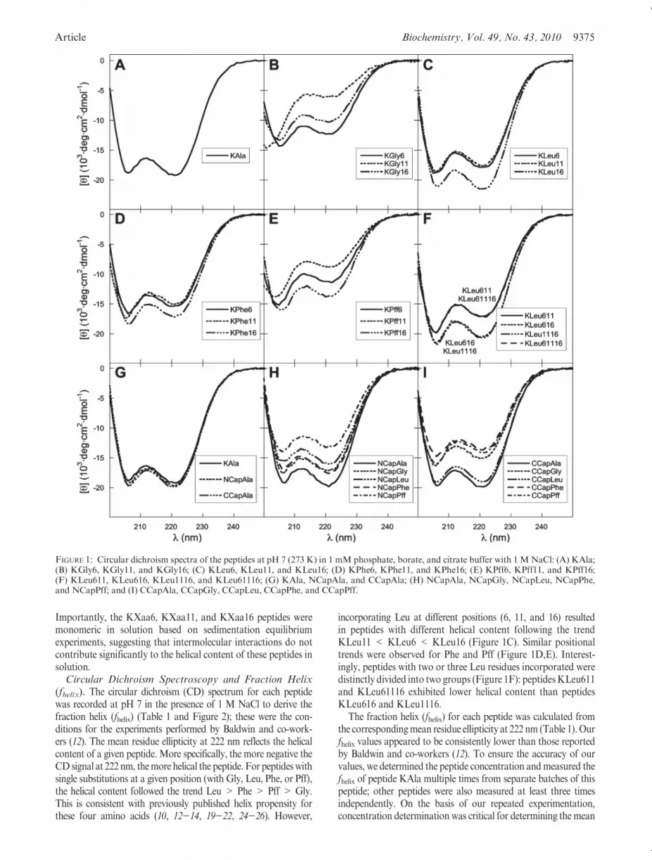
Importantly, the KXaa6, KXaa11, and KXaa16 peptides weremonomeric in solution based on sedimentation equilibriumexperiments, suggesting that intermolecular interactions do notcontribute significantly to the helical content of these peptides insolution.Circular Dichroism Spectroscopy and Fraction Helix
(fhelix). The circular dichroism (CD) spectrum for each peptidewas recorded at pH 7 in the presence of 1 M NaCl to derive thefraction helix (fhelix) (Table 1 and Figure 2); these were the con-ditions for the experiments performed by Baldwin and co-work-ers (12). The mean residue ellipticity at 222 nm reflects the helicalcontent of a given peptide. More specifically, the more negative theCD signal at 222 nm, themore helical the peptide. For peptideswithsingle substitutions at a given position (with Gly, Leu, Phe, or Pff),the helical content followed the trend Leu > Phe > Pff > Gly.This is consistent with previously published helix propensity forthese four amino acids (10, 12-14, 19-22, 24-26). However,
incorporating Leu at different positions (6, 11, and 16) resultedin peptides with different helical content following the trendKLeu11 < KLeu6 < KLeu16 (Figure 1C). Similar positionaltrends were observed for Phe and Pff (Figure 1D,E). Interest-ingly, peptides with two or three Leu residues incorporated weredistinctly divided into two groups (Figure 1F): peptidesKLeu611and KLeu61116 exhibited lower helical content than peptidesKLeu616 and KLeu1116.
The fraction helix (fhelix) for each peptide was calculated fromthe correspondingmean residue ellipticity at 222 nm (Table 1). Ourfhelix values appeared to be consistently lower than those reportedby Baldwin and co-workers (12). To ensure the accuracy of ourvalues, we determined the peptide concentration andmeasured thefhelix of peptide KAla multiple times from separate batches of thispeptide; other peptides were also measured at least three timesindependently. On the basis of our repeated experimentation,concentration determination was critical for determining the mean
FIGURE 1: Circular dichroism spectra of the peptides at pH 7 (273 K) in 1 mM phosphate, borate, and citrate buffer with 1 MNaCl: (A) KAla;(B) KGly6, KGly11, and KGly16; (C) KLeu6, KLeu11, and KLeu16; (D) KPhe6, KPhe11, and KPhe16; (E) KPff6, KPff11, and KPff16;(F) KLeu611, KLeu616, KLeu1116, and KLeu61116; (G) KAla, NCapAla, and CCapAla; (H) NCapAla, NCapGly, NCapLeu, NCapPhe,and NCapPff; and (I) CCapAla, CCapGly, CCapLeu, CCapPhe, and CCapPff.

9376 Biochemistry, Vol. 49, No. 43, 2010 Cheng et al.
residue ellipticity, and thus fhelix, of a peptide. Interestingly, themolar absorptivity (ε, also known as the extinction coefficient) isknown to differ by 15% in different aqueous solutions (98). Toensure consistency, we have performed concentration determina-tion of all peptide stock solutions in 6Mguanidinium chloride (98)and used aliquots of the stock to prepare the corresponding CDsamples. Importantly, our experimentally determined mean resi-due ellipticity at 222 nm and fhelix for KAla are within error of thevalues reported by Waters (25), despite being different from thevalues published previously by Baldwin (12).Helix Propensity of Leu, Phe, and Pff. The helix propen-
sity (w), N-capping parameter (n), and C-capping parameter (c)for Ala, Gly, and Lys were derived from globally fitting the fhelixof peptides KAla, KGly6, KGly11, KGly16, NCapAla, CCa-pAla, NCapGly, and CCapGly based on modified Lifson-Roigtheory (12, 26, 34, 38, 81) using the least-squares method (Table 2).For parameters that converged to negative probabilities(which carry no physical meaning), the values were set to thosepublished by Baldwin (35). The apparent helix propensities forLeu, Phe, and Pff at the various positions were initially derivedfrom the fhelix values of the corresponding peptides based onmodified Lifson-Roig theory (12, 26, 34, 38, 81) (Figure 2).These initial derivations were performed using one peptide foreach amino acid at one given position. For example, the apparenthelix propensities for Leu at positions 6, 11, and 16 were derivedfrom peptides KLeu6, KLeu11, and KLeu16, respectively.Interestingly, the helix propensity of Leu (wLeu) appeared to be
the same at positions 6 and 11 but was considerably higher atposition 16. Similarly, the helix propensity of Phe (and Pff)appeared to be the same at positions 6 and 11 (for each aminoacid) but was significantly higher at position 16. Therefore, wederived a global w6,11 for positions 6 and 11 and a separate w16 forposition 16 for the three amino acids Leu, Phe, and Pff (Table 3).The resulting helix propensityw6,11 for the amino acids followed thetrend Leu > Phe > Pff (consistent with known helix propensitytrends Leu>Phe and Phe>Pff) (10, 12-14, 19-22, 24-26). Thesame trend was observed for the values of w16.
The calculated fraction helix values using the parametersdetermined in this study fit all the experimentally measureddata to a similar degree compared to Baldwin parameters(12, 35, 90) based on the sum of the squares of the differencebetween calculated and experimental values (Table 4). Thelower the sum of squares, the better the calculated valuesmatched the experimental values. The experimentally mea-sured fhelix values of the peptides studied byBaldwin (12, 35, 90)were more accurately calculated using Baldwin parameterscompared to our parameters. However, the fhelix values ofpeptides investigated in this study were more accuratelycalculated using our parameters compared to Baldwin para-meters (12, 35, 90) (Table 4).
The apparent high helix propensity for all three amino acidswhen they are placed near the C-terminus could be due to specificstabilizing Xaa-Lys (i, iþ 3) interactions with the terminal Lys,because intrahelical (i, i þ 3) interactions can be energeticallyfavorable (21, 22, 25, 40-66). Also, intrahelical (i, i þ 4)interactions have been observed between aromatic and basicresidues in analogous Ala-based peptides (50, 51, 53, 60, 65).Accordingly, Xaa-Lys (i, i þ 3) interaction could be possible inour studies.Using the helix propensity at positions 6 and 11 as thehelix propensity at position 16, we calculated the putativeXaa-Lys (i, i þ 3) interaction energetics near the C-terminus(Table 3). The putative stabilizing interaction appears to followthe trend Pff ∼ Leu > Phe. Hydrophobic interactions betweenthe four hydrophobic methylenes on the Lys side chain and theseresidues would be one possible explanation for the putativeinteraction trend. The hydrophobicity of Pff is higher than thatof Phe, whereas the aliphatic Leu side chain is slightly morehydrophobic than Phe, with more flexibility to interact with (andpack against) the Lysmethylenes. However, it was unclear if suchPhe-Lys (i, i þ 3) and Leu-Lys (i, i þ 3) sequence patterns areprevalent in natural protein structures.
Table 2: Statistical Mechanical Helix Formation Parameters for Host
Peptide Residues Derived from Experimentally Measured Fraction Helix
Values Based on Modified Lifson-Roig Theorya
residue w n c
Ala 1.44( 0.01 1.00b 0.801( 0.343
Gly 0.00119( 0.00948 2.85( 0.40 0.88b
Lys 1.06( 0.03 0.79b 2.85( 1.45
acetyl 9.52( 1.47
carboxyamide 1.30b
aThese statistical mechanical helix formation parameters are derivedfrom experimental data from eight peptides: KAla, KGly6, KGly11,KGly16, NCapAla, CCapAla, NCapGly, and CCapGly; for sequences,see Table 1. bThe parameter initially converged to a negative probability(which carries no physical meaning); therefore, the value was set to thatpublished by Baldwin (35).
FIGURE 2: Helix propensity for Leu,Phe, andPff at various positions(6, 11, and 16) derived from circular dichroism data at 222 nm of thecorresponding Ala-based peptides based on the modified Lifson-Roig theory (12, 26, 34, 38, 81).
Table 3: Apparent Helix Propensities (w), Free Energies of Helix Forma-
tion (ΔGhelix formation), and Putative Xaa16-Lys (i, i þ 3) Interaction
Energetics for Leu, Phe, and Pff
amino acid w
ΔGhelix formation
(cal/mol)a
putative Xaa16-Lys
(i, i þ 3) interaction
energy (cal/mol)b
Leu (6,11) 1.09( 0.04 -46.8( 20.3
Leu (16) 2.41( 0.23 -477 ( 54 -804( 48
Phe (6, 11) 0.573( 0.037 302( 36
Phe (16) 0.940( 0.047 33.6( 27.8 -521 ( 43
Pff (6, 11) 0.135 ( 0.013 1090( 50
Pff (16) 0.310( 0.052 636( 99 -790( 130
aΔGhelix formation = -RT ln(w). bThe putative Xaa16-Lys (i, i þ 3)interaction energetics are derived from the corresponding experimentallymeasured fhelix values (Table 1) by the nesting block method (41, 66, 99),using the helix propensity at positions 6 and 11 as the helix propensity atposition 16.

Article Biochemistry, Vol. 49, No. 43, 2010 9377
Exploring Natural Protein Structures. The nonredundantprotein structure database PDBselect (April 2009, 25% thresh-old) (100, 101) was surveyed for Leu-Lys (i, i þ 3) and Phe-Lys(i, iþ 3) sequence patterns in the context of various helix-relatedstructures (Table 5). The definition of the helical conformationwas based on backbone dihedrals (102, 103), and only heliceswithmore than six residues were considered to be a helix to avoid“helices” with fewer than one turn (66). A total of 4418 proteinchains and 666086 residues were considered involving 17622helices and 236790 helical residues. Approximately 33.7% of theresidues in the database were helical (236790 of 666086), a resultsimilar to those of earlier analyses (1-3). To gauge the signifi-cance of the occurrences for Leu-Lys (i, iþ 3) and Phe-Lys (i, iþ3), the pair propensities for the sequence patterns in the variousstructures were derived by dividing the occurrence by thecorresponding expected occurrence. The expected occurrencewas obtained by bootstrapping the residues in the individualstructural context of each amino acid. Bootstrapping was per-formed 100000 times for these cases to yield standard deviationsfor the expected occurrence. This enabled the calculation ofstandard deviations for the pair propensities and Z values(Table 5), which was used to derive the P values. Propensitiesgreater than unity represent occurrences that are higher thanexpected on the basis of residue occurrences in the correspondingstructural context, whereas propensities less than unity indicateoccurrences lower than the expected value. To survey Leu-Lys (i,i þ 3) and Phe-Lys (i, i þ 3) sequence patterns in various helix-related structures, we considered the conformation of six-residuesegments: i - 1 through i þ 4. The conformation of each residuein the segment was categorized as helix (h), nonhelix (or coil, c),or any structure (including helix and nonhelix, a). The conforma-tion of the six-residue segment is designated with the conforma-tion of residues i (Leu or Phe) and i þ 3 (Lys) boldfaced andunderlined. For example, the conformation of the sequencepattern Leu-Lys (i, i þ 3) in all protein structures would bedesignated aaaaaa; the conformation of the same sequencepattern within only helical structures (including termini) wouldbe designated ahhhha.
The pair propensity for both Leu-Lys (i, i þ 3) and Phe-Lys(i, iþ 3) sequences was close to unity when considering all proteinstructures in the survey [aaaaaa (Table 5)], suggesting indiffer-ence for both sequence patterns in protein structures. In thecontext of natural helices (ahhhha), the two patterns exhibitedpair propensities less than unity, suggesting a less than expectedoccurrence for both patterns in helices. Because the exceptionally
high statisticalmechanical helix propensity (w) was observed nearthe C-terminus of the helical peptides for both Leu and Phe (videsupra), we then focused on surveying the C-terminal ends ofnatural helices. Five different helix terminating scenarios wereconsidered: hhhhhc, hhhhca, hhhcaa, hhcaaa, and hcaaaa(Table 5). Interestingly, only helices terminating immediately afterLysiþ3 (hhhhhc) or immediately after Leui/Phei (hhcaaa) exhibitedpair propensity values significantly greater than unity, suggesting
Table 4: Sum of Squares of the Differences between Experimentally Measured and Calculated fhelix Values for Leu-, Phe-, and Ala-Containing Ala-Based
Peptides Based on Modified Lifson-Roig Theory Using Various Parameter Sets
peptide
Baldwin 1994 parameters,
calculated sum of squareseBaldwin 1995 parameters,
calculated sum of squareseBaldwin 1996 parameters,
calculated sum of squares fparameters from this study,
calculated sum of squarese
Baldwin 1994 (10 peptidesa) 0.0290 0.0156 0.0148 0.0686
Baldwin 1995 (8 peptidesb) 0.0261 0.0458 0.0063 0.1035
Baldwin 1996 (11 peptidesc) 0.0293 0.0156 0.0154 0.0692
This study (22 peptidesd) 0.1017 0.1113 0.1897 0.0274
all (34 peptidesa,b,c,d) 0.1354 0.1596 0.2051 0.1348
aTen Ala-based peptides containing Ala, Lys, Leu, and Phe studied by Baldwin (12): YG, YGG, YGGG, YGG-G8, YGG-G13 YGG-G18, YGG-1L,YGG-1F, YGG-3L, and 2L-GGY. bEight Ala-based peptides containing Ala, Lys, and Leu studied by Baldwin (35): YG, YGG, YGGG, YGG-G8, YGG-G13, YGG-G18, XAK, and CCZ. cEleven Ala-based peptides containing Ala, Lys, Leu, and Phe studied by Baldwin (90): YGAK, YGGAK, YGGGAK,YGG-G7, YGG-G12, YGG-G17, YG-ZC17, YGG-1L, YGG-3L, 2L-GGY, and YGG-1F. dTwenty-two Ala-based peptides containing Leu, Phe and Alainvestigated in this study: KAla, NCapAla, CCapAla, KGly6, KGly11, KGly16, NCapGly, CCapGly, KPhe6, KPhe11, KPhe16, NCapPhe, CCapPhe,KLeu6,KLeu11,KLeu16,KLeu611,KLeu616,KLeu1116,KLeu61116,NCapLeu, andCCapLeu (Table 1). eThe experimental fhelix was derived from theCDdata at 222 nm as determined by Baldwin (12). fThe experimental fhelix was derived from the CD data at 222 nm as determined by Baldwin (90).
Table 5: Statistical Analysis for Leu-Lys and Phe-Lys (i, i þ 3) Sequence
Patterns in Various Structural Patterns
i i þ 3 structurea occurrencebpair
propensitycZ
valuedP
valuee
Leu Lys aaaaaa 3476 0.949( 0.016 -3.06 2.21� 10-3
Leu Lys ahhhha 1285 0.848( 0.022 -6.14 8.16� 10-10
Leu Lys hhhhhc 234 1.68 ( 0.14 8.01 1.15� 10-15
Leu Lys hhhhca 117 1.12( 0.11 1.20 2.30� 10-1
Leu Lys hhhcaa 131 1.23( 0.12 2.37 1.78� 10-2
Leu Lys hhcaaa 188 2.05 ( 0.21 10.1 6.77� 10-24
Leu Lys hcaaaa 69 1.19( 0.16 1.42 1.56� 10-1
Phe Lys aaaaaa 1737 1.02( 0.02 1.02 3.08� 10-1
Phe Lys ahhhha 508 0.908( 0.043 -1.46 1.44� 10-1
Phe Lys hhhhhc 70 1.43 ( 0.20 3.01 2.61� 10-3
Phe Lys hhhhca 31 0.837( 0.136 -1.00 3.17� 10-1
Phe Lys hhhcaa 32 0.856( 0.139 -0.890 3.73� 10-1
Phe Lys hhcaaa 70 1.63 ( 0.25 4.15 3.32� 10-5
Phe Lys hcaaaa 49 1.19( 0.19 1.23 2.19� 10-1
aThe various structural patterns for a six-residue segment from residuesi- 1 through iþ 4; residues i and iþ 3 are underlined. The structure for eachamino acid is determined by the backbone dihedrals (φ and ψ) to be eitherhelix (h) or nonhelix (c). When the structure of the residue is not restricted,the letter a is used to represent any structure. The φ and ψ definition forhelix follows a relative inclusive criterion (102, 103). bThe occurrence of thesequence and structure pattern in the nonredundant protein structuredatabase PDBselect (April 2009, 25% threshold) (100, 101). cThe occur-rence of the Leu-Lys or Phe-Lys (i, iþ 3) pairs divided by the correspondingexpected value. The expected value was obtained by bootstrapping thecorresponding individual structural pattern for positions i and iþ 3, therebyremoving bias due to amino acid usage in the structure of interest. Becausecomplete bootstrapping was performed 100000 times, this enabled thecalculation of a standard deviation for the expected value and thuspropensity. dThe difference between the occurrence and the expected valuedivided by the standard deviation for the expected value. In other words,the number of standard deviations that separate the occurrence and theexpected value; a positive number means the occurrence is larger thanthe expected value, whereas a negative number means the opposite. eTheprobability that the occurrence and expected occurrence are the same basedon the standard deviation obtained from bootstrapping assuming a Gaussiandistribution.

9378 Biochemistry, Vol. 49, No. 43, 2010 Cheng et al.
Phe-Lys (i, i þ 3) and Leu-Lys (i, i þ 3) sequences occurred morethan expected in these two structural contexts. Furthermore, thesefour sequence structural patterns were particularly significant onthe basis of the considerably highly positive Z values and smallP values. Accordingly, these sequence structural patterns wereexamined in more detail.
There were 234 occurrences of the Leu-Lys (i, i þ 3) sequencepatterns with Lys as the most C-terminal residue with helicaldihedrals (hhhhhc) (Table 5 and Figure 3); however, no Leu-Lysinteractions were apparent in these cases (Figure 3). Nonetheless,there were three cases of Leu-Ala-Ala-Lys sequence, which isthe exact sequence in our experimental studies (Figure 4). Therewere 70 occurrences of the Phe-Lys (i, i þ 3) sequence patterns
with Lys as the most C-terminal residue with helical dihedrals(hhhhhc); however, none of the sequences were the Phe-Ala-Ala-Lys sequence. Furthermore, no Phe-Lys interactions wereapparent (Figure 3). Interestingly, the relative placement of theLeu/Phe four residues upstream fromGly in hhhhhc is consistentwith previously observed Schellman motifs (102, 111).
There were 188 occurrences of the Leu-Lys (i, i þ 3) sequencepatterns with Leu as the most C-terminal residue with helicaldihedrals (hhcaaa) (Table 5 and Figure 3); however, none ofthe sequences were Leu-Ala-Ala-Lys. The most prevalent struc-ture (84 of 188) involved the Schellman motif with a residue inthe left-handed helix conformation (Figure 3). Unfortunately,this is unlikely the case for the peptides in this study with two
FIGURE 3: Two-helix C-terminal structural scenarios (with occurrences higher than expected) for Leu-Lys (i, i þ 3) (top row) and Phe-Lys(i, i þ 3) (bottom row) sequence patterns. There is only one type of backbone conformation for the hhhhhc structures, whereas there are severaldifferent types of backbone conformations for the hhcaaa structures. The backbone φ-ψ plots, the number of occurrences, average backbonedihedral angles, the most frequently observed amino acids for each position, and the overlay of the three-dimensional structures for each type aredepicted.

Article Biochemistry, Vol. 49, No. 43, 2010 9379
intervening Ala residues between Leu and Lys. There were 70occurrences of the Phe-Lys (i, iþ 3) sequence patternswithPhe asthe most C-terminal residue with helical dihedrals (hhcaaa)(Table 5 and Figure 3). The most prevalent structures (27 of70) involved a residue in the left-handed helix conformation, andnone of the sequences were Phe-Ala-Ala-Lys. In these structures,a hydrophobic residue was frequently present immediately afterthe left-handed Gly, consistent with the Schellman motif at theC-terminus of natural protein helices (102, 111). Because a left-handed helix conformation was involved for the sequencepatterns with Leui/Phei as the most C-terminal residue withhelical dihedrals (hhcaaa), these sequence structural patterns arenot likely to be relevant to this study (involving right-handed
intervening Ala residues). In comparison, the Leu-Lys (i, i þ 3)and Phe-Lys (i, i þ 3) sequence patterns with Lys as the mostC-terminal residuewith helical dihedrals (hhhhhc) would bemorerelevant to the experimentally measured high statistical mechan-ical helix propensity of Leu and Phe. However, no specific sidechain interactions between Leu/Phe and Lys were observed inthese structures to explain the high helix forming parametersmeasured experimentally.
These initial survey results considered only the residues at the iand i þ 3 positions, ignoring the intervening sequences. It wouldbe logical to ask how frequently the specific Leu-Ala-Ala-Lys andPhe-Ala-Ala-Lys sequences occur in natural structures. Accord-ingly, we performed surveys on these two specific sequences
FIGURE 4: Helix-related structural scenarios for Leu-Ala-Ala-Lys (top row) and Phe-Ala-Ala-Lys (bottom row) sequence patterns. There arethree structural scenarios for the Leu-Ala-Ala-Lys sequence, whereas there are two structural scenarios for the Phe-Ala-Ala-Lys sequence. Thebackbone φ-ψ plots, the number of occurrences, average backbone dihedral angles, and the overlay of the three-dimensional structures for eachtype are depicted.

9380 Biochemistry, Vol. 49, No. 43, 2010 Cheng et al.
(Table 6 and Figure 4). The occurrences were low for bothsequences, and extremely low for Phe-Ala-Ala-Lys. Importantly,both sequences seemed to be present in helical structures; 18 ofthe 25 sequence patterns were involved in helices for Leu-Ala-Ala-Lys (Figure 4). More specifically, Leu-Ala-Ala-Lys occurredmostly within a helix (14 of 25), and somewhat at the C-terminalend of a helix (4 of 25), but not at the N-terminal end of a helix.Two of the five Phe-Ala-Ala-Lys occurrences were at theC-terminal end of a helix with clear Lys-Phe cation-π interac-tions (112) (Figure 4). The Leu/Phe-Ala-Ala-Lys sequence pat-terns do not occur very frequently in natural helices and are notfound at the N-terminus of helices. Despite the low overalloccurrences and propensities, both sequences are found at theC-termini of natural helices.
DISCUSSION
Our CD data show that incorporating the same amino acid atdifferent positions results in varying helical contents (Figure 1).Interestingly, there is a general positional trend for the helicalcontent of the peptides: KXaa16 > KXaa6 > KXaa11 (exceptfor Gly). Glycine is peculiar in being capable of adopting variousleft-handed structures with φ>0 due to the achiral nature of theresidue. Because these conformations are not typically accessibleto natural chiral amino acids, there will be no further discussionregarding Gly. Importantly, lower fhelix values should be ob-served upon substitution of Ala with Leu, Phe, or Pff in themiddle of the peptide (position 11) compared to substitution atthe ends of the peptide (positions 6 and 16) for two reasons. First,all three amino acids have lower helix propensities than the hostresidue Ala, leading to lower helicities. Second, substituting Alawith a more helix breaking amino acid (than Ala) in the center of
the peptide attenuates the helical content more than placing suchamino acids near the termini because the termini are inherentlyweakly helical due to end fraying (38, 113, 114). The differencebetween substituting the Ala at positions 16 and 6 is most likelydue to the asymmetric geometry of the R-helix. In particular,the side chain CR-Cβ vectors inherently project toward theN-terminus because of the chirality of the amino acids, backbonedihedrals, and helix handedness. This geometry results in fre-quent side chain shielding of upstream helix hydrogen bonds,thereby stabilizing the helical conformation. As such, the N-terminus ismore stable than theC-terminus inR-helices (114-119).Although the modified Lifson-Roig theory apparently accountsfor this asymmetry by introducing capping parameters n andc (12, 26, 34, 38, 81), the exceptionally high helical content forpeptides with non-Ala residues at position 16 cannot be fullyrationalized with the capping parameters alone. Therefore, thisrequired us to invoke the exceptionally high helix propensity forthe residue at position 16, and the apparent helix propensity forthe same amino acid follows the trend w16 > w6 = w11. Theseresults are in sharp contrast to those published by Stellwagen andco-workers (92), apparently showing positional independenceand additivity of amino acid replacements on helix stability atanalogous positions. However, Stellwagen and co-workers artifi-cially normalized the CD signals for all peptides, attemptingdirect comparison between peptides without invoking any statis-tical mechanical theory. Furthermore, the monomeric helicalpeptideswere assumed to be in a two-state equilibrium, leading tomost likely invalid van’t Hoff analyses.
Our results show that helix propensity for an amino acid (Leu,Phe, or Pff) is similar at positions 6 and 11, but different atposition 16 in a 19-residue Ala-based peptide. In other words,helix propensity for an amino acid is similar near the N-terminusand center of the peptide chain, but different near the C-terminusof the peptide chain. These results appear to somewhat contradictthe results from Serrano and co-workers in a different Ala-basedsystem (94-96). Serrano and co-workers found that helixpropensity varies when placed at the first three N-terminalresidues of the peptide chain, at the last three C-terminal residuesof the peptide chain, and in the middle of the helical peptide (orseven residues from the ends of the peptide chain) (94-96).However, the fundamental difference between the two systems isthe variance in the immediate surrounding residues ((2 aminoacids). These neighboring residues change according to the guestposition in Serrano’s system (94-96) but remain constant in ourBaldwin-based system (12). Therefore, the results from the twostudies are valid in each of the corresponding contexts and do notcontradict one another.
The less than expected occurrence for both Leu-Lys and Phe-Lys (i, iþ 3) sequence patterns in natural protein helices [ahhhha(Table 5)] is consistent with the amphiphilic nature of helices inglobular proteins (103, 120-124). Helices residing on the surfaceof globular proteins would require a hydrophilic face and ahydrophobic face opposite from one another. Leu and Phe areconsidered to be hydrophobic, whereas Lys is considered to behydrophilic. Because the geometry of an ideal helix has 3.6residues per turn, amino acids three residues apart should havesimilar characteristics (i.e., both hydrophobic or both hydro-philic). Therefore, it is not surprising that both Leu-Lys and Phe-Lys (i, iþ 3) sequences occur less than expected in natural proteinhelices. In contrast, bothLeu-Lys andPhe-Lys (i, iþ 3) sequencesoccur more than expected near the C-terminus of natural proteinhelices involving the structures hhhhhc and hhcaaa (Table 5).
Table 6: Statistical Analysis for Leu-Ala-Ala-Lys and Phe-Ala-Ala-Lys
Sequence Patterns in Various Structural Patterns
sequence from
i to i þ 3 structurea occurrenceb propensitycZ
valuedP
valuee
Leu-Ala-Ala-Lys aaaaaa 25 1.22( 0.27 1.00 3.17� 10-1
Leu-Ala-Ala-Lys hhhhhh 14 0.896( 0.226 -0.41 6.82� 10-1
Leu-Ala-Ala-Lys hhhhhc 3 1.45( 1.00 0.65 5.16� 10-1
Leu-Ala-Ala-Lys hhhhca 1 0.423( 0.274 -0.89 3.73� 10-1
Phe-Ala-Ala-Lys aaaaaa 5 0.530( 0.171 -1.45 1.47� 10-1
Phe-Ala-Ala-Lys hhhhhh 1 0.180( 0.074 -1.97 4.88� 10-2
Phe-Ala-Ala-Lys hhhhca 2 2.39( 2.58 1.28 2.01� 10-1
aThe various structural patterns for a six-residue segment from residuei - 1 to i þ 4; residues i and i þ 3 are underlined. The structure for eachamino acid is determined by the backbone dihedrals (φ and ψ) to be eitherhelix (h) or nonhelix (c). When the structure of the residue is not restricted,the letter a is used to represent any structure. The φ and ψ definition forhelix follows a relative inclusive criterion (102, 103). bThe occurrence of thesequence and structure pattern in the nonredundant protein structuredatabase PDBselect (April 2009, 25% threshold) (100, 101). cThe occur-rence for the Leu-Ala-Ala-Lys or Phe-Ala-Ala-Lys sequences divided by thecorresponding expected value. The expected value was obtained by boot-strapping the corresponding individual structural pattern for positions ithrough i þ 3, thereby removing bias due to amino acid usage in thestructure of interest. Because complete bootstrapping was performed 10000times, this enabled the calculation of a standard deviation for the expectedvalue and thus propensity. dThe difference between the occurrence and theexpected value divided by the standard deviation for the expected value.In other words, the number of standard deviations that separate theoccurrence and the expexted value; a positive number means the occurrenceis larger than the expected value, whereas a negative number meansthe opposite. eThe probability that the occurrence and expected occur-rence are the same based on the standard deviation obtained frombootstrapping assuming a Gaussian distribution.

Article Biochemistry, Vol. 49, No. 43, 2010 9381
However, the hhhhhc structures involve one residue beyond Lys,and Lys is the last residue in our peptides. Therefore, therelevancy of the hhhhhc structures may be somewhat debatablebut cannot be completely ruled out. Unfortunately, the hhcaaastructures involve a left-handed conformation (φ > 0) forposition i þ 1 or i þ 2 (Figure 3), which is extremely rare forthe Ala residue in our peptides. Therefore, the hhcaaa structuresare most likely irrelevant to our peptides. In contrast, the specificsequences in our study, Leu-Ala-Ala-Lys and Phe-Ala-Ala-Lys,do not occur frequently in nature (Table 6), suggesting that Ala-based peptides most likely do not represent the situation innatural protein helices. Nonetheless, Ala-based peptides are stillgood minimalist model systems for fundamental studies.
There are several possible explanations for the apparent posi-tional dependence for the helix propensities in our studies. First, Lysis preferred at theC-cap andC1positions in protein helices, whereasLeu is preferred at the C3 and C4 positions (111, 125-127). Thesegeneral preferences are consistent with potential intrahelical Xaa-Lys (i, i þ 3) interactions in our peptide, and intrahelical (i, i þ 3)interactions are well-documented (21, 22, 25, 40-66). Second, theC-terminal end of a helix may adopt a 310-helix, because 310-helicesare observed at the C-terminal end of protein R-helices (2, 128).Furthermore, Ala-based peptides have been shown to adopt amixture of R- and 310-helices (129-131), with the C-terminal endadopting a 310-helix for a 21-residue peptide (130). Interestingly, thethree-residue per turn geometry of the 310-helix could be furtherstabilized by an intrahelical (i, i þ 3) interaction as proposed byDoig (132). Analogous intrahelical Xaa-Lys (i, iþ 3) interactionshave been implied by Doig (60) and invoked by Serrano (22) forR-helices. Third, residues near the terminus of the peptide chaincould exhibit different helix propensities. This proximity to theterminus of the peptide chain is different from being positioned atthe ends of helices (or capping effects). On the basis of the statisticalmechanical models that describe the multistate nature for theequilibrium of monomeric helical peptides, conformational stateswith helices terminating in the center of the peptide do exist, andcapping effects can account for them (12, 26, 34, 38, 81). However,these capping effects alone cannot account for the high helicalcontent for peptides with non-Ala residues incorporated near theC-terminus of the peptide chain in this study. Therefore, the highC-terminal helix propensity observed in this studymaybe due to theproximity of position 16 to the C-terminus of the peptide chain(the fourth residue from the C-terminus of the peptide chain). Incontrast, position 6 near the N-terminus is farther from theN-terminus of the peptide chain, and thereby unaffected. Thisappears to be consistent with molecular dynamics simulations onpolymers, revealing high main chain translational mobility near thetermini based on mean-square-displacement analysis (133, 134).Also, this highmain chain translationalmobility is no longerpresentby the fifth monomer unit from the terminus (133).
Helices are rarely present at the ends of the protein chain innatural proteins. Furthermore, the residues at the termini ofnatural proteins are generally structurally disordered and un-resolved in atomic-resolution structures. Nonetheless, the up-stream and downstream protein chain can create differentstructural contexts compared to the N- and C-termini of thepeptide chain, respectively. In particular, the steric restrictionswould be higher and the conformational entropy should belower, in the presence of the upstream or downstream proteinchain compared to their absence (or near the termini of thepeptide chain). This would account for the molecular mechanicsresults showing a difference in solvent exposure, the number of
van der Waals interactions, and configurational entropy forresidues near the N-terminus compared to internal residues (94).Therefore, the helix propensity should be different on the basis ofthe proximity to the ends of the peptide chain as shown inprevious studies (94-96) and the C-terminus (position 16) in thisstudy. The lack of this chain terminal effect near the N-terminus(position 6) in this study suggests that the effect is diminished atthe sixth residue from the end of the peptide chain, consistentwith simulation results on a polymer (133). The length depen-dence of helix propensity in Ala-based peptides (85, 91) may alsobe due to this chain terminal effect, because the proportion ofresidues in the proximity of the ends of the peptide chain changeswith chain length. Because the chain terminal effect would notexist at internal positions of a protein, no position dependence inhelix propensity was observed in an internal helix of a protein (93).Overall, the chain terminal effect appears to explain the high w16
compared to w6,11 in this study, the positional dependence of helixpropensity in previous studies of Ala-based peptides (94-96), thelength dependence of helix propensity (85, 91), and the positionindependence in a protein helix (93).
CONCLUSIONS
Circular dichroism spectroscopy coupled with calculationsbased on modified Lifson-Roig theory (12, 26, 34, 38, 81) hasbeen used to determine the helix propensities for Leu, Phe, and Pffat different positions of the same helical peptide. IncorporatingLeu, Phe, or Pff at various positions of the same peptide resulted invarying helical contents. The helicity of the substituted hostpeptide follows the general trend KXaa16 >KXaa6>KXaa11.Position dependence of the helix propensities was observed forLeu, Phe, and Pff, with exceptionally high helix propensity atposition 16 (close to the C-terminus) compared to positions 6 and11. The unique behavior for these non-Ala residues at position 16may be due to specific C-cap interactions, adoption of a 310-helixnear the C-terminus perhaps with Xaa-Lys (i, iþ 3) interactions,or the chain terminal effect. Nonetheless, it appears that helixpropensity is generally position-independent except in the presenceof alternative structures or in the proximity of either terminus ofthe peptide chain. These results should facilitate the design ofhelical peptides, proteins, and non-natural foldamers.
ACKNOWLEDGMENT
We thank the Computer and Information Networking Centerat National Taiwan University for the support of the high-performance computing facilities. We thank National ScienceFoundation (REU, CHE0453206) for supporting Nathan S.Hutson, who participated in this project. We thank ProfessorCheu-Pyeng Cheng (Department of Chemistry, National TsingHua University, Hsinchu, Taiwan, China) and Professor AlisonMcCurdy (Department of Chemistry and Biochemistry, CaliforniaState University, Los Angeles, CA) for helpful discussions.
SUPPORTING INFORMATION AVAILABLE
Details of the synthesis and characterization of the peptides.This material is available free of charge via the Internet at http://pubs.acs.org.
REFERENCES
1. Chou, P. Y., and Fasman, G. D. (1974) Conformational parametersfor amino-acids in helical, β-sheet, and random coil regions calcu-lated from proteins. Biochemistry 13, 211–222.

9382 Biochemistry, Vol. 49, No. 43, 2010 Cheng et al.
2. Barlow, D. J., and Thornton, J. M. (1988) Helix geometry inproteins. J. Mol. Biol. 201, 601–619.
3. We recently conducted survey on a nonredundant protein structuredatabase PDBselect {December 2003, 25% threshold [(1994) ProteinSci. 3, 522-524]}. Using hydrogen bonding to define the R-helixconformation as described by Kabsch and Sander [(1983) Biopoly-mers 22, 2577-2637], we found 101210 R-helical residues of 341444total residues; 29.6% of all residues were R-helical.
4. Gellman, S. H. (1998) Foldamers: A manifesto. Acc. Chem. Res. 31,173–180.
5. Hill, D. J., Mio, M. J., Prince, R. B., Hughes, T. S., andMoore, J. S.(2001) A field guide to foldamers. Chem. Rev. 101, 3893–4011.
6. Sanford, A. R., and Gong, B. (2003) Evolution of helical foldamers.Curr. Org. Chem. 7, 1649–1659.
7. Cheng, R. P. (2004) Beyond de novo protein design: De novo designof non-natural folded oligomers. Curr. Opin. Struct. Biol. 14, 512–520.
8. Hecht, S., and Huc, I., Eds. (2007) Foldamers. Structure Properties,and Applications, Wiley-VCH, Weinheim, Germany.
9. Platzer, K. E. B., Ananthanarayanan, V. S., Andreatta, R. H., andScheraga, H. A. (1972)Helix-coil stability constants for the naturallyoccurring amino acids in water. IV. Alanine parameters fromrandom poly(hydroxypropylglutamine-co-L-alanine). Macromole-cules 5, 177–187.
10. Sueki, M., Lee, S., Powers, S. P., Denton, J. B., Konishi, Y., andScheraga, H. A. (1984)Helix-coil stability constants for the naturallyoccurring amino acids in water. 22. Histidine parameters fromrandom poly[(hydroxybutyl)glutamine-co-L-histidine]. Macromole-cules 17, 148–155.
11. Marqusee, S., Robbins, V. H., and Baldwin, R. L. (1989) Unusuallystable helix formation in short alanine-based peptides. Proc. Natl.Acad. Sci. U.S.A. 86, 5286–5290.
12. Chakrabartty, A., Kortemme, T., and Baldwin, R. L. (1994) Helixpropensities of the amino acids measured in alanine-based peptideswithout helix-stabilizing side-chain interactions. Protein Sci. 3,843–852.
13. O’Neil, K. T., and DeGrado, W. F. (1990) A thermodynamic scalefor the helix-forming tendencies of the commonly occurring aminoacids. Science 250, 646–651.
14. Lyu, P. C., Liff, M. I., Marky, L. A., and Kallenbach, N. R. (1990)Side chain contributions to the stability of R-helical structure inpeptides. Science 250, 669–673.
15. Spek, E. J., Olson, C. A., Shi, Z., and Kallenbach, N. R. (1999)Alanine is an intrinsic R-helix stabilizing amino acid. J. Am. Chem.Soc. 121, 5571–5572.
16. Kemp, D. S., Boyd, J. G., and Muendel, C. C. (1991) The helical sconstant for alanine in water derived from template-nucleatedhelices. Nature 352, 451–454.
17. Kemp, D. S., Oslick, S. L., and Allen, T. J. (1996) The structure andenergetics of helix formation by short templated peptides in aqueoussolution. 3. Calculation of the helical propogation constant s fromthe template stability constants t/c for Ac-Hel1-Alan-OH, n = 1-6.J. Am. Chem. Soc. 118, 4249–4255.
18. Serrano, L., Sancho, J., Hirshberg, M., and Fersht, A. R. (1992)R-Helix stability in proteins I. Empirical correlations concerningsubstitution of side-chains at the N and C-caps and the replacementof alanine by glycine or serine at solvent-exposed surfaces. J. Mol.Biol. 227, 544–559.
19. Horovitz, A., Matthews, J. M., and Fersht, A. R. (1992) R-Helixstability in proteins II. Factors that influence stability at an internalposition. J. Mol. Biol. 227, 560–568.
20. Blaber, M., Zhang, X.-j., and Matthews, B. W. (1993) Structuralbasis of amino acid R helix propensity. Science 260, 1637–1640.
21. Park, S.-H., Shalongo, W., and Stellwagen, E. (1993) Residue helixparameters obtained from dichroic analysis of peptides of definedsequence. Biochemistry 32, 7048–7053.
22. Mu~noz, V., and Serrano, L. (1994) Elucidating the folding problemof helical peptides using empirical parameters. Nat. Struct. Biol. 1,399–409.
23. Zhou, N. E., Kay, C. M., Sykes, B. D., and Hodges, R. S. (1993) Asingle-stranded amphipathic R-helix in aqueous solution: Design,structural characterization, and its application for determiningR-helical propensities of amino acids. Biochemistry 32, 6190–6197.
24. Myers, J. K., Pace, C. N., and Scholtz, J. M. (1997) A directcomparison of helix propensity in proteins and peptides. Proc. Natl.Acad. Sci. U.S.A. 94, 2833–2837.
25. Butterfield, S. M., Patel, P. R., and Waters, M. L. (2002) Contribu-tion of aromatic interactions to R-helix stability. J. Am. Chem. Soc.124, 9751–9755.
26. Chiu, H.-P., Suzuki, Y., Gullickson, D., Ahmad, R., Kokona, B.,Fairman, R., and Cheng, R. P. (2006) Helix propensity of highlyfluorinated amino acids. J. Am. Chem. Soc. 128, 15556–15557.
27. Chiu, H.-P., and Cheng, R. P. (2007) Chemoenzymatic synthesis of(S)-hexafluoroleucine and (S)-tetrafluoroleucine.Org. Lett. 9, 5517–5520.
28. Serrano, L., and Fersht, A. R. (1989) Capping and R-helix stability.Nature 342, 296–299.
29. Lyu, P. C., Zhou,H.X., Jelveh,N.,Wemmer,D. E., andKallenbach,N. R. (1992) Position-dependent stabilizing effects in R-helices:N-Terminal capping in synthetic model peptides. J. Am. Chem.Soc. 114, 6560–6562.
30. Lyu, P. C., Wemmer, D. E., Zhou, H. X., Pinker, R. J., andKallenbach, N. R. (1993) Capping interactions in isolated R helices:Position-dependent substitution effects and structure of a serine-capped peptide helix. Biochemistry 32, 421–425.
31. Zhou, H. X., Lyu, P. C., Wemmer, D. E., and Kallenbach, N. R.(1994) Structure of a C-terminal R-helix cap in a synthetic peptide.J. Am. Chem. Soc. 116, 1139–1140.
32. Bell, J. A., Becktel, W. J., Sauer, U., Baase, W. A., and Matthews,B.W. (1992)Dissection of helix capping in T4 lysozyme by structuraland thermodynamic analysis of six amino acid substitutions at Thr59. Biochemistry 31, 3590–3596.
33. Chakrabartty, A., Doig, A. J., and Baldwin, R. L. (1993) Helixcapping propensities in peptides parallel those in proteins. Proc.Natl. Acad. Sci. U.S.A. 90, 11332–11336.
34. Doig, A. J., Chakrabartty, A., Klingler, T. M., and Baldwin, R. L.(1994) Determination of free-energies of N-capping in R-helicesby modification of the Lifson-Roig helix-coil theory to includeN- and C-capping. Biochemistry 33, 3396–3403.
35. Doig, A. J., and Baldwin, R. L. (1995) N- and C-capping preferencesof all 20 amino acids in R-helical peptides. Protein Sci. 4, 1325–1336.
36. Forood, B., Feliciano, E. J., andNambiar, K. P. (1993) Stabilizationof R-helical structures in short peptides via end capping. Proc. Natl.Acad. Sci. U.S.A. 90, 838–842.
37. Zhukovsky, E. A., Mulkerrin, M. G., and Presta, L. G. (1994)Contribution to global protein stabilization of the N-capping boxin human growth hormone. Biochemistry 33, 9856–9864.
38. Andersen,N.H., andTong,H. (1997) Empirical parameterization ofa model for predicting peptide helix/coil equilibrium populations.Protein Sci. 6, 1920–1936.
39. Schneider, J. P., and DeGrado, W. F. (1998) The design of efficientR-helical C-capping auxiliaries. J. Am. Chem. Soc. 120, 2764–2767.
40. Marqusee, S., and Baldwin, R. L. (1987) Helix stabilization byGlu-...Lysþ salt bridges in short peptides of de novo design. Proc.Natl. Acad. Sci. U.S.A. 84, 8898–8902.
41. Scholtz, J. M., Qian, H., Robbins, V. H., and Baldwin, R. L. (1993)The energetics of ion-pair and hydrogen-bonding interactions in ahelical peptide. Biochemistry 32, 9668–9676.
42. Huyghues-Despointes, B. M. P., Scholtz, J. M., and Baldwin, R. L.(1993) Helical peptides with three pairs of Asp-Arg and Glu-Argresidues in different orientations and spacings. Protein Sci. 2, 80–85.
43. Padmanabhan, S., and Baldwin, R. L. (1994) Helix-stabilizing inter-action between tyrosine and leucine or valine when the spacing in i,iþ4. J. Mol. Biol. 241, 706–713.
44. Padmanabhan, S., and Baldwin, R. L. (1994) Tests for helix-stabiliz-ing interations between various nonpolar side chains in alanine-based peptides. Protein Sci. 3, 1992–1997.
45. Stapley, B. J., Rohl, C. A., and Doig, A. J. (1995) Addition of sidechain interactions to modified Lifson-Roig helix-coil theory: Appli-cation to energetics of phenylalanine-methionine interactions. Pro-tein Sci. 4, 2383–2391.
46. Luo, P., andBaldwin, R. L. (2002)Origin of the different strengths ofthe (i,iþ4) and (i,iþ3) leucine pair interactions in helices. Biophys.Chem. 96, 103–108.
47. Gans, P. J., Lyu, P. C., Manning, M. C., Woody, R. W., andKallenbach, N. R. (1991) The helix-coil transition in heterogeneouspeptides with specific side-chain interactions: Theory and compar-ison with CD spectral data. Biopolymers 31, 1605–1614.
48. Lyu, P. C., Gans, P. J., and Kallenbach, N. R. (1992) Energeticcontribution of solvent-exposed ion pairs to alpha-helix structure.J. Mol. Biol. 223, 343–350.
49. Mayne, L., Englander, S. W., Qiu, R., Yang, J., Gong, Y., Spek,E. J., and Kallenbach, N. R. (1998) Stabilizing effect of a multiplesalt bridge in a prenucleated peptide. J. Am. Chem. Soc. 120, 10643–10645.
50. Olson, C. A., Shi, Z., andKallenbach, N.R. (2001) Polar interactionswith aromatic side chains in R-helical peptides: CH 3 3 3O H-bondingand cation-π interactions. J. Am. Chem. Soc. 123, 6451–6452.

Article Biochemistry, Vol. 49, No. 43, 2010 9383
51. Shi, Z., Olson, C. A., Bell, A. J., Jr., and Kallenbach, N. R. (2001)Stabilization of R-helix structure by polar side-chain interactions:Complex salt bridges, cation-π interactions, and C-H 3 3 3OH-bonds.Biopolymers 60, 366–380.
52. Olson, C. A., Spek, E. J., Shi, Z., Vologodskii, A., and Kallenbach,N.R. (2001) Cooperative helix stabilization by complexArg-Glu saltbridges. Proteins: Struct., Funct., Genet. 44, 123–132.
53. Shi, Z., Olson, C. A., and Kallenbach, N. R. (2002) Cation-πinteraction in model R-helical peptides. J. Am. Chem. Soc. 124,3284–3291.
54. Shi, Z., Olson, C. A., Bell, A. J., Jr., and Kallenbach, N. R. (2002)Non-classical helix stabilizing interactions: C-H 3 3 3O H-bondingbetween Phe and Glu side chains in R-helical peptides. Biophys.Chem. 101-102, 267–279.
55. Merutka, G., Shalongo, W., and Stellwagen, E. (1991) A modelpeptide with enhanced helicity. Biochemistry 30, 4245–4248.
56. Merutka,G., and Stellwagen, E. (1991) Effect of amino acid ion pairson peptide helicity. Biochemistry 30, 1591–1594.
57. Stellwagen, E., Park, S. H., Shalongo, W., and Jain, A. (1992) Thecontribution of residue ion-pairs to the helical stability of a modelpeptide. Biopolymers 32, 1193–1200.
58. Fisinger, S., Serrano, L., and Lacroix, E. (2001) Computationalestimation of specific side chain interaction energies in R helices.Protein Sci. 10, 809–818.
59. Smith, J. S., and Scholtz, J. M. (1998) Energetics of polar side-chaininteractions in helical peptides: Salt effects on ion pairs and hydrogenbonds. Biochemistry 37, 33–40.
60. Andrew, C. D., Penel, S., Jones, G. R., and Doig, A. J. (2001)Stabilizing nonpolar/polar side-chain interactions in the R-helix.Proteins: Struct., Funct., Genet. 45, 449–455.
61. Andrew, C. D., Bhattacharjee, S., Kokkoni, N., Hirst, J. D., Jones,G. R., and Doig, A. J. (2002) Stabilizing interactions betweenaromatic and basic side chains in R-helical peptides and proteins.Tyrosine effects on helix circular dichroism. J. Am. Chem. Soc. 124,12706–12714.
62. Iqbalsyah, T.M., andDoig, A. J. (2005) Pairwise coupling in anArg-Phe-Met triplet stabilizes R-helical peptide via shared rotamer pre-ferences. J. Am. Chem. Soc. 127, 5002–5003.
63. Iqbalsyah, T. M., and Doig, A. J. (2005) Anticooperativity in aGlu-Lys-Glu salt bridge triplet in an isolated R-helical peptide.Biochemistry 44, 10449–10456.
64. Errington, N., and Doig, A. J. (2005) A phosphoserine-lysine saltbridge within an R-helical peptide, the strongest R-helix side-chaininteraction measured to date. Biochemistry 44, 7553–7558.
65. Tsou, L. K., Tatko, C. D., andWaters, M. L. (2002) Simple cation-πinteraction between a phenyl ring and a protonated amine stabilizesan R-helix in water. J. Am. Chem. Soc. 124, 14917–14921.
66. Cheng,R. P., Girinath, P., andAhmad,R. (2007) Effect of lysine sidechain length on intra-helical glutamate-lysine ion pairing interac-tions. Biochemistry 46, 10528–10537.
67. Kirshenbaum, K., Zuckermann, R. N., and Dill, K. A. (1999)Designing polymers that mimic biomolecules. Curr. Opin. Struct.Biol. 9, 530–535.
68. Seebach, D., and Matthews, J. L. (1997) β-Peptides: A surprise atevery turn. Chem. Commun., 2015–2022.
69. Cheng, R. P., Gellman, S. H., andDeGrado,W. F. (2001) β-Peptides:From structure to function. Chem. Rev. 101, 3219–3232.
70. Moore, J. S. (1997) Shape-persistent molecular architectures ofnanoscale dimension. Acc. Chem. Res. 30, 402–413.
71. Sanford, A. R., Yamato, K., Yang, X., Yuan, L., Han, Y., andGong, B. (2004) Well-defined secondary structures. Information-storingmolecualr duplexes and helical foldamers based on unnaturalpeptide backbones. Eur. J. Biochem. 271, 1416–1425.
72. Huc, I. (2004) Aromatic oligoamide foldamers. Eur. J. Org. Chem.,17–29.
73. Arvidsson, P. I., Rueping, M., and Seebach, D. (2001) Design,machine synthesis, and NMR-solution structure of a β-heptapeptideforming a salt-bridge stabilised 314-helix in methanol and in water.Chem. Commun., 649–650.
74. Rueping, M., Mahajan, Y. R., Jaun, B., and Seebach, D. (2004)Design, synthesis and structural investigations of a β-peptide form-ing a 314-helix stabilized by electrostatic interactions.Chem.;Eur. J.10, 1607–1615.
75. Cheng, R. P., and DeGrado, W. F. (2001) De novo design of amonomeric helical β-peptide stabilized by electrostatic interactions.J. Am. Chem. Soc. 123, 5162–5163.
76. Hart, S. A., Bahadoor, A. B. F., Matthews, E. E., Qiu, X. J., andSchepartz, A. (2003) Helix macrodipole control of β3-peptide 14-helixstability in water. J. Am. Chem. Soc. 125, 4022–4023.
77. Raguse, T. L., Lai, J. R., and Gellman, S. H. (2003) Environment-independent 14-helix formation in short β-peptides: Striking abalance between shape control and functional diversity. J. Am.Chem. Soc. 125, 5592–5593.
78. Raguse, T. L., Lai, J. R., andGellman, S.H. (2002) Evidence that theβ-peptide 14-helix is stabilized by β3-residues with side-chain branch-ing adjacent to the β-carbon atom.Helv. Chim. Acta 85, 4154–4164.
79. Kritzer, J. A., Tirado-Rives, J., Hart, S. A., Lear, J. D., Jorgensen,W. L., and Schepartz, A. (2005) Relationship between side chainstructure and 14-helix stability of β3-peptides in water. J. Am. Chem.Soc. 127, 167–178.
80. Zimm, B. H., and Bragg, J. K. (1959) Theory of the phase transitionsbetween helix and random coil in polypeptide chains. J. Chem. Phys.31, 526–535.
81. Lifson, S., and Roig, A. (1961) On the theory of helix-coil transitionin polypeptides. J. Chem. Phys. 34, 1963–1974.
82. Mu~noz, V., and Serrano, L. (1995) Elucidating the folding problemof helical peptides using empirical parameters. 2. Helix macrodipoleeffects and rational modification of the helical content of naturalpeptides. J. Mol. Biol. 245, 275–296.
83. Mu~noz, V., and Serrano, L. (1995) Elucidating the folding problemof helical peptides using empirical parameters. 3. Temperature andpH-dependence. J. Mol. Biol. 245, 297–308.
84. Renold, P., Tsang, K. Y., Shimizu, L. S., and Kemp, D. S. (1996)For short alanine-lysine peptides the helical propensities of lysineresidues (s values) are strongly temperature dependent. J. Am. Chem.Soc. 118, 12234–12235.
85. Job,G. E.,Kennedy,R. J.,Heitmann, B.,Miller, J. S.,Walker, S.M.,and Kemp, D. S. (2006) Temperature- and length-dependent ener-getics of formation of polyalanine helices in water: Assignment ofwAla(n, T ) and temperature-dependent CD ellipticity standards. J.Am. Chem. Soc. 128, 8227–8233.
86. Moreau, R. J., Schubert, C. R., Nasr, K. A., T€or€ok,M.,Miller, J. S.,Kennedy, R. J., and Kemp, D. S. (2009) Context-independent,temperature-dependent helical propensities for amino acid residues.J. Am. Chem. Soc. 131, 13107–13116.
87. Luo, P., and Baldwin Robert, L. (1997) Mechanism of helix induc-tion by trifluoroethanol: A framework for extrapolating the helix-forming properties of peptides from trifluorethanol/water mixturesback to water. Biochemistry 36, 8413–8421.
88. Luo, P., and Baldwin, R. L. (1999) Interaction between water andpolar groups of the helix backbone: An important determinant ofhelix propensities. Proc. Natl. Acad. Sci. U.S.A. 96, 4930–4935.
89. Scheraga, H. A., Vila, J. A., and Ripoll, D. R. (2002) Helix-coiltransitions re-visited. Biophys. Chem. 101-102, 255–265.
90. Rohl, C. A., Chakrabartty, A., and Baldwin, R. L. (1996) Helixpropagation and N-cap propensities of the amino acids measured inalanine-based peptides in 40 volume percent trifluoroethanol. Pro-tein Sci. 5, 2623–2637.
91. Kennedy, R. J., Tsang, K.-Y., and Kemp, D. S. (2002) Consistenthelicities from CD and template t/c data for N-templated polyala-nines: Progress towards resolution of the alanine helicity problem.J. Am. Chem. Soc. 124, 934–944.
92. Merutka, G., and Stellwagen, E. (1990) Positional independence andadditivity of amino acid replacements on helix stability in mono-meric peptides. Biochemistry 29, 894–898.
93. Ermolenko, D. N., Richardson, J. M., andMakhatadze, G. I. (2003)Noncharged amino acid residues at the solvent-exposed positions inthe middle and at the C terminus of the R-helix have the same helicalpropensity. Protein Sci. 12, 1169–1176.
94. Petukhov,M.,Mu~noz, V., Yumoto,N., Yoshikawa, S., and Serrano,L. (1998) Position dependence of non-polar amino acid intrinsichelical propensities. J. Mol. Biol. 278, 279–289.
95. Petukhov,M., Uegaki, K., Yumoto, N., Yoshikawa, S., and Serrano,L. (1999) Position dependence of amino acid intrinsic helical propen-sities II: Non-charged polar residues: Ser, Thr, Asn, and Gln. ProteinSci. 8, 2144–2150.
96. Petukhov, M., Uegaki, K., Yumoto, N., and Serrano, L. (2002)Amino acid intrinsic R-helical propensities III: Positional depen-dence at several positions of C terminus. Protein Sci. 11, 766–777.
97. Fields, G. B., and Noble, R. L. (1990) Solid-phase peptide-synthesisutilizing 9-fluorenylmethoxycarbonyl amino-acids. Int. J. Pept.Protein Res. 35, 161–214.
98. Pace, C. N., Vajdos, F., Fee, L., Grimsley, G., and Gray, T. (1995)How to measure and predict the molar absorption coefficient of aprotein. Protein Sci. 4, 2411–2423.
99. Robert, C. H. (1990) A hierarchical nesting approach to describe thestability of R-helices with side-chain interactions. Biopolymers 30,335–347.

9384 Biochemistry, Vol. 49, No. 43, 2010 Cheng et al.
100. Hobohm, U., and Sander, C. (1994) Enlarged representative set ofprotein structures. Protein Sci. 3, 522–524.
101. Griep, S., and Hobohm, U. (2010) PDBselect 1992-2009 andPDBfilter-select. Nucleic Acids Res. 38, D318–D319.
102. Gunasekaran, K., Nagarajaram, H. A., Ramakrishnan, C., andBalaram, P. (1998) Stereochemical punctuation marks in proteinstructures: Glycine and proline containing helix stop signals.J. Mol. Biol. 275, 917–932.
103. Engel, D. E., and DeGrado, W. F. (2004) Amino acid propensitiesare position-dependent throughout the length of R-helices. J. Mol.Biol. 337, 1195–1205.
104. Kabsch, W., and Sander, C. (1983) Dictionary of protein secondarystructure: Pattern-recognition of hydrogen-bonded and geometricalfeatures. Biopolymers 22, 2577–2637.
105. Efron, B., and Gong, G. (1983) A leisurely look at the bootstrap, thejackknife, and cross-validation. Am. Stat. 37, 36–48.
106. Kuebler, R. R., and Smith, H. (1976) in Statistics: A beginning,pp 302, John Wiley & Sons, Inc., New York.
107. Klugh, H. E. (1970) in Statistics: The essentials for research, pp 350,John Wiley & Sons, Inc., New York.
108. Edelhoch, H. (1967) Spectroscopic determination of tryptophan andtyrosine in proteins. Biochemistry 6, 1948–1954.
109. Chakrabartty, A., Kortemme, T., Padmanabhan, S., and Baldwin,R. L. (1993) Aromatic side-chain contribution to far-ultravioletcircular-dichroism of helical peptides and its effect on measurementof helix propensities. Biochemistry 32, 5560–5565.
110. Padmanabhan, S., Marqusee, S., Ridgeway, T., Laue, T. M., andBaldwin, R. L. (1990) Relative helix-forming tendencies of nonpolaramino acids. Nature 344, 268–270.
111. Aurora,R., andRose,G.D. (1998)Helix capping.Protein Sci. 7, 21–38.112. Gallivan, J. P., andDougherty, D. A. (1999) Cation-π interactions in
structural biology. Proc. Natl. Acad. Sci. U.S.A. 96, 9459–9464.113. Charkrabartty, A., Schellman, J. A., and Baldwin, R. L. (1991)
Large differences in the helix propensities of alanine and glycine.Nature 351, 586–588.
114. Shalongo, W., Dugad, L., and Stellwagen, E. (1994) Distribution ofhelicity within the model peptide acetyl(AAQAA)3amide. J. Am.Chem. Soc. 116, 8288–8293.
115. Tirado-Rives, J., and Jorgensen, W. L. (1991) Molecular dynamicssimulations of the unfolding of anR-helical analogue of ribonucleaseA S-peptide in water. Biochemistry 30, 3864–3871.
116. Soman, K. V., Karimi, A., and Case, D. A. (1991) Unfolding of anR-helix in water. Biopolymers 31, 1351–1361.
117. Brooks, C. L., III, and Case, D. A. (1993) Simulations of peptideconformational dynamics and thermodynamics. Chem. Rev. 93,2487–2502.
118. Hirst, J. D., and Brooks, C. L., III (1995) Molecular-dynamics simula-tions of isolated helices of myoglobin. Biochemistry 34, 7614–7621.
119. Young, W. S., and Brooks, C. L., III (1996) A microscopic view ofhelix propagation: N- andC-terminal helix growth in alanine helices.J. Mol. Biol. 259, 560–572.
120. Kanehisa, M. I., and Tsong, T. Y. (1980) Local hydrophobicitystabilizes secondary structures in proteins. Biopolymers 19, 1617–1628.
121. Cid, H., Bunster, M., Arriagada, E., and Campos, M. (1982)Prediction of secondary structure of proteins by means of hydro-phobicity profiles. FEBS Lett. 150, 247–254.
122. Wuilmart, C., and Urbain, J. (1984) R secondary structures gene-rate weak but recurrent periodicity in proteins. Eur. J. Biochem. 139,35–40.
123. Horne, D. S. (1988) Prediction of protein helix content from anautocorrelation analysis of sequence hydrophobicities. Biopolymers27, 451–477.
124. Reyes, V. E., Phillips, L., Humphreys, R. E., and Lew, R. A. (1989)Prediction of protein helices with a derivative of the strip-of-helixhydrophobicity algorithm. J. Biol. Chem. 264, 12854–12858.
125. Richardson, J. S., and Richardson, D. C. (1988) Amino acidpreferences for specific locations at the ends of R helices. Science240, 1648–1652.
126. Dasgupta, S., and Bell, J. A. (1993) Design of helix ends. Amino acidpreferences, hydrogen bonding, and electrostatic interactions. Int. J.Pept. Protein Res. 41, 499–511.
127. Kumar, S., and Bansal, M. (1998) Dissecting R-helices: Position-specific analysis of R-helices in globular proteins. Proteins 31, 460–476.
128. Baker, E. N., and Hubbard, R. E. (1984) Hydrogen bonding inglobular proteins. Prog. Biophys. Mol. Biol. 44, 97–179.
129. Miick, S. M., Martinez, G. V., Fiori, W. R., Todd, A. P., andMillhauser, G. L. (1992) Short alanine-based peptides may form310-helices and not R-helices in aqueous solution. Nature 359,653–655.
130. Fiori, W. R., Miick, S. M., and Millhauser, G. L. (1993) Increasingsequence length favors R-helix over 310-helix in alanine-based pep-tides: Evidence for a length-dependent structural transition. Bio-chemistry 32, 11957–11962.
131. Bolin, K. A., and Millhauser, G. L. (1999) R and 310: The splitpersonality of polypeptide helices. Acc. Chem. Res. 32, 1027–1033.
132. Sun, J. K., andDoig, A. J. (1998) Addition of side-chain interactionsto 310-helix/coil and R-helix/310-helix/coil theory. Protein Sci. 7,2374–2383.
133. Lyulin, A. V., and Michels, M. A. J. (2002) Molecular dynamicssimulation of bulk atactic polystyrene in the vicinity of Tg. Macro-molecules 35, 1463–1472.
134. Eslami, H., and M€uller-Plathe, F. (2009) Structure and mobility ofpoly(ethylene terephthalate): A molecular dynamics simulationstudy. Macromolecules 42, 8241–8250.

S1
Supporting Information
Positional Effects on Helical Ala-Based Peptides
Richard P. Cheng,* Prashant Girinath, Yuta Suzuki, Hsiou-Ting Kuo, Hao-Chun Hsu, Wei-Ren Wang, Po-An Yang, Donald Gullickson, Cheng-Hsun Wu, Marc J. Koyack, Hsien-Po Chiu,
Yi-Jen Weng, Pier Hart, Bashkim Kokona, Robert Fairman, Tzu-En Lin, Olivia Barrett
KAla (Ac-Tyr-Gly-Gly-Lys-Ala-Ala-Ala-Ala-Lys-Ala-Ala-Ala-Ala-Lys-Ala-Ala-Ala-Ala-Lys-NH2). The peptide was synthesized using 733.9 mg (0.150 mmol) of Fmoc-PAL-PEG-PS resin. The synthesis gave 966.1 mg of resin (72.3% yield). The cleavage yielded 289.4 mg of crude peptide (99.4% yield, 58.1% purity). The peptide was purified by preparative RP-HPLC using a C18 column to 99.5% purity. Retention time on analytical RP-HPLC was 23.8 min. Retention time on GPC was 32.2 min (monomeric). The identity of the peptide was confirmed by MALDI-TOF mass spectrometry. Calculated for C75H128N24O21 [MH+]: 1701.976; observed: 1701.969. Apparent molecular weight from sedimentation equilibrium analysis: 1450±100. KLeu6 (Ac-Tyr-Gly-Gly-Lys-Ala-Leu-Ala-Ala-Lys-Ala-Ala-Ala-Ala-Lys-Ala-Ala-Ala-Ala-Lys-NH2). The peptide was synthesized using 213.4 mg (0.051 mmol) of Fmoc-PAL-PEG-PS resin. The synthesis gave 279.7 mg of resin (71.1% yield). The cleavage yielded 160.3 mg of crude peptide (>99% yield, 43.1% purity). The peptide was purified by preparative RP-HPLC using a C18 column to 99.2% purity. Retention time on analytical RP-HPLC was 26.3 min. The identity of the peptide was confirmed by MALDI-TOF mass spectrometry. Calculated for C78H134N24O21 [MH+]: 1744.023; observed: 1744.001. Apparent molecular weight from sedimentation equilibrium analysis: 2120±230. KLeu11 (Ac-Tyr-Gly-Gly-Lys-Ala-Ala-Ala-Ala-Lys-Ala-Leu-Ala-Ala-Lys-Ala-Ala-Ala-Ala-Lys-NH2). The peptide was synthesized using 489.9 mg (0.100 mmol) of Fmoc-PAL-PEG-PS resin. The synthesis gave 648.8 mg of resin (72.8% yield). The cleavage yielded 178.6 mg of crude peptide (99.5% yield, 59.7% purity). The peptide was purified by preparative RP-HPLC using a C18 column to 99.7% purity. Retention time on analytical RP-HPLC was 26.8 min. The identity of the peptide was confirmed by MALDI-TOF mass spectrometry. Calculated for C78H134N24O21 [MH+]: 1744.023; observed: 1744.168. Apparent molecular weight from sedimentation equilibrium analysis: 1800±180. KLeu16 (Ac-Tyr-Gly-Gly-Lys-Ala-Ala-Ala-Ala-Lys-Ala-Ala-Ala-Ala-Lys-Ala-Leu-Ala-Ala-Lys-NH2). The peptide was synthesized using 224.1 mg (0.052 mmol) of Fmoc-PAL-PEG-PS resin. The synthesis gave 252.6 mg of resin (35.5% yield). The cleavage yielded 71.3 mg of crude peptide (>99% yield, 51.3% purity). The peptide was purified by preparative RP-HPLC using a C18 column to 99.6% purity. Retention time on analytical RP-HPLC was 26.9 min. The identity of the peptide was confirmed by MALDI-TOF mass spectrometry. Calculated for C78H134N24O21 [MH+]: 1744.023; observed: 1743.935. Apparent molecular weight from sedimentation equilibrium analysis: 3100±450. Although the apparent molecular weight exceeds a monomer, model analysis revealed that this peptide is a monomer in solution, because the data was not fit better using dimers or other larger species.

S2
KLeu611 (Ac-Tyr-Gly-Gly-Lys-Ala-Leu-Ala-Ala-Lys-Ala-Leu-Ala-Ala-Lys-Ala-Ala-Ala-Ala-Lys-NH2). The peptide was synthesized using 278.5 mg (0.05 mmol) of Fmoc-PAL-PEG-PS resin. The synthesis gave 341.3 mg of resin (64.5% yield). The cleavage yielded 102.0 mg of crude peptide (>99% yield, 58.3% purity). The peptide was purified by preparative RP-HPLC using a C18 column to 99.9% purity. Retention time on analytical RP-HPLC was 29.5 min. The identity of the peptide was confirmed by MALDI-TOF mass spectrometry. Calculated for C81H140N24O21 [MH+]: 1786.070; observed: 1785.062. Apparent molecular weight from sedimentation equilibrium analysis: 2010±210. KLeu616 (Ac-Tyr-Gly-Gly-Lys-Ala-Leu-Ala-Ala-Lys-Ala-Ala-Ala-Ala-Lys-Ala-Leu-Ala-Ala-Lys-NH2). The peptide was synthesized using 278.9 mg (0.05 mmol) of Fmoc-PAL-PEG-PS resin. The synthesis gave 339.7 mg of resin (62.3% yield). The cleavage yielded 104.5 mg of crude peptide (>99% yield, 54.9% purity). The peptide was purified by preparative RP-HPLC using a C18 column to 99.9% purity. Retention time on analytical RP-HPLC was 30.1 min. The identity of the peptide was confirmed by MALDI-TOF mass spectrometry. Calculated for C81H140N24O21 [MH+]: 1786.070; observed: 1785.546. Apparent molecular weight from sedimentation equilibrium analysis: 1770±210. KLeu1116 (Ac-Tyr-Gly-Gly-Lys-Ala-Ala-Ala-Ala-Lys-Ala-Leu-Ala-Ala-Lys-Ala-Leu-Ala-Ala-Lys-NH2). The peptide was synthesized using 279.1 mg (0.05 mmol) of Fmoc-PAL-PEG-PS resin. The synthesis gave 371.0 mg of resin (74.3% yield). The cleavage yielded 111.1 mg of crude peptide (>99% yield, 59.2% purity). The peptide was purified by preparative RP-HPLC using a C18 column to 99.9% purity. Retention time on analytical RP-HPLC was 31.1 min. The identity of the peptide was confirmed by MALDI-TOF mass spectrometry. Calculated for C81H140N24O21 [MH+]: 1786.070; observed: 1785.253. Apparent molecular weight from sedimentation equilibrium analysis: 2190±190. KLeu61116 (Ac-Tyr-Gly-Gly-Lys-Ala-Leu-Ala-Ala-Lys-Ala-Leu-Ala-Ala-Lys-Ala-Leu-Ala-Ala-Lys-NH2). The peptide was synthesized using 279.8 mg (0.05 mmol) of Fmoc-PAL-PEG-PS resin. The synthesis gave 309.1 mg of resin (28.7% yield). The cleavage yielded 79.8 mg of crude peptide (>99% yield, 59.2% purity). The peptide was purified by preparative RP-HPLC using a C18 column to 99.4% purity. Retention time on analytical RP-HPLC was 33.4 min. The identity of the peptide was confirmed by MALDI-TOF mass spectrometry. Calculated for C84H146N24O21 [MH+]: 1828.117; observed: 1827.109. Apparent molecular weight from sedimentation equilibrium analysis: 1450±170. KPhe6 (Ac-Tyr-Gly-Gly-Lys-Ala-Phe-Ala-Ala-Lys-Ala-Ala-Ala-Ala-Lys-Ala-Ala-Ala-Ala-Lys-NH2). The peptide was synthesized using 218.2 mg (0.050 mmol) of Fmoc-PAL-PEG-PS resin. The synthesis gave 248.4 mg of resin (37.3% yield). The peptide was purified by preparative RP-HPLC using a C18 column to 99.8% purity. Retention time on analytical RP-HPLC was 26.6 min. The identity of the peptide was confirmed by MALDI-TOF mass spectrometry. Calculated for C81H132N24O21 [MH+]: 1778.008; observed: 1778.139. Apparent molecular weight from sedimentation equilibrium analysis: 1680±110.

S3
KPhe11 (Ac-Tyr-Gly-Gly-Lys-Ala-Ala-Ala-Ala-Lys-Ala-Phe-Ala-Ala-Lys-Ala-Ala-Ala-Ala-Lys-NH2). The peptide was synthesized using 220.4 mg (0.050 mmol) of Fmoc-PAL-PEG-PS resin. The synthesis gave 244.4 mg of resin (25.3% yield). The cleavage yielded 82.4 mg of crude peptide (>99% yield, 31.3% purity). The peptide was purified by preparative RP-HPLC using a C18 column to 98.9% purity. Retention time on analytical RP-HPLC was 27.1 min. The identity of the peptide was confirmed by MALDI-TOF mass spectrometry. Calculated for C81H132N24O21 [MH+]: 1778.008; observed: 1778.139. Apparent molecular weight from sedimentation equilibrium analysis: 1500±100. KPhe16 (Ac-Tyr-Gly-Gly-Lys-Ala-Ala-Ala-Ala-Lys-Ala-Ala-Ala-Ala-Lys-Ala-Phe-Ala-Ala-Lys-NH2). The peptide was synthesized using 219.9 mg (0.050 mmol) of Fmoc-PAL-PEG-PS resin. The synthesis gave 262.8 mg of resin (47.9% yield). The cleavage yielded 101.3 mg of crude peptide (>99% yield, 29.2% purity). The peptide was purified by preparative RP-HPLC using a C18 column to 99.3% purity. Retention time on analytical RP-HPLC was 27.1 min. The identity of the peptide was confirmed by MALDI-TOF mass spectrometry. Calculated for C81H132N24O21 [MH+]: 1778.008; observed: 1778.691. Apparent molecular weight from sedimentation equilibrium analysis: 1740±120. KPff6 (Ac-Tyr-Gly-Gly-Lys-Ala-Pff-Ala-Ala-Lys-Ala-Ala-Ala-Ala-Lys-Ala-Ala-Ala-Ala-Lys-NH2). The peptide was synthesized using 218.4 mg (0.050 mmol) of Fmoc-PAL-PEG-PS resin. The synthesis gave 273.0 mg of resin (53.8% yield). The cleavage yielded 59.7 mg of crude peptide (>99% yield, 37.0% purity). The peptide was purified by preparative RP-HPLC using a C18 column to 98.8% purity. Retention time on analytical RP-HPLC was 28.9 min. The identity of the peptide was confirmed by MALDI-TOF mass spectrometry. Calculated for C81H127N24O21F5 [MH+]: 1867.961; observed: 1866.952. Apparent molecular weight from sedimentation equilibrium analysis: 1950±130. KPff11 (Ac-Tyr-Gly-Gly-Lys-Ala-Ala-Ala-Ala-Lys-Ala-Pff-Ala-Ala-Lys-Ala-Ala-Ala-Ala-Lys-NH2). The peptide was synthesized using 220.4 mg (0.050 mmol) of Fmoc-PAL-PEG-PS resin. The synthesis gave 278.5 mg of resin (50.6% yield). The cleavage yielded 66.0 mg of crude peptide (98.9% yield, 17.0% purity). The peptide was purified by preparative RP-HPLC using a C18 column to 98.8% purity. Retention time on analytical RP-HPLC was 29.4 min. The identity of the peptide was confirmed by MALDI-TOF mass spectrometry. Calculated for C81H127N24O21F5 [MH+]: 1867.961; observed: 1867.936. Apparent molecular weight from sedimentation equilibrium analysis: 2700±250. Although the apparent molecular weight exceeds a monomer, model analysis shows approximately 5% of the peptide may be a higher order aggregate in solution, so the major species in solution is a monomer. KPff16 (Ac-Tyr-Gly-Gly-Lys-Ala-Ala-Ala-Ala-Lys-Ala-Ala-Ala-Ala-Lys-Ala-Pff-Ala-Ala-Lys-NH2). The peptide was synthesized using 216.8 mg (0.050 mmol) of Fmoc-PAL-PEG-PS resin. The synthesis gave 276.3 mg of resin (49.2% yield). The cleavage yielded 165.6 mg of crude peptide (>99% yield, 19.2% purity). The peptide was purified by preparative RP-HPLC using a C18 column to 98.7% purity. Retention time on analytical RP-HPLC was 30.2 min. The identity of the peptide was confirmed by MALDI-TOF mass spectrometry. Calculated for C81H127N24O21F5 [MH+]: 1867.961; observed: 1867.031. Apparent molecular weight from sedimentation equilibrium analysis: 1810±160.

S4
KGly6 (Ac-Tyr-Gly-Gly-Lys-Ala-Gly-Ala-Ala-Lys-Ala-Ala-Ala-Ala-Lys-Ala-Ala-Ala-Ala-Lys-NH2). The peptide was synthesized using 300.0 mg (0.06 mmol) of Fmoc-PAL-PEG-PS resin. The synthesis gave 336.4 mg of resin (32.5% yield). The cleavage yielded 104.9 mg of crude peptide (>99% yield). The peptide was purified by preparative RP-HPLC using a C18 column to 99.4% purity. Retention time on analytical RP-HPLC was 21.9 min. The identity of the peptide was confirmed by MALDI-TOF mass spectrometry. Calculated for C74H126N24O21 [MH+]: 1687.961; observed: 1687.792. Apparent molecular weight from sedimentation equilibrium analysis: 1780±150. KGly11 (Ac-Tyr-Gly-Gly-Lys-Ala-Ala-Ala-Ala-Lys-Ala- Gly-Ala-Ala-Lys-Ala-Ala-Ala-Ala-Lys-NH2). The peptide was synthesized using 513.3 mg (0.10 mmol) of Fmoc-PAL-PEG-PS resin. The synthesis gave 630.7 mg of resin (61.6% yield). The cleavage yielded 179.2 mg of crude peptide (>99% yield). The peptide was purified by preparative RP-HPLC using a C18 column to 99.6% purity. Retention time on analytical RP-HPLC was 21.7 min. The identity of the peptide was confirmed by MALDI-TOF mass spectrometry. Calculated for C74H126N24O21 [MH+]: 1687.961; observed: 1687.340. KGly16 (Ac-Tyr-Gly-Gly-Lys-Ala-Ala-Ala-Ala-Lys-Ala-Ala-Ala-Ala-Lys-Ala-Gly-Ala-Ala-Lys-NH2). The peptide was synthesized using 255 mg (0.05 mmol) of Fmoc-PAL-PEG-PS resin. The synthesis gave 231 mg of resin. The cleavage yielded 82.5 mg of crude peptide (>99% yield). The peptide was purified by preparative RP-HPLC using a C18 column to 99.4% purity. Retention time on analytical RP-HPLC was 20.9 min. The identity of the peptide was confirmed by MALDI-TOF mass spectrometry. Calculated for C74H126N24O21 [MH+]: 1687.961; observed: 1687.838. Apparent molecular weight from sedimentation equilibrium analysis: 1830±150. NCapAla (Ac-Ala-Ala-Lys-Ala-Ala-Ala-Ala-Lys-Ala-Ala-Ala-Ala-Lys-Ala-Ala-Gly-Gly-Tyr-NH2). The peptide was synthesized using 248.3 mg (0.05 mmol) of Fmoc-PAL-PEG-PS resin. The synthesis gave 301.0 mg of resin (59.2 % yield). The cleavage yielded 69.8 mg of crude peptide (>99% yield). The peptide was purified by preparative RP-HPLC using a C18 column to 99.4% purity. Retention time on analytical RP-HPLC was 23.5 min. The identity of the peptide was confirmed by MALDI-TOF mass spectrometry. Calculated for C69H116N22O20 [MH+]: 1573.881; observed: 1573.708. NCapLeu (Ac-Leu-Ala-Lys-Ala-Ala-Ala-Ala-Lys-Ala-Ala-Ala-Ala-Lys-Ala-Ala-Gly-Gly-Tyr-NH2). The peptide was synthesized using 238.1 mg (0.05 mmol) of Fmoc-PAL-PEG-PS resin. The synthesis gave 295.0 mg of resin (64.9 % yield). The cleavage yielded 61.1 mg of crude peptide (96.0 % yield). The peptide was purified by preparative RP-HPLC using a C18 column to 99.6% purity. Retention time on analytical RP-HPLC was 27.9 min. The identity of the peptide was confirmed by MALDI-TOF mass spectrometry. Calculated for C72H122N22O20 [MH+]: 1615.928; observed: 1615.821. NCapPhe (Ac-Phe-Ala-Lys-Ala-Ala-Ala-Ala-Lys-Ala-Ala-Ala-Ala-Lys-Ala-Ala-Gly-Gly-Tyr-NH2). The peptide was synthesized using 248.6 mg (0.05 mmol) of Fmoc-PAL-PEG-PS resin. The synthesis gave 282.1 mg of resin (36.1 % yield). The cleavage yielded 72.6 mg of crude peptide (>99% yield). The peptide was purified by preparative RP-HPLC using a C18 column to

S5
99.7% purity. Retention time on analytical RP-HPLC was 27.5 min. The identity of the peptide was confirmed by MALDI-TOF mass spectrometry. Calculated for C75H120N22O20 [MH+]: 1649.913; observed: 1649.825. NCapPff (Ac-Pff-Ala-Lys-Ala-Ala-Ala-Ala-Lys-Ala-Ala-Ala-Ala-Lys-Ala-Ala-Gly-Gly-Tyr-NH2). The peptide was synthesized using 123.3 mg (0.026 mmol) of Fmoc-PAL-PEG-PS resin. The synthesis gave 142.4 mg of resin (36.0 % yield). The cleavage yielded 36.1 mg of crude peptide (>99% yield). The peptide was purified by preparative RP-HPLC using a C18 column to 98.7% purity. Retention time on analytical RP-HPLC was 30.8 min. The identity of the peptide was confirmed by MALDI-TOF mass spectrometry. Calculated for C75H115F5N22O20 [MH+]: 1739.886; observed: 1739.908 NCapGly (Ac-Gly-Ala-Lys-Ala-Ala-Ala-Ala-Lys-Ala-Ala-Ala-Ala-Lys-Ala-Ala-Gly-Gly-Tyr-NH2). The peptide was synthesized using 238.1 mg (0.05 mmol) of Fmoc-PAL-PEG-PS resin. The synthesis gave 290.8 mg of resin (62.3 % yield). The cleavage yielded 69.8 mg of crude peptide (>99% yield). The peptide was purified by preparative RP-HPLC using a C18 column to 99.4% purity. Retention time on analytical RP-HPLC was 24.3 min. The identity of the peptide was confirmed by MALDI-TOF mass spectrometry. Calculated for C68H114N22O20 [MH+]: 1559.866; observed: 1559.514. CCapAla (Ac-Tyr-Gly-Gly-Ala-Ala-Lys-Ala-Ala-Ala-Ala-Lys-Ala-Ala-Ala-Ala-Lys-Ala-Ala-NH2). The peptide was synthesized using 245.5 mg (0.05 mmol) of Fmoc-PAL-PEG-PS resin. The synthesis gave 289.6 mg of resin (50.2 % yield). The cleavage yielded 64.4 mg of crude peptide (>99% yield). The peptide was purified by preparative RP-HPLC using a C18 column to 99.4% purity. Retention time on analytical RP-HPLC was 24.9 min. The identity of the peptide was confirmed by MALDI-TOF mass spectrometry. Calculated for C69H116N22O20 [MH+]: 1573.881; observed: 1573.668. CCapLeu (Ac-Tyr-Gly-Gly-Ala-Ala-Lys-Ala-Ala-Ala-Ala-Lys-Ala-Ala-Ala-Ala-Lys-Ala-Leu-NH2). The peptide was synthesized using 245.8 mg (0.05 mmol) of Fmoc-PAL-PEG-PS resin. The synthesis gave 634.1 mg of resin (>99 % yield). The cleavage yielded 50.7 mg of crude peptide. The peptide was purified by preparative RP-HPLC using a C18 column to 98.5% purity. Retention time on analytical RP-HPLC was 28.8 min. The identity of the peptide was confirmed by MALDI-TOF mass spectrometry. Calculated for C72H122N22O20 [MH+]: 1615.928; observed: 1615.637. CCapPhe (Ac-Tyr-Gly-Gly-Ala-Ala-Lys-Ala-Ala-Ala-Ala-Lys-Ala-Ala-Ala-Ala-Lys-Ala-Phe-NH2). The peptide was synthesized using 241.3 mg (0.05 mmol) of Fmoc-PAL-PEG-PS resin. The synthesis gave 368.3 mg of resin (>99 % yield). The cleavage yielded 39.0 mg of crude peptide. The peptide was purified by preparative RP-HPLC using a C18 column to 99.0% purity. Retention time on analytical RP-HPLC was 29.0 min. The identity of the peptide was confirmed by MALDI-TOF mass spectrometry. Calculated for C75H120N22O20 [MH+]: 1649.913; observed: 1649.360. CCapPff (Ac-Tyr-Gly-Gly-Ala-Ala-Lys-Ala-Ala-Ala-Ala-Lys-Ala-Ala-Ala-Ala-Lys-Ala-Pff-NH2). The peptide was synthesized using 247.7 mg (0.05 mmol) of Fmoc-PAL-PEG-PS resin.

S6
The synthesis gave 258.5 mg of resin (11.1 % yield). The cleavage yielded 38.3 mg of crude peptide (>99% yield). The peptide was purified by preparative RP-HPLC using a C18 column to 98.7% purity. Retention time on analytical RP-HPLC was 31.7 min. The identity of the peptide was confirmed by MALDI-TOF mass spectrometry. Calculated for C69H116N22O20 [MH+]: 1739.886; observed: 1738.973. CCapGly (Ac-Tyr-Gly-Gly-Ala-Ala-Lys-Ala-Ala-Ala-Ala-Lys-Ala-Ala-Ala-Ala-Lys-Ala-Gly-NH2). The peptide was synthesized using 244.2 mg (0.05 mmol) of Fmoc-PAL-PEG-PS resin. The synthesis gave 171.0 mg of resin. The cleavage yielded 17.2 mg of crude peptide (>99% yield). The peptide was purified by preparative RP-HPLC using a C18 column to 98.5% purity. Retention time on analytical RP-HPLC was 24.2 min. The identity of the peptide was confirmed by MALDI-TOF mass spectrometry. Calculated for C68H114N22O20 [MH+]: 1559.866; observed: 1559.150.


