Embed Size (px)
Citation preview

Predicting Outcomes in Patients with Diabetic Foot Ulcers
Supervisors: Professor Robert Fitridge (Principal Supervisor) Dr Prue Cowled Associate Professor Joseph Dawson
Guilherme Pena
A thesis submitted to the University of Adelaide in
fulfilment of the requirements for the degree of
Doctor of Philosophy in Medicine
Discipline of Surgery / Adelaide Medical School
November2021

1
Table of Contents
Thesis Declaration...................................................................................................... 2
Acknowledgements .................................................................................................... 3
Thesis outline ............................................................................................................. 4
Chapter 1: Pathophysiology and Principles of Management of the Diabetic Foot ...... 9
Chapter 2: The Patient Presenting with Chronic Limb-Threatening Ischaemia. Does
Diabetes Influence Presentation, Limb Outcomes and Survival? ............................. 52
Chapter 3: Diabetic Foot and Lower Limb Amputations: Underestimated Problem
with a Cost to Health System and to the Patient ...................................................... 71
Chapter 4: Evaluation of a Novel 3-Dimensional Wound Measurement Device for
Assessment of Diabetic Foot Ulcers......................................................................... 78
Chapter 5: Micronutrient Status in Diabetic Patients with Foot Ulcers...................... 99
Chapter 6: Factors Associated with Key Outcomes in Diabetes-Related Foot
Disease: a Prospective Observational Study ......................................................... 118
Chapter 7: General Discussion .............................................................................. 141
Appendix 1: Baseline and Follow up Datasets ....................................................... 151
Appendix 2: Data Dictionary ................................................................................... 161
Appendix 3: The Diabetic Foot. From Ulcer to Infection ......................................... 193
Appendix 4: Assessment of a Smartphone-Based Application for Diabetic Foot Ulcer
Measurement ......................................................................................................... 210

2
Thesis Declaration
I certify that this work contains no material which has been accepted for the award of
any other degree or diploma in my name, in any university or other tertiary institution
and, to the best of my knowledge and belief, contains no material previously
published or written by another person, except where due reference has been made
in the text. In addition, I certify that no part of this work will, in the future, be used in a
submission in my name, for any other degree or diploma in any university or other
tertiary institution without the prior approval of the University of Adelaide and where
applicable, any partner institution responsible for the joint-award of this degree.
I acknowledge that copyright of published works contained within this thesis resides
with the copyright holder(s) of those works. I also give permission for the digital
version of my thesis to be made available on the web, via the University’s digital
research repository, the Library Search and also through web search engines,
unless permission has been granted by the University to restrict access for a period
of time.
I acknowledge the support I have received for my research through the provision of
an Australian Government Research Training Program Scholarship.

3
Acknowledgements
I would first like to thank my principal supervisor, Professor Robert Fitridge, whose
expertise, support, and enthusiasm was invaluable in developing and completing this
project. I must also thank my co-supervisors, Prue Cowled and Joseph Dawson,
your friendly guidance and expert advice have been invaluable throughout all stages
of the research.
I would like to acknowledge my co-authors for their wonderful collaboration and Ruth
Battersby for assisting in data collection and data management. I could not finish this
thesis without your brilliant help.
Many thanks to all participants that took part in the study and enabled this research to
be possible.
In addition, I would like to thank my family in Brazil. Although being geographically
very far they have always been so close and supportive – Muito obrigado!! Finally, I
could not have completed this project without the ultimate support of my partner,
Lucy.
4
Thesis outline
This thesis aims to expand the knowledge about the natural history, predictive
factors of key outcomes and management of diabetic patients with foot ulcers.
In Chapter 1 we present a broad review of the topic emphasising the
pathophysiology and principles of management of the diabetic foot. It has been
published as a chapter of the book “Mechanisms of Vascular Disease: A reference
book for Vascular Specialists”. This is a core text for Vascular Training in
Australasia.
The subsequent chapters are a series of published manuscripts covering different
topics associated with diabetes-related foot disease. In Chapter 2 we present a
review exploring the differences in presentation, anatomy of vascular disease and
outcomes of treatment of chronic lower limb threatening Ischaemia between diabetic
and non-diabetic patients. Chapter 3 describes both the burden of lower limb
amputation (minor and major) in Australia and New Zealand using the Australasia
Vascular Audit (AVA) database and the financial costs of diabetic patients
undergoing amputations in our institution.
The following research gaps were identified during the literature review process and
were factored into the research design:
(1) Lack of standardisation in assessment of foot wounds. Assessment of wounds
is an integral part of assessment of patients with diabetic foot disease. Having
a method of objective wound assessment is especially important when
performing research involving wound management. We were aware of a
prototype device (WoundVue® camera) that was developed by the machine
learning department at The University of Adelaide in collaboration with a local

5
technology company - LBT Innovations Limited (Adelaide, South Australia). We
proposed to validate the WoundVue® for assessment of diabetic foot wounds.
Research question: Is the WoundVue camera capable of reliably assessing
foot wounds in diabetic patients?
(2) Lack of information about prevalence of micronutrient deficiency in the diabetic
foot population. Anecdotal data from patients seen at the foot clinic at the
Queen Elizabeth hospital suggested that a high prevalence of mineral and
vitamin deficiency exists in this population.
Research question: What is the prevalence of micronutrient deficiency in
diabetic patients with foot ulcers?
(3) Paucity of prospective research assessing factors associated with key
outcomes in diabetic foot disease. A new classification system for threatened
limbs was proposed in 2014 by the Society for Vascular Surgery and provides
a comprehensive assessment of limb status. This classification, named WIfI, is
particularly applicable to diabetic patients with foot ulcers and therefore was
endorsed by the International Working Group on the Diabetic Foot. Despite
multiple researchers using this WIfI classification, there were a lack of
prospective trials assessing the value of this scoring system on assessment of
patients with diabetic foot ulcers. We decided to include the WIfI system in the
prospective trial assessing factors associated with negative outcomes in
diabetic foot disease.
Research question: What are the factors associated with risk of major
amputation, mortality, wound healing completion and amputation free survival
in diabetic foot disease?

6
Chapters 4 to 6 address the aforementioned specific research questions. Chapter 4
describes a study using the WoundVue® system, which is a prototype device that
uses the principles of stereophotogrammetry to provide a 3-dimensional assessment
of wounds. The study showed that the WoundVue® camera is capable of providing
accurate and reproducible wound measurements and it has the potential to be a
valuable adjunct in diabetic foot wound care. We report in Chapter 5 the prevalence
of micronutrient deficiencies in diabetic patients with foot ulcers. It is well known that
micronutrients, such as vitamin C and zinc, have an important role in wound healing.
However, micronutrient deficiencies are not commonly assessed in clinical practice
for diabetic patients with foot ulcers. Knowing the prevalence of micronutrient
deficiency could reveal a potential target for intervention. In Chapter 6 we present
the results of a prospective observational study assessing factors associated with
key outcomes in diabetic foot disease: namely major amputation, mortality,
completion of wound healing and amputation-free survival. As part of this study, we
have prospectively validated the WIfI classification system.
Finally, in Chapter 7 we summarise the main findings of this thesis and discuss its
implications for clinical practice and scientific research.
The appendix chapters include the research baseline and follow up questionnaires;
data dictionary and two published manuscripts about diabetic-related foot disease
that G Pena contributed as co-author.

7

8

9
Chapter 1: Pathophysiology and Principles of Management
of the Diabetic Foot
Guilherme Pena1,2, David G. Armstrong3, Joseph L. Mills4, Robert Fitridge1,2
1The University of Adelaide, Adelaide, Australia
2Department of Vascular and Endovascular Surgery, Royal Adelaide Hospital, Adelaide. SA, Australia
3Southwestern Academic Limb Salvage Alliance (SALSA) at Keck School of Medicine, USC, California, USA
4Vascular and Endovascular Surgery, DeBakey Department of Surgery, Baylor College of Medicine, Houston, Texas, USA
Pena G., Armstrong D.G., Mills J.L., Fitridge R. (2020) Pathophysiology and Principles of Management of the Diabetic Foot. In: Fitridge R. (eds) Mechanisms of Vascular Disease. Springer, Cham. doi: 10.1007/978-3-030-43683-4_26
Key Learning points
• Diabetic foot complications are the most common cause of “non-traumatic”
lower limb amputation. Eighty five percent of these amputations are preceded
by foot ulceration.
• Diabetic neuropathy is the most prevalent chronic complication of diabetes,
affecting at least half of all diabetic patients during their lifetime. The
pathogenesis of diabetic neuropathy is complex, multifactorial and not fully
understood. Metabolic abnormalities that are implicated in the pathogenesis of
diabetic neuropathy including non-enzymatic glycosylation of neural
structures, malfunction of polyol metabolism, activation of the hexosamine
pathway and protein kinase C (PKC) isoforms.
• Approximately 50% of patients with a diabetic foot ulcer have coexisting PAD.
PAD in diabetes tends to occur more distally than smoking-related PAD and is

10
particularly common below the knee. These atherosclerotic lesions tend to be
multilevel with a high prevalence of long occlusions.
• Conventional methods of assessing tissue perfusion in the peripheral
circulation are frequently unreliable in patients with diabetes, and therefore it
is challenging to determine the perfusion deficit in patients with diabetic foot
ulceration.
• WIfI classification should be used for limb staging in the diabetic foot and is
based on grading Wound, Ischaemia and foot Infection on a scale from 0 to 3.
WIfI can be used to assess the risk of major limb amputation at 1 year and
also the need for revascularisation.
• The principles of management of diabetic patients with foot ulcers include
offloading, wound management, management of infection, assessment of
perfusion and revascularisation if required.
• Negative pressure wound therapy should be considered in patients with
diabetes and a post-operative (surgical) wound on the foot. infection severity
guides the choice of the empiric antibiotic regimen and its route of
administration. The presence of osteomyelitis has important diagnostic,
therapeutic and prognostic implications. The current IWGDF guidelines
recommend treatment of diabetic foot osteomyelitis with antibiotic therapy for
no longer than 6 weeks.
• Assessment of perfusion in the diabetic foot can be challenging. The level of
perfusion required to heal a foot ulcer depends on multiple factors such as
ulcer size and location, presence/extent of gangrene and infection. The Global
Vascular Guidelines on the management of chronic limb-threatening

11
Ischaemia recommends an evidence-based approach to diabetic patients with
foot ulceration.
• Charcot neuroarthropathy is a serious but frequently missed condition in
people with diabetic neuropathy. The hallmark of this condition is a warm,
swollen and erythematous foot which can easily be misinterpreted as acute
infection, gout or osteomyelitis. Early treatment of Charcot requires
immobilization and non-weight- bearing in a cast until the acute inflammatory
process subsides.
Introduction
Diabetes is a major public health challenge worldwide, which is associated with a
variety of complications including cardiovascular, kidney, eye and foot disease. It is
an important cause of mortality, morbidity, cost (to health systems and the patient)
and disability worldwide. The number of adults living with diabetes worldwide has
quadrupled over the last 35 years and will continue to rise(1). In 2013, approximately
382 million people had diabetes and this number is expected to rise to 592 million by
2035(2).
Diabetic foot may be defined as infection, ulceration or destruction of tissues of the
foot associated with neuropathy and/or peripheral artery disease in the lower
extremity of people with diabetes(3). It is estimated that patients with diabetes have
a 34% lifetime risk of developing a foot ulcer with more than 50% of these ulcers
becoming infected and many of those requiring hospitalisation(4, 5). The cost of care
for diabetic patients with a lower extremity ulcer is a major economic burden for both
society and individual patients(6–10). These costs are significantly increased when

12
ulcers became infected or when patients needed prolonged inpatient treatment or
amputation(11). Diabetic foot complications are the most common cause of non-
traumatic lower limb amputation internationally(5, 12). A history of foot ulcer is
significantly associated with negative outcomes. The risk of death at 5 years for a
patient with a diabetic foot ulcer is 2.5 times as high as the risk for a patient with
diabetes who does not have a foot ulcer(13). Of all amputations in diabetic patients,
85% are preceded by foot ulceration which subsequently deteriorates to gangrene or
infection(14, 15).
Mortality after diabetes-related amputation is notoriously high; 70% at 5 years for all
patients with diabetes and 74% at 2 years for those undergoing dialysis(5, 16). More
than three quarters of patients with diabetic foot ulcers can achieve primary healing
within 1 year(5, 17, 18). Unfortunately, patients with a DFU history have a high risk of
re-ulceration(17). Approximately 40% of patients have a recurrence within 1 year of
the ulcer healing, almost 60% within 3 years, and 65% within 5 years(5). Thus, it
may be more useful to think of patients who have achieved wound closure as being
in remission rather than being healed(5). Foot ulcers in people with DM have a
serious impact on health-related quality of life, particularly with respect to physical
functioning and role limitations due to physical and emotional issues(19, 20). They
also represent a major use of health resources, incurring costs not only for
dressings, but also staff costs, tests and investigations, antibiotics and specialist
footwear.
Pathophysiology of the Diabetic Foot
The pathogenesis of foot ulceration is complex and requires an appreciation of the
role of several contributory factors, including peripheral neuropathy, peripheral

13
arterial disease (PAD), biomechanical problems including limited joint mobility and
susceptibility to infection.
Neuropathy and biomechanical abnormality
Diabetic neuropathy is one of the most prevalent chronic complications of diabetes,
affecting at least half of all diabetic patients during their lifetime(21). It creates a
substantial burden on both the affected patients and the healthcare system(22, 23).
The pathogenesis of diabetic neuropathy is complex, multifactorial and not fully
understood. There are several metabolic abnormalities that are implicated in the
pathogenesis of diabetic neuropathy including non-enzymatic glycosylation of neural
structures, malfunction of polyol metabolism and activation of the hexosamine
pathway and protein kinase C (PKC) isoforms. Collectively these metabolic
abnormalities cause an imbalance in the mitochondrial redox state and lead to
excess formation of mitochondrial and cytosolic reactive oxygen species (ROS)
promoting neuronal damage(21, 24–26). The polyol pathway is probably the most
studied of these metabolic abnormalities. Excess glucose is converted to sorbitol by
aldose reductase and results in osmotic imbalance in the cell. This activates a
compensatory efflux of myoinositol and taurine. Myoinositol is an essential
component of sodium/potassium (Na/K) ATPase and its loss impairs normal nerve
physiology.
The increase in aldose reductase activity, which also depletes cellular stores of
NADPH, is needed for nitric oxide generation and regeneration of the essential
antioxidant glutathione. This results in the generation of cytoplasmic ROS and
consequent cellular dysfunction(21).

14
Another factor thought to contribute to the pathogenesis of diabetic neuropathy is
related to impaired insulin signalling. Although insulin is not involved in glucose
uptake into neurons, it has been demonstrated that it has important neurotrophic
effects promoting neuronal growth and survival. Reduction of this neurotrophic
signalling due to insulin deficiency in Type 1 diabetes promotes cellular injury. The
state of relative insulin deficiency (due to peripheral insulin resistance), could in part
also be contributing to the aetiology of DN in Type 2 diabetes(22, 27). Recently there
has been interest in understanding the bioenergetic profile of the peripheral nervous
system, especially the interaction between axons and Schwann cells (SC) and their
association with neuropathy. There is growing evidence the SCs are critical sensors
of axon activity and provide energy for axon activity(28, 29). It is speculated that in
diabetes SCs not only lose their ability to provide energy to myelinated and
unmyelinated axons but also transfer toxic lipid species to the axons they
contact(22).
The relative importance of the multiple pathways implicated in the pathogenesis of
diabetic neuropathy varies with cell type, disease profile and time. As a likely
consequence of differences in the underlying mechanisms, tight glucose control can
reduce neuropathy in type 1 diabetic patients but appears not to be as efficacious in
type 2 patients(30, 31).
Diabetic neuropathy affects the proximal and distal, somatic and autonomic nerves.
Most relevant to the pathophysiology of diabetic foot ulcers is sensory neuropathy
with loss of protective sensation (LOPS), motor neuropathy resulting in foot deformity
and autonomic neuropathy associated with sudomotor dysfunction contributing to dry
skin which is more prone to cracking and wound development. Peripheral
neuropathy must be severe before leading to LOPS and when present, increases
15
vulnerability to physical and thermal trauma(4). With an inability to detect the pain
signals that warn of impending tissue trauma and impaired ability to distribute forces
that are applied to the plantar surface, the insensate foot is exposed to increased
pressures that hasten tissue damage leading to ulceration(32). Motor neuropathy is
believed to lead to weakness preferentially affecting the intrinsic muscles of the foot,
thus causing imbalance between flexors and extensors of the toes (intrinsic minus
foot). Atrophy of the small muscles responsible for metatarsophalangeal plantar
flexion is thought to lead to the development of hammer toes, claw toes, prominent
metatarsal heads, and pes cavus. These structural deformities and restriction of joint
mobility are commonly associated with areas of increased peak plantar
pressures(32–34).
Assessment of gait and dynamic plantar pressures are valuable to help understand
the biomechanical abnormalities that contribute to the formation and persistence of
diabetic foot ulceration. Recent studies have shown that diabetic patients with foot
ulcers have distinguishing gait parameters including reduced range of movement of
joints; higher vertical and horizontal ground reaction forces and slower walking
speeds with smaller step lengths(35) and higher plantar pressures than diabetic
controls with no ulceration(36). This provides supportive evidence of the importance
of pressure-offloading in the management of diabetic foot ulcers.
Peripheral artery disease (PAD)
PAD is common in patients with diabetes(37–39), and approximately 50% of patients
with a diabetic foot ulcer have coexisting PAD(18, 40, 41). PAD in diabetes occurs
predominantly in the infra-inguinal vasculature and is dissimilar to PAD in patients
without diabetes in its characteristics, treatment and outcomes. The atherosclerotic

16
lesions tend to be multilevel and particularly severe in the below knee vessels
(popliteal and tibial arteries), with a high prevalence of long occlusions(38, 42). The
predilection for multiple crural vessel involvement combined with extensive calf
arterial calcification increases the technical challenges associated with
revascularisation using either open bypass or endovascular techniques(41).
Furthermore, in patients with diabetes, a similar degree of anatomical arterial
disease can result in a more severe perfusion deficit because of paucity of collateral
vessels as well as the influence of physiological factors associated with diabetes,
such as arteriolar shunting(43). The presence of PAD amongst patients with foot
ulceration is associated with adverse outcomes such as poor wound healing and
higher rates of lower extremity amputation(17).
Assessing foot perfusion is particularly challenging in patients with diabetes. This
population commonly lacks typical symptoms of vascular insufficiency such as
claudication or rest pain(44). However, assessment of perfusion is an essential step
in the management of patients with diabetic foot ulceration, in order to estimate the
risk of amputation, likelihood of wound healing without vascular intervention, and
likely benefit of revascularisation.
Foot perfusion needs to be measured and then assessed in terms of global and
regional perfusion deficits rather than as an absolute measurement. Quantification of
blood flow required to heal a foot lesion depends on several factors including the
presence of infection, extent of tissue loss, abnormal mechanical loading of the foot
during walking, and co-morbidities such as renal failure(45–47). Conventional
methods of assessing tissue perfusion in the peripheral circulation are frequently
unreliable in patients with diabetes and it may therefore be difficult to determine the
perfusion deficit in patients with foot ulceration (see below).

17
In summary, the combination of foot deformity, loss of protective sensation, dry skin,
inadequate off-loading, and repetitive minor trauma can lead to tissue damage and
ulceration. Once an ulcer has formed, healing may be delayed or not occur,
particularly if significant ischaemia is present.
Clinical assessment of the Diabetic Foot
History and Physical examination
A thorough history and physical examination of each patient presenting with diabetic
foot pathology should include a history of duration of diabetes and adequacy of
diabetic control, significant medical co-morbidities and a history of pedal wounds,
prior amputations, and lower extremity vascular interventions(48). A history of foot
tingling, burning and/or numbness, can help to identify those patients with
neuropathy. A history of claudication or other walking impairment, ischemic rest pain,
and nonhealing wounds are highly suggestive of periphery artery disease. Physical
examination should follow a systematic approach and the patient should be
examined including both feet. Foot deformities (e.g., claw toes, hammer toes), bony
prominences and limited joint mobility should be noted as they contribute to a high
risk of ulceration. It is important to examine between all of the toes. Footwear must
be inspected for appropriateness. Any wounds must be carefully assessed, and
accurate documentation made regarding wound location, size, depth, characteristic
of the wound base and margins, exudate (amount and type) and the presence and
severity of infection.
The possibility of peripheral neuropathy and vascular insufficiency must be
assessed. Sensory examination using the 10 g (5.07 Semmes-Weinstein)

18
monofilament and/or tuning fork (128 Hz) is important for the assessment of
pressure perception, vibration perception and tactile sensation. Lower extremity
vascular examination should include palpation of lower extremity pulses (i.e.,
femoral, popliteal, dorsalis pedis, and posterior tibial), auscultation for femoral bruits,
and inspection of the legs and feet. Pulse palpation is necessary but not sufficient to
assess perfusion.
Assessment of foot perfusion
Ankle-Brachial Index (ABI)
ABI is the ratio of systolic pressure at the ankle to that in the arm; if the arm
pressures are disparate, the higher of the two should be used as the denominator. It
is a quick, simple and non-invasive test used to document PAD(49). In addition to
reflecting the presence of PAD, the ABI also is an indicator of generalized
atherosclerosis(50, 51). Patients with ABI ≤0.90 are diagnosed with PAD. Diabetic
patients with an ABI above 0.9 may possibly have PAD and should undergo further
assessment if clinical suspicion is present. Values >1.40 are abnormal and indicate
that the arteries are calcified and not able to be compressed, which is more common
among individuals with diabetes mellitus and/or advanced chronic kidney
disease(52, 53). In the setting of definite or suspected incompressible ABI values,
additional testing should be undertaken. Individuals with diabetes frequently have
calcium deposition in the arterial media; a condition known as medial arterial
calcification (MAC), which most commonly affect the calf arteries(54). This condition
causes arterial wall stiffness, which results in vessels that are more difficult to
occlude in the calf and ankle. The consequence is an artefactually high ankle

19
pressure and ABI(55, 56). This should also be suspected even with near normal
pressures if the Doppler arterial waveforms are blunted.
Toe Pressure and Toe:Brachial Index
An alternative to ABI is to measure toe pressures (TPs) and the toe:brachial
pressure index (TBI). These may be more useful measures of perfusion in the
diabetic patient because MAC frequently spares the pedal arteries(57, 58). Toe
pressures are obtained by placing a cuff around the base of the toe, ideally the
hallux, with a digital flow sensor beyond the cuff. Toe pressures may be measured
by photoplethysmography (detecting pulsatile flow and producing a pulse wave
curve) or laser Doppler (detecting changes in wavelength when the laser encounters
red blood cells)(59). TBI is the ratio between toe pressure and the highest of the two
brachial pressures. A TBI ≥0.75 is generally considered within the normal range,
whilst a TBI <0.25 is consistent with severe PAD(60, 61). An absolute systolic toe
pressure of 30 mm Hg or greater has been correlated with a significantly higher
probability of foot ulcer healing in diabetic patients(45).
Doppler waveform assessment
Audio and visual analyses of Doppler waveforms are useful tools for assessment of
the presence of PAD. A normal Doppler waveform in the lower extremities has a
characteristic triphasic pattern, composed primarily of a systolic forward-flow phase,
a late-systolic reverse flow phase, and a smaller, diastolic forward-flow phase(21).
Detection of a triphasic pedal Doppler arterial waveform with a hand-held Doppler
provides strong evidence for the absence of PAD(62). The presence of monophasic

20
flow with an isolated forward systolic waveform with diminished amplitude is usually
associated with significant PAD.
Transcutaneous oxygen pressure (TcPO2) and skin perfusion pressure (SPP)
Transcutaneous oxygen tension (TcPO2) measures the transfer of oxygen
molecules to the skin surface, allowing objective quantification of the degree of limb
perfusion(63). TcPO2 maps the actual oxygen supply available for the skin tissue
cells and it also responds to microcirculatory events. The measured PO2 in the
dermis is displayed in millimetres of mercury, with a normal healthy value in the foot
for an individual breathing normobaric air being >50 mmHg(63, 64). Skin perfusion
pressure (SPP) is the blood pressure that is required to restore flow to capillaries
following controlled occlusion and subsequent flow return.
TcPO2 and SPP values can be used to predict the presence of vascular disease and
the likely success of healing an ulcer and major/minor amputations with or without
revascularization. TcPO2 measurements with an oxygen challenge are also utilized
as an indicator of whether or not hyperbaric therapy will be likely to be beneficial in
wound healing(60). TcPO2 levels of less than 25 mmHg are indicative of severely
reduced blood flow to the area of evaluation and strongly suggest that
revascularization will be required to achieve healing. Patients with a TcPO2 ≥ 25
mmHg and SPP ≥ 40 mmHg have a higher likelihood of wound healing compared to
wounds with evidence of a more severe perfusion deficit(45, 65, 66).

21
Diagnosis of Osteomyelitis
The accurate diagnosis of foot sepsis and in particular osteomyelitis (OM) in the
diabetic foot is important for planning adequate treatment and affects prognosis. The
prevalence of bone involvement is variable, depending on the context. It is found in
approximately 60% of patients hospitalized for a DFI and 10–20% of apparently less
severe infections presenting in an outpatient setting(67, 68). The differentiation of
bone infection from soft tissue infection may be challenging, as can be the
differentiation of bone infection from non-infectious bone disorders such as acute
gout and acute Charcot neuroarthropathy. To further complicate foot assessment,
these conditions may co-exist with OM or foot sepsis, particularly when an adjacent
ulcer is present.
A definitive diagnosis of OM ideally requires both the presence of histological
findings consistent with bone infection (acute or chronic inflammatory cells, necrosis)
and the isolation of bacteria from an aseptically obtained bone sample(69). However,
bone biopsy is frequently not able to be performed, most commonly due to
commencement of antibiotics prior to review of the foot. The clinician may need to
rely on clinical, laboratory and imaging findings for diagnosis. The presence of
exposed bone, bone palpable with a probe (“probe to bone test”); erythematous and
indurated (“sausage”) toe, especially with an ulcer, an ulcer that is deep, failure to
heal despite offloading or wound location over a bony prominence and the presence
of a soft tissue sinus are highly suggestive of OM.
The probe to bone test (PBT) is a useful clinical diagnostic tool for diagnosing
osteomyelitis. PBT is performed by using a sterile probe to gently explore the wound.
If the probe encounters a hard or gritty substance that is presumed to be bone or
joint space, the test is considered positive and this greatly increases the likelihood of

22
osteomyelitis in a high-risk population where there is a high pre-test probability to
diagnose OM(67). The test also useful to rule out OM, as a negative probe-to-bone
test in a patient at low risk strongly argues against the diagnosis of osteomyelitis(67,
68). Occasionally, a viscous exudate (joint fluid) may be found discharging from a
sinus, supporting a diagnosis of joint infection. Imaging diagnosis of osteomyelitis
usually begins with plain radiographs which provide an inexpensive, widely available
tool for initial evaluation and are often adequate for imaging the foot in patients with
suspected diabetic foot osteomyelitis (DFO). Radiographic signs of osteomyelitis
include decreased bone density, lytic changes and cortical erosion, trabecular
destruction, bone necrosis, Brodie abscess, sclerosis, and periosteal reaction. X-ray
has low sensitivity especially in the early stages of osteomyelitis as radiological
changes may be delayed for up to 4 weeks following infection. Comparison with
previous films or repeat radiographs at 2–6 weeks may be useful. If these studies
are negative and clinical suspicion remains high, the patient will need additional
imaging. MRI is the preferred advanced imaging modality for diagnosing
osteomyelitis due to good sensitivity and specificity and good spatial resolution for
assessment of both soft tissues and bone. In situations where MRI is contraindicated
or unavailable, a nuclear medicine scan such as leukocyte scan preferably combined
with a bone scan is the best alternative. Other imaging modalities may also be
helpful in the diagnosis of OM(69, 70)(Table 1).

23
Table 1: Imaging modalities for diagnosis of Osteomyelitis
Imaging modality Advantages Disadvantages
X Ray Readily available and inexpensive Low sensitivity in early stages of osteomyelitis
May be used to monitor response to antibiotic treatment
Specificity is limited by difficulty differentiating infection from Charcot’s arthropathy and other pathologies (eg gout)
Can also reveal presence of radio-opaque foreign bodies, gas in soft tissues, calcified arteries ,fractures or bony abnormalities
MRI Preferred advanced imaging modality for diagnosing osteomyelitis
Reduced performance with severe ischaemia
Does not use radiation Not all patients are suitable for MRI
Excellent spatial resolution and is very useful for evaluation of bone marrow as well as of soft tissue structures. Good for detection of sinus tracts, deep tissue necrosis, abscesses and other inflammatory changes
Nuclear medicine scan
More sensitive than radiographs for detecting osteomyelitis during early stages of the disease
Poor specificity and low resolution of images
Labelled leucocyte scintigraphy with either indium-111 (111In) or technetium-99 (99mTc), improves specificity
Includes a 24-hour waiting period before imaging can begin as well as low resolution of the images
White blood cell-labelled single-photon emission computed tomography can be combined with computed tomography (99mTc WBC labelled-SPECT/CT) imaging provide good spatial resolution
Limited availability
PET (PET/CT) Excellent spatial resolution Limited availability
Does not require blood processing High costs
Risk classification/ Staging of the diabetic foot
Based on a thorough history, physical examination and ABI/toe pressures, each
patient should be carefully assessed and assigned to a specific foot risk stage. Limb
staging is important to provide risk stratification of patients with respect to disease
natural history (risk of amputation, likelihood of wound healing, and likely benefit
from revascularisation). Staging also allows meaningful comparison of different
treatment strategies. Various classification systems are used to stratify diabetic
patients with foot complications in an attempt to predict the outcomes of likelihood of

24
ulcer healing and risk of lower limb amputation and thus help plan treatment
strategies.
The Meggit-Wagner wound classification system was previously widely used, and it
is based on assessment of ulcer depth and the presence of osteomyelitis or
gangrene(71). The drawback of the Wagner classification system is that it does not
specifically address two critically important parameters in diabetic foot: ischaemia
and infection(72).
The University of Texas classification grades ulcers based on depth. Each grade is
then staged according to the presence of infection, ischaemia or both. However, this
classification lacks adequate assessment of infection and ischaemia as they are
included only as dichotomised variables(73, 74). The SINBAD system grades ulcer
site, area and depth, the presence of sepsis, arterial disease and neuropathy as
dichotomised variables(75). The IWGDF recommends the use of SINBAD as a
primary triage and audit tool for the diabetic foot(66). SINBAD unfortunately lacks
mandatory perfusion assessment.
WIfI Classification
In 2014 the Society for Vascular Surgery proposed a Lower Extremity Threatened
Limb Classification System which represents a synthesis of multiple previously
published classification schemes that focussed on diabetic foot ulcers and pure
ischaemia models. This classification is referred as WIfI and is based on grading
each of the three major factors (Wound, Ischaemia and foot Infection) on a scale
from 0 to 3, where 0 represents none, 1 mild, 2 moderate, and 3 severe(77).

25
Wounds are classified from grade 0 through grade 3 based on size, depth, severity,
location and anticipated difficulty achieving wound healing (Table 2). Advanced
gangrene with an unsalvageable foot is classified as WIfI clinical stage 5.
Classification of ischaemia is based on ABI, Toe pressure (TP) or transcutaneous
oxygen saturation (TcPO2) (Table 3), with preference given to toe pressures,
especially in patients with diabetes. Diabetic patients may have falsely elevated ABIs
due to MAC and in this situation TP or TcPO2 measurements are preferred for
assessment of perfusion. Patients with TP < 30 mmHg have severe ischaemia and
are likely to require revascularization to achieve wound healing and limb salvage.
WIfI incorporates the classification used by the Infectious Diseases Society of
America (the “infection” part of the PEDIS classification) to assess severity of
infection (Table 4).
Table 2: WIfI wound grading
Grade Ulcer Gangrene
0 No ulcer
No gangrene Ischaemic rest pain (requires typical symptoms + ischaemia grade 3); no wound
1
Small, Shallow ulcer(s) on distal leg or foot; no exposed bone, unless limited to distal phalanx
No gangrene Minor tissue loss. Salvageable with simple digital amputation (1 or 2 digits) or skin coverage
2
Deeper ulcer with exposed bone, joint or tendon: generally not involving the heel; shallow heel ulcer, without calcaneal involvement Gangrenous changes limited to
digits Major tissue loss salvageable with multiple (≥3) digital amputations or standard transmetatarsal amputation (TMA) +/- skin coverage
3
Extensive deep ulcer involving forefoot and/or midfoot; deep, full thickness heel ulcer +/- calcaneal involvement Extensive gangrene involving
forefoot and / or midfoot; full thickness heel necrosis +/-
calcaneal involvement
Extensive tissue loss salvageable only with complex foot reconstruction or nontraditional TMA (Chopart or Lisfranc); flap coverage or complex management needed for large soft tissue defect.

26
Table 3: WIfI Ischaemia grading
Grade ABI Ankle systolic pressure TP,TcP02
0 ≥0.80 > 100 mmHg ≥60 mmHg
1 0.6-0.79 70-100 mmHg 40-59 mmHg
2 0.4-0.59 50 - 70 mmHg 30-39 mmHg
3 ≤0.39 < 50 mmHg < 30 mmHg TP = Toe pressure, TcPO2 = Transcutaneous oxygen pressure. If TP and ABI measurements result in different grades,
TP will be the primary determinant of ischemia grade.
Table 4: WIfI infection grading
Grade Clinical manifestation of infection
0 No symptoms or signs of infection
Infection present, as defined by the presence of at least 2 of the following items: a) Local swelling or induration b) Erythema > 0.5 to ≤ 2cm around the ulcer c) Local tenderness or pain d) Local warmth e) Purulent discharge (thick, opaque to white, or sanguineous secretion).
1
Local infection involving only skin and the subcutaneous tissue (without involvement of deeper tissues and without systemic signs as described below). Exclude other causes of inflammatory response of the skin (eg, trauma, gout, acute Charcot neuro-osteoarthropathy, fracture, thrombosis, venous stasis)
2 Local infection (as described above) with erythema >2 cm, or involving structures deeper than skin and subcutaneous tissues (eg, abscess, osteomyelitis, septic arthritis, fasciitis), and No systemic inflammatory response signs (SIRS)
3
Local infection (as described above) with the signs of SIRS, as manifested by two or more of the following: a) Temperature >38°C or <36°C b) Heart rate > 90 beats/min c) Respiratory rate >20 breaths/min or PaCO2 < 33 mmHg d) White cell count > 12,000
or < 4000cu/mm or 10% immature (band) forms.
Once the patient has been scored under the three categories, the appropriate
spectrum score is then derived to give an overall amputation risk. Spectrum scores
deemed low risk, moderate risk and high risk for limb amputation at 1 year are
categorized as clinical stage 2, stage 3 and stage 4 disease, respectively, although
the definitions of ‘low’, ‘moderate’ and ‘high’ risk are not given.
The three categories (wound, ischemia, and foot infection) with four grades of
severity produces a grid with 64 theoretically possible clinical combinations (WIfI

27
classes). A Delphi consensus of the members of the Society of Vascular Surgery
Lower Extremity Guidelines Committee assigned a risk category to each of the
combinations in regard to risk of limb amputation at 1 year (very low risk, low risk,
moderate risk or high risk) and benefit of revascularisation (very low benefit, low
benefit, moderate benefit or high benefit) for each of the possible combinations
(Table 5). Several reported series have been published with analysis of outcomes of
patients with threatened limb, including diabetic foot patients, based on WIfI clinical
stage validating this model(47, 77–83).
Table 5: WIfI clinical stages
Estimate risk of amputation at 1 year
Ischaemia 0 Ischaemia 1 Ischaemia 2 Ischaemia 3
Wound 0 VL VL L M VL L M H L L M H L M M H
Wound 1 VL VL L M VL L M H L M H H M M H H
Wound 2 L L M H M M H H M H H H H H H H
Wound 3 M M H H H H H H H H H H H H H H
fI 0 fI1 fI2 fI3 fI 0 fI1 fI2 fI3 fI 0 fI1 fI2 fI3 fI 0 fI1 fI2 fI3
Estimate likelihood of benefit of revascularisation (assuming infection can be controlled first)
Ischaemia 0 Ischaemia 1 Ischaemia 2 Ischaemia 3
Wound 0 VL VL VL VL VL L L M L L M M M H H H
Wound 1 VL VL VL VL L M M M M H H H H H H H
Wound 2 VL VL VL VL M M H H H H H H H H H H
Wound 3 VL VL VL VL M M M H H H H H H H H H
fI 0 fI1 fI2 fI3 fI 0 fI1 fI2 fI3 fI 0 fI1 fI2 fI3 fI 0 fI1 fI2 fI3
fI, foot Infection
VL = Very low = Clinical Stage 1
L = Low = Clinical stage 2
M = Moderate = Clinical stage 3
H = High = Clinical stage 4

28
Principles of Management
The goals of treatment of the patient with a diabetic foot ulcer are to achieve wound
healing, avoid amputations (particularly major), improve quality of life and prevent
ulcer recurrence. In order to achieve these goals, establishment of a multidisciplinary
team to manage diabetic foot pathology is considered to be the best practice
strategy(14, 84, 85). This integrated approach acknowledges that no one specialist
possesses all the expertise and knowledge to optimally manage the patient. In
particular, the management of patients with chronic and complex wounds requires
input from a number of healthcare professionals. The multidisciplinary team may
include, but is not limited to, podiatrists, vascular surgeons, orthopaedic surgeons,
vascular interventionalists, endocrinologists, infectious disease specialists, diabetes
educators, wound care nurses, orthotists, radiologists and dieticians.
At initial clinical assessment of a patient with a diabetic foot, a decision needs to be
made whether the patient is suitable for outpatient management or if admission to
hospital is required for intravenous antibiotics, surgical debridement and/or
revascularisation. The principles of management of diabetic patients with foot ulcers
include offloading, wound management, management of infection and
revascularisation if required. Figure 1 summarizes an approach to patients with
diabetic foot ulcers.

29
Figure 1: A systematic approach to the assessment and management of the diabetic foot.
Offloading
People with diabetes should wear appropriate footwear that fits, protects and
accommodates the shape of their feet in order to prevent ulceration. For patients
with a plantar diabetic foot ulcer, prescription of appropriate offloading devices to
heal the ulcer is recommended. There are numerous products available to assist in
redistributing pressure over a larger weight bearing area thus providing offloading.
The gold standard for treatment of a heel or neuropathic plantar forefoot ulcer

30
without ischemia or uncontrolled infection is a non-removable knee-high device. This
could be a total contact cast (TCC) or removable cast walker made irremovable(86,
87). There is strong evidence that pressure-relief devices that cannot be removed
are associated with faster healing of ulcers than are removable devices. However, in
situations where frequent wound care or wound review is required (such as
ischaemic or heavily exudative ulcers) or if active infection is present, non-removable
offloading device are not suitable and a removable walker needs to be
considered(66, 86, 88).
When knee-high devices are contraindicated or not tolerated by people with diabetic
foot ulcers, other offloading devices such as forefoot offloading shoe, cast shoe, or
custom-made temporary shoe should be considered. When removable offloading
devices are prescribed, potential issues with patient adherence must be anticipated
and strategies put in place to improve patient compliance(5).
Elective foot surgery such as Achilles tendon lengthening, digital flexor tenotomy and
joint arthroplasty may be considered for recalcitrant forefoot plantar ulcers and
prevention of ulcer recurrence in appropriate high risk patients(86). Once a plantar
ulcer is healed, the use of footwear that has a demonstrated plantar pressure-
relieving effect during walking is indicated to reduce the risk of re-ulceration.
Offloading for non-plantar ulcer depends on the type and location of the wound and
various modalities can be considered, including shoe modifications, temporary
footwear, toe spacers and orthoses(86).
Wound management
The basic principles of wound management include regular cleaning with sterile
water or saline, debridement if necessary to remove debris, slough, necrotic and

31
infected matter from the wound surface, and dressing with a sterile, inert dressing
with the aim of controlling exudate and maintaining a warm, moist environment to
promote healing(66, 89, 90). Debridement may be undertaken using physical (e.g.,
surgical, sharp or hydro-debridement), biological (larvae), autolytic (hydrogels) or
biochemical (enzymes) methods. Depending upon the severity of the foot wound,
urgent surgical debridement may be required to drain necrotic tissue and pus. This
also permits adequate assessment of the extent of infection and enables deep
specimen(s) to be obtained for culture to determine the true causative organisms
and their antibiotic sensitivities. As a general rule, all necrotic and infected tissue
should be removed, ensuring that no bone is left exposed, while leaving part of the
wound open to allow drainage.
Bone resection and minor amputation is often necessary when there is osteomyelitis,
extensive soft tissue necrosis and/or deep abscess present. Minor amputation may
consist of simple removal of a toe, ray amputation (toe and metatarsal), or
transmetatarsal amputation. Once infection is under control and the necessary
surgical drainage/debridement has been performed, attention to the long-term
function of the foot is a key issue. Patients who have undergone previous surgeries
or amputations may have biomechanical consequences that can potentially result in
an unstable foot or lead to a foot prone to re-ulceration(70).
Wound care is an essential aspect in the management of diabetic foot ulcers and
post-operative wounds. There are several types of dressings available from basic
wound contact dressings to more advanced gels, films, and antimicrobial dressings.
Dressings aim to control exudate, maintain a warm and moist environment to
promote healing, control the growth of microorganisms and protect the wound(91). In
general, selection of dressings should principally be made on the basis of exudate

32
control, comfort and cost(90, 92–94). However, in noninfected neuro-ischaemic
diabetic foot ulcers that are difficult to heal despite best standard care, sucrose-
octasulfate-impregnated dressings should be considered(95). It is recognised that
nutrition has important role in wound healing and that in individuals with DFUs and
chronic infection are high risk for macro and micronutrient deficiency. However there
is no robust evidence so far that intervention aimed at correcting the nutritional
status improves wound healing in diabetic patients with foot ulcers(90).
Negative pressure wound therapy (NPWT) assists in wound management by
physical and biological responses that influence wound healing(96, 97). An RCT
demonstrated benefit of NPWT compared to standard care in both the time to
healing and the proportion of wounds healed for complex post-operative diabetic foot
wounds(98, 99).
Hyperbaric oxygen therapy has theoretical benefit as an adjunct in wound healing in
the diabetic foot. Treatment involves placing the patient in a compression chamber,
increasing the environmental pressure within the chamber, and administering 100%
oxygen for respiration. While HBOT might be of benefit in nonhealing diabetic ulcers
there is insufficient evidence of a benefit in long term follow up and it does not
appear to reduce minor amputation rate in people with foot ulcers due to
diabetes(100, 101). The International Working Group in Diabetic Foot recommend
that HBOT might be considered as adjunct therapy, however stated that further
research is necessary to determine which patient group might benefit most from this
treatment and also to establish cost-effectiveness(66, 90).
33
Management of infection
While most DFIs are relatively superficial at presentation, microorganisms can
spread contiguously to subcutaneous tissues, including fascia, tendons, muscle,
joints and bone, and infection can become limb- or life-threatening(70). The system
proposed by the Infectious Diseases Society of America (the “infection” part of the
PEDIS classification), which is also incorporated into the WIfI classification,
categorises the severity of infection in the diabetic foot. Infection severity guides the
choice of the empiric antibiotic regimen and its route of administration and helps the
clinician to determine the need for hospitalisation, the requirement and timing for
surgery and influence the likelihood of amputation. Table 6 describes features
associated with more serious foot infection and potential need for admission to
hospital. Mild infections are usually treated with oral antibiotics while limb and life-
threatening infections requires intravenous antibiotic therapy and may need surgical
debridement.

34
Table 6: Characteristics suggesting a more serious diabetic foot infection and potential indications for admission
Wound specific
Wound Penetrates to subcutaneous tissues (e.g, fascia, tendon, muscle, Joint, bone)
Cellulitis Extensive (>2cm), distant from ulceration or rapidly progressive
Local signs Severe inflammation or induration, crepitus, bullae, discoloration, necrosis, or gangrene, ecchymoses or petechiae, new anaesthesia.
General
Presentation Acute onset/worsening or rapidly progressive
Systemic signs Leukocytosis, very high C-reactive protein or erythrocyte sedimentation rate, severe/worsening hyperglycaemia, acidosis, deterioration of renal function, electrolytes abnormalities.
Complicating features
Presence of a foreign body, puncture wound, deep abscess, arterial or venous insufficiency, lymphoedema, immunosuppressive illness or treatment, lack of home support), and unable to comply with the required outpatient treatment regimen
Current treatment Progression while on apparently appropriate antibiotic or supportive therapy
adapted from IWGDF—Guidelines (2019)(66).
The empirical antibiotic regimen should be based on the anticipated spectrum of
infecting organisms and local protocol. S. aureus and beta-haemolytic streptococci
are widely recognized as pathogens in acute DFIs. In chronic wounds, especially in
the setting of prior antimicrobial therapy, infections are more frequently polymicrobial
and the causative pathogens are more diverse, often including aerobic gram-
negative bacilli and obligate anaerobic bacteria(102). A major problem in treating
DFIs has been the increased rate of isolation of antibiotic resistant pathogens,
particularly methicillin-resistant S. aureus (MRSA). Therefore, depending upon
patient risk and local prevalence of MRSA an antimicrobial agent active against
these bacteria should be added to the empirical regimen.

35
It is of paramount importance to collect and process specimens for culture
appropriately. Superficial wound swabs are easy to obtain, however they frequently
grow contaminants and are less likely to yield the true pathogens. Specimens should
be obtained only after cleansing and debriding the wound and ideally should include
tissue obtained by curettage or biopsy(102).
In some chronic infections, such as osteomyelitis, if deemed safe, it is advisable to
discontinue antibiotic therapy for at least a few days before obtaining deep cultures
or bone biopsies because prior antibiotic therapy can cause false-negative results.
As stated previously, surgery remains a cornerstone of treatment for many deep
infections. Bone resection and minor amputations are often required when there is
osteomyelitis and/or extensive soft tissue infection present. A specimen of proximal
bone should be obtained at the time of surgery for analysis by culture and
histopathology. The wound should be washed, and a clean instrument used when
collecting these specimens to reduce the risk of contamination. These results have
clinical implications as patients with residual bone infection require a longer duration
of postoperative antibiotics and may carry an increased risk for re-amputation(103).
Table 7 summarises the Infectious Diseases Society of America and IWGDF
recommendations(66) for the duration of antibiotic treatment according to the clinical
presentation. Management of osteomyelitis has been an area of controversy
particularly in relation to selection of patients for non-operative management and
duration of antibiotic treatment if surgery is not performed.
Osteomyelitis has traditionally been treated with prolonged (≥3 months) course of
antibiotics. However more recent trials have demonstrated that shorter period of
treatment (6 weeks) may be as effective(104–106) and associated with significantly

36
fewer adverse effects related to antibiotics such as diarrhoea(105). As a result, the
most recent IWGDF guidelines in 2019 recommend 6 weeks of antibiotic therapy
for patients who do not undergo resection of infected bone(66). A key issue is to
appropriately select patients in whom non-operative management is safe and likely
to be successful. Patients with infection confined to a small, forefoot lesion; without
severe or necrotizing soft-tissue infections or significant peripheral arterial disease
are more likely to respond well to non-surgical treatment. Other factors such as
fitness for surgery, likely foot function if surgery is undertaken and patient preference
needs to be considered when deciding treatment(107).
Table 7: Suggested Route, Setting, and Duration of Antibiotic Therapy, by Clinical Presentation
From 2012 Infectious diseases society of America clinical practice guideline for the diagnosis and treatment of diabetic foot infections(96) * The IWGDF Guidance on the diagnosis and management of foot infections in persons with diabetes recommends 6 weeks of antibiotic therapy for patients who do not undergo resection of infected bone and no more than a week of antibiotic therapy if all infected bone is resected.
Site of Infection, by
Severity or Extent
Route of
AdministrationSetting
Duration of
therapy
Soft tissue only
Mild Topical or oral Outpatient
1-2 wk; may
extend up to 4 wk
if slow to resolve
ModerateOral (or initial
parenteral)
Outpatient
/inpatient1-3wk
Severe
Initial parenteral,
switch to oral
when possible
Inpatient, then
outpatient2-4 wk
Bone or Joint
No residual infected
tissue (eg,
postamputation)
Parenteral or oralInpatient, then
outpatient2-5 d
Residual infected soft
tissue (but not bone)Parenteral or oral
Inpatient, then
outpatient1-3 wk
Residual infected
(but viable) bone
Initial parenteral,
then consider
oral switch
Inpatient, then
outpatient4-6 wk
No surgery, or
residual dead bone
postoperatively
Initial parenteral,
then consider
oral switch
Inpatient, then
outpatient≥ 3 mo

37
Lower limb arterial revascularisation
All patients with diabetic foot ulceration should be evaluated for the presence of
peripheral artery disease at the time of presentation by clinical assessment and
basic non-invasive testing (most frequently, ABI and toe waveforms and pressures).
All diabetic patients with a foot ulcer and PAD should be considered for vascular
imaging and possibly revascularisation. Colour duplex ultrasound, CT-angiography,
MR-angiography or intra-arterial digital subtraction angiography can be used to
obtain anatomical information of the arterial system which is essential when planning
revascularisation(45).
The aim of revascularisation in the patient with DFU is to treat the perfusion deficit by
restoring direct flow to at least one of the foot arteries, preferably the artery that
supplies the anatomical region of the wound. The level of perfusion required to heal
a foot ulcer depends on multiple factors such as ulcer size and location,
presence/extent of gangrene and infection(108). A patient with a shallow, uninfected
toe ulcer is likely to need less perfusion to heal the foot compared to a patient with
extensive tissue necrosis who is likely to require much better perfusion to achieve
wound healing(108). As a general rule, a toe pressure ≥30 mmHg; or, a TcPO2 ≥25
mmHg should be achieved post revascularisation(45).
The decision regarding the revascularisation technique is complex and the clinician
must take into consideration the morphology and length of the arterial lesion,
availability of autogenous venous conduit, patient comorbidities and available
expertise. Revascularisation is increasingly attempted by endovascular means in the
first instance. However open bypass remains an effective approach particularly for
patients with severe ischaemia, long segment disease and major tissue loss who
have available vein conduit and acceptable operative risk(108). Patients with more
38
advanced tissue loss (higher WIfI stage) need significantly more reinterventions after
endovascular therapy to heal the foot(109). In addition to revascularisation,
aggressive medical therapy and cardiovascular risk management including support
for cessation of smoking, treatment of hypertension and prescription of a statin and
antiplatelets should be ensured in diabetic patients with PAD. A detailed evidence-
based assessment and treatment algorithm has recently been published as part of
the Global Vascular Guidelines for chronic limb threatening ischaemia document in
2019(110).
Charcot neuroarthropathy
Charcot arthropathy occurs in 1–2% patients with diabetes and peripheral
neuropathy. It is characterised by pathological fractures, joint dislocation, deformity
and severe destruction of the foot(111). It has serious implications for the patient as
it may result in significant foot deformity, ulceration, and subsequent limb loss. The
pathogenesis of Charcot arthropathy appears to be multifactorial with a genetic
predisposition, altered levels of neuropeptides (calcitonin gene-related peptide
[CGRP] and nitric oxide) in the foot, increased inflammatory cytokines and
disordered bone turnover contributing to the condition(112).
Acute Charcot arthropathy presents with a warm, swollen and erythematous foot
which can easily be misinterpreted as acute infection, gout or osteomyelitis. The
absence of ulceration, lack of other signs of infection and a WCC within normal
range favours the diagnosis of Charcot foot over an infective process. Plain
radiography is the initial imaging modality for assessment of Charcot foot. If such
imaging is normal and the clinical suspicion is high, MRI or nuclear imaging can be
useful as they are more sensitive for assessment of bone pathology.

39
Typically, the affected individual has preserved blood flow in the foot with good pedal
pulses. Repetitive cumulative injuries to an insensate foot may progress into the
destructive stage of Charcot arthropathy and lead to gross foot deformity. The
hallmark deformity of this condition is midfoot collapse, described as a “rocker-
bottom” foot. The process leading to gross deformities of the foot and/or ankle is
relatively painless given the neuropathy.
The most important strategies for management of active Charcot foot are early
diagnosis, offloading and immobilisation. The use of total contact cast (TCC) is
considered the treatment of choice. The cast needs to be changed every 1–2 weeks
to accommodate the decreasing oedema. Patients presenting with a very swollen
foot may be immobilised in a backslab until the initial swelling subsides (with bed rest
and immobilisation). Opinion varies in relation to whether patients should be
weightbearing or not and regarding the length of time that the cast should be applied.
Casting should be continued until resolution of the erythema, swelling, warmth and
improvement in radiological signs which may take several months. Antiresorptive
therapy (bisphosphonates) and calcitonin have been used in the acute phase
however there is lack of conclusive evidence for the benefit of these adjunct
therapies. After the TCC has been removed, different offloading modalities can be
used, including the Charcot restraint orthotic walker (CROW).
Surgical procedures may be performed to correct bone alignment, excise exostoses
and relieve areas of high pressure. The goal for treatment for patient with chronic
Charcot changes is to maintain a stable foot free from ulceration and infection, which
frequently requires significant orthopaedic, podiatry and orthotics input(113).

40
Prevention
Diabetic foot disease is potentially preventable, and every effort should be made to
ensure that high-risk patients are identified and receive early treatment of foot
complications. The International Working Group for the Diabetic Foot emphasizes
the importance of prevention of foot problems(66). Successful efforts to prevent and
treat diabetic foot complications depend upon a well-organised multidisciplinary
approach. Ideally a foot care programme should provide the following:
1. Education for people with diabetes, their caregivers and for healthcare staff.
2. A system to detect all high-risk patients.
3. Measures to reduce risk of foot ulceration, such as podiatric maintenance care
and appropriate footwear.
4. Prompt and effective treatment of any foot complication.
5. Focussed care of patients in diabetic foot remission to maximize ulcer-free,
hospital-free and activity-rich days.
6. Auditing all aspects of the diabetic foot to identify problems and ensure that local
practice meets accepted standards of care.
Diabetic foot ulcers should be seen as a chronic potentially limb-threatening
condition and strategies should be designed to meet the needs of patients requiring
chronic care, rather than simply responding to acute problems when they occur.
Aggressive preventive strategies should aim to provide an efficient and cost-effective
solution to a challenging and costly disease process.

41
Conclusion
Diabetic foot complications are a major public health challenge worldwide and one of
the ten major causes of disability worldwide. Unfortunately, the number of people
affected by diabetic foot pathology is likely to continue to rise due to the population
ageing and the globally increasing incidence of diabetes.
The prevention of diabetic foot ulcers is essential to reduce the risks to the patient
and the resultant economic burden to society. Once an ulcer has developed the
management is complex and requires a multidisciplinary team approach to optimise
outcomes. Treatment should be evidence-based and may include offloading, wound
management, management of infection and revascularisation.
Significant and exciting advances in the management of diabetic foot have occurred
in the past decades. There has been the development and implementation of
international treatment guidelines for the diabetic foot, such as the IWGDF
guidelines, and growing implementation of diabetic foot programs across the globe.
There is now a better understanding of the pathophysiology of the diabetic foot which
allows development of therapeutic interventions.
There are innovative technologies, such as negative pressure wound therapy and
endovascular interventions, that have been adopted and have changed the
management of diabetic foot. This is an evolving field and there are several new
technologies which have the potential to improve outcomes of patients with foot
complications.
Despite all the recent advances, much remains to be done. Continuous investment in
prevention, management and research of the diabetic foot syndrome is of paramount
importance.

42
References
1. NCD Risk Factor Collaboration. Worldwide trends in diabetes since 1980: a pooled analysis
of 751 population-based studies with 4.4 million participants. Lancet. 2016;387:1513–30.
2. Guariguata L, Whiting DR, Hambleton I, Beagley J, Linnenkamp U, Shaw JE. Global
estimates of diabetes prevalence for 2013 and projections for 2035. Diabetes Res Clin Pract.
2014;103:137–49.
3. Bakker K, Apelqvist J, Lipsky BA, Van Netten JJ. The 2015 IWGDF guidance documents on
prevention and management of foot problems in diabetes: development of an evidence-based
global consensus. Diabetes Metab Res Rev. 2016;32(Suppl 1):2–6.
4. Singh N, Armstrong DG, Lipsky BA. Preventing foot ulcers in patients with diabetes. JAMA.
2005;293:217–28. https://doi.org/10.1001/jama.293.2.217.
5. Armstrong DG, Boulton AJM, Bus SA. Diabetic foot ulcers and their recurrence. N Engl J
Med. 2017;376:2367–75.
6. Rice JB, Desai U, Cummings AK, Birnbaum HG, Skornicki M, Parsons NB. Burden of diabetic
foot ulcers for medicare and private insurers. Diabetes Care. 2014;37:651–8.
7. Driver VR, Fabbi M, Lavery LA, Gibbons G. The costs of diabetic foot: the economic case for
the limb salvage team. J Am Podiatr Med Assoc. 2010;100:335–41.
8. Ragnarson Tennvall G, Apelqvist J. Health-economic consequences of diabetic foot lesions.
Clin Infect Dis. 2004;39(Suppl 2):S132–9.
9. Kerr M, Rayman G, Jeffcoate WJ. Cost of diabetic foot disease to the National Health Service
in England. Diabet Med. 2014;31:1498–504.
10. Nussbaum SR, Carter MJ, Fife CE, DaVanzo J, Haught R, Nusgart M, et al. An economic
evaluation of the impact, cost, and Medicare policy implications of chronic nonhealing
wounds. Value Health. 2018;21:27–32.
11. Petrakis I, Kyriopoulos IJ, Ginis A, Athanasakis K. Losing a foot versus losing a dollar; a
systematic review of cost studies in diabetic foot complications. Expert Rev Pharmacoecon
Outcomes Res. 2017;17:165–80.
12. Boulton AJ, Vileikyte L, Ragnarson-Tennvall G, Apelqvist J. The global burden of diabetic foot
disease. Lancet. 2005;366:1719–24.
13. Walsh JW, Hoffstad OJ, Sullivan MO, Margolis DJ. Association of diabetic foot ulcer and
death in a population-based cohort from the United Kingdom. Diabet Med. 2016;33:1493–8.
14. Apelqvist J, Larsson J. What is the most effective way to reduce incidence of amputation in
the diabetic foot? Diabetes Metab Res Rev. 2000;16(Suppl 1):S75–83.

43
15. Lepantalo M, Apelqvist J, Setacci C, Ricco JB, de Donato G, Becker F, et al. Chapter V:
diabetic foot. Eur J Vasc Endovasc Surg. 2011;42(Suppl 2):S60–74.
16. Lavery LA, Hunt NA, Ndip A, Lavery DC, Van Houtum W, Boulton AJ. Impact of chronic
kidney disease on survival after amputation in individuals with diabetes. Diabetes Care.
2010;33:2365–9.
17. Ghanassia E, Villon L, Thuan Dit Dieudonne JF, Boegner C, Avignon A, Sultan A. Long-term
outcome and disability of diabetic patients hospitalized for diabetic foot ulcers: a 6.5-year
follow-up study. Diabetes Care. 2008;31:1288–92.
18. Prompers L, Schaper N, Apelqvist J, Edmonds M, Jude E, Mauricio D, et al. Prediction of
outcome in individuals with diabetic foot ulcers: focus on the differences between individuals
with and without peripheral arterial disease. The EURODIALE Study. Diabetologia.
2008;51:747–55.
19. Nabuurs-Franssen MH, Huijberts MS, Nieuwenhuijzen Kruseman AC, Willems J, Schaper
NC. Health-related quality of life of diabetic foot ulcer patients and their caregivers.
Diabetologia. 2005;48:1906–10.
20. Ribu L, Rustoen T, Birkeland K, Hanestad BR, Paul SM, Miaskowski C. The prevalence and
occurrence of diabetic foot ulcer pain and its impact on health-related quality of life. J Pain.
2006;7:290–9.
21. Strandness DE Jr, Schultz RD, Sumner DS, Rushmer RF. Ultrasonic flow detection. A useful
technic in the evaluation of peripheral vascular disease. Am J Surg. 1967;113:311–20.
22. Feldman EL, Nave K-A, Jensen TS, Bennett DL. New horizons in diabetic neuropathy:
mechanisms, bioenergetics, and pain. Neuron. 2017;93:1296–313.
23. Lazzarini PA, Pacella RE, Armstrong DG, van Netten JJ. Diabetes-related lower-extremity
complications are a leading cause of the global burden of disability. Diabet Med.
2018;35:1297–9.
24. Greene DA, Stevens MJ, Feldman EL. Diabetic neuropathy: scope of the syndrome. Am J
Med. 1999;107:2s–8s.
25. Vinik AI, Nevoret ML, Casellini C, Parson H. Diabetic neuropathy. Endocrinol Metab Clin
North Am. 2013;42:747–87.
26. Tesfaye S, Chaturvedi N, Eaton SE, Ward JD, Manes C, Ionescu-Tirgoviste C, et al. Vascular
risk factors and diabetic neuropathy. N Engl J Med. 2005;352:341–50.
27. Kim B, Feldman EL. Insulin resistance in the nervous system. Trends Endocrinol Metab.
2012;23:133–41.
28. Domenech-Estévez E, Baloui H, Repond C, Rosafio K, Médard J-J, Tricaud N, et al.
Distribution of monocarboxylate transporters in the peripheral nervous system suggests
putative roles in lactate shuttling and myelination. J Neurosci. 2015;35:4151–6.

44
29. Fünfschilling U, Supplie LM, Mahad D, Boretius S, Saab AS, Edgar J, et al. Glycolytic
oligodendrocytes maintain myelin and long-term axonal integrity. Nature. 2012;485:517–21.
30. Pop-Busui R, Boulton AJM, Feldman EL, Bril V, Freeman R, Malik RA, et al. Diabetic
neuropathy: a position statement by the American Diabetes Association. Diabetes Care.
2017;40:136–54.
31. Callaghan BC, Little AA, Feldman EL, Hughes RA. Enhanced glucose control for preventing
and treating diabetic neuropathy. Cochrane Database Syst Rev 2012;6:CD007543.
32. Dinh TL, Veves A. A review of the mechanisms implicated in the pathogenesis of the diabetic
foot. Int J Low Extrem Wounds. 2005;4:154–9.
33. van Schie CH, Vermigli C, Carrington AL, Boulton A. Muscle weakness and foot deformities in
diabetes: relationship to neuropathy and foot ulceration in Caucasian diabetic men. Diabetes
Care. 2004;27:1668–73.
34. Mueller MJ, Diamond JE, Delitto A, Sinacore DR. Insensitivity, limited joint mobility, and
plantar ulcers in patients with diabetes mellitus. Phys Ther. 1989;69:453–9.
35. Fernando ME, Crowther RG, Lazzarini PA, Sangla KS, Buttner P, Golledge J. Gait
parameters of people with diabetes-related neuropathic plantar foot ulcers. Clin Biomech.
2016;37:98–107.
36. Fernando ME, Crowther RG, Lazzarini PA, Yogakanthi S, Sangla KS, Buttner P, et al. Plantar
pressures are elevated in people with longstanding diabetes-related foot ulcers during follow-
up. PLoS One. 2017;12:e0181916.
37. Tapp RJ, Balkau B, Shaw JE, Valensi P, Cailleau M, Eschwege E. Association of glucose
metabolism, smoking and cardiovascular risk factors with incident peripheral arterial disease:
the DESIR study. Atherosclerosis. 2007;190:84–9.
38. Jude EB, Oyibo SO, Chalmers N, Boulton AJ. Peripheral arterial disease in diabetic and
nondiabetic patients: a comparison of severity and outcome. Diabetes Care. 2001;24:1433–7.
39. Paneni F, Beckman JA, Creager MA, Cosentino F. Diabetes and vascular disease:
pathophysiology, clinical consequences, and medical therapy: part I. Eur Heart J.
2013;34:2436–43.
40. Prompers L, Huijberts M, Apelqvist J, Jude E, Piaggesi A, Bakker K, et al. High prevalence of
ischaemia, infection and serious comorbidity in patients with diabetic foot disease in Europe.
Baseline results from the Eurodiale study. Diabetologia. 2007;50:18–25.
41. Hinchliffe RJ, Brownrigg JR, Andros G, Apelqvist J, Boyko EJ, Fitridge R, et al. Effectiveness
of revascularization of the ulcerated foot in patients with diabetes and peripheral artery
disease: a systematic review. Diabetes Metab Res Rev. 2016;32(Suppl 1):136–44.

45
42. Graziani L, Silvestro A, Bertone V, Manara E, Andreini R, Sigala A, et al. Vascular
involvement in diabetic subjects with ischemic foot ulcer: a new morphologic categorization of
disease severity. Eur J Vasc Endovasc Surg. 2007;33:453–60.
43. Forsythe RO, Brownrigg J, Hinchliffe RJ. Peripheral arterial disease and revascularization of
the diabetic foot. Diabetes Obes Metab. 2015;17:435–44.
44. Dolan NC, Liu K, Criqui MH, Greenland P, Guralnik JM, Chan C, et al. Peripheral artery
disease, diabetes, and reduced lower extremity functioning. Diabetes Care. 2002;25:113–20.
45. Hinchliffe RJ, Brownrigg JR, Apelqvist J, Boyko EJ, Fitridge R, Mills JL, et al. IWGDF
guidance on the diagnosis, prognosis and management of peripheral artery disease in
patients with foot ulcers in diabetes. Diabetes Metab Res Rev. 2016;32(Suppl 1):37–44.
46. Mills JL Sr. Update and validation of the Society for Vascular Surgery wound, ischemia, and
foot infection threatened limb classification system. Semin Vasc Surg. 2014;27:16–22.
47. Zhan LX, Branco BC, Armstrong DG, Mills JL Sr. The Society for Vascular Surgery lower
extremity threatened limb classification system based on wound, ischemia, and foot infection
(WIfI) correlates with risk of major amputation and time to wound healing. J Vasc Surg.
2015;61:939–44.
48. Boulton AJM, Armstrong DG, Kirsner RS, Attinger CE, Lavery LA, Lipsky BA, et al. Diagnosis
and Management of Diabetic Foot Complications, vol. 2018. Arlington: American Diabetes
Association; 2018. p. 1–20.
49. Carter SA. Indirect systolic pressures and pulse waves in arterial occlusive diseases of the
lower extremities. Circulation. 1968;37:624–37.
50. Fowkes FG, Murray GD, Butcher I, Heald CL, Lee RJ, Chambless LE, et al. Ankle brachial
index combined with Framingham risk score to predict cardiovascular events and mortality: a
meta-analysis. JAMA. 2008;300:197–208.
51. Newman AB, Siscovick DS, Manolio TA, Polak J, Fried LP, Borhani NO, et al. Ankle-arm
index as a marker of atherosclerosis in the cardiovascular health study. Cardiovascular Heart
Study (CHS) Collaborative Research Group. Circulation. 1993;88:837–45.
52. Gerhard-Herman MD, Gornik HL, Barrett C, Barshes NR, Corriere MA, Drachman DE, et al.
2016 AHA/ACC guideline on the Management of Patients with Lower Extremity Peripheral
Artery Disease: a report of the American College of Cardiology/American Heart Association
task force on clinical practice guidelines. Circulation. 2017;135:e726–e79.
53. Aboyans V, Ricco JB, MEL B, Bjorck M, Brodmann M, Cohnert T, et al. 2017 ESC guidelines
on the diagnosis and treatment of peripheral arterial diseases, in collaboration with the
European Society for Vascular Surgery (ESVS): document covering atherosclerotic disease of
extracranial carotid and vertebral, mesenteric, renal, upper and lower extremity arteries
endorsed by: the European Stroke Organization (ESO)The Task Force for the Diagnosis and
46
Treatment of Peripheral Arterial Diseases of the European Society of Cardiology (ESC) and of
the European Society for Vascular Surgery (ESVS). Eur Heart J. 2018;39:763–816.
54. Everhart JE, Pettitt DJ, Knowler WC, Rose FA, Bennett PH. Medial arterial calcification and
its association with mortality and complications of diabetes. Diabetologia. 1988;31:16–23.
55. Potier L, Abi Khalil C, Mohammedi K, Roussel R. Use and utility of ankle brachial index in
patients with diabetes. Eur J Vasc Endovasc Surg. 2011;41:110–6.
56. Hyun S, Forbang NI, Allison MA, Denenberg JO, Criqui MH, Ix JH. Ankle-brachial index,
toebrachial index, and cardiovascular mortality in persons with and without diabetes mellitus.
J Vasc Surg. 2014;60:390–5.
57. Young MJ, Adams JE, Anderson GF, Boulton AJ, Cavanagh PR. Medial arterial calcification
in the feet of diabetic patients and matched non-diabetic control subjects. Diabetologia.
1993;36:615–21.
58. Brooks B, Dean R, Patel S, Wu B, Molyneaux L, Yue DK. TBI or not TBI: that is the question.
Is it better to measure toe pressure than ankle pressure in diabetic patients? Diabet Med.
2001;18:528–32.
59. Widmer LW, Vikatmaa P, Aho P, Lepantalo M, Venermo M. Reliability and repeatability of toe
pressures measured with laser Doppler and portable and stationary photoplethysmography
devices. Ann Vasc Surg. 2012;26:404–10.
60. Andersen CA. Noninvasive assessment of lower extremity hemodynamics in individuals with
diabetes mellitus. J Vasc Surg. 2010;52:76s–80s.
61. Williams DT, Price P, Harding KG. The influence of diabetes and lower limb arterial disease
on cutaneous foot perfusion. J Vasc Surg. 2006;44:770–5.
62. Alavi A, Sibbald RG, Nabavizadeh R, Valaei F, Coutts P, Mayer D. Audible handheld Doppler
ultrasound determines reliable and inexpensive exclusion of significant peripheral arterial
disease. Vascular. 2015;23:622–9.
63. Quigley FG, Faris IB. Transcutaneous oxygen tension measurements in the assessment of
limb ischaemia. Clin Physiol. 1991;11:315–20.
64. Fife CE, Smart DR, Sheffield PJ, Hopf HW, Hawkins G, Clarke D. Transcutaneous oximetry in
clinical practice: consensus statements from an expert panel based on evidence. Undersea
Hyperb Med. 2009;36:43–53.
65. Brownrigg JR, Hinchliffe RJ, Apelqvist J, Boyko EJ, Fitridge R, Mills JL, et al. Performance of
prognostic markers in the prediction of wound healing or amputation among patients with foot
ulcers in diabetes: a systematic review. Diabetes Metab Res Rev. 2016;32(Suppl 1):128–35.
66. International Working Group on the Diabetic Foot. 2019 IWGDF guidelines on the prevention
and management of diabetic foot disease 2019. Available from:
https://iwgdfguidelines.org/guidelines/guidelines/. Accessed 20 July 2019.

47
67. Lavery LA, Armstrong DG, Peters EJ, Lipsky BA. Probe-to-bone test for diagnosing diabetic
foot osteomyelitis: reliable or relic? Diabetes Care. 2007;30:270–4.
68. Grayson ML, Gibbons GW, Balogh K, Levin E, Karchmer AW. Probing to bone in infected
pedal ulcers. A clinical sign of underlying osteomyelitis in diabetic patients. JAMA.
1995;273:721–3.
69. Berendt AR, Peters EJ, Bakker K, Embil JM, Eneroth M, Hinchliffe RJ, et al. Diabetic foot
osteomyelitis: a progress report on diagnosis and a systematic review of treatment. Diabetes
Metab Res Rev. 2008;24(Suppl 1):S145–61.
70. Lipsky BA, Aragon-Sanchez J, Diggle M, Embil J, Kono S, Lavery L, et al. IWGDF guidance
on the diagnosis and management of foot infections in persons with diabetes. Diabetes Metab
Res Rev. 2016;32(Suppl 1):45–74.
71. Wagner FW Jr. The dysvascular foot: a system for diagnosis and treatment. Foot Ankle.
1981;2:64– 122.
72. Oyibo SO, Jude EB, Tarawneh I, Nguyen HC, Harkless LB, Boulton AJ. A comparison of two
diabetic foot ulcer classification systems: the Wagner and the University of Texas wound
classification systems. Diabetes Care. 2001;24:84–8.
73. Armstrong DG, Lavery LA, Harkless LB. Validation of a diabetic wound classification system.
The contribution of depth, infection, and ischemia to risk of amputation. Diabetes Care.
1998;21:855–9.
74. Lavery LA, Armstrong DG, Harkless LB. Classification of diabetic foot wounds. J Foot Ankle
Surg. 1996;35:528–31.
75. Ince P, Abbas ZG, Lutale JK, Basit A, Ali SM, Chohan F, et al. Use of the SINBAD
classification system and score in comparing outcome of foot ulcer management on three
continents. Diabetes Care. 2008;31:964–7.
76. Mills JL Sr, Conte MS, Armstrong DG, Pomposelli FB, Schanzer A, Sidawy AN, et al. The
Society for Vascular Surgery Lower Extremity Threatened Limb Classification System: risk
stratification based on wound, ischemia, and foot infection (WIfI). J Vasc Surg. 2014;59:220–
34.e1–2.
77. Cull DL, Manos G, Hartley MC, Taylor SM, Langan EM, Eidt JF, et al. An early validation of
the Society for Vascular Surgery lower extremity threatened limb classification system. J Vasc
Surg. 2014;60:1535–41.
78. Darling JD, McCallum JC, Soden PA, Guzman RJ, Wyers MC, Hamdan AD, et al. Predictive
ability of the Society for Vascular Surgery wound, ischemia, and foot infection (WIfI)
classification system after first-time lower extremity revascularizations. J Vasc Surg.
2017;65:695–704.
48
79. Darling JD, McCallum JC, Soden PA, Meng Y, Wyers MC, Hamdan AD, et al. Predictive
ability of the Society for Vascular Surgery wound, ischemia, and foot infection (WIfI)
classification system following infrapopliteal endovascular interventions for critical limb
ischemia. J Vasc Surg. 2016;64:616–22.
80. Causey MW, Ahmed A, Wu B, Gasper WJ, Reyzelman A, Vartanian SM, et al. Society for
Vascular Surgery limb stage and patient risk correlate with outcomes in an amputation
prevention program. J Vasc Surg. 2016;63:1563.e2–73.e2.
81. Beropoulis E, Stavroulakis K, Schwindt A, Stachmann A, Torsello G, Bisdas T. Validation of
the wound, ischemia, foot infection (WIfI) classification system in nondiabetic patients treated
by endovascular means for critical limb ischemia. J Vasc Surg. 2016;64:95–103.
82. Ward R, Dunn J, Clavijo L, Shavelle D, Rowe V, Woo K. Outcomes of critical limb ischemia in
an urban, safety net hospital population with high WIfI amputation scores. Ann Vasc Surg.
2017;38:84–9.
83. Hicks CW, Canner JK, Karagozlu H, Mathioudakis N, Sherman RL, Black JH, et al. The
Society for Vascular Surgery wound, ischemia, and foot infection (WIfI) classification system
correlates with cost of care for diabetic foot ulcers treated in a multidisciplinary setting. J Vasc
Surg. 2018;67:1455–62.
84. Larsson J, Apelqvist J, Agardh CD, Stenstrom A. Decreasing incidence of major amputation
in diabetic patients: a consequence of a multidisciplinary foot care team approach? Diabet
Med. 1995;12:770–6.
85. Reiber GE, Raugi GJ. Preventing foot ulcers and amputations in diabetes. Lancet.
2005;366:1676–7.
86. Bus SA, Armstrong DG, van Deursen RW, Lewis JE, Caravaggi CF, Cavanagh PR. IWGDF
guidance on footwear and offloading interventions to prevent and heal foot ulcers in patients
with diabetes. Diabetes Metab Res Rev. 2016;32(Suppl 1):25–36.
87. van Netten JJ, Lazzarini PA, Armstrong DG, Bus SA, Fitridge R, Harding K, et al. Diabetic
foot Australia guideline on footwear for people with diabetes. J Foot Ankle Res. 2018;11:2.
88. Bus SA, van Deursen RW, Armstrong DG, Lewis JE, Caravaggi CF, Cavanagh PR. Footwear
and offloading interventions to prevent and heal foot ulcers and reduce plantar pressure in
patients with diabetes: a systematic review. Diabetes Metab Res Rev. 2016;32(Suppl 1):99–
118.
89. Game FL, Apelqvist J, Attinger C, Hartemann A, Hinchliffe RJ, Londahl M, et al. Effectiveness
of interventions to enhance healing of chronic ulcers of the foot in diabetes: a systematic
review. Diabetes Metab Res Rev. 2016;32(Suppl 1):154–68.

49
90. Game FL, Attinger C, Hartemann A, Hinchliffe RJ, Londahl M, Price PE, et al. IWGDF
guidance on use of interventions to enhance the healing of chronic ulcers of the foot in
diabetes. Diabetes Metab Res Rev. 2016;32(Suppl 1):75–83.
91. Hilton JR, Williams DT, Beuker B, Miller DR, Harding KG. Wound dressings in diabetic foot
disease. Clin Infect Dis. 2004;39(Suppl 2):S100–3.
92. Storm-Versloot MN, Vos CG, Ubbink DT, Vermeulen H. Topical silver for preventing wound
infection. Cochrane Database Syst Rev. 2010;(3):CD006478.
93. Norman G, Dumville JC, Mohapatra DP, Owens GL, Crosbie EJ. Antibiotics and antiseptics
for surgical wounds healing by secondary intention. Cochrane Database Syst Rev.
2016;(3):CD011712.
94. Wu L, Norman G, Dumville JC, O’Meara S, Bell-Syer SE. Dressings for treating foot ulcers in
people with diabetes: an overview of systematic reviews. Cochrane Database Syst Rev.
2015;(7):CD010471.
95. Edmonds M, Lazaro-Martinez JL, Alfayate-Garcia JM, Martini J, Petit JM, Rayman G, et al.
Sucrose octasulfate dressing versus control dressing in patients with neuroischaemic diabetic
foot ulcers (explorer): an international, multicentre, double-blind, randomised, controlled trial.
Lancet Diabetes Endocrinol. 2018;6:186–96.
96. Huang C, Leavitt T, Bayer LR, Orgill DP. Effect of negative pressure wound therapy on wound
healing. Curr Probl Surg. 2014;51:301–31.
97. Dumville JC, Hinchliffe RJ, Cullum N, Game F, Stubbs N, Sweeting M, et al. Negative
pressure wound therapy for treating foot wounds in people with diabetes mellitus. Cochrane
Database Syst Rev. 2013;(1):CD010318.
98. Armstrong DG, Lavery LA. Negative pressure wound therapy after partial diabetic foot
amputation: a multicentre, randomised controlled trial. Lancet. 2005;366:1704–10.
99. Apelqvist J, Armstrong DG, Lavery LA, Boulton AJ. Resource utilization and economic costs
of care based on a randomized trial of vacuum-assisted closure therapy in the treatment of
diabetic foot wounds. Am J Surg. 2008;195:782–8.
100. Kranke P, Bennett MH, Martyn-St JM, Schnabel A, Debus SE, Weibel S. Hyperbaric oxygen
therapy for chronic wounds. Cochrane Database Syst Rev. 2015;(6):CD004123.
101. Santema KTB, Stoekenbroek RM, Koelemay MJW, Reekers JA, van Dortmont LMC, Oomen
A, et al. Hyperbaric oxygen therapy in the treatment of ischemic lower- extremity ulcers in
patients with diabetes: results of the DAMO2CLES multicenter randomized clinical trial.
Diabetes Care. 2018;41:112–9.
102. Spichler A, Hurwitz BL, Armstrong DG, Lipsky BA. Microbiology of diabetic foot infections:
from Louis Pasteur to ‘crime scene investigation’. BMC Med. 2015;13:2.

50
103. Kowalski TJ, Matsuda M, Sorenson MD, Gundrum JD, Agger WA. The effect of residual
osteomyelitis at the resection margin in patients with surgically treated diabetic foot infection.
J Foot Ankle Surg. 2011;50:171–5.
104. Rod-Fleury T, Dunkel N, Assal M, Rohner P, Tahintzi P, Bernard L, et al. Duration of post-
surgical antibiotic therapy for adult chronic osteomyelitis: a single-centre experience. Int
Orthop. 2011;35:1725–31.
105. Tone A, Nguyen S, Devemy F, Topolinski H, Valette M, Cazaubiel M, et al. Six-week versus
twelveweek antibiotic therapy for nonsurgically treated diabetic foot osteomyelitis: a
multicenter open-label controlled randomized study. Diabetes Care. 2015;38:302–7.
106. Vouillarmet J, Moret M, Morelec I, Michon P, Dubreuil J. Application of white blood cell
SPECT/CT to predict remission after a 6 or 12 week course of antibiotic treatment for diabetic
foot osteomyelitis. Diabetologia. 2017;60:2486–94.
107. Lipsky BA. Treating diabetic foot osteomyelitis primarily with surgery or antibiotics: have we
answered the question? Diabetes Care. 2014;37:593–5.
108. Mills JL. Lower limb ischaemia in patients with diabetic foot ulcers and gangrene: recognition,
anatomic patterns and revascularization strategies. Diabetes Metab Res Rev. 2016;32(Suppl
1):239–45.
109. Kobayashi N, Hirano K, Yamawaki M, Araki M, Sakai T, Sakamoto Y, et al. Characteristics
and clinical outcomes of repeat endovascular therapy after infrapopliteal balloon angioplasty
in patients with critical limb ischemia. Catheter Cardiovasc Interv. 2018;91:505–14.
110. Conte MS, Bradbury AW, Kolh P, White JV, Dick F, Fitridge R, et al. Global vascular
guidelines on the management of chronic limb-threatening ischemia. Eur J Vasc Endovasc
Surg. 2019;58:S1–S109.e33.
111. Armstrong DG, Peters EJG. Charcot’s arthropathy of the foot. J Am Podiatr Med Assoc.
2002;92:390–4.
112. Johnson-Lynn SE, McCaskie AW, Coll AP, Robinson AHN. Neuroarthropathy in diabetes:
pathogenesis of Charcot arthropathy. Bone Joint Res. 2018;7:373–8.
113. Trepman E, Nihal A, Pinzur MS. Current topics review: Charcot neuroarthropathy of the foot
and ankle. Foot Ankle Int. 2005;26:46–63.
.

51

52
Chapter 2: The Patient Presenting with Chronic Limb-
Threatening Ischaemia. Does Diabetes Influence
Presentation, Limb Outcomes and Survival?
Robert Fitridge1,2, Guilherme Pena1,2, Joseph L. Mills3
1The University of Adelaide, Adelaide, Australia
2Department of Vascular and Endovascular Surgery, Royal Adelaide Hospital, Adelaide. SA, Australia
3Vascular and Endovascular Surgery, DeBakey Department of Surgery, Baylor College of Medicine, Houston, Texas, USA
Fitridge R, Pena G, Mills JL. The patient presenting with chronic limb-threatening ischaemia. Does diabetes influence presentation, limb outcomes and survival? Diabetes Metab Res Rev. 2020 Mar;36 Suppl 1:e3242. doi: 10.1002/dmrr.3242
Abstract
Peripheral arterial disease (PAD) confers an elevated risk of major amputation and
delayed wound healing in diabetic patients with foot ulcers. The major international
vascular societies recently developed evidence-based guidelines for the assessment
and management of patients with chronic limb-threatening ischaemia (CLTI). CLTI
represents the cohort of diabetic and non-diabetic patients who have PAD which is of
sufficient severity to delay wound healing and increase amputation risk. Diabetic
patients with CLTI are more likely to present with tissue loss, infection and have less
favourable anatomy for revascularisation than those without diabetes. Although
diabetes is not consistently reported as a strong independent risk factor for limb loss,
major morbidity and mortality in CLTI patients, it is impossible in clinical practice to
isolate diabetes from comorbidities, such as end-stage renal disease and coronary
artery disease which occur more commonly in diabetic patients. Treatment of CLTI in
the diabetic patient is complex and should involve a multi-disciplinary team to

53
optimize outcomes. Clinicians should use an integrated approach to management
based on patient risk assessment, an assessment of the severity of the foot
pathology and a structured anatomical assessment of arterial disease as suggested
by the Global Vascular Guidelines for CLTI.
Introduction
Peripheral arterial disease (PAD) is present in a significant proportion of patients
seen with diabetic foot ulcers (DFU). In the Eurodiale study, 49% of the subjects with
diabetic foot ulcers were diagnosed with PAD(1) and the presence of arterial disease
was significantly associated with delayed wound healing and major amputation.
Individuals who presented with PAD and infection had significantly increased risk of
non-healing of their wounds(1, 2).
Assessment of the severity of PAD in the patient with DFU and the perfusion
required to heal a foot ulcer in a timely way, remains a key challenge to clinicians
managing these patients.
The concept of critical limb ischaemia (CLI) was developed by an expert working
group in 1982, at which time the overwhelming majority of patients presented with
smoking-related arterial disease, and relatively few patients were diabetic. Patients
with diabetes were in fact excluded from this original definition of CLI. CLI was
defined as rest pain with an ankle pressure of <40 mmHg, or tissue loss with an
ankle pressure (AP) of <60mmHg(3). However, in 1993 Peter Bell’s group in
Leicester found that 49% of non-diabetic patients assessed as having CLI did not
meet this haemodynamic definition of CLI(4).
54
Subsequently, the second European Consensus Document on Chronic Critical Leg
Ischaemia added a toe pressure (TP) of less than or equal to 30 mmHg(5).
TASC/ TASC II
In 2000, the TransAtlantic Inter-society Consensus (TASC) working group
recommended a more inclusive haemodynamic definition of chronic critical limb
ischaemia, with an AP<50-70 mmHg, Toe Pressure (TP)< 30-50 mmHg or TcPO2 <
30-50 mmHg(6).
TASC2 in 2007 stated that “ischemic rest pain most commonly occurs below an AP
<50 mmHg or a TP< 30 mmHg”. In patients with tissue loss, an AP<70 mmHg or
TP< 50 mmHg were described as the perfusion cut-offs for CLI. The authors
acknowledged that there is not complete consensus regarding haemodynamic
parameters required to confirm the diagnosis of CLI(7).
Definition and Rationale for Chronic Limb-threatening Ischaemia (CLTI)
Recently, all the major global Vascular Societies (European Society for Vascular
Surgery, Society for Vascular Surgery and the World Federation of Vascular
Societies) collaborated in the development of evidence-based guidelines for the
assessment and management of patients with critical limb ischaemia. The Writing
Group of the Global Vascular Guidelines on the Management of Chronic Limb
Threatening Ischemia (CLTI) includes experts from all the international Vascular
Societies and experts in other specialties managing patients with PAD and diabetic
foot pathology. The writing group have proposed the term CLTI to represent the
cohort of diabetic and non-diabetic patients who have PAD which was potentially of
sufficient severity as to delay wound healing and increase amputation risk.

55
CLTI defines rest pain as foot pain of greater than 2 weeks duration with an ABI <
0.4, AP < 50 mmHg, TP < 30 mmHg, TcPO2 < 30 mmHg or flat waveforms on pulse
volume recordings.
The concept of CLTI in patients with tissue loss (ulceration or gangrene) present for
at least 2 weeks recognises that there is a broad range of perfusion deficits which
are associated with delayed wound healing and also increased risk of major
amputation. This guideline has thus not used an absolute cut-off for perfusion, but
uses the WIfI system to assess risk of major amputation at 1 year and also the
likelihood of benefit from revascularisation (open or endovascular) for a particular
foot lesion, assuming that infection is able to be adequately treated. WIfI introduced
the concept that better perfusion was needed to heal more complex wounds and this
has been adopted in the definition of CLTI(8). The WIfI system is a limb-disease
burden classification, analogous to tumor, Nodes and Metastasis (TNM) for cancer
staging and does not take into account the severity of co-morbidities such as renal
failure/dialysis, heart failure etc. Patient co-morbidities/ patient risk and anatomical
complexities for revascularisation have also been incorporated into the Global
Vascular Guideline on the Management of CLTI, as they are needed for clinical
decision making in addition to limb staging(9).
Proposed approach to assessment and management of the patient with CLTI
The GVG guideline for CLTI proposes a three-step integrated approach to
assessment and management of the patient with rest pain or tissue loss based on
Patient risk estimation, Limb staging (limb Threat severity) and ANatomic pattern of
disease (GLASS-Global Limb Anatomic Staging System) [=PLAN].

56
Patient Risk Estimation: How do we define the high-risk patient with CLTI?
The Global Guideline for CLTI has defined the high-risk patient as one in whom the
anticipated peri-procedural mortality is ≥ 5% or estimated 2-year survival is ≤ 50%.
Given comorbidities frequently associated with patients who present with diabetes
including ESRD and CAD, it is likely that a higher proportion of diabetic patients will
be included in this high-risk group.
The estimation of perioperative risks of revascularisation, life expectancy,
amputation-free survival and quality of life is of paramount importance in the
management of patients with CTLI. Several risk stratification tools have been
developed and can be used to assist clinicians to make decisions regarding the best
therapeutic approach for patients presenting with CTLI.
Limb Staging
The WIfI classification system is used for limb staging. This classification is based on
grading each of the three major factors (Wound, Ischaemia and Foot Infection) on a
scale from 0 to 3, where 0 represents none, 1 mild, 2 moderate, and 3 severe.(8)
Diabetic patients commonly present with more severe tissue loss and/or infection
and therefore are more likely to be included in more advanced WIfI stages (stage 3
and 4).
Anatomical Pattern of Disease
Patients with CLTI generally present with multilevel disease, although a proportion
do present with severe tibial artery disease in isolation.
The anatomic pattern of disease should be assessed using an integrated, limb-
based anatomical staging system, incorporating this multi-level disease profile. The
Global Guidelines have proposed an approach, “Global Limb Anatomical Staging

57
System” (GLASS) which evaluates the complexity of a preferred target arterial
pathway from groin to foot for revascularisation. The target artery path (TAP) is
particularly relevant to endovascular approaches to revascularisation.
Diabetic patients in particular tend to have severe below knee atherosclerosis, often
associated with extensive calcification(10-14). This pattern of disease is likely to
represent a GLASS stage III or High Complexity Disease stage, which would be
associated with an expected higher risk of early technical failure and lower mid-term
limb-based patency following revascularisation.
Whether open bypass or endovascular therapy is assessed as the best initial option
for patients with CTLI remains controversial. The decision regarding the
revascularisation technique is complex and the clinician must take into consideration
the morphology and length of lesion(s) in the target artery path (TAP), availability of
autogenous venous conduit (for bypass), patient risk/comorbidities, severity of limb
and foot pathology (WIfI stage) and the expertise of the clinicians managing the
patient.
Advances in endovascular therapies during the past decade have broadened the
options for treating peripheral vascular disease percutaneously and revascularisation
is increasingly attempted by endovascular means in the first instance. Results of
ongoing randomised controlled trials such as the BEST-CLI, BASIL-2 and BASIL-3
will provide powerful data that will help to shape a much-needed evidence-based
approach to CTLI.
58
Anatomical Differences in atherosclerosis between diabetic and non-diabetic
patients
Patients presenting with CLTI generally have multi-level occlusive disease.
Compared to patients without diabetes, PAD occurs predominantly in the infra-
inguinal vasculature in diabetic patients. The atherosclerotic lesions tend to be
multilevel and particularly severe in the profunda femoris and the below knee
vessels, such as the below knee popliteal and tibial arteries, with a high prevalence
of long segment occlusions(10-15). The predilection for multiple infra-popliteal vessel
involvement combined with extensive arterial calcification increases the technical
challenges associated with revascularisation using either open bypass or
endovascular techniques(16, 17).
There are also difficulties in the diagnostic approach to PAD in diabetic patients. This
population commonly lacks typical symptoms of vascular insufficiency such as
claudication or rest pain(18). Not uncommonly diabetic patients present with de novo
tissue loss and no prior diagnosis of PAD. Conventional methods for assessing
tissue perfusion in the peripheral circulation are frequently unreliable in patients with
diabetes, most often due to medial calcinosis or the presence of foot wounds, and it
may therefore be difficult to determine the perfusion deficit in the foot (global and
regional) in patients with ulceration. As previously mentioned, the concept of
perfusion deficit has been incorporated into the WIfI classification and it is based on
the concept that the amount of blood flow required to heal a foot lesion likely
depends on several factors including the presence of infection, extent of tissue loss,
abnormal mechanical loading of the foot during walking and co-morbidities such as
renal failure(17, 19, 20).

59
Microcirculation in Diabetes
Diabetic microangiopathy has historically been considered to be an important cause
of poor healing of a diabetic foot ulcer. However, the concept of preferential
occlusion of small vessels in diabetic patients rendering a poorer prognosis with
limited revascularisation options has been shown to be incorrect(17, 21, 22).
Although it has been shown that occlusive disease of the microcirculation is not a
complication of diabetes, it is known that there are structural and, most importantly,
functional changes in the microcirculation in diabetic patients. The structural changes
include a reduction of capillary size, thickening of the basement membrane, and
arteriolar hyalinosis (thickening of the walls of arterioles by deposition of hyaline
material)(23, 24). These changes are more pronounced in capillary beds exposed to
high hydrostatic load, such as the lower limbs. The functional microcirculatory
changes are related to dysfunction of vascular endothelial cells and vascular smooth
muscle cells, impaired nerve-axon reflex and the presence of arteriovenous shunts
that result in increased maldistribution of blood flow between the nutritional
capillaries and subpapillary vessels. These changes are more pronounced in the
presence of diabetic neuropathy and impact the ability of the microcirculation to
vasodilate in periods of stress or injury(22, 24, 25). Interestingly, it has been
demonstrated that microcirculatory dysfunction can improve considerably, but not
completely return to normal, with successful bypass surgery(26). The presence of
microcirculatory impairment in diabetic patients should not preclude revascularisation
and in general PAD should be considered the most important cause of impaired
perfusion to the foot(17).

60
Wound healing in diabetic patients
Wound healing is a dynamic process consisting of continuous, overlapping phases:
haemostasis, inflammation, proliferation, and remodelling. This complex and
interactive process involves soluble mediators, blood cells, extracellular matrix, and
parenchymal cells(27). While acute wounds go through the linear progression of
overlapping biological and molecular stages, chronic non-healing wounds such as
DFU fail to proceed through the normal temporal sequence of tissue repair. These
chronic wounds are often characterized by disorganized healing phases within the
wound, excessive inflammation (including elevated levels of proteases, reactive
oxygen species (ROS), and inflammatory cytokines), the presence of senescent cell
populations with impaired proliferative and secretory capacities, and also by
defective mesenchymal stem cells(28).
Multiple factors can lead to impaired wound healing in diabetic patients with foot
wounds. These factors can be categorized into intrinsic factors (e.g. neuropathy,
ischaemia, other complicating systemic effects due to diabetes) and extrinsic factors
(e.g. wound infection, callus formation, and excessive pressure to the site)(23).
Among the intrinsic aspects there are several molecular and cellular factors that
contribute to wound healing deficiencies in individuals with diabetes including
decreased or impaired growth factor production, impaired angiogenesis,
macrophage function, collagen accumulation, epidermal barrier function, quantity of
granulation tissue, keratinocyte and fibroblast migration and proliferation, and
imbalance between the accumulation of extracellular matrix components and their
remodelling by matrix metalloproteinases (MMPs)(29).

61
Excessive pressure (associated with neuropathy, foot deformity and callus), reduced
tissue oxygenation (secondary to PAD and microcirculatory changes) and the
presence and severity of infection are critical factors contributing to delayed wound
in DFU. Aggressive offloading, assessment and treatment of perfusion deficit and
attention to infection are imperative in the management of these ulcers.
Today there is an increasing recognition that the presence of a biofilm in diabetic foot
wounds significantly impacts and often stalls wound healing. A biofilm can be defined
as a coherent cluster of bacterial cells imbedded in a biopolymer matrix which shows
increased tolerance to antimicrobials and resists the antimicrobial properties of the
host defence compared with planktonic cells(30). As stated in this definition, bacteria
living in biofilms are well protected against antibiotics and host defences, and they
thereby become extremely difficult to eradicate(31, 32). Biofilms have been shown to
be ubiquitous in clinically infected and non‐healing DFUs(33). There is evidence that
metabolic activities of the biofilm and the recruitment of cells that consume oxygen
establish and maintain localised low oxygen tensions in a wound and may contribute
to wound chronicity(34, 35).
In summary several pathogenic abnormalities, ranging from reduced tissue
perfusion; disease-specific intrinsic molecular and cellular flaws in the complex
healing process, to extrinsic factors due to infection/critical colonisation; and
continued trauma, may contribute to failure of healing in diabetic foot ulcers.
Outcomes of CLTI in contemporary practice
CLTI implies not only an increased risk of limb loss but also an increased risk of
cardiovascular morbidity and mortality. An observational cohort study using data

62
from the Swedish National Quality Registry for Vascular surgery (10,617 cases of
CLTI treated between 2008 and 2013) found a mortality of 21% at one year and 41%
at 3 years. Amputation-free survival was 70% at one year and 51% at 3 years(36). A
study utilising Dutch National registries, for patients who were treated between 1998
and 2010 (13,470 with CLI) described mortality of 23.9 to 27.3% at one year and
57.1 to 60.3% at 5 years(37).
How does diabetes influence outcomes in patients presenting with chronic
limb-threatening ischaemia?
This is an important question which has been explored in a number of studies with
conflicting results. Several studies have identified lower rates of clinical success
(amputation free survival and clinical improvement)(38), higher rates of
amputation(10, 39-41), graft failure(41, 42), and even mortality(10, 39) for diabetic
patients after lower extremity revascularisation. These studies, however, are
heterogenous, have assessed different outcomes, and some have included patient
populations with claudication.
30 day outcomes following intervention for CLTI (Table 1):
Data from the Finland National Vascular Registry (Finnvasc registry) of 3,925
infrainguinal open surgical procedures performed for CLI from 1991 until 1999 were
analysed. 30-day post-operative outcomes included 3.1% mortality, 6.3% major
amputation and 9.2% death and/or amputation. These outcomes were associated
with(1) a patient history of diabetes,(2) coronary artery disease (CAD),(3) foot
gangrene and(4) urgent operation. Patients presenting with 3 or more of these

63
factors had a greater than 5.5% 30-day mortality and >15% risk of major amputation
or death(43).
Data for 4,984 individuals from the 2007 to 2009 National Surgical Quality
Improvement Program (NSQIP) were used to assess factors associated with 30-day
perioperative mortality, major morbidity, and a composite end point of morbidity and
mortality following infra-inguinal bypass surgery for CTLI. In the derivation data set
(n = 3275), the 30-day mortality rate was 2.9% and the rate of any major morbidity
was 19.1%. Diabetes, which was present in 52% of the study population, was not an
independent predictor of morbidity and mortality(44).
A more recent study using The American College of Surgeons NSQIP vascular
module included 8,887 patients undergoing open or endovascular revascularisation
for CLTI between 2011 and 2014. Overall, 54% of patients were diabetic. Diabetic
patients were younger, more likely to present with sepsis, to have an open or
infected wound, to have baseline renal dysfunction or be dialysis-dependent. There
was no difference in 30-day mortality after open intervention (3.1% in diabetics
versus 2.8% in non-diabetic patients) or endovascular intervention (2.6 versus 2.1%)
and no difference in rate of major amputation (4.7% in both open groups and 5.4
versus 5.7% following endovascular treatment) in diabetic patients versus those
without diabetes(45).
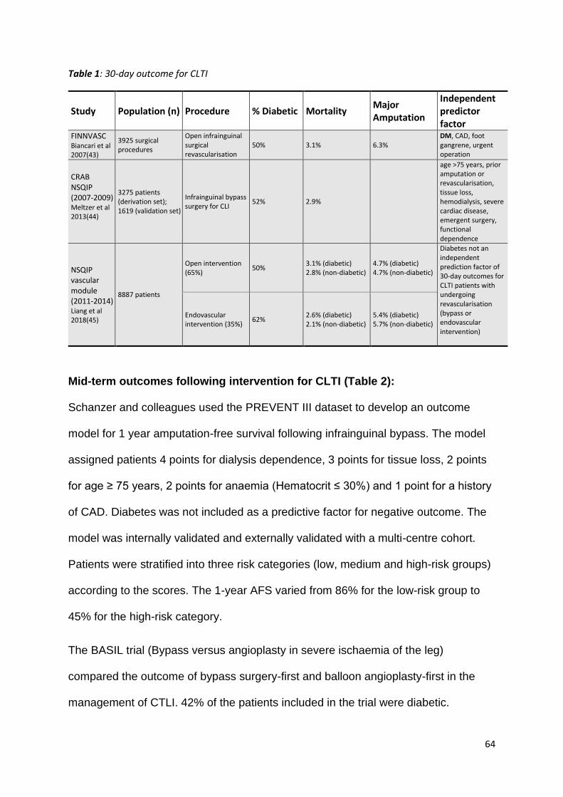
64
Table 1: 30-day outcome for CLTI
Mid-term outcomes following intervention for CLTI (Table 2):
Schanzer and colleagues used the PREVENT III dataset to develop an outcome
model for 1 year amputation-free survival following infrainguinal bypass. The model
assigned patients 4 points for dialysis dependence, 3 points for tissue loss, 2 points
for age ≥ 75 years, 2 points for anaemia (Hematocrit ≤ 30%) and 1 point for a history
of CAD. Diabetes was not included as a predictive factor for negative outcome. The
model was internally validated and externally validated with a multi-centre cohort.
Patients were stratified into three risk categories (low, medium and high-risk groups)
according to the scores. The 1-year AFS varied from 86% for the low-risk group to
45% for the high-risk category.
The BASIL trial (Bypass versus angioplasty in severe ischaemia of the leg)
compared the outcome of bypass surgery-first and balloon angioplasty-first in the
management of CTLI. 42% of the patients included in the trial were diabetic.
Study Population (n) Procedure % Diabetic Mortality Major Amputation
Independent predictor factor
FINNVASC Biancari et al 2007(43)
3925 surgical procedures
Open infrainguinal surgical revascularisation
50% 3.1% 6.3% DM, CAD, foot gangrene, urgent operation
CRAB NSQIP (2007-2009) Meltzer et al 2013(44)
3275 patients (derivation set); 1619 (validation set)
Infrainguinal bypass surgery for CLI
52% 2.9%
age >75 years, prior amputation or revascularisation, tissue loss, hemodialysis, severe cardiac disease, emergent surgery, functional dependence
NSQIP vascular module (2011-2014) Liang et al 2018(45)
8887 patients
Open intervention (65%)
50% 3.1% (diabetic) 2.8% (non-diabetic)
4.7% (diabetic) 4.7% (non-diabetic)
Diabetes not an independent prediction factor of 30-day outcomes for CLTI patients with undergoing revascularisation (bypass or endovascular intervention)
Endovascular intervention (35%)
62% 2.6% (diabetic) 2.1% (non-diabetic)
5.4% (diabetic) 5.7% (non-diabetic)

65
Amputation-free survival at 1 and 3 years was 68% and 57% in those patients
assigned to surgery first and 71% and 52% respectively in those assigned to
angioplasty-first for management of CTLI(46). A predictive model of overall survival
at 2 years after randomisation was created using regression model of baseline
covariates. The predictive model failed to identify DM as a predictor of AFS following
revascularisation after correcting for renal function, severity of below-knee arterial
disease (Bollinger score) and other co-morbidities(47).
Simons et al described a predictive model for 1-year AFS after lower limb extremity
bypass using the USA Vascular Quality Initiative (VQI) database (2003–
2012).(48) The overall 1-year AFS was 74% and on Cox regression model diabetes
was considered an independent risk factor for amputation or death (HR, 1.2; 95% CI,
1.1–1.4). However, bedbound status (HR, 4.4; 95% CI, 2.7–7.0), followed by dialysis
dependence (HR, 2.5; 95% CI, 2.1–2.9) had the largest magnitude of effect on the
risk of amputation or death. Details of the described predictive models can be found
in table 2.
Hicks at al also used the VQI database (2008-2014) and compared outcomes
following lower extremity bypass and endovascular intervention at and below the
knee in patients with DM versus patients without DM with CTLI(49). 2566 patients
were included, including 500 patients (19%) undergoing bypass surgery (DM 355 vs
non-DM 145) and 2066 patients (81%) undergoing endovascular treatment (DM
1463 vs non-DM 603). On multivariate analysis, there were no significant
differences in 1-year major amputation or mortality comparing patients with DM
versus patients without DM for either bypass surgery or endovascular intervention.
A recent study examined Medicare records of 72,199 patients diagnosed with
primary CLTI in 2011 and the clinical outcomes over 4 years were assessed(50).

66
54% of the patients were diabetic and 60% received revascularisation treatment
during follow up. 29% of the patients died or underwent major amputation in the first
year after diagnosis of severe ischaemia. In a multivariate model of the association
of baseline variables on outcomes, diabetes had a significant association with
mortality (HR 1.09 IC 1.06- 1.11) but not with major amputation. However, diabetes
was not included in a reduced multivariate model identifying the strongest predictors
of mortality and major amputation(50).
Table 2: Contemporary mid-term outcomes of patients presenting with CLTI
NR = not reported
Conclusion
There is little doubt that diabetes affects the presentation, diagnosis, and
management of PAD in many ways. Although diabetes itself does not seem to
consistently be a strong independent risk factor for morbidity and mortality in CTLI
patients undergoing revascularisation, it is impossible in clinical practice to isolate
diabetes from the comorbidities frequently occurring in diabetes. Diabetes is a major
Study Population (n) % Diabetic Procedure
Amputation free survival
Independent predictor factor
1 year 3 years
PREVENT III Schanzer 2008(51)
953 patients (PIII derivation set); 451 patients (PIII validation set); 716 patients (external validation)
64% Infrainguinal vein bypass for CLI
72.5% - 77% NR Dialysis, Tissue loss, Age ≥ 75, HCT ≤ 30%, CAD
BASIL Adam et al 2005(46) Bradbury et al 2010(52)
452 patients randomised to bypass surgery first or balloon angioplasty first revascularisation strategy
42%
Surgery first (50%) 68% 57%
Age, presence of tissue loss, serum creatinine, number of ankle pressure measurements detectable, maximum ankle pressure measured, a history of MI or angina, a history of stroke or TIA, below knee Bollinger angiogram score, body mass index, and smoking status
Angioplasty first (50%) 71% 52%
VQI Simons et al 2016(48)
7754 patients (VQI derivation dataset), 1404 patients (external validation dataset)
56% Nonemergency infrainguinal bypass for CLI
74% NR
Age, Tissue loss, CHF, DM, Creatinine, Ambulatory status, weight, bypass conduit use, bypass target, vessel, antiplatelet agent on discharge

67
risk factor for ESRD(53) and CAD(54). Diabetic patients are more likely to present
with tissue loss, non-healing wounds, and to have less favourable anatomy for
revascularisation(16, 18). The presence of diabetes however should not preclude
treatment of patients with CTLI and the three-step integrated PLAN approach
suggested by GVG guideline for CLTI should be implemented whenever possible.
References
1. Singh N, Armstrong DG, Lipsky BA. Preventing foot ulcers in patients with diabetes. Jama.
2005;293(2):217-28.
2. Abbott CA, Carrington AL, Ashe H, Bath S, Every LC, Griffiths J, et al. The North-West
Diabetes Foot Care Study: incidence of, and risk factors for, new diabetic foot ulceration in a
community-based patient cohort. Diabet Med. 2002;19(5):377-84.
3. Kerr M, Barron E, Chadwick P, Evans T, Kong WM, Rayman G, et al. The cost of diabetic foot
ulcers and amputations to the National Health Service in England. Diabet Med. 2019.
4. Boulton AJ, Vileikyte L, Ragnarson-Tennvall G, Apelqvist J. The global burden of diabetic foot
disease. Lancet. 2005;366(9498):1719-24.
5. Schaper NC, Van Netten JJ, Apelqvist J, Lipsky BA, Bakker K. Prevention and management
of foot problems in diabetes: a Summary Guidance for Daily Practice 2015, based on the IWGDF
Guidance Documents. Diabetes Metab Res Rev. 2016;32 Suppl 1:7-15.
6. Kantor J, Margolis DJ. Efficacy and prognostic value of simple wound measurements.
Archives of Dermatology. 1998;134(12):1571-4.
7. Sheehan P, Jones P, Caselli A, Giurini JM, Veves A. Percent change in wound area of
diabetic foot ulcers over a 4-week period is a robust predictor of complete healing in a 12-week
prospective trial. Diabetes Care. 2003;26(6):1879-82.
8. Hinchliffe RJ, Brownrigg JR, Apelqvist J, Boyko EJ, Fitridge R, Mills JL, et al. IWGDF
guidance on the diagnosis, prognosis and management of peripheral artery disease in patients with
foot ulcers in diabetes. Diabetes Metab Res Rev. 2016;32 Suppl 1:37-44.
9. Keast DH, Bowering CK, Evans AW, Mackean GL, Burrows C, D'Souza L. MEASURE: A
proposed assessment framework for developing best practice recommendations for wound
assessment. Wound Repair Regen. 2004;12(3 Suppl):S1-17.
10. Sugama J, Matsui Y, Sanada H, Konya C, Okuwa M, Kitagawa A. A study of the efficiency
and convenience of an advanced portable Wound Measurement System (VISITRAK). J Clin Nurs.
2007;16(7):1265-9.
11. Chang AC, Dearman B, Greenwood JE. A comparison of wound area measurement
techniques: visitrak versus photography. Eplasty. 2011;11:e18-e.
12. Bills JD, Berriman SJ, Noble DL, Lavery LA, Davis KE. Pilot study to evaluate a novel three-
dimensional wound measurement device. Int Wound J. 2016;13(6):1372-7.
13. Khoo R, Jansen S. The Evolving Field of Wound Measurement Techniques: A Literature
Review. Wounds. 2016;28(6):175-81.

68
14. Williams KJ, Sounderajah V, Dharmarajah B, Thapar A, Davies AH. Simulated Wound
Assessment Using Digital Planimetry versus Three-Dimensional Cameras: Implications for Clinical
Assessment. Annals of vascular surgery. 2017;41:235-40.
15. Jorgensen LB, Sorensen JA, Jemec GB, Yderstraede KB. Methods to assess area and
volume of wounds - a systematic review. Int Wound J. 2016;13(4):540-53.
16. Filko D, Cupec R, Nyarko EK. Wound measurement by RGB-D camera. Mach Vision Appl.
2018;29(4):633-54.
17. Hartley R, Zisserman A. Multiple view geometry in computer vision: Cambridge university
press; 2003.
18. Koo TK, Li MY. A Guideline of Selecting and Reporting Intraclass Correlation Coefficients for
Reliability Research. Journal of chiropractic medicine. 2016;15(2):155-63.
19. Wysocki AB. Wound measurement. Int J Dermatol. 1996;35(2):82-91.
20. Constantine RS, Bills JD, Lavery LA, Davis KE. Validation of a laser-assisted wound
measurement device in a wound healing model. Int Wound J. 2016;13(5):614-8.
21. Davis KE, Constantine FC, Macaslan EC, Bills JD, Noble DL, Lavery LA. Validation of a laser-
assisted wound measurement device for measuring wound volume. J Diabetes Sci Technol.
2013;7(5):1161-6.
22. Jorgensen LB, Skov-Jeppesen SM, Halekoh U, Rasmussen BS, Sorensen JA, Jemec GBE,
et al. Validation of three-dimensional wound measurements using a novel 3D-WAM camera. Wound
Repair Regen. 2018;26(6):456-62.
23. Chanussot-Deprez C, Contreras-Ruiz J. Telemedicine in wound care: a review. Adv Skin
Wound Care. 2013;26(2):78-82.
24. Authority. ACoSaQiHCaNHP. Australian Atlas of Healthcare Variation. Sydney2015. p. 329-
34.
25. Schubert V, Zander M. Analysis of the measurement of four wound variables in elderly
patients with pressure ulcers. Advances in wound care: the journal for prevention and healing.
1996;9(4):29-36.
26. Plassmann P, Melhuish JM, Harding KG. Methods of measuring wound size: a comparative
study. Ostomy Wound Manage. 1994;40(7):50-2, 4, 6-60.

69
70

71
Chapter 3: Diabetic Foot and Lower Limb Amputations:
Underestimated Problem with a Cost to Health System and
to the Patient
Guilherme Pena1,2, Prue Cowled1, Joseph Dawson2, Brenton Johnson2, Robert Fitridge1,2
1The University of Adelaide, Adelaide, Australia
2Department of Vascular and Endovascular Surgery, Royal Adelaide Hospital, Adelaide. SA, Australia
Pena G, Cowled P, Dawson J, Johnson B, Fitridge R. Diabetic foot and lower limb amputations: underestimated problem with a cost to health system and to the patient. ANZ Journal of Surgery. 2018 Jul;88(7-8):666-7. doi: 10.1111/ans.14436
Diabetes is a major public health challenge worldwide, which is associated with a
variety of complications including cardiovascular, kidney, eye and foot disease. The
absolute number and the percentage of the population with diagnosed diabetes
continues to rise. It is projected that in 2035 there will be 592million people
worldwide with diabetes(1).
In Australia, the prevalence of diabetes has more than doubled over the past three
decades, with an estimate of 1.7 million people living with diabetes in 2013. It is
predicted that this number will increase by 40% in the next 20 years(1). Diabetes
imposes a substantial burden on Australian healthcare expenditure and
approximately 10% of all inpatient hospital episodes in Australia have diabetes as
either the principal or an additional diagnosis(2).
It is known that individuals with diabetes have an increased risk of foot ulceration
and lower limb amputations when compared to people without diabetes(3). Lazzarini
described the burden of diabetes-related foot disease faced by the Australian
population as the second largest of the four main diabetes complications in terms of

72
burden of disease and numbers of people affected. In contrast, foot disease ranked
a distant last compared to the other diabetes complications with regard to
government funding(4).
Diabetic foot lesions are frequently the result of a patient having two or more risk
factors, with diabetic peripheral neuropathy and ischaemia playing vital roles.
Neuropathy leads to reduced sensation in the foot often foot deformity, which results
in an altered biomechanical pattern. This results in high risk of ulceration even
following minor trauma (e.g from ill-fitting shoes). Peripheral arterial disease is a risk
factor for impaired wound healing and subsequent lower extremity amputation(5)
This report describes amputation data from the Australian Vascular Audit (AVA) over
the 6-year period 2010-2015 and total admission and rehabilitation costs for patients
who underwent amputation at the Royal Adelaide Hospital. The AVA has been in
operation since January 2010 and is the official audit of the Australian and New
Zealand Society for Vascular Surgery(6). It is however self-reporting and voluntary
so only the procedures entered are amenable to data analysis. Data entry is known
to be incomplete especially in private hospitals.
A total of 20,669 amputations were recorded in the AVA between 2010-2015. Most of
those procedures (13,515) were minor amputations – toes or forefoot amputation;
12,115 procedures were performed in Australia and 1,400 in New Zealand. Amongst
patients who underwent minor amputations, 79% had diabetes and 62% had a
smoking history. The median length of hospital stay for patients undergoing minor
amputations was 11 days, one day longer than the average length of stay for stroke
patients in South Australia(7).
Approximately 35% of the recorded procedures (7154) were major amputations. 5,795
were performed in Australia with an above knee/below knee amputation (AKA/BKA)

73
ratio of 0.74. 1,359 procedures were carried out in New Zealand with an AKA/BKA
ratio of 1.02. This ratio is important as there is an increased prosthetic rehabilitation
rate in patients undergoing BKA versus those undergoing an AKA. Sixty one percent
of patients who had a major amputation were diabetic and 67% had a smoking history.
The median length of hospital stay for a patient undergoing major amputation was 20
days.
The distribution of patients undergoing amputation for diabetes-related disease is
heterogeneous. Data from the Australia Commission of Safety and Quality in Health
Care shows that the average number of admissions varies across states and
territories, from 19 per 100,000 people aged 18 years and over in Tasmania, to 65 per
100,000 in the Northern Territory. This difference is related to risk factors for diabetes-
related amputations, including the incidence of diabetes, distribution of Indigenous
population, socioeconomic status and geographical remoteness(8).
Cost analysis of 325 amputations conducted at the Royal Adelaide Hospital in the
2015-2016 financial year revealed an average total admission cost of $A 18,153 for
minor amputations and $A 35,016 for major amputations. Fifty two percent of patients
who had a major amputation were discharged directly to a rehabilitation facility. The
average length of stay in rehabilitation across two Local Health Networks in Adelaide
was 27 days with an average cost of $A 1,233 per day. Therefore, the total direct cost
for a patient who had a major amputation and went to a rehabilitation facility was $A
68,307. These costs were obtained using the Power Performance Manager (PPM)
Patient Costing Database and included all direct and indirect costs associated the
acute patient admission and inpatient rehabilitation. It is important to mention that
acute inpatient stay and inpatient rehabilitation are only a small component of lifetime
health care costs after amputation. In addition to these, there are costs with outpatient

74
visits, prosthetic charges and indirect costs from amputation-related work
absenteeism, reduced productivity, reduced labour force participation from chronic
disability, costs of a carer and premature mortality. There are few publications about
costs associated with lower limb amputation. Mackenzie reported that direct health-
care costs for the first two years after major amputation following injury was US$
91,106(9).
Diabetic foot disease is potentially preventable and every effort should be made to
ensure that high-risk patients are identified and receive early treatment of foot
complications. The International Working Group for the Diabetic Foot emphasizes
the importance of prevention of foot problems. Successful efforts to prevent and treat
diabetic foot complications depend upon a well-organised multidisciplinary team
comprised of general practitioners, nurses, podiatrists, vascular surgeons,
endocrinologists, infectious diseases specialists and orthopaedic surgeons. Ideally a
foot care programme should provide the following: 1 Education for people with
diabetes, their carers and for healthcare staff. 2 A system to detect all high-risk
patients. 3 Measures to reduce risk of foot ulceration, such as podiatric maintenance
care and appropriate footwear. 4 Prompt and effective treatment of any foot
complication. 5 Auditing of all aspects of the service to identify problems and ensure
that local practice meets accepted standards of care(5).
Diabetic foot ulcers should be seen as a chronic potentially limb-threatening
condition and strategies should be designed to meet the needs of patients requiring
chronic care, rather than simply responding to acute problems when they occur5.
Aggressive preventative medicine should aim to provide an efficient and cost-
effective solution to a challenging and costly disease process.

75
References
1. Guariguata L, Whiting DR, Hambleton I, Beagley J, Linnenkamp U, Shaw JE. Global
estimates of diabetes prevalence for 2013 and projections for 2035. Diabetes Res Clin Pract.
2014; 103: 137-49.
2. Australian Institute of Health & Welfare (AIHW): Diabetes. Australia Government; 2016.
Available from: https://www.aihw.gov.au/reports-statistics/health-conditions-disability-
deaths/diabetes/overview. Accessed 20 August 2017.
3. Kvitkina T, Narres M, Claessen H et al. Incidence of lower extremity amputation in the
diabetic compared to the non-diabetic population: a systematic review protocol. Syst Rev.
2015; 4: 74.
4. Lazzarini PA, Gurr JM, Rogers JR, Schox A, Bergin SM. Diabetes foot disease: the Cinderella
of Australian diabetes management?. J Foot Ankle Res. 2012; 5: 24.
5. Bus SA, Netten JJ, Lavery LA et al. IWGDF Guidance on the prevention of foot ulcers in at‐
risk patients with diabetes. Diabetes Metab Res Rev. 2016; 32: 16-24.
6. Beiles CB, Bourke BM. Validation of Australian data in the Australasian vascular audit. ANZ J
Surg. 2014; 84: 624-7.
7. SA health: Length of stay times over the decades. Government of South Australia; 2016.
Available from: http://transforminghealth.sa.gov.au/length-stay-times-decades/. Accessed 18
August 2017.
8. Australian Commission on Safety and Quality in Health Care and National Health
Performance Authority. Australian Atlas of Healthcare Variation. Sydney: ACSQHC, 2015.
9. MacKenzie EJ, Castillo RC, Jones AS, et al. Health-care costs associated with amputation or
reconstruction of a limb-threatening injury. J Bone Joint Surg 2007; 89: 1685

76

77

78
Chapter 4: Evaluation of a Novel 3-Dimensional Wound
Measurement Device for Assessment of Diabetic Foot
Ulcers
Guilherme Pena1,2, Beatrice Kuang1,2, Zygmunt Szpak3, Prue Cowled2, Joseph Dawson1,2, Robert Fitridge1,2
1Department of Vascular and Endovascular Surgery, Royal Adelaide Hospital, Adelaide, South Australia, Australia
2Discipline of Surgery, The University of Adelaide, Adelaide, South Australia, Australia
3Australian Institute for Machine Learning, School of Computer Science, The University of Adelaide, Adelaide, South Australia, Australia
.
Pena G, Kuang B, Szpak Z, Cowled P, Dawson J, Fitridge R. Evaluation of a Novel Three-Dimensional Wound Measurement Device for Assessment of Diabetic Foot Ulcers. Adv Wound Care (New Rochelle). 2020 Nov;9(11):623-631. doi: 10.1089/wound.2019.0965.
Abstract
Objective
The initial wound measurement and regular monitoring of diabetic foot ulcers (DFU)
is critical to assess treatment response. There is no standardised, universally
accepted assessment method to characterise DFU. To address this need, a novel
topographic imaging system has been developed. Our study aims to assess the
reliability and practicality of the WoundVue® camera technology in the assessment
of DFU.
Approach
The WoundVue® system consists of two infrared cameras and an infrared projector,
and is able to produce a 3D reconstruction of the wound.
Fifty-seven diabetic foot wounds were photographed from two different angles and
distances using the WoundVue® camera. Wound area, volume and maximum depth

79
were measured for assessment of reliability. Thirty-one of these wounds also had
area calculated using the Visitrak™ system and a correlation between the area
obtained using both systems was assessed.
Results
WoundVue® images analysis showed excellent intra and inter-rater agreement for
area, volume and maximum depth (all ICC > 0.970). Good agreement was found for
area measurement using the WoundVue® camera and Visitrak™ system (ICC
0.842). The average percentage difference between measures obtained using the
WoundVue from different angles for assessment of different sizes and shapes of
wounds were 2.9% (95 CI 0.3%-5.4%), 12.9% (95% CI 9.6%-35.7%) and 6.2% (95%
CI 2.3% - 14.7%) for area, maximum depth and volume respectively.
Innovation
This is the first human trial evaluating this novel 3-Dimensional wound measurement
device.
Conclusion
The WoundVue® system is capable of recreating a 3D model of DFU and produces
consistent data.

80
Introduction
Foot problems in diabetes are common and carry a substantial physical,
physiological and financial burden for affected patients. It is estimated that patients
with diabetes have an almost 25% lifetime risk of developing a foot ulcer and
approximately 2% of patients develop new foot ulcers each year(1, 2). More than
50% of these ulcers become infected and many requiring hospitalisation(1). Diabetic
foot care accounts for a substantial proportion of healthcare expenditure and the
majority of this expenditure arises through prolonged and severe ulceration(3).
Diabetic foot complications are the most common cause of “non-traumatic” lower
limb amputation and it has been estimated that on a global scale a lower limb is lost
every 20 seconds as a consequence of diabetes(4).
The treatment of diabetic ulcers is complex and requires a multidisciplinary team.
The principles of management include wound care, management of infection,
revascularisation if required, and offloading, with the aim of achieving expeditious
wound healing, prevention of ulcer recurrence, and ultimately avoiding
amputations(5).
As part of wound management, it is essential to obtain accurate and reproducible
wound measurements. A thorough initial wound assessment provides baseline data
about the status of the wound and is important in developing a treatment plan(6).
Clinical Problem Addressed
Repeated wound measurements at clinical encounters is valuable for assessing the
effectiveness of treatment and can be a predictor of longer-term ulcer healing. In a
prospective study of patients with diabetic foot ulcers, the percent change in foot
ulcer area after 4 weeks of observation was a good predictor of healing at 12

81
weeks(7). Recent recommendations from the International Working Group for the
Diabetic Foot recommend consideration of revascularisation if the wound in patients
who were initially assessed as having adequate perfusion has not significantly
improved within 6 weeks of optimal wound care(8).Thus accurate wound
assessment is an essential component of diabetic foot management.
The ideal method of wound measurement should be practical, comfortable for the
patient, accurate and most importantly reproducible. In clinical practice, it is
important to be able to reassess wounds regularly to track changes in size, depth
and appearance over time. The measurement methods most commonly used include
simple ruler assessment, acetate tracing, and digital imaging methods(9). Ruler-
based techniques to calculate area are simple and inexpensive but inconsistent and
are not very reliable for irregular or large wounds. Acetate tracing is performed by
placing a transparent film over the wound and tracing the outline with a permanent
marker, allowing more accurate area calculation when the wound is irregular. The
area can be obtained by placing the wound trace on a metric grid and counting the
number of squares of a known area. However, it is time consuming and inaccuracies
may arise when deciding the value of partial squares. Alternatively, the wound
outline can be retraced onto a digital tablet, which calculates the area(9). One
example of digital planimetry device that has been validated is the Visitrak™ system.
(Smith & Nephew Wound Management, Inc, Largo, Florida)(10). Digital photography
is also commonly used. The wound can be photographed with a ruler or a marker of
known dimensions placed at the skin near the wound edge and the image
transferred to a computer and planimetric software can be used to calculate the
area. However, inaccuracies due to parallax may occur(11).

82
More recently new methods of wound measurement using laser scanners,
stereophotogrammetry and structured light technique have become available.
Eykona®, Silhouette® and the inSight® cameras are examples of these new
technologies with the potential to provide a more comprehensive evaluation of the
wound including assessment of volume(12-15). The WoundVue® system is a new
prototype device that uses the principle of stereophotogrammetry to provide a 3-
dimensional assessment of the wound (Figures1 and 2).
The purposes of this study were to determine reliability of the WoundVue® camera
for area, volume and maximum depth measurements and to assess the agreement
between area measurements obtained using the WoundVue® camera and the
Visitrak™ system.
Figure 1: WoundVue® camera.
The system consists of a Microsoft Surface Pro Tablet, 3D-camera, a pair of LED lights and tailor-made software.

83
Figure 2: 3D reconstruction of foot wound.
The left panel is an image of the wound and the right panel shows the concomitant 3D reconstruction and metric measurements.
Material and Methods
Ethical approval was granted from Central Adelaide Local Health Network Human
Research Ethics Committee and written informed consent was obtained from all
participants.
Patients with DFU were enrolled from multidisciplinary diabetic foot clinics at the
Queen Elizabeth Hospital and Lyell McEwin Health Service or admitted under the
Vascular Surgery service at the Royal Adelaide Hospital, all within the metropolitan
Adelaide region, South Australia, from June to November 2018.
Digital documentation of wounds was obtained by one of the clinicians involved in
the study (GP) using the WoundVue® camera. GP received a formal 30-minute
training session on the device prior to the research. The WoundVue® system is a
prototype device developed by the machine learning department at The University of
Adelaide in collaboration with LBT Innovations Limited (Adelaide, South Australia).
The camera system consists of two infrared cameras which image the wound from
two different vantage points and an infrared projector which casts a textured light
pattern onto the wound. The textured light pattern facilitates matching pixels in the
first image with corresponding pixels in the second image. Once corresponding
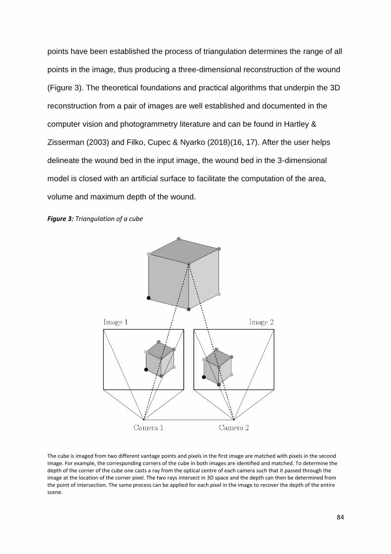
84
points have been established the process of triangulation determines the range of all
points in the image, thus producing a three-dimensional reconstruction of the wound
(Figure 3). The theoretical foundations and practical algorithms that underpin the 3D
reconstruction from a pair of images are well established and documented in the
computer vision and photogrammetry literature and can be found in Hartley &
Zisserman (2003) and Filko, Cupec & Nyarko (2018)(16, 17). After the user helps
delineate the wound bed in the input image, the wound bed in the 3-dimensional
model is closed with an artificial surface to facilitate the computation of the area,
volume and maximum depth of the wound.
Figure 3: Triangulation of a cube
The cube is imaged from two different vantage points and pixels in the first image are matched with pixels in the second image. For example, the corresponding corners of the cube in both images are identified and matched. To determine the depth of the corner of the cube one casts a ray from the optical centre of each camera such that it passed through the image at the location of the corner pixel. The two rays intersect in 3D space and the depth can then be determined from the point of intersection. The same process can be applied for each pixel in the image to recover the depth of the entire scene.

85
Fifty-seven wounds were photographed with the WoundVue® camera from two
slightly different angles and distances for assessment of intra-rater reliability. All the
photos were taken by the same clinician (GP) from an appropriate position where the
target ulcer was placed close to the centre of the frame for both right and left
cameras. The objective was to assess consistency in measurements when the
photos were taken in satisfactory but not identical circumstances. Subsequently, two
clinicians (GP and BK) independently assessed all the images and outlined wound
edges. Thirty-one wounds that were photographed also had area measured using
Visitrak™ system by the same clinician involved in the study (GP).
Wound images were downloaded and processed by the Australian Institute for
Machine Learning (AIML - University of Adelaide) where area, volume and maximum
depth measurements were obtained.
Statistics
Intraclass correlation coefficient (ICC) estimates and their 95% confident intervals
were calculated. In accordance with Ko and Li (18) recommendations, ICC values
>0.90 were indicative of excellent agreement, whereas values between 0.75–0.90
and 0.50–0.75 portrayed good and moderate agreement, respectively.
A two-way mixed effects, absolute agreement, measurement model was used for
assessment of Woundvue® intra and inter-rater reliability and a one-way random
effect model was used to assess the agreement between the different instruments
(WoundVue® and Visitrak™).
One sample T test of the differences of measurements obtained using the
WoundVue® system was performed to assess average percentage difference

86
between wound dimensions acquired from photos taken from slightly different
angles.
All statistical analysis was performed using SPSS statistical package version 25
(IBM SPSS Statistics for Windows, Version 25.0. Armonk, NY: IBM Corp.).
Results
Fifty-seven diabetic foot wounds of different shapes and sizes were photographed
using the WoundVue® Camera. The average wound area obtaining was 5.7cm2,
ranging from 0.4cm2 (small digital ulcer) to 30.0cm2 (forefoot amputation wound).
Wound maximum depth and volume were on average 0.6cm (max 5.6cm and min
0.02cm) and 1.2cm3 (max 14.9cm3 and min 0.003cm3) respectively.
Excellent intra-rater (GP) reliability was found for area [ICC 0.995 (95% CI 0.991 –
0.997)], volume [ICC 0.988 (95%CI 0.979 – 0.993)] and maximum depth [ICC 0.984
(95% CI 0.975 – 0.990)] measurements obtained with the WoundVue® camera
between the 2 different images taken of the same wounds (Figure 4).

87
Figure 4: Intra-rater reliability of the WoundVue®.
Similarly, excellent inter-rater reliability for area [ICC 0.983 (95% CI 0.971 – 0.990)],
volume [ICC 0.978 (95%CI 0.962 – 0.988)] and maximum depth [ICC 0.975 (95% CI
0.956 – 0.986)] was achieved when images were analysed by two different
assessors.
The average percentage difference between measures obtained using the
WoundVue® from different angles for assessment of different sizes and shapes of

88
wounds were 2.9% (95 CI 0.3%-5.4%), 12.9% (95% CI 9.6%-35.7%) and 6.2% (95%
CI 2.3% - 14.7%) for area, maximum depth and volume respectively.
Thirty-one wounds that were photographed also had areas measured using
Visitrak™. The reason for the fact that not all wounds photographed had areas
measured with Visitrak™ is that this device was only available in one of the clinics
initially and then became accessible to other settings during the research. A good
agreement was found for area measurement using the WoundVue® camera and
Visitrak™ system [ICC 0.842 (95% CI 0.700 – 0.920)] (Figure 5).
Figure 5: Correlation between WoundVue® and Visitrak for area measurement.
Discussion
An accurate and reliable measurement method of wound area and depth is important
for appropriate wound documentation, assessment of progress and determining the
efficacy of treatment. An ideal measurement method should be consistent, easy to
learn and use, cost efficient and comfortable for patients(19).

89
With the advent of new technologies, 3D cameras have become available and they
have the potential to afford a more comprehensive assessment of wounds, providing
not only measures of surface area and circumference but also wound volume.
There are 3D measurement systems commercially available. However, they have
not yet had a major impact in clinical practice. A major limiting factor for their
widespread adoption remains the significant cost of purchase and maintenance of
such devices. Examples are the Eykona camera® (Fuel 3D, Oxford UK), Silhouette®
(Aranz, Christchurch, New Zealand) and inSight® (eKare Inc, Fairfax, USA)(12, 14).
These systems use different technologies to assess wound dimensions. The Eykona
®camera uses what is known as "photometric stereo" to construct a 3D model of the
wound. Photometric stereo involves taking a sequence of photographs of the wound
while holding the camera stationary but illuminating the wound from different
directions. It turns out that one can construct a 3D model of the wound by analyzing
the shading patterns in the series of wound bed images. The Eykona® device
requires the user to place a bespoke calibration target near the wound bed. The
Silhouette® device projects three red laser fan-beams onto the wound. One
positions the device such that the three red laser lines intersect and form a star
shape on the wound bed. Silhouette uses knowledge of the orientation and position
of the lasers’ beams to determine the depth of pixels in the image that the laser falls
on. That is, one obtains minimal depth information since one only has depth
corresponding to three thin lines that project onto the wound. Consequently, one can
expect a significant variance in depth and volume measurement since any three
lines of the wound-bed will lead to different reconstructions. The InSight® and
WoundVue® device are similar in that they both projecting an infrared structured
light pattern onto the wound to aid in constructing a 3D model and construct a
90
volume measurement by taking into account the entire wound bed. However, the
devices use different camera lenses, dot patterns and algorithms to build the 3D
models and produce the measurements. The choice of lenses is crucial since it
influences the minimum wound size that a device handle.
This study describes an initial evaluation of the WoundVue® camera in human
subjects. The device proved to be practical and provides a reliable method of wound
assessment. It requires minimal training to operate the camera and software, and it
can be easily deployed in routine clinical practice.
Results demonstrated excellent intra-rater reliability for area, volume and maximum
depth assessment for a range of wound shapes and sizes. Photos taken in the same
clinical encounter but with the device positioned in different angles and distance from
the wound provided consistent results with ICC above 0.980 for all the parameters.
In addition, when the photos were analysed by two assessors there was excellent
correlation for all parameters.
Wound area measurements obtained with WoundVue® demonstrated a good
correlation with wound area obtained using the Visitrak™ system, with ICC 0.842. As
demonstrated in figure 5 the correlation between the two systems was less strong for
larger wounds. This was not surprising as the two systems use different methods to
assess surface area. The WoundVue® system evaluates surface area of the wound
taking into consideration all the irregularities of the wound bed while Visitrak™ film
grid, although malleable, is unable to conform precisely to the wound surface.
The proportional difference for area assessment using photos of real wounds with
different shapes and sizes was from different angles was very low (2.9%, 95 CI
0.3%-5.4%), demonstrating high value of the WoundVue® camera for area
91
assessment. There was more variability for assessment of the other parameters. The
highest variability was for maximum depth, 12.9% (95% CI 9.6%-35.7%). This can
be explained by the fact that the study included several superficial wounds for which
any variance in depth measurement resulted in large proportional difference in this
parameter. Examples are wounds that were less than 3mm in depth for which 1mm
of difference between measurements, whilst not clinically important, resulted in more
than 30% variance. The proportional difference in volume between measurements
was 6.2% (95% CI 2.3% - 14.7%). This variance is viewed as acceptable considering
that images were taken from slightly different angles and the cohort comprised of
real diabetic foot wounds with different shapes and sizes. The considerable
variability in depth measurement, especially for superficial wounds, also affected
volume measurements.
Assessment of wound volume is challenging. Slight movements of the patient or
camera operator can change the appearance of a wound, especially if there is an
undermined segment of the wound. Localisation of the wound on a curved part of the
body (e.g. the heel) can make it difficult to correctly estimate wound size(15). The
commercially available 3D cameras have been shown to be accurate for area
assessment, however they have not been sufficiently validated in terms of accuracy
and reliability for volume assessment. Several validation studies were done in a
controlled environment using artificial or animal wound models(12, 14, 20, 21) which
are much less complex than real wounds in clinical settings. Therefore, these results
should be looked at with caution. A study using wound models compared volume
measurement by water displacement technique with measurement obtained using
Eykona® and Silhouette® devices. Both devices significantly overestimated wound
volume compared to water displacement.(14) More recently, Jørgensen at al.

92
assessed 48 real wounds using a novel 3D-WAM camera (prototype device) and
compared wound volume obtained with the 3D device with volume obtained by
injecting gel into wound cavity and a high agreement between methods was
reported(22).
There are many advantages associated with the use of a 3D camera system such as
WoundVue®. It is a practical, efficient and comprehensive way to assess wound
size. It is non-invasive and thus causes no discomfort to patients. As the wound bed
is not touched during assessment there is no risk of infection, and importantly, cross-
infection. The potential for using this system in Telemedicine is exciting(23). This is
particularly important in countries such as Australia where distance from expert
assessment can be a critical factor in providing good care. In Australia, the
prevalence of diabetic foot disease is high in remote geographic areas especially
among the indigenous population(24). Frequently, patients live hundreds of
kilometres from specialised multidisciplinary foot clinics. The use of a reliable 3D
camera system that provides high quality imaging and measurements would allow
enhanced communication between local teams (general practitioners and community
nurses) and wound care specialists (specialist nurses, podiatrists and vascular
surgeons). A 3D photo of a wound is able to transmit more nuanced information that
often cannot be easily captured in a wound description. As a result, routine follow-up
care could be performed in a remote area with close liason with a multi-disciplinary
service.
In addition to being able to provide wound dimensions, the WoundVue® core
machine learning algorithms have been adapted to interpret tissue types. This
innovative technology allows for an even more comprehensive wound assessment
measuring, for example, the percentage of wound area with non-viable tissue in the

93
base. Further studies need to be undertaken to validate this feature currently offered
by the WoundVue® system.
Limitations of the current study include a small sample size, lack of visitrak™
assessment in all patients and the fact that we did not compare the volume and
maximum depth measurements obtained with WoundVue® camera with other
methods. Ideally wound depth should have been measured using ruler or a depth
probe and wound volume with saline or alginate cast. However, these methods can
cause patient discomfort, are impractical to be performed in a busy diabetic foot
clinic and lack reproducibility(25, 26). It would also be valuable to compare the
measurements obtained with other 3D wound camera systems available. However,
unfortunately the research team did not have access to other devices and none of
the 3D camera devices commercially available has been proven to be particularly
accurate for depth and volume assessment.
There are also limitations related to the device. As with all methods of digital wound
measurement, the WoundVue® camera is unable to assess undermined parts of a
wound, and the presence of debris and clot in a wound may affect volume
measurement. Furthermore, assessment of wounds at points of curvature of the
body represent significant challenges and some wounds may not be suitable for 3D
reconstruction. The WoundVue® Camera demonstrated restrictions for measuring
depth and volume of shallow, flat wounds. However, for this type of wound, area
assessment is clinically much more relevant.
Currently the WoundVue® camera does not provide the dimensions instantly and the
device needs to be connected to specific software on a desktop computer to
generate the 3D model of the wound and wound measurements. Future

94
development of the system should allow for immediate wound assessment within the
device without the need to connect to external software.
Innovation
This was the first human study evaluating the WoundVue® system. This system is
capable of recreating a reliable 3D model of diabetic foot wounds, providing wound
measurements. It has the potential to be a valuable adjunct in diabetic foot wound
care as digital images are ideal for monitoring wounds over time and for telemedicine
application.
Key findings
• WoundVue® camera has demonstrated excellent consistency for wound
measurement in relation to area, maximum depth and volume.
• Good correlation for area assessment between WoundVue® and Visitrak™,
although this correlation was less strong for larger wounds.
• 3-Dimensional wound measurement devices, such as WoundVue® camera,
has the potential to be a valuable adjunct in diabetic foot wound care.

95
References
1. Singh N, Armstrong DG, Lipsky BA. Preventing foot ulcers in patients with diabetes. Jama.
2005;293(2):217-28.
2. Abbott CA, Carrington AL, Ashe H, Bath S, Every LC, Griffiths J, et al. The North-West
Diabetes Foot Care Study: incidence of, and risk factors for, new diabetic foot ulceration in a
community-based patient cohort. Diabetic medicine : a journal of the British Diabetic Association.
2002;19(5):377-84.
3. Kerr M, Barron E, Chadwick P, Evans T, Kong WM, Rayman G, et al. The cost of diabetic foot
ulcers and amputations to the National Health Service in England. Diabetic medicine : a journal of the
British Diabetic Association. 2019;36(8):995-1002.
4. Boulton AJ, Vileikyte L, Ragnarson-Tennvall G, Apelqvist J. The global burden of diabetic foot
disease. Lancet (London, England). 2005;366(9498):1719-24.
5. Schaper NC, Van Netten JJ, Apelqvist J, Lipsky BA, Bakker K. Prevention and management
of foot problems in diabetes: a Summary Guidance for Daily Practice 2015, based on the IWGDF
Guidance Documents. Diabetes Metab Res Rev. 2016;32 Suppl 1:7-15.
6. Kantor J, Margolis DJ. Efficacy and prognostic value of simple wound measurements.
Archives of Dermatology. 1998;134(12):1571-4.
7. Sheehan P, Jones P, Caselli A, Giurini JM, Veves A. Percent change in wound area of
diabetic foot ulcers over a 4-week period is a robust predictor of complete healing in a 12-week
prospective trial. Diabetes Care. 2003;26(6):1879-82.
8. Hinchliffe RJ, Brownrigg JR, Apelqvist J, Boyko EJ, Fitridge R, Mills JL, et al. IWGDF
guidance on the diagnosis, prognosis and management of peripheral artery disease in patients with
foot ulcers in diabetes. Diabetes Metab Res Rev. 2016;32 Suppl 1:37-44.
9. Keast DH, Bowering CK, Evans AW, Mackean GL, Burrows C, D'Souza L. MEASURE: A
proposed assessment framework for developing best practice recommendations for wound
assessment. Wound repair and regeneration : official publication of the Wound Healing Society [and]
the European Tissue Repair Society. 2004;12(3 Suppl):S1-17.
10. Sugama J, Matsui Y, Sanada H, Konya C, Okuwa M, Kitagawa A. A study of the efficiency
and convenience of an advanced portable Wound Measurement System (VISITRAK). J Clin Nurs.
2007;16(7):1265-9.
11. Chang AC, Dearman B, Greenwood JE. A comparison of wound area measurement
techniques: visitrak versus photography. Eplasty. 2011;11:e18-e.
12. Bills JD, Berriman SJ, Noble DL, Lavery LA, Davis KE. Pilot study to evaluate a novel three-
dimensional wound measurement device. Int Wound J. 2016;13(6):1372-7.

96
13. Khoo R, Jansen S. The Evolving Field of Wound Measurement Techniques: A Literature
Review. Wounds : a compendium of clinical research and practice. 2016;28(6):175-81.
14. Williams KJ, Sounderajah V, Dharmarajah B, Thapar A, Davies AH. Simulated Wound
Assessment Using Digital Planimetry versus Three-Dimensional Cameras: Implications for Clinical
Assessment. Annals of vascular surgery. 2017;41:235-40.
15. Jorgensen LB, Sorensen JA, Jemec GB, Yderstraede KB. Methods to assess area and
volume of wounds - a systematic review. Int Wound J. 2016;13(4):540-53.
16. Filko D, Cupec R, Nyarko EK. Wound measurement by RGB-D camera. Mach Vision Appl.
2018;29(4):633-54.
17. Hartley R, Zisserman A. Multiple view geometry in computer vision (2nd ed.): Cambridge
university press; 2004.
18. Koo TK, Li MY. A Guideline of Selecting and Reporting Intraclass Correlation Coefficients for
Reliability Research. Journal of chiropractic medicine. 2016;15(2):155-63.
19. Wysocki AB. Wound measurement. Int J Dermatol. 1996;35(2):82-91.
20. Constantine RS, Bills JD, Lavery LA, Davis KE. Validation of a laser-assisted wound
measurement device in a wound healing model. Int Wound J. 2016;13(5):614-8.
21. Davis KE, Constantine FC, Macaslan EC, Bills JD, Noble DL, Lavery LA. Validation of a laser-
assisted wound measurement device for measuring wound volume. Journal of diabetes science and
technology. 2013;7(5):1161-6.
22. Jorgensen LB, Skov-Jeppesen SM, Halekoh U, Rasmussen BS, Sorensen JA, Jemec GBE,
et al. Validation of three-dimensional wound measurements using a novel 3D-WAM camera. Wound
repair and regeneration : official publication of the Wound Healing Society [and] the European Tissue
Repair Society. 2018;26(6):456-62.
23. Chanussot-Deprez C, Contreras-Ruiz J. Telemedicine in wound care: a review. Advances in
skin & wound care. 2013;26(2):78-82.
24. Australian Commission on Safety and Quality in Health Care and National Health
Performance Authority. Australian Atlas of Healthcare Variation. Sydney: ACSQHC, 2015.
25. Schubert V, Zander M. Analysis of the measurement of four wound variables in elderly
patients with pressure ulcers. Advances in wound care: the journal for prevention and healing.
1996;9(4):29-36.
26. Plassmann P, Melhuish JM, Harding KG. Methods of measuring wound size: a comparative
study. Ostomy/wound management. 1994;40(7):50-2, 4, 6-60

97

98

99
Chapter 5: Micronutrient Status in Diabetic Patients with
Foot Ulcers
Guilherme Pena1,2, Beatrice Kuang1,2, Prue Cowled2, Stuart Howell3, Joseph Dawson1,2, Ross Philpot4, Robert Fitridge1,2
1Department of Vascular and Endovascular Surgery, Royal Adelaide Hospital, Adelaide, South Australia, Australia
2Discipline of Surgery, The University of Adelaide, Adelaide, South Australia, Australia
3Data Management and Analysis Centre, School of Population Health, The University of Adelaide, Adelaide, South Australia 5005, Australia
4Department of Infectious Disease, The Queen Elizabeth Hospital, Adelaide, South Australia
Pena G, Kuang B, Cowled P, Howell S, Dawson J, Philpot R, Fitridge R. Micronutrient Status in Diabetic Patients with Foot Ulcers. Adv Wound Care (New Rochelle). 2020 Jan;9(1):9-15. doi: 10.1089/wound.2019.0973.
Abstract
Objective: To explore the prevalence of micronutrient deficiencies in patients with
diabetic foot ulcers and correlate this with foot disease severity and other clinical
factors.
Approach: Prospective cohort study of diabetic patients with foot ulcers seen in
multidisciplinary foot clinics across Adelaide or admitted to the Vascular Surgery unit
at the Royal Adelaide Hospital between February 2017 and September 2018. A total
of 131 patients were included in the study. Plasma serum[MOU1] levels of vitamin A, C,
D, E; copper, zinc and ferritin were measured. Demographic and clinical data
including BMI, smoking status, duration of diabetes, HbA1c and WIfI score were
obtained.
Results: The most prevalent nutritional deficiency found was Vitamin D affecting
55.7% of patients. Suboptimal levels of vitamin C affected 73% of patients
comprising of marginal levels in 22.2% and deficient levels in 50.8%. Zinc deficiency,

100
vitamin A deficiency and low ferritin levels were present in 26.9%, 10.9% and 5.9%
of patients respectively. There was no correlation between BMI, grip strength,
duration of diabetes, HbA1c or smoking status with micronutrient deficiency.
Increased severity of diabetic foot disease was associated with lower vitamin C
levels (p = 0.02).
Innovation: This study has demonstrated that the deficiency of micronutrients,
especially Vitamin D, vitamin C, Zinc and Vitamin A, is common in diabetic patients
with foot ulcers.
Conclusions: The prevalence of micronutrient deficiency is high in diabetic
population with foot ulcers/wounds. Special concerns regarding the high prevalence
of vitamin C and zinc deficiency given their roles in wound healing. Although further
research needs to be performed to determine the clinical implications of our findings,
micronutrient deficiency should be considered in diabetic patients with foot wounds.
Introduction
Diabetic foot complications carry a substantial physical, psychological and financial
burden for the patients and community. People who suffer from diabetes have a
lifetime risk of nearly 25% of developing a foot ulcer and more than 50% of patients’
ulcer will develop infection(1). A history of foot ulcer is significantly associated with
negative outcomes. Approximately 85% of all amputations in diabetic patients are
preceded by foot ulceration which subsequently deteriorates to foot infection or
gangrene(2).
Diabetic foot complications are recognised as the most common cause of non-
traumatic lower limb amputation internationally. Worldwide it is estimated that every

101
20 seconds a lower limb amputation is performed as a consequence of diabetes(3).
The challenge to heal a diabetic foot ulcer is compounded by the high rate of re-
ulceration once healed. Approximately 40% of patients have recurrence of an ulcer
within 1 year after healing, almost 60% within 3 years, and 65% within 5 years(3).
Multifactorial efforts should be made to give affected patients the best chance to heal
foot ulcers. Traditionally this has taken the form of local wound care, debridement,
offloading, attention to infection and revascularisation if required. However, there are
many other patient-related factors that are frequently overlooked that may influence
wound healing (Table 1). One of the factors most commonly neglected is patient
nutrition.
Table 8: Factors considered to affect wound healing
Clinical Problem Addressed
Wound healing is a complex, dynamic, interactive process involving soluble
mediators, blood cells, extracellular matrix and parenchymal cells. Wound healing
has three phases that overlap in time; inflammation, tissue formation, and tissue
remodelling(4). The relationship between nutrition and wound healing has been
Poor or impaired perfusion Infection Smoking and alcoholism Aging Chronic diseases (e.g, diabetes, chronic kidney disease, AIDS) Malnutrition Medication (e.g. glucocorticoid steroids, non-steroidal anti-inflammatory drugs, chemotherapy) Obesity Oedema Presence of foreign body Venous insufficiency

102
recognized for centuries. It is widely known that macronutrient malnutrition,
especially protein, adversely affects wound healing. Equally important are
micronutrients, which are critical components of cellular metabolism. A number of
vitamins and minerals play a significant role in the immune system and wound
healing, particularly important are Vitamin C, Vitamin A and Zinc(5). Despite their
roles, they are not routinely measured or monitored in clinical practice.
The primary goal of this study was to assess the prevalence of vitamin and
micronutrient deficiency in diabetic patients with foot ulcers seen either in an
outpatient setting within multidisciplinary foot clinics, or inpatients admitted to our
Vascular service. The secondary goal was to correlate micronutrient levels with
disease severity and other clinical factors, namely duration of diabetes, HbA1c
levels, grip strength and smoking status.
Material and Methods
This study is part of a major project assessing factors influencing outcomes in
patients with diabetic foot disease. Ethics has been obtained from Central Adelaide
Local Health Network ethics committee and written consent was obtained from all
participants. Subjects consisted patients seen at Multidisciplinary Foot Clinics at The
Queen Elizabeth Hospital and Lyell McEwin Hospital, or admitted under the Vascular
Surgery service at the Royal Adelaide Hospital, all within the Adelaide metropolitan
area of South Australia. Eligibility criteria included being diabetic, age ≥ 18 years,
able to have follow ups in Adelaide and presence of foot ulcer(s).
A total of 131 patients were recruited for the study between February 2017 and
September 2018. Plasma levels of Vitamin A, C, D, E; copper, zinc and ferritin were
measured at recruitment. All the samples were of venous blood taken by a

103
phlebologist in hospital or outpatient pathology collection centre. Specimens were
handled and transported as per collection guide and all processed by SA Pathology.
Table 2 shows reference levels used for analysis. Demographic information and
clinical data were prospectively obtained during patient assessment including age,
gender, weight, height, BMI and smoking status. In addition, grip strength and WIfI
score were also recorded. Grip strength is a measurement of muscle function as
indicator of functional as well as nutritional status(6). The WIfI is a validated
classification which stratifies patients with threatened lower extremity, including
patients with diabetic foot ulcers, based on three major factors that impact
amputation risk and clinical management: Wound, Ischemia, and foot Infection(7).
Table 9: Reference levels of micronutrients
Vitamin A <0.7 µmol/L Deficient
≥0.7 µmol/L Non-deficient
Vitamin C
< 11.4 µmol/L Deficient
11.4 - 22.7 µmol/L Marginal
> 22.7 µmol/L Adequate
Vitamin D < 60nmol/L Deficient
≥ 60 nmol/L Non-deficient
Vitamin E < 12 µmol/L Deficient
≥ 12 µmol/L Non-deficient
Zinc < 9 µmol/L Deficient
≥ 9 µmol/L Non-deficient
Copper < 10 µmol/L Deficient
≥ 10 µmol/L Non-deficient
Ferritin < 30 µg/L Deficient
≥ 30 µg/L Non-deficient* *Ferritin is an acute phase reactant and significantly higher cut-off levels for ferritin are used to define iron-deficiency accompanied by inflammation
The association between the nutrients most commonly cited as important for wound
healing, namely vitamin C, vitamin A and Zinc, and smoking status, grip strength,
duration of diabetes (since diagnosis), HbA1c levels and burden of diabetic foot
disease assessed by WIfI was assessed.

104
Statistics
Continuous measures are summarised as means with standard deviations and
medians with interquartile range. Categorical measures are presented as counts and
percentages. Associations between nutrient deficiencies and continuous predictors
were determined using the Kruskal-Wallis test (Vitamin C deficiency) or Wilcoxon
test (remaining nutrients) as appropriate. The associations between nutrient
deficiency and categorical predictors were assessed using Pearson’s Chi square or
Fisher’s Exact test as appropriate. There were no formal statistical assessments for
Vitamin E or Copper deficiencies as all patients were in the normal range for these
nutrients.
Results
One hundred and thirty-one patients were enrolled in the study. The characteristics
of the participants are shown in Table 3. Figure 1 summarizes the prevalence of
micronutrient deficiencies. The elements most frequently found to be deficient, in
descending order were: vitamin D, vitamin C, zinc, ferritin and vitamin A. None of the
patients had low levels of vitamin E or copper.

105
Table 10: Summary of participant characteristics
Variable Level Number Percent
Gender Female 27 20.6 Male 104 79.4
Smoking status
----------------- Current 31 23.7
Ex-smoker 62 47.3 Never 38 29
WIfI category
----------------- 1 25 19.1
2 32 24.4 3 33 25.2 4 41 31.3
Variable Mean SD
Age (years) 66.3 13.1 Duration of diabetes (years) 16.4 10.7 HbA1c (%) 8.8 4.4 BMI 29.4 6.1 Grip Strength (Kg) 29.1 9.6
Figure 1: Percentage of participants with vitamin and mineral deficiencies
Twenty seven percent of patients had normal levels of vitamin C. The remainder had
suboptimum levels with just over half of all the patients having low or no measurable
plasma levels of this vitamin (Figure 2).

106
Figure 2: Vitamin C levels
There was no association between the duration of diabetes, HbA1c levels, grip
strength, smoking habits and BMI and levels of vitamin A, C and Zinc. Patients with a
higher burden of foot disease as assessed by the WIfI score had lower levels of
vitamin C (p = 0.02) and higher ferritin level (p = 0.004). Lower grip strength and
smoking habit were associated with lower vitamin D levels (p = 0.02 and 0.01
respectively) (Table 4).
Table 11: Association between micronutrients and clinical parameters
Smoking status
(P value)1
Grip strength
(P value)
Duration of
diabetes
(P Value)
HbA1C
(P value)
WIfI score*
(P Value)
Vitamin C 0.13 0.77 0.21 0.70 0.02
Vitamin A 1.0 0.39 0.46 0.82 0.06
Zinc 0.30 0.87 0.98 0.41 0.05
Ferritin 0.34 0.63 0.78 0.47 0.004
Vitamin D 0.02 0.01 0.21 0.19 0.73
1 Assessed as Current smoker or non-smoker *Fisher’s exact test (Pearson’s Chisquare test otherwise)
There were no formal statistical assessment for Vitamin E or Copper deficiencies as all patients were in the
normal range for these nutrients

107
Discussion
Vitamins and minerals are required in small amounts, yet they are critical to cellular
metabolism, including the wound healing process. This study has demonstrated that
micronutrient deficiencies are very common in diabetic patients with foot
ulcers/wound.
Vitamin D deficiency was the most common deficiency detected, and its prevalence
is consistent with previous reports(8). This was expected as vitamin D deficiency is
recognised as a global public health problem, usually related to sunscreen use and
sun avoidance behaviours(9). Diabetic patients with foot ulcers may be at particular
risk of vitamin D deficiency due to reduced level of physical activity which is often
considered a surrogate for the amount of time spent outdoors and therefore sun
exposure. Vitamin D is well recognised for its role in the bone homeostasis.
However, vitamin D signalling has also many extra-skeletal effects. These include
regulation of cell proliferation, immune and muscle function, skin differentiation, and
reproduction, as well as vascular and metabolic properties(10). Despite the
abundance of preclinical data regarding vitamin D and skin cell interaction, there is
no good quality evidence to support a substantial role in wound healing. The
association between reduced levels of vitamin D and smoking habits is consistent
with previous reports, however the mechanism of this association is unclear(11, 12).
Vitamin D deficiency was associated with lower grip strength. This can be explained
by the fact that lower muscular strength is one of the aspects of sarcopenia and
frailty that commonly accompany aging. It is well known that geriatric patients are
higher risk of vitamin D deficiency due to a lower sunshine exposure and a reduced
capacity of the older skin to synthesize vitamin D under the influence of UV light(13).

108
The prevalence of deficiencies in vitamin C, Zinc and Vitamin A were higher than
anticipated, and concerning given the pivotal roles these nutrients play in wound
healing. Iron status was assessed by ferritin levels which is a marker of iron stores in
the body. Six percent of the patients had ferritin less than 30 µg/l and were
considered deficient. However, the prevalence of iron deficiency is likely higher than
identified by our study. Ferritin is an acute phase reactant, and a significantly higher
cut-off level for ferritin is used to define iron-deficiency accompanied by
inflammation(14). This explain why there was a positive correlation between ferritin
levels and WIfI score, for which one of the components is presence and severity of
infection. None of the study patients had biochemical deficiency of copper or vitamin
E. Deficiency of copper and vitamin E in adults is extremely rare and generally
related to physiological abnormalities such as malabsorption syndrome. It is not
anticipated that deficiency of these elements will be a consequence of simple
reduced intake(15, 16).
Vitamin C (ascorbic acid) is an important water-soluble vitamin, essential for
collagen, carnitine and neurotransmitter biosynthesis(17). Vitamin C is a co-factor for
prolyl and lysyl hydroxylase, two essential enzymes in the collagen biosynthesis
pathway, and as such ensures the maintenance of normal mature collagen networks
in humans. Hydroxyproline serves to stabilize the triple helix and hydroxylysine is
necessary for formation of the intermolecular crosslinks in collagen which is critical to
the biological functions of this protein(18). This failure of collagen synthesis in
deficiency of vitamin C leads to the manifestations characteristic of scurvy(17, 18).
An interesting experiment that validates the role of vitamin C in skin haemostasis
was conducted in 1939 by John Crandon, a second-year surgical resident at Boston
City Hospital. Crandon commenced a vitamin C-free diet and published a detailed

109
description of the dramatic changes he experienced during this period. Notably, at
about 6 months into the experiment, his appendectomy scar from years ago began
to disintegrate. In addition, a back incision, performed as part of the experiment,
failed to heal and a biopsy demonstrated lack of “intercellular substance”. Following
the administration of IV vitamin C, the wounds rapidly healed(19).
Only 27% of patients in the current study had optimum levels of vitamin C, 22% had
marginal levels and 51% were deficient. Approximately 1 in 5 patients had non-
measurable levels. These findings were surprising given this vitamin is found widely
in fruits and vegetables. Although low plasma ascorbic acid levels do not necessarily
indicate scurvy, serum levels have a linear relationship with vitamin C intake and
clinical cases of scurvy always have low or no measurable plasma ascorbic acid(20).
Many common conditions are thought to result in pro-oxidant states that contribute to
low vitamin C levels, including cigarette smoking, diabetes and acute illnesses(21,
22) In this study there was no correlation between Vitamin C levels and smoking
history, diabetic control (measured by HbA1c) or duration of diabetes. Patients with
more advanced foot disease burden assessed by WIfI classification had lower
vitamin C levels. This could be related to the effect of inflammation and acute illness
on plasma vitamin C levels or could be related to lower vitamin intake in the group
with more advanced disease. It is known that diabetic foot disease has a profound
impact on patients’ quality of life. Diabetic foot ulceration is associated with restricted
mobility, social isolation and reduced self-esteem(23). It can be assumed that
patients with more severe disease burden assessed by the WIfI score would
experience more drastic impact on mobility and social life and therefore would be
less likely to have a diet rich in fresh fruit and vegetables.

110
It is well documented that patients with scurvy present with weakening of connective
tissues and poor wound healing(17, 18). However, there are no definitive data
showing that increasing the vitamin C concentration directly enhances its
biochemical or molecular function in human tissues, or that higher vitamin C levels
confer a wound-healing benefit in patients without scurvy. There is a lack of evidence
from well-designed studies to support the theory that vitamin C supplementation
above the recommended daily allowances improves wound healing. However, it
does make sense to treat patients with reduced levels, especially in the presence of
chronic ulcers or large wounds.
Zinc is the second most abundant trace element in the human body after iron(24)
and its main sources are animal products and seafood. It is an essential trace
element crucial for the function of more than 300 enzymes and it is important for
cellular processes such as cell division and apoptosis(25). Zinc plays an important
role in wound healing as it serves as a cofactor in numerous transcription factors and
enzyme systems including zinc-dependent matrix metalloproteinases. Matrix
metalloproteinases are a group of calcium-dependent zinc-containing enzymes that
are involved in the degradation of extra-cellular matrix (ECM). Metalloproteinases
and their inhibitors, are essential for the regulation of ECM degradation and
deposition during wound repair (26). In this study, 27% of diabetic patients with foot
ulcers had low levels of this mineral. There was no correlation of zinc levels with
diabetic duration and control, grip strength or smoking status. The patients with more
advanced foot disease tended to have lower zinc levels and this correlation nearly
reached statistical significance (p = 0.05). It is important to note that serum zinc
concentrations may not fully reflect the physiological zinc status in an individual and
factors such as inflammation may affect plasma levels.

111
The role of oral supplementation of zinc in wound healing is controversial. A
frequently cited randomised controlled trial (RCT) from 1967 demonstrated
decreased wound healing time by 43% in patients with pilonidal sinus wounds
receiving oral zinc sulphate supplements (27). Serum zinc concentrations in
participants in this study were not measured. A more recent RCT with patients with
diabetic foot ulcers demonstrated a statistically significant improvement in wound
healing following 12 weeks supplementation of zinc(28). However, both studies had
small sample size and did not select patients based on zinc parameters.
Vitamin A is an essential, dietary, fat-soluble vitamin which has multiple functions
including an important role in wound healing. It is involved in epithelial
differentiation and proliferation, stimulation of angiogenesis, collagen synthesis,
and fibroplasia. Vitamin A also has a unique ability to reverse the inhibitory effects
of glucocorticosteroids on wound healing(29). Vitamin A is available in the human
diet in two forms: preformed vitamin A, found in food from animal sources, and
provitamin A carotenoids, such as beta-carotene present in fruit and vegetables.
Most of the vitamin A in the body is stored in the liver, and the plasma contains
only approximately 1% of the total body reserve. Levels can be reduced in the
setting of inflammation and liver disease. Vitamin A deficiency is a public health
problem in low-income countries, especially in Africa and South-East Asia, where
it is the leading cause of preventable blindness. Although vitamin A deficiency is
considered rare in developed countries, 11% of the diabetic patients with foot
ulcer included in this study had low retinol levels (<0.7µmol/l) and 1 patient had
severe deficiency (<0.35µmol/l).
The reason for low vitamin levels in the studied patients is likely multifactorial, with
poor intake playing a major role. To ensure adequate micronutrient intake, a diverse

112
diet is required. Micronutrient-rich foods include fruit and vegetables, meat and dairy,
seafood, nuts and seeds. Maintaining a rich and diverse diet may be particularly
difficult for low-income households as it may be less affordable than a more energy-
dense diet. It is known that diabetic ulcers are associated with reduced quality of life,
decrease mobility and social isolation(23). This potentially impairs the ability of
affected patients to go shopping frequently for fresh food. This is particularly
important for vitamin C which is found in many fruits and vegetables. Vitamin C
levels in food depend on transport, storage and cooking practices(30). In addition,
there may be insufficient education about nutrition, poor cooking skills and a heathy
diet rich in vegetables and fruits may also be perceived as not palatable or boring.
This study’s primary aim was to assess the prevalence of micronutrient deficiency in
diabetic patients with foot ulcers. We acknowledge that there are many potential
confounders that may influence the micronutrient levels and would be ideal to have a
detailed nutritional history from the patients including history of malabsorption
syndromes, dietary intake, supplement use. It would be also interesting to compare
the results with control groups of individuals without diabetes and a group of diabetic
patients without history of foot ulcers. The sample size of this study was large and
selection criteria was broad to include whole heterogeneity of a diabetic population
with foot ulcers. Although further research needs to be performed to determine the
clinical implications of our findings, vitamin and mineral deficiency should be
considered in all diabetic patients with foot wounds. Foot ulcers are a common and
challenging complication of diabetes and constitute a substantial burden for these
patients. The management should based on intensive multi-modality therapy aiming
to achieve wound healing, reduce risk of re-ulceration an improved quality of life.

113
Innovation
This study has demonstrated that the prevalence of micronutrient deficiency,
especially vitamin D, vitamin C, Zinc and vitamin A, is high in diabetic patients with
foot ulcers. Currently there is no good quality evidence to support micronutrient
supplementation to improve wound healing and this study did not assess any
correlation with outcomes. However, in light of the physiological role of some
micronutrients, especially Vitamins C, A and Zinc, the complexity of the diabetic foot
disease and the high prevalence of micronutrient deficiency found in this study, we
suggest assessing the levels of these vitamins and minerals in patients with diabetic
foot ulcers and considering supplementary treatment if deficiency is found.
Key findings
• Suboptimal levels of vitamin C affected 73% of diabetic patients with foot
ulcers comprising of marginal levels in 22.2% and deficient levels in 50.8%.
• Zinc deficiency was found in approximately 27% of the patients
• Vitamin A deficiency was present in approximately 11% of the patients.
• Although further research needs to be performed to determine the clinical
implications of our findings, micronutrient deficiency should be considered in
diabetic patients with foot wounds.
References
1. Singh N, Armstrong DG, Lipsky BA. Preventing foot ulcers in patients with diabetes. Jama.
2005;293(2):217-28.
2. Apelqvist J, Larsson J. What is the most effective way to reduce incidence of amputation in
the diabetic foot? Diabetes Metab Res Rev. 2000;16 Suppl 1:S75-83.
3. Armstrong DG, Boulton AJM, Bus SA. Diabetic Foot Ulcers and Their Recurrence. N Engl J
Med. 2017;376(24):2367-75.

114
4. Singer AJ, Clark RA. Cutaneous wound healing. N Engl J Med. 1999;341(10):738-46.
5. Guo S, Dipietro LA. Factors affecting wound healing. J Dent Res. 2010;89(3):219-29.
6. Norman K, Stobaus N, Gonzalez MC, Schulzke JD, Pirlich M. Hand grip strength: outcome
predictor and marker of nutritional status. Clin Nutr. 2011;30(2):135-42.
7. Mills JL, Sr., Conte MS, Armstrong DG, Pomposelli FB, Schanzer A, Sidawy AN, et al. The
Society for Vascular Surgery Lower Extremity Threatened Limb Classification System: risk
stratification based on wound, ischemia, and foot infection (WIfI). Journal of vascular surgery.
2014;59(1):220-34.e1-2.
8. Daly RM, Gagnon C, Lu ZX, Magliano DJ, Dunstan DW, Sikaris KA, et al. Prevalence of
vitamin D deficiency and its determinants in Australian adults aged 25 years and older: a national,
population-based study. Clin Endocrinol (Oxf). 2012;77(1):26-35.
9. van Schoor N, Lips P. Worldwide vitamin D status. Vitamin D: Elsevier; 2018. p. 15-40.
10. Carmeliet G, Bouillon R, Bilezikian J, Marcocci C, Bikle D, White JH, et al. Skeletal and extra-
skeletal actions of vitamin D: Current evidence and outstanding questions. 2018.
11. Brot C, Jørgensen NR, Sørensen OH. The influence of smoking on vitamin D status and
calcium metabolism. European journal of clinical nutrition. 1999;53(12):920.
12. Krall EA, Dawson-Hughes B. Smoking increases bone loss and decreases intestinal calcium
absorption. Journal of bone and mineral research : the official journal of the American Society for
Bone and Mineral Research. 1999;14(2):215-20.
13. Visser M, Lips P, Deeg DJH. Low Vitamin D and High Parathyroid Hormone Levels as
Determinants of Loss of Muscle Strength and Muscle Mass (Sarcopenia): The Longitudinal Aging
Study Amsterdam. The Journal of Clinical Endocrinology & Metabolism. 2003;88(12):5766-72.
14. Camaschella C. Iron-deficiency anemia. N Engl J Med. 2015;372(19):1832-43.
15. Halfdanarson TR, Kumar N, Li CY, Phyliky RL, Hogan WJ. Hematological manifestations of
copper deficiency: a retrospective review. Eur J Haematol. 2008;80(6):523-31.
16. Institute of Medicine Panel on Dietary AaR, Compounds. Vitamin E. Dietary Reference
Intakes for Vitamin C, Vitamin E, Selenium, and Carotenoids. Washington (DC): National Academies
Press (US); 2000.
17. Naidu KA. Vitamin C in human health and disease is still a mystery? An overview. Nutr J.
2003;2:7.
18. Boyera N, Galey I, Bernard BA. Effect of vitamin C and its derivatives on collagen synthesis
and cross-linking by normal human fibroblasts. Int J Cosmet Sci. 1998;20(3):151-8.
19. Crandon JH, Lund CC, Dill DB. Experimental Human Scurvy. New England Journal of
Medicine. 1940;223(10):353-69.

115
20. Jacob RA, Sotoudeh G. Vitamin C function and status in chronic disease. Nutr Clin Care.
2002;5(2):66-74.
21. Schectman G, Byrd JC, Hoffmann R. Ascorbic acid requirements for smokers: analysis of a
population survey. Am J Clin Nutr. 1991;53(6):1466-70.
22. Sinclair AJ, Taylor PB, Lunec J, Girling AJ, Barnett AH. Low Plasma Ascorbate Levels in
Patients with Type 2 Diabetes Mellitus Consuming Adequate Dietary Vitamin C. Diabetic Medicine.
1994;11(9):893-8.
23. Kinmond K, McGee P, Gough S, Ashford R. 'Loss of self': a psychosocial study of the quality
of life of adults with diabetic foot ulceration. J Tissue Viability. 2003;13(1):6-8, 10, 2 passim.
24. Scrimshaw NS, Young VR. The requirements of human nutrition. Sci Am. 1976;235(3):50-64.
25. Jansen J, Karges W, Rink L. Zinc and diabetes — clinical links and molecular mechanisms.
The Journal of Nutritional Biochemistry. 2009;20(6):399-417.
26. Caley MP, Martins VL, O'Toole EA. Metalloproteinases and Wound Healing. Adv Wound Care
(New Rochelle). 2015;4(4):225-34.
27. Pories WJ, Henzel JH, Rob CG, Strain WH. Acceleration of wound healing in man with zinc
sulphate given by mouth. Lancet. 1967;1(7482):121-4.
28. Momen-Heravi M, Barahimi E, Razzaghi R, Bahmani F, Gilasi HR, Asemi Z. The effects of
zinc supplementation on wound healing and metabolic status in patients with diabetic foot ulcer: A
randomized, double-blind, placebo-controlled trial. Wound Repair Regen. 2017;25(3):512-20.
29. Wicke C, Halliday B, Allen D, Roche NS, Scheuenstuhl H, Spencer MM, et al. Effects of
steroids and retinoids on wound healing. Arch Surg. 2000;135(11):1265-70.
30. Levine M, Rumsey SC, Daruwala R, Park JB, Wang Y. Criteria and recommendations for
vitamin C intake. Jama. 1999;281(15):1415-23.

116

117

118
Chapter 6: Factors Associated with Key Outcomes in
Diabetes-Related Foot Disease: a Prospective
Observational Study
Guilherme Pena1,2; Beatrice Kuang1,2; Suzanne Edwards3, Prue Cowled2, Joseph Dawson1,2, Robert Fitridge1,2
1Department of Vascular and Endovascular Surgery, Royal Adelaide Hospital, Adelaide, South Australia, Australia
2Discipline of Surgery, The University of Adelaide, Adelaide, South Australia, Australia
3Faculty of Health and Medical Sciences, The University of Adelaide, Adelaide, South Australia, 5005
Pena G, Kuang B, Edwards S, Cowled P, Dawson J, Fitridge R. Factors Associated
With Key Outcomes in Diabetes Related Foot Disease: A Prospective Observational
Study. Eur J Vasc Endovasc Surg. 2021 Aug;62(2):233-240. doi:
10.1016/j.ejvs.2021.04.002.
Abstract
Aims: Diabetic foot disease is a serious and common complication of diabetes
mellitus. The aim of this study was to assess limb and patient factors associated with
key clinical outcomes in diabetic patients with foot ulcers.
Methods: This was a prospective observational study of diabetic patients with foot
wounds admitted to a major tertiary teaching hospital in South Australia or seen at
associated multidisciplinary foot clinics between February 2017 and December 2018.
Patient demographic and clinical data were collected, including limb status severity
assessed by the WIfI system and grip strength. Participants were followed-up for 12
months. The primary outcomes were major amputation, mortality, amputation-free
survival and completion of healing of the index wound within 1 year.
Results: A total of 153 participants were recruited and outcome data was obtained for
152 subjects. Forty-two participants underwent revascularisation during the research

119
period. Eighteen participants (11.8%) suffered major amputation of the index limb and
sixteen (10.5%) died during follow-up. Complete wound healing was achieved in 106
(70%) of participants. There was a statistically significant association between WIfI
stage and major amputation (Subdistribution Hazard Ratio (SHR) =2.75); mortality
(Hazard Ratio (HR)=2.60); amputation-free survival (Odds Ratio (OR)=0.32) and
wound healing (SHR=0.69). There was also a statistically significant association
between time to healing and grip strength (SHR=0.50), and previous amputations
(major or minor) (SHR=0.57).
Conclusions: This prospective study supports the ability of the WIfI classification
system to predict 1-year key clinical outcomes in diabetic population with foot ulcers.
It also demonstrated that grip strength may be a useful predictor of wound healing.
Key words: Diabetic foot; Chronic limb threatening ischemia; Amputation; Infection;
Wound
Introduction
Diabetic foot disease is common worldwide and it is an important cause of mortality,
morbidity and cost to health systems, patients and their families(1, 2).
It is estimated that people with diabetes have a 19-34% lifetime risk of developing a
foot ulcer, with an annual incidence of 2%(3, 4). Diabetic foot problems account for
more hospital admissions than any other long-term complications seen in patients with
diabetes mellitus and are the most common cause of ‘non-traumatic’ lower limb
amputation internationally. It has been estimated that every 30 seconds a lower limb
is amputated somewhere in the world as a consequence of diabetes, and the majority

120
(up to 85%) of diabetic amputations are reported to be preceded by a foot ulcer(1, 5,
6).
Estimating key clinical outcomes, such as wound healing, major amputation and death
is important to help guide management and target interventions for diabetic foot
disease. There are several limb-specific factors (e.g. foot ischemia and infection) and
patient comorbidities (e.g. congestive heart failure and end-stage renal disease) that
have been shown to influence outcomes. Multiple classification systems exploring
these factors have been proposed for assessment of diabetic foot disease. The most
appropriate classification depends on the purpose of the assessment, the setting in
which it is being made and the availability of resources(7).
One of these classification systems is ‘WIfI’. This system was proposed in 2014 by the
Society for Vascular Surgery and provides a comprehensive description of limb‐related
disease burden and is endorsed by the International Working Group on the Diabetic
Foot(8, 9). WIfI is also recommended by the Global Vascular Guidelines on the
management of Chronic Limb-Threatening Ischemia (CLTI)(10). The WIfI system is
based on assessment of three key factors: wound (W), ischemia (I), and foot
infection(fI). The three key components of WIfI, each with four grades of severity
(none, mild, moderate, and severe), result in 64 potential limb presentations that have
been assigned to one of four clinical stages (1 to 4) on the basis of anticipated
amputation risk and benefit of revascularisation (Figure 1)(11).
The aim of this study was to prospectively assess limb and patient factors associated
with major amputation, mortality, amputation-free survival and wound healing, in a
cohort of diabetic people with foot ulcers.

121
Figure 1: WIfI classification system
Summary of WIfI classification system. Adapted from Mills et al [11]
Methods
This was a prospective observational study. Diabetic people with foot wounds admitted
to the Royal Adelaide Hospital or seen at associated multidisciplinary foot (MDF)
clinics at Royal Adelaide Hospital, Queen Elizabeth Hospital or Lyell McEwin Hospital
in Adelaide, South Australia between February 2017 and December 2018 were eligible
to participate. The exclusion criteria was inability to participate in follow-up within
Adelaide. The study was approved by local institutional review boards and written
consent was obtained from all participants.

122
At recruitment, participant demographic and clinical data were collected. Photography
of the index wound was taken using a WoundVue® 3D camera(12). Limb status
severity was assessed using the WIfI system. Toe pressure (TP, mmHg), measured
by a doppler technician using a manual PPG unit (Hadeco Smartdop 45™), was used
to determine the degree of ischaemia.
We assessed a component of physical frailty using grip strength, which was measured
at the baseline visit with an isometric dynamometer (TTM Advanced Hand
Dynamometer, Tokyo, Japan), according to a standardised protocol. Three
measurements were made from the participant's dominant hand and the mean of the
measurements obtained. Grip strength was stratified by gender and body mass index
(BMI) for analysis as per Fried phenotype criteria(13).
Data on index wound status, treatments administered, and outcomes (wound healing
or amputation) were collected during follow-up visits at MDF clinics for up to 12
months. The frequency of follow-up visits was determined by clinical need. The
multidisciplinary care given to participants followed the recommendations provided in
the guidelines published by the International Working Group in Diabetic Foot(9).
Offloading methods applied were usually determined by podiatrist and/or orthotist
assessing the patient and the device was provided by the health service. Decision
about the revascularisation was made by the treating Vascular Surgeon taking into
consideration the limb status, morphology and length of the arterial lesion, availability
of autogenous venous conduit, patient comorbidities and preference.
If patients had been discharged from our clinic at 1-year post recruitment, information
regarding survival and limb status was obtained by contacting patients, general
practitioners or by interrogation of medical notes and surgical databases.

123
The outcomes of interest were major amputation, mortality, amputation-free survival
(defined as patient alive without amputation of index leg at trans-tibial level or above)
and completion of healing of the index wound within 1 year of recruitment.
Statistical analysis
There are four outcomes in this analysis: time to death, time to major amputation, time
to healing and amputation-free survival. A priori covariates included in all the adjusted
models in this analysis were: Age, Grip Strength by sex/BMI (low vs adequate),
estimated Glomerular Filtration Rate (eGFR) (eGFR < 30 ml/min vs eGFR ≥ 30ml/min),
HbA1c value, previous arterial intervention, and previous amputation (minor or major).
Kaplan-Meier curves were created for time to death versus WIfI stage (then versus
Wound, Ischemia and Foot Infection). Cumulative Incidence Functions were created
for time to major amputation and time to healing versus WIfI stage (then versus
Wound, Ischemia and Foot Infection). Cox Proportional Hazards models were
performed for time to death versus WIfI stage (unadjusted and adjusted), accounting
for censoring. Models with predictors: Wound, Ischaemia and Foot infection for time
to death outcome were also performed. Unadjusted and final adjusted Cox
proportional hazards models were created.
Using a Fine and Gray approach(14), competing risks models were performed for time
to major amputation and time to healing, adjusting for the competing risk of death. The
same process was used as with the Cox proportional hazard models: an unadjusted
model with predictor WIfI stage (then Wound, Ischaemia and Foot Infection) and an
adjusted model with a priori confounders as above.

124
Binary logistic models of amputation-free survival (Yes/No) versus WIfI stage (then
Wound, Ischemia and Foot Infection) were performed, firstly as unadjusted models
then as an adjusted model with the confounders listed previously.
Binary logistic regressions were performed for the above outcomes of interest versus
the predictors: revascularisation and WifI stage (and ischaemia grade in a second
model).
The statistical software used were SAS 9.4 (SAS Institute Inc., Cary, NC, USA) and
Stata Statistical Software: Release 15.1 College Station, TX: StataCorp LP.
Results
A total of 153 participants were recruited from February 2017 to December 2018. They
were followed up for up to 1 year and follow-up finished in December 2019. One
participant was lost to follow-up and outcome data was obtained for 152 subjects.
The baseline characteristics of participants are detailed on Table 1. The distribution
of participants according to WIfI stage was: 19.6% stage 1; 25.5% stage 2; 25.5%
stage 3; and 29.4% stage 4. A total of 42 participants underwent revascularisation
during the research period. Approximately ¾ of the interventions were endovascular
and the femoropopliteal segment was the segment most frequently treated (Figure
2). Adjusting for degree of ischaemia, participants who did not have revascularisation
had odds of dying within the follow up period of 5.5 times that of patients who did
have revascularisation (Odds Ratio=5.5, 95% CI: 1.3, 24.0). No other statistically
significant differences in outcomes were found between revascularised and non
revascularised participants when adjusting for WIfI clinical stage or degree of
ischaemia.

125
Table 1: Baseline characteristics of participants
Variable Median (IQR) Mean (SD) n (%) (N=152)
Age 65.5 (13.3)
Gender – Female 31 (20.4)
Distribution by WIfI stage: 1 30 (19.6)
2 39 (25.5)
3 39 (25.5)
4 45 (29.4)
Grip strength in dominant hand (kg) 29.9 (10.3)
Grip strength – poor for BMI/sex 63(47.4)
eGFR: ≥ 30 ml/min/1.73m2 136 (89.5)
≤ 30 ml/min/1.73m2 16 (10.5)
Years since diagnosis of diabetes 15 (9.5, 20)
HbA1c (mmol/mol) 64 (52, 81)
HbA1c (%) 8 (6.9, 9.6)
Previous amputation (minor or major) 47 (30.9)
Previous arterial intervention 18 (11.8)
Smoking status - Previous 65 (43.1)
- Current 38 (25.2)
- Never 48 (31.8)
Figure 2: Distribution of revascularised participants according to ischemia grade, WIfI clinical stage, general type of procedure and level of intervention

126
Major Amputation
Eighteen participants (11.8%) suffered a major amputation of the affected limb within
1 year from presentation. Fourteen of those were staged as WIfI 4 at initial
presentation, which represented 31.1% of the WIfI 4 cohort. Two patients WIfI 3
(5.3% of WIfI 3 cohort) and one patient WIfI 2 (2.6%) and one WIfI 1 (3.5%) lost the
index limb during follow-up. There was a statistically significant association between
time to major amputation and WIfI stage, adjusting for confounding factors and
accounting for the competing risk of death (P value=0.031). For every one unit
increase in WIfI stage, the subdistribution hazard of major amputation increases by
2.75 times (SHR =2.75, 95% confidence interval (CI): 1.10, 6.88, global P value =
0.031). Patients with WIfI stage 4 had a disproportionally higher risk of amputation
within 1 year of recruitment compared with other stages (Figure 3). Using the
categorical model and adjusting for confounders, patients with WIfI=4 had a
subdistribution hazard of major amputation 8.74 times than that of patients with WIfI=
3 (SHR=8.74, 95% CI: 1.13, 67.33, comparison P value = 0.038).
The SHR of time to major amputation for individual components of WIfI classification
were 5.91 (95% CI: 2.64, 13.25, P value < 0.0001) for Wound; 1.37 (95% CI: 0.95,
1.96, P value = 0.089) for Ischemia and 1.23 (95% CI: 0.67, 2.25, P value = 0.497)
for Foot Infection.
No a priori confounders had a significant association with risk of major amputation.

127
Figure 3: Cumulative Incidence Functions for time to major amputation by WIfI stage
Mortality
Sixteen participants (10.5%) died within 1 year of recruitment. There was a statistically
significant association between time to death and WIfI stage, adjusting for confounding
factors. For every one unit increase in WIfI stage, the hazard of dying increases by
2.60 times (HR=2.60, 95% CI: 1.19, 5.69, global P value = 0.0161) (Fig 4). Hazard
Ratio of death for individual components of WIfI classification were 1.68 (95% CI: 0.86,
3.29, P value = 0.1311) for Wound; 1.75 (95% CI: 1.21, 2.55, P value = 0.0033) for
Ischemia and 1.15 (95% CI: 0.72, 1.85, P value = 0.5612) for Foot Infection.
HbA1c percentage was also significantly associated with time to death (HR = 1.11,
95% CI: 1.03, 1.18, P value = 0.0040).

128
Figure 4: Kaplan-Meir Estimator of overall Survival Function: by WIfI stage
Amputation Free Survival
One hundred and nineteen participants (78.3%) were alive and had not undergone
major amputation of the index leg at 1 year.
There was a statistically significant association between amputation-free survival and
WIfI stage, adjusting for a priori confounders. For a one unit increase in WIfI stage,
the odds of having amputation-free survival decreases by 68% (OR = 0.32, 95% CI:
0.17, 0.61, global P value = 0.0005). Using the categorical model and adjusting for
confounders, patients with WIfI=1 had odds of having an amputation-free survival
22.40 times that of patients with WIfI=4 (OR = 22.40, 95% CI: 2.80, 179.2,
comparison P value=0.0034).
WIfI components Wound (OR = 0.28, 95% CI: 0.14, 0.49, P value < 0.0001) and
Ischemia (OR = 0.55, 95% CI: 0.40, 0.76, P value = 0.0002) had statistically significant

129
association with amputation free survival. No confounders had a significant
association with amputation-free survival.
Wound healing
Complete wound healing occurred in 106 participants (69.7%). There was a
statistically significant association between time to healing and WIfI stage, adjusting
for other covariates and accounting for the competing risk of death. For every one
unit increase in WIfI stage, the subdistribution hazard of healing decreases by 31%
(SHR=0.69, 95% CI: 0.57, 0.84, global P value <0.0001) (Fig. 5). The SHRs of
healing for individual components of WIfI classification were 0.56 (95% CI: 0.42,
0.76, P value <0.0001) for Wound; 0.67 (95% CI: 0.56, 0.81, P value <0.0001) for
Ischemia and 0.97 (95% CI: 0.82, 1.15, P value = 0.732) for Foot Infection.
There was also a statistically significant association between time to healing and grip
strength (SHR=0.50, 95% CI: 0.32, 0.78, global P value=0.002) (Fig. 5) and previous
amputations (major or minor) (SHR=0.57, 95% CI: 0.35, 0.95, global P value=0.032).

130
Figure 5: Cumulative Incidence Functions for time to healing by WIfI stage and grip strength
Discussion
Foot ulcers in patients with diabetes are common, resulting in considerable suffering
and are associated with a high rate of amputation and mortality. Defining prognostic
factors for this condition is important to guide health care professionals, and patients,
on the management of diabetic foot disease. This prospective study has

131
demonstrated that among diabetic patients with foot ulceration the WIfI stage is an
important prognostic factor associated with critical outcomes, namely major
amputation, mortality, amputation free survival and wound healing. It also revealed
an association between grip strength and wound healing.
The WIfI system was proposed as a risk stratification tool for assessment of the
threatened lower extremity and was aimed to be particularly applicable to patients
with diabetes. A compilation of published data containing 10 trials and nearly 3000
patients assessing 1-year amputation risk according to WIfI stage was included in
the 2019 Global guidelines on Chronic Limb Threatening Ischaemia(10). Overall WIfI
clinical stage 1 patients exhibited a very low amputation risk (median risk = 0%),
Stage 2 and stage 3 patients were at intermediate risk of amputation (median risk
9% and 9.4% respectively) and stage 4 patients had significant highest amputation
risk (Median 29%). All the studies were retrospective analyses of databases, and 7
out of 10 studies were centred on populations with CLTI (which includes patients
with diabetic foot ulcers and associated ischaemia(15-20). Only two studies included
any patients presenting with threatened limbs(21, 22) and a single study focused on
patients with diabetic foot ulcers (DFUs) treated in a multidisciplinary setting(23).
Our study was designed to prospectively evaluate patients presenting specifically
with DFUs. Consistent with those prior reports, we demonstrated a significant
correlation between 1-year amputation risk and WIfI stage, with stage 4 patients
having significantly higher risk than patients with less advanced disease. However, in
our cohort, patients with stage 1 and 2 had a low risk of limb loss and patient with
WIfI 3 were at intermediate risk.
Eighteen out of 26 participants with ischemia grade 3 at recruitment, underwent
revascularisation. Those with grade 3 ischaemia that did not undergo

132
revascularisation were not considered appropriate candidates for limb salvage
intervention, were not anatomically suitable for revascularisation, or refused
intervention. Five participants with ischemia grade 0 at recruitment had therapeutic
arterial intervention. Those participants failed to initially heal the wound despite
appropriate management of infection, wound care, and offloading and had significant
arterial disease on imaging of arterial anatomy. Patients that underwent
revascularisation had better outcomes in terms of mortality compared with patients
with similar ischaemia grades who were not revascularised. This was expected as
the patients who underwent therapeutic intervention were a selected group of
participants who were considered to be candidates for and to benefit from
intervention (selection bias). Some participants with limited life expectancy and
prohibitive periprocedural risks were not revascularised despite a high degree of
ischemia. The comparison between outcomes of revascularised and non
revascularised participants is merely illustrative. However, it is not possible to reach
any scientific conclusion about the value of revascularisation for the following
reasons: small sample size; patient selection bias and heterogeneity of participants
within the same WIfI clinical stage (some participants with ischaemia predominant
disease and some with wound or infection predominant disease).
Wound status was the individual component of the WIfI stage most strongly
associated with amputation risk (HR = 5.91). A possible explanation is that, in
general, the wound status is the least reversible of the three components of the WIfI.
Infection can be treated promptly with antibiotics and surgical debridement, and
ischemia can be managed with an endovascular or surgical revascularisation
procedure. Over the past few decades, there have been significant advancements in
endovascular techniques, allowing arterial intervention in patients considered not
133
suitable for open repair. However, there is no quick intervention to reverse wound
status and its improvement usually depends on controlling the other aspects
assessed by WIfI (ischemia / Infection) as well as aggressive offloading and
evidence-based topical wound care.
WIfI stage was also associated with mortality. None of the participants who
presented with WIfI stage 1 died within 1 year of recruitment, while mortality
occurred in 17.8% of the participants with WIfI stage 4. It is well known that presence
of DFUs is significantly associated with mortality(24-26). The risk of death at 5 years
for a patient with a diabetic foot ulcer is 2.5 times as high as the risk for a patient with
diabetes who does not have a foot ulcer(25). Our study demonstrated that not only
the presence, but the severity of diabetic foot disease assessed by the WIfI stage, is
associated with all-cause mortality. Notably, the WIfI component ischemia had the
strongest association with mortality. This can partially be explained by the high
prevalence of cardiovascular and cerebrovascular disease in diabetic patients with
DFUs and peripheral arterial disease. However, previous studies have demonstrated
that even after adjusting for comorbidities such as ischaemic heart disease,
congestive heart failure, cerebrovascular disease, peripheral arterial disease and
chronic kidney disease, the presence of DFUs remained an independent risk factor
for death(25).
Due to the association of WIfI with major amputation and mortality, the WIfI stage
was also found to be a strong predictor of 1-year amputation free survival in the DFU
population. Patients with WIfI 4 were at particular high risk of dying or suffering limb
loss during follow-up.
The WIfI stage was not specifically designed to predict wound healing. However, it is
not surprising that a significant correlation between WIfI stage and healing was

134
observed as the classification system assesses three major factors considered
important for healing.
Overall, 70% of the foot ulcers achieved primary healing within 1 year in our study
and this is consistent with published data(4). As shown in figure 3, the WIfI system
demonstrated a good ability to stratify patients according to likelihood of healing. As
the WIfI stage increased, patients had a uniform decrease in completion of wound
healing within 1 year of recruitment. This association had been demonstrated in
retrospective studies and overall there is an indication that patients with more
advanced WIfI stages (stages 3 and 4) have a significant delay in time to wound
healing(22, 23, 27). In contrast, wound healing was achieved equally well across
WIfI classes in a study published by Vartanian et al. assessing 91 threatened limbs
with neuroischemic ulcers (68% with diabetes)(28). However, in this study measures
of ischemia were not available for approximately 1/3 of patients and there was likely
significantly underestimation WIfI class for the overall cohort.
Participants who had previous amputations, both minor and major, were less likely to
achieve complete wound healing during follow-up. This is likely related to increased
pressure and contractures associated with biomechanical compensation following a
partial foot amputation(29) resulting in changes in gait and plantar pressure
distribution following contralateral major amputation(30). The increase in local
pressure and changes in foot shape make offloading (which is the most important
intervention for neuropathic DFUs) more challenging.
An interesting finding of this research was the association between grip strength and
wound healing. The authors are not aware of any published study examining this
correlation. Patients with low grip strength as measured by the Fried phenotype
135
criteria(13) were 50% less likely to have their wounds healed compared with those
with adequate grip strength when corrected for other factors. Grip strength is a
measure of muscle function and its association with health outcomes including
mortality, disability, post-operative complications, and increased length of stay has
been extensively studied(31). A direct causal relationship between grip strength and
wound healing is unlikely. It is more likely that grip strength reflects other variables
that are potentially causal, such as nutritional status, sarcopenia and frailty(13, 32-
34).
Muscle weakness is one of the components of frailty. Frailty is a biological syndrome
more commonly seen with aging and represents a decrease in reserve and
resistance to stressors, resulting from cumulative declines across multiple
physiologic systems, and causing vulnerability to adverse outcomes(13, 32).
Alterations in the homeostatic balance in frailty include increased pro-inflammatory
markers, decreased antioxidants, and decreased anabolic hormones which may
contribute to impaired wound healing(35). Grip strength is only one of the tests to
assess physical performance in frailty. Other tests commonly used are chair stand
and gait speed assessment. However, the presence of foot wounds and use of
offloading devices precludes the appropriateness of these tests in this patient cohort.
The use of grip strength measurement as a predictor of wound healing is appealing.
It is a quick, inexpensive, and non-invasive test that can be easily performed in
outpatient and inpatient settings. Although a clear protocol for measurement is
required to ensure reliability of the method, minimal training and portable,
inexpensive equipment is necessary to conduct grip strength test.
Particular strengths of this study are the prospective design and the broad inclusion
criteria of “all comers” with diabetes and foot ulcers. There was good representation

136
of patients with all stages of limb severity assessed by the WIfI classification.
Outcome data achieved was excellent with only for one subject lost to follow-up.
We recognise several limitations with this research. It represents data from a single
vascular service in Australia and therefore results may not be generalised to other
populations and institutions. However, our service treats a large population, and as
such, patients were recruited from wide geographical and socioeconomic groups.
Analyses were based on WIFI stage at initial assessment and limbs were not
systematically restaged after procedures or at follow-up encounters. WIfI stage is not
static and as the patient's clinical course evolves, with resolving cellulitis or improved
perfusion after revascularisation, the status of the limb and consequently the WIfI
stage might change. Previous reports suggest that WIfI restaging is an important tool
for predicting limb loss(36). In addition, we did not assess recurrence of ulceration.
Unfortunately, patients with a DFU history have a high risk of re-ulceration and
approximately 40% of patients have a recurrence within 1 year of the ulcer healing(4,
37). It would have been interesting to complement the WIfI classification by
assessing the anatomical pattern of arterial disease using the Global Anatomic
Staging System (GLASS) grades(10). However, to assess the outcome of
revascularisation considering WIfI and GLASS grades would require a much larger
sample size.
In summary, our data support the concept that the WIfI classification is associated
with important clinical outcomes in diabetic foot disease. It also demonstrated that
grip strength assessment by using an isometric hand dynamometer may be useful in
predicting wound healing.

137
Acknowledgment
The authors would like to acknowledge Ruth Battersby for her assistance in data
collection and data management. The authors received no financial support for the
research, authorship, and/or publication of this article.
References
1. Boulton AJ, Vileikyte L, Ragnarson-Tennvall G, Apelqvist J. The global burden of diabetic foot
disease. Lancet. 2005;366(9498):1719-24.
2. Collaboration NRF. Worldwide trends in diabetes since 1980: a pooled analysis of 751
population-based studies with 4·4 million participants. Lancet. 2016;387(10027):1513-30.
3. Singh N, Armstrong DG, Lipsky BA. Preventing foot ulcers in patients with diabetes. Jama.
2005;293(2):217-28.
4. Armstrong DG, Boulton AJM, Bus SA. Diabetic Foot Ulcers and Their Recurrence. N Engl J
Med. 2017;376(24):2367-75.
5. Bharara M, Mills JL, Suresh K, Rilo H, Armstrong DG. Diabetes and landmine‐related
amputations: a call to arms to save limbs. Int Wound J. 2009;6(1):2-3.
6. Pecoraro RE, Reiber GE, Burgess EM. Pathways to diabetic limb amputation: basis for
prevention. Diabetes care. 1990;13(5):513-21.
7. Monteiro-Soares M, Boyko EJ, Jeffcoate W, Mills JL, Russell D, Morbach S, et al. Diabetic
foot ulcer classifications: A critical review. Diabetes Metab Res Rev. 2020;36(S1):e3272.
8. Hinchliffe RJ, Forsythe RO, Apelqvist J, Boyko EJ, Fitridge R, Hong JP, Katsanos K, Mills JL,
Nikol S, Reekers J, Venermo M. Guidelines on diagnosis, prognosis, and management of
peripheral artery disease in patients with foot ulcers and diabetes (IWGDF 2019 update).
Diabetes Metab Res Rev. 2020;36:e3276.
9. Schaper NC, van Netten JJ, Apelqvist J, Bus SA, Hinchliffe RJ, Lipsky BA, IWGDF Editorial
Board. Practical Guidelines on the prevention and management of diabetic foot disease
(IWGDF 2019 update). Diabetes Metab Res Rev. 2020;36:e3266.
10. Conte MS, Bradbury AW, Kolh P, White JV, Dick F, Fitridge R, et al. Global vascular
guidelines on the management of chronic limb-threatening ischemia. J Vasc Surg.
2019;69(6):3S-125S.e40.
11. Mills JL, Sr., Conte MS, Armstrong DG, Pomposelli FB, Schanzer A, Sidawy AN, et al. The
Society for Vascular Surgery Lower Extremity Threatened Limb Classification System: risk

138
stratification based on wound, ischemia, and foot infection (WIfI). J Vasc Surg.
2014;59(1):220-34.e1-2.
12. Pena G, Kuang B, Szpak Z, Cowled P, Dawson J, Fitridge R. Evaluation of a Novel Three-
Dimensional Wound Measurement Device for Assessment of Diabetic Foot Ulcers. Adv
Wound Care. 2020 Nov 1;9(11):623-31.
13. Fried LP, Tangen CM, Walston J, Newman AB, Hirsch C, Gottdiener J, et al. Frailty in Older
Adults: Evidence for a Phenotype. J Gerontol A Biol Sci Med Sci. 2001;56(3):M146-M57.
14. Competing Risk Analysis. Available from
https://www.publichealth.columbia.edu/research/population-health-methods/competing-risk-
analysis. Accessed April 15 2020
15. Darling JD, McCallum JC, Soden PA, Guzman RJ, Wyers MC, Hamdan AD, et al. Predictive
ability of the Society for Vascular Surgery Wound, Ischemia, and foot Infection (WIfI)
classification system after first-time lower extremity revascularisations. J Vasc Surg.
2017;65(3):695-704.
16. Darling JD, McCallum JC, Soden PA, Meng Y, Wyers MC, Hamdan AD, et al. Predictive
ability of the Society for Vascular Surgery Wound, Ischemia, and foot Infection (WIfI)
classification system following infrapopliteal endovascular interventions for critical limb
ischemia. J Vasc Surg. 2016;64(3):616-22.
17. Cull DL, Manos G, Hartley MC, Taylor SM, Langan EM, Eidt JF, et al. An early validation of
the Society for Vascular Surgery lower extremity threatened limb classification system. J Vasc
Surg. 2014;60(6):1535-41.
18. Ward R, Dunn J, Clavijo L, Shavelle D, Rowe V, Woo K. Outcomes of Critical Limb Ischemia
in an Urban, Safety Net Hospital Population with High WIfI Amputation Scores. Ann Vasc
Surg. 2017;38:84-9.
19. Beropoulis E, Stavroulakis K, Schwindt A, Stachmann A, Torsello G, Bisdas T. Validation of
the Wound, Ischemia, foot Infection (WIfI) classification system in nondiabetic patients treated
by endovascular means for critical limb ischemia J Vasc Surg. 2016;64(1):95-103.
20. Tokuda T, Hirano K, Sakamoto Y, Mori S, Kobayashi N, Araki M, et al. Use of the Wound,
Ischemia, foot Infection classification system in hemodialysis patients after endovascular
treatment for critical limb ischemia J Vasc Surg. 2018;67(6):1762-8.
21. Causey MW, Ahmed A, Wu B, Gasper WJ, Reyzelman A, Vartanian SM, et al. Society for
Vascular Surgery limb stage and patient risk correlate with outcomes in an amputation
prevention program. J Vasc Surg. 2016;63(6):1563e2-73e2.
22. Zhan LX, Branco BC, Armstrong DG, Mills JL, Sr. The Society for Vascular Surgery lower
extremity threatened limb classification system based on Wound, Ischemia, and foot Infection
139
(WIfI) correlates with risk of major amputation and time to wound healing. J Vasc Surg.
2015;61(4):939-44.
23. Mathioudakis N, Hicks CW, Canner JK, Sherman RL, Hines KF, Lum YW, et al. The Society
for Vascular Surgery Wound, Ischemia, and foot Infection (WIfI) classification system predicts
wound healing but not major amputation in patients with diabetic foot ulcers treated in a
multidisciplinary setting. J Vasc Surg. 2017;65(6):1698-705.e1.
24. Martins-Mendes D, Monteiro-Soares M, Boyko EJ, Ribeiro M, Barata P, Lima J, et al. The
independent contribution of diabetic foot ulcer on lower extremity amputation and mortality
risk. J Diabetes Complications. 2014;28(5):632-8.
25. Walsh J, Hoffstad O, Sullivan M, Margolis D. Association of diabetic foot ulcer and death in a
population‐based cohort from the United Kingdom. Diabetic Medicine. 2016;33(11):1493-8.
26. Brennan MB, Hess TM, Bartle B, Cooper JM, Kang J, Huang ES, et al. Diabetic foot ulcer
severity predicts mortality among veterans with type 2 diabetes. J Diabetes Complications.
2017;31(3):556-61.
27. Hicks CW, Canner JK, Karagozlu H, Mathioudakis N, Sherman RL, Black JH, et al. The
Society for Vascular Surgery Wound, Ischemia, and foot Infection (WIfI) classification system
correlates with cost of care for diabetic foot ulcers treated in a multidisciplinary setting. J
Vascular Surg. 2018;67(5):1455-62.
28. Vartanian SM, Robinson KD, Ofili K, Eichler CM, Hiramoto JS, Reyzelman AM, et al.
Outcomes of neuroischemic wounds treated by a multidisciplinary amputation prevention
service. Ann Vasc Surg. 2015;29(3):534-42.
29. Armstrong DG, Lavery LA. Plantar pressures are higher in diabetic patients following partial
foot amputation. Ostomy Wound Manage. 1998;44(3):30-2, 34, 36 passim.
30. Kanade R, Van Deursen RWM, Price P, Harding K. Risk of plantar ulceration in diabetic
patients with single-leg amputation. Clin Biomech. 2006;21(3):306-13.
31. Bohannon RW. Hand-grip dynamometry predicts future outcomes in aging adults. J Geriatr
Phys Ther. 2008;31(1):3-10.
32. Clegg A, Young J, Iliffe S, Rikkert MO, Rockwood K. Frailty in elderly people. Lancet.
2013;381(9868):752-62.
33. Rouland A, Fourmont C, Sberna AL, Aho Glele LS, Mouillot T, Simoneau I, et al. Malnutrition
in type 2 diabetic patients does not affect healing of foot ulcers. Acta Diabetol.
2019;56(2):171-6.
34. Norman K, Stobäus N, Gonzalez MC, Schulzke J-D, Pirlich M. Hand grip strength: outcome
predictor and marker of nutritional status. Clin Nutr. 2011;30(2):135-42.

140
35. Gould L, Abadir P, Brem H, Carter M, Conner-Kerr T, Davidson J, et al. Chronic wound repair
and healing in older adults: current status and future research. Wound Repair Regen.
2015;23(1):1-13.
36. Leithead C, Novak Z, Spangler E, Passman MA, Witcher A, Patterson MA, et al. Importance
of postprocedural Wound, Ischemia, and foot Infection (WIfI) restaging in predicting limb
salvage. J Vasc Surg. 2018;67(2):498-505.
37. Ghanassia E, Villon L, Thuan Dit Dieudonne JF, Boegner C, Avignon A, Sultan A. Long-term
outcome and disability of diabetic patients hospitalized for diabetic foot ulcers: a 6.5-year
follow-up study. Diabetes Care. 2008;31(7):1288-92.

141
Chapter 7: General Discussion
Diabetic foot disease is a severe public health issue, and its incidence has increased
due to the worldwide prevalence of diabetes mellitus and the prolonged life
expectancy of diabetic patients(1, 2). Diabetic foot disease is associated with
negative outcomes such as decreased quality of life, major amputation and mortality.
Notably 85% of all amputations in diabetic patients are preceded by a foot ulceration
which subsequently deteriorates to a severe gangrene or infection(3, 4).
Australia currently has over 1.2 million people diagnosed with diabetes(5). Recent
reports suggest Australia has a high incidence of diabetic foot disease related
hospitalisation and amputation compared to international populations(6). In fact,
Australia has the second highest rate of diabetes related amputations in the
developed world(7).
Diabetes related lower limb complications (DRLECs) (i.e., neuropathy either alone or
with foot ulcers or amputations) are responsible for considerable global disability
burden. A study published in 2020 estimated that, in 2016, approximately 131 million
people (1.8% of the global population) had DRLECs. An estimated 16.8 million YLDs
(years lived with disability) (2.1% global YLDs) were caused by DRLECs, including,
2.5 million from foot ulcers and 1.5 million from amputation, which equates to 59% of
the diabetes YLDs (28.6 million). Interestingly, when compared with all 271 causes in
the GBD (Global Burden of Disease) 2016, DRLECs would rank between the 10th
leading cause (falls 2.35%) and the 11th leading cause (chronic obstructive pulmonary
disease 2.06%)(8). The unfortunate reality is that diabetic foot disease affects
disproportionately more people who are already vulnerable. Indigenous status, poor
socioeconomic condition and geographical remoteness are important risk factors for
diabetes-related amputations of the lower limb(9).
142
Despite all the burden caused by diabetic foot disease, it is still an underestimated
problem. Compared to other major problems such as stroke, ischaemic heart
disease, breast cancer, diabetic foot disease attracts much less attention and it is
rarely in the spotlight. There is a need for more action and more research in this
area.
We established a diabetic foot research program in 2017. Initially we performed a
broad review of the topic, focusing on the pathophysiology and management of
diabetic foot disease(10) and the differences in presentation and treatment outcomes
between diabetic and non-diabetic patients presenting with chronic limb threatening
ischaemia(11). Different from what was anticipated, diabetes itself does not seem to
be a strong independent risk factor for morbidity and mortality in CTLI patients
undergoing revascularisation. However, it is impossible in clinical practice to isolate
diabetes from frequently associated comorbid conditions such as chronic kidney
disease and coronary artery disease. In addition, diabetic patients are more likely to
present with advanced tissue loss, non-healing wounds, and to have less favourable
anatomy for revascularisation which makes treatment more challenging.
We performed an analysis of the burden of amputations in Australia and New Zealand
using the Australasian Vascular Audit (AVA) database and the financial costs
associated admissions for diabetic-related foot disease in the Central Adelaide Local
Health Network(12). It was demonstrated that amputation cause a high burden to the
health care system with more than 20.000 amputation procedures entered in the AVA
database in the 6-year period (2010-2015). Approximately 2/3 of those procedures
were minor amputation (distal to ankle joint) and, as expected, most of the amputations
were performed in diabetic patients. An interesting finding was that a lower rate of
above knee amputation compared below knee amputation in Australian than in New
143
Zealand. Unfortunately, there was not enough clinical information on the AVA
database to allow us to speculate the reasons for higher ratio of above knee
amputation in New Zealand. Notable data emerging from this paper that highlights the
burden of diabetic-foot related disease to the health system, is the fact that patients
that were admitted to hospital and underwent a “simple” toe amputation stayed longer
on average than patients who had a stroke.
Key gaps in assessment and management of diabetes related foot complications were
identified in our literature review and were factored into the research design. The
following research questions were addressed in this project.
• Is the WoundVue camera capable of reliably assessing foot wounds in
diabetic patients?
• What is the prevalence of micronutrient deficiency in the diabetic foot
population?
• What factors at presentation are associated with risk of major
amputation, mortality, wound healing, and amputation free survival in
diabetic foot disease?
This was a prospective observational study. We recruited 153 diabetic people with foot
wounds admitted to the Royal Adelaide Hospital or seen at associated multidisciplinary
foot clinics at Royal Adelaide Hospital, Queen Elizabeth Hospital or Lyell McEwin
Hospital in Adelaide. Patients were followed up at multidisciplinary foot (MDF) clinics
for up to 12 months. Details of the methodology and outcomes of the research and
results were given in the previous chapters.
Multiple challenges were faced during the research. Recruiting patients in a busy MDF
clinic, ensuring that datasets were completed and that participants had blood tests

144
done; dealing with technical problems of the WoundVue® camera; and organising
follow ups are examples of the challenges faced. The most difficult task was obtaining
follow up and outcome information. Because patients were frequently discharged from
MDF clinics to the care of GP or podiatry before completion of wound healing, active
investigation for outcome data had to be done by contacting patients, general
practitioners or by interrogation of medical notes and surgical databases. This was
only possible with the help of the clinical research officer Ruth Battersby.
It would have been beneficial to establish a research clinic where we could see the
participants who were discharged from the regular MDF clinic. This would allow us to
assess not only completeness of wound healing but also the rate of healing and to
obtain objective documentation of the index wounds using the WoundVue® camera at
all stages of healing. However, this would require significant logistics and funding that
were not available at time.
Nevertheless, excellent follow up data was achieved, and outcome data was
unavailable for only a single participant. The research has provided interesting findings
that may translate in improved strategies in the management of the diabetic foot
disease.
We demonstrated that WoundVue® camera is capable of recreating a 3D model of
diabetic foot wounds and provide reliable wound measurements (Chapter 4).
Technological advances such as the WoundVue® device have the potential to be
valuable adjuncts in diabetic foot wound care as digital images are ideal for
monitoring wounds over time and for telemedicine application. We had a positive
experience with this particular device, and it has been shown to be a practical and
more objective way to assess wound size. LBT Innovation is currently looking for
suitable partners to commercialise this product for the market. Whether we are going

145
to see the WoundVue camera on the market is still unknown. However, we believe
that for an imaging device to be broadly adopted for wound assessment it needs to
be low-cost, user-friendly and widely available to be utilised by nurses, podiatrist and
medical practitioners in both inpatient and outpatient settings. The relatively low cost,
universal availability, and the rapid evolution of smartphones in terms of computer
power, improved machine learning algorithms and imaging technology makes it the
ideal device for wound assessment. Multiple apps have been developed for this
purpose and Appendix 4 describe a related research project performed by our group
using one of those apps.
As a result of the WoundVue® project, we developed a positive relationship with
researchers from Australian Institute for Machine Learning (AIML). Recently our
group submitted a grant application to assess novel virtual/augmented reality
platform for telehealth diabetic foot assessment and treatment. This new technology
will allow a specialist team in a city hub to “see through the eyes” of rural health
workers while assessing and treating foot wounds. Creating a new model of patient
consultation allowing detailed clinical examination via telehealth is especially
relevant in these difficult times of the global COVID 19 pandemic.
We found an alarming rate of micronutrient deficiency in diabetic patients with foot
ulcers, especially concerning low levels of vitamin C, zinc and vitamin A (Chapter 5).
This finding has drawn considerable attention and different groups have contacted
us and some aim to replicate the project. Valuable feedback was received from
researchers from the Centre for Free Radical Research (University of Otago,
Christchurch, NZ)(13). They mentioned that vitamin C is an extremely labile
molecule, and blood samples need to be carefully handled to prevent its rapid
oxidation and degradation(14-16). They were concerned about the high prevalence

146
of vitamin C deficiency found in our study and commented that in their experience
clinical laboratories may not use optimal methods to both collect blood and assay the
samples, which would interfere with results. In our cohort of patients, all the samples
were venous blood samples taken by phlebologists in hospital or in SA Pathology
outpatient collection centres. SA Pathology is the state-wide pathology provider for
the public health sector in South Australia and it is an institution recognised for the
highest standards of medical testing and research. All specimens were handled and
transported as per their collection guide which states: ‘’Handle with urgency, must be
protected from light - wrap in aluminium foil. Place immediately on ice. Samples must
be centrifuged and frozen within two hours of collection”. Specimens not collected
and processed using this protocol were rejected by the laboratory (only one
laboratory performs this test). We thus believe that our results are an accurate
representation of vitamin C levels in this cohort of patients, and we strongly
encourage other groups to replicate our study and assess the prevalence of
micronutrient deficiency (including vitamin C). We recognise that the gold standard
assay to measure vitamin C is high pressure liquid chromatography (HPLC) with
electrochemical detection(16). The findings related to vitamin C deficiency would
benefit from validation, preferably using HPLC. However, this test in not available
routinely and it is associated with high costs. In addition, a randomised controlled
trial is needed to provide evidence on the effectiveness of micronutrient
supplementation, especially vitamin C, on healing of diabetic-related foot wounds.
Given the findings of this research, our group now routinely measures Vitamin C, A
and Zinc levels in all diabetic patients with non-healing foot wounds. Micronutrient
assays is relatively expensive particularly compared to the costs of micronutrient
supplements. Given the high prevalence of deficiency found in this research, one

147
could argue that supplements should be routinely offered to the patients rather than
propose testing. However, we believe that optimal nutrient intakes are those that
promote health, while minimizing risk of excess. Micronutrient supplements should
be used to help individuals meet a nutrient requirement or to treat a diagnosed
deficiency. Ideally, a dietitian should be part of the multidisciplinary team looking
after diabetic patients with foot ulcers.
The main goal of this research was to prospectively assess limb and patient factors
associated with major amputation, mortality, amputation-free survival and wound
healing, in a cohort of diabetic people with foot ulcers. As detailed in chapter 6 we
demonstrated that the WIfI stage was a strong predictor of negative outcomes with
participants with initial limb stage WIfI 4 having disproportionally higher risk of dying
and having a major amputation within 1 year of recruitment compared to the other
stages. Unquestionably, identifying predictors factors is only the first step towards
improving patient care. We hope that the findings of this research will help to develop
targeted strategies and further research aiming to improve the care of diabetic patients
with foot ulcers. It seems that more timely identification and aggressive intervention is
required for patients with limb status WIfI 4, given the poor outcome noticed in this
group of patients.
Another interesting finding of this research was the association between grip strength
and wound healing. This opens the opportunity for a more broad, integrative approach
for these patients. It would be interesting to investigate if nutrition and exercise therapy
aiming to improve strength and muscle mass would have an impact in wound healing.
The recent release of the Global Guidelines for treatment of chronic limb threatening
ischaemia is a hallmark for evidence-based strategies in the management of CLTI

148
including diabetic patients with tissue loss and associated PVD(17). Unfortunately, at
the time we designed the research project, this guideline which includes the Global
Anatomic Staging System (GLASS) had not yet been released. It would have been
valuable to use this scoring system prospectively to characterise the anatomy of
vascular disease of the participants who underwent revascularisation. We suggest
that future research assessing revascularisation strategies take into consideration
the limb status as assessed by the WIfI classification and arterial anatomy status as
assessed by the GLASS system. Currently our group is conducting a trial assessing
the value of the Siemens Syngo Parenchyma Blood Volume (PVB) as an adjunct in
endovascular revascularisation procedures for diabetic patients with foot tissue loss.
The PBV software indicates the distribution of blood in lesions and surrounding
tissue by means of color-coded cross-sectional blood volume maps. Based on this
blood volume information, physicians can evaluate changes in perfusion caused by
treatment. We are utilising the GLASS system to characterise the anatomy of
vascular disease and the WIfI system to assess the limb status. It is expected that
PBV assessment will enable us to assess how much perfusion is required to heal a
wound for different WIfI configurations.
Certainly, there have been great efforts in the past years in the field of diabetic-
related foot disease. Examples of new evidence-based interventions were mentioned
in this thesis and can be found in the most recent International Working Group on
Diabetic Foot guidelines(18-22). Locally, it is important to mention the release of the
Foot Forward program which is a new Australian program designed to help people
with diabetes understand the importance of getting their feet checked. This program
aims to prevent diabetes-related amputations by educating patients and health
professionals in the detection of foot problems early when they are treatable(23).

149
Only with continuous investment in education and scientific tools we will move
towards ending diabetes-related amputation.
References
1. Zhang P, Lu J, Jing Y, Tang S, Zhu D, Bi Y. Global epidemiology of diabetic foot ulceration: a
systematic review and meta-analysis. Annals of Medicine. 2017;49(2):106-16.
2. Armstrong DG, Boulton AJM, Bus SA. Diabetic Foot Ulcers and Their Recurrence. N Engl J
Med. 2017;376(24):2367-75.
3. Lepantalo M, Apelqvist J, Setacci C, Ricco JB, de Donato G, Becker F, et al. Chapter V:
Diabetic foot. Eur J Vasc Endovasc Surg. 2011;42 Suppl 2:S60-74.
4. Apelqvist J, Larsson J. What is the most effective way to reduce incidence of amputation in
the diabetic foot? Diabetes Metab Res Rev. 2000;16 Suppl 1:S75-83.
5. Australian Institute of Health and Welfare 2020. Diabetes. Cat. no. CVD 82. Canberra:
AIHW.Available from: https://www.aihw.gov.au/reports/diabetes/diabetes. Accessed 04 April 2021
6. Zhang Y, van Netten JJ, Baba M, Cheng Q, Pacella R, McPhail SM, et al. Diabetes-related
foot disease in Australia: a systematic review of the prevalence and incidence of risk factors, disease
and amputation in Australian populations. Journal of Foot and Ankle Research. 2021;14(1):8.
7. Lazzarini PA, van Netten JJ, Fitridge RA, Griffiths I, Kinnear EM, Malone M, et al. Pathway to
ending avoidable diabetes‐related amputations in Australia. Medical Journal of Australia.
2018;209(7):288-90.
8. Zhang Y, Lazzarini PA, McPhail SM, van Netten JJ, Armstrong DG, Pacella RE. Global
Disability Burdens of Diabetes-Related Lower-Extremity Complications in 1990 and 2016. Diabetes
Care. 2020;43(5):964-74.
9. Australian Commission on Safety and Quality in Health Care and National Health
Performance Authority. Australian Atlas of Healthcare Variation. Sydney: ACSQHC, 2015. Sydney
2015. http://www.safetyandquality.gov.au/atlas
10. Pena G., Armstrong D.G., Mills J.L., Fitridge R. (2020) Pathophysiology and Principles of
Management of the Diabetic Foot. In: Fitridge R. (eds) Mechanisms of Vascular Disease. Springer,
Cham.
11. Fitridge R, Pena G, Mills JL. The patient presenting with chronic limb‐threatening ischaemia.
Does diabetes influence presentation, limb outcomes and survival? Diabetes/metabolism research
and reviews. 2020;36:e3242.
150
12. Pena G, Cowled P, Dawson J, Johnson B, Fitridge R. Diabetic foot and lower limb
amputations: underestimated problem with a cost to health system and to the patient. ANZ Journal of
Surgery. 2018;88(7-8):666-7.
13. Pullar JM, Vissers MC. Re:“Micronutrient Status in Diabetic Patients with Foot Ulcers” by
Pena et al. Advances in Wound Care. 2021 Jan 1;10(1):49-50.
14. Pullar JM, Bayer S, Carr AC. Appropriate handling, processing and analysis of blood samples
is essential to avoid oxidation of vitamin C to dehydroascorbic acid. Antioxidants. 2018;7(2):29.
15. Michels AJ, Frei B. Myths, artifacts, and fatal flaws: identifying limitations and opportunities in
vitamin C research. Nutrients. 2013;5(12):5161-92.
16. Washko PW, Welch RW, Dhariwal KR, Wang Y, Levine M. Ascorbic acid and
dehydroascorbic acid analyses in biological samples. Analytical biochemistry. 1992;204(1):1-14.
17. Conte MS, Bradbury AW, Kolh P, White JV, Dick F, Fitridge R, et al. Global vascular
guidelines on the management of chronic limb-threatening ischemia. Journal of Vascular Surgery.
2019;69(6):3S-125S.e40.
18. Rayman G, Vas P, Dhatariya K, Driver V, Hartemann A, Londahl M, et al. Guidelines on use
of interventions to enhance healing of chronic foot ulcers in diabetes (IWGDF 2019 update).
Diabetes/Metabolism Research and Reviews. 2020;36(S1):e3283.
19. Schaper NC, van Netten JJ, Apelqvist J, Bus SA, Hinchliffe RJ, Lipsky BA, et al. Practical
Guidelines on the prevention and management of diabetic foot disease (IWGDF 2019 update).
Diabetes/metabolism research and reviews. 2020;36:e3266.
20. Lipsky BA, Senneville É, Abbas ZG, Aragón‐Sánchez J, Diggle M, Embil JM, et al. Guidelines
on the diagnosis and treatment of foot infection in persons with diabetes (IWGDF 2019 update).
Diabetes/Metabolism Research and Reviews. 2020;36:e3280.
21. Hinchliffe RJ, Forsythe RO, Apelqvist J, Boyko EJ, Fitridge R, Hong JP, et al. Guidelines of
the international writing group on the diabetic foot on diagnosis. Diabetes/Metabolism Research and
Reviews. 2020:e3276.
22. Bus SA, Lavery LA, Monteiro‐Soares M, Rasmussen A, Raspovic A, Sacco IC, et al.
Guidelines on the prevention of foot ulcers in persons with diabetes (IWGDF 2019 update).
Diabetes/Metabolism Research and Reviews. 2020;36.
23. Diabetes Australia. Foot Forward for diabetes 2020 Available from: https://footforward.org.au.
Accessed 20 April 2021.

151
Appendix 1: Baseline and Follow up Datasets

152

153
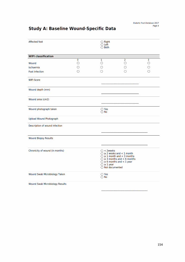
154

155

156

157

158

159

160

161
Appendix 2: Data Dictionary
The following fields should be completed by the treating surgeon. Most fields are mandatory. Those that are not have been assigned an asterisk; however, where possible this information should also be provided.
Baseline Clinical Data
Date of Birth
Identifying and definitional attributes Definition: The day, month and year of a person’s birth
Context: To accurately calculate age
Guide for use
Obligation: Mandatory
How to answer on form: Enter day, month and year (dd/mm/yyyy)
Gender
Identifying and definitional attributes Definition: Whether patient is male or female
Context: The patient’s gender is an important demographic indicator; generally men outnumber women
Guide for use
Obligation: Mandatory
How to answer on form: Tick appropriate box
Race
Identifying and definitional attributes Definition: Ethnic group of the patient, as selected by the patient
Context: The patient’s race is an important demographic indicator as the prevalence of Diabetes varies according to race/ethnic background
Guide for use
Obligation: Mandatory
How to answer on form: Tick appropriate box
Duration of diabetes
Identifying and definitional attributes Definition: How many years since participant was diagnosed with diabetes
Context: Duration of diabetes is an important prognostic indicator
Guide for use
Obligation: Mandatory
How to answer on form: Enter number of years since the diagnosis of diabetes

162
Diabetes control strategy Identifying and definitional attributes
Definition: Treatment strategy for diabetes at baseline – may include no treatment, diet; oral medications or Insulin
Context: Management strategy for diabetes is an important indicator of severity of the Disease
Guide for use
Obligation: Mandatory
How to answer on form: Tick appropriate box
Most recent HbA1C measurement Identifying and definitional attributes
Definition: HbA1c is glycosylated haemoglobin and reflects the average blood glucose over the lifespan of the red blood cells containing it
Context: HbA1c test shows an average of your blood glucose level over the past 10 –12 weeks
Guide for use
Obligation: Optional
How to answer on form: Enter value of HbA1C (%)
Date of most recent HbA1C test Identifying and definitional attributes Definition: Date when the most recent HbA1c test was performed
Context: To assess need for repeat HbA1C test
Guide for use
Obligation: Optional
How to answer on form: Enter date of the most recent HbA1C test
Height
Identifying and definitional attributes Definition: Height of person measured in centimetres
Context: Patient height will be used to calculate BMI and to help calculate glomerular filtration rate
Guide for use
Obligation: Mandatory
How to answer on form: Enter height of patient in whole centimetres (cm)
Weight
Identifying and definitional attributes Definition: Weight of person measured in kilograms
Context: Patient height will be used to calculate BMI and to help calculate glomerular filtration rate
Guide for use
Obligation: Mandatory
How to answer on form: Enter weight of patient in whole kilograms (KG)
Smoking status

163
Identifying and definitional attributes
Whether or not the patient smokes, or has ever smoked. Divided into following categories: never, ceased >12 months ago, ceased within last 12 months or current
Definition:
Context: Smoking status is a strong predictor of wound healing and presence of peripheral vascular disease
Guide for use
Obligation: Mandatory
How to answer on form: Tick appropriate box
Pack years if current or past smoker Identifying and definitional attributes Pack year describes the number of cigarettes a person has smoked over time.
Definition: One pack year is defined as 20 manufactured cigarettes (one pack) smoked
per day for one year
Context: To help determine how predictive smoking is for outcome
Guide for use
Obligation: Optional
How to answer on form:
Example: If patient smoked 1.5 packs per day for 26 years, this would equal 39 pack years: 1 pack per day x 26 years = 26 pack years
0.5 pack per day x 26 years = 13 pack years
26 pack years + 13 pack years = 39 pack years
Enter calculated number
History of Stroke/TIA Identifying and definitional attributes Definition: If patient has previously had a stroke/TIA
Context: Positive history for Stroke/TIA may be a marker for arterial disease and influence wound healing
Guide for use
Obligation: Mandatory
How to answer on form: Tick appropriate box

164
Cardiac assessment Identifying and definitional attributes Series of symptoms associated with cardiac fitness:
1. Asymptomatic with normal ECG
2. Asymptomatic but with MI>6months ago
Occult MI on ECG
Fixed deficit on stress test (tetrafosmin (TF) / dobutamine stress echo (DSE)
3. Any one of :
Stable angina
No angina but reversible perfusion defect on TF or DSE
Definition: Significant silent ischaemia (1% of time) on Holter monitoring
Ejection fraction (EF) 25-45%
Controlled ectopy or asymptomatic arrhythmia
History of congestive heart failure (CHF) which is now controlled
4. Any of :
Unstable angina
Symptomatic or poorly controlled arrhythmia
Poorly controlled or recurrent CHF
EF<25%; MI within 6 months
5. Unknown
Context: To determine whether cardiac status is correlated with outcomes in
Patients with diabetic foot ulcers
Guide for use
Obligation: Mandatory
How to answer on form: Tick one box

165
Respiratory assessment Identifying and definitional attributes Series of symptoms associated with respiratory fitness:
1. Asymptomatic
Normal CXR
Pulmonary function test (PFTs) more than 80% predicted
2. Asymptomatic or mild exertional dyspnoea
Mild parenchymal changes
PFTs 65-80% predicted
3. Between 2 and 4
Definition: 4. Any of:
Dyspnoeic at rest of minimal exercise
Vital capacity less than 1.85 litres
FEV1<1.2litres or <35% of predicted maximal
Voluntary ventilation <50% predicted
pCO2>45mmHg
Supplemental O2 use necessary
Pulmonary hypertension
5. Unknown
Context: To determine whether respiratory status is correlated with outcomes in
Patients with diabetic foot ulcers
Guide for use
Obligation: Mandatory
How to answer on form: Tick one box
Previous minor amputation Identifying and definitional attributes Definition: If patient has previously had toes/forefoot amputation
Context: Minor amputation alters the biomechanics of the foot and is a prognostic factor for further amputation
Guide for use
Obligation: Mandatory
How to answer on form: Tick one box
If answer is yes, enter details about previous minor amputation (date; which side and which toes)
Previous major amputation Identifying and definitional attributes
Definition: If patient has previously had amputation above the ankle level. (this includes below knee amputation; through knee amputation; above knee amputation or hip disarticulation
Context: Major amputation is a prognostic factor for further amputation
Guide for use
Obligation: Mandatory
How to answer on form: Tick appropriate box
If answer is yes, enter details about previous major amputation (date; which side and which operation was performed)

166
Antiplatelet
Identifying and definitional attributes
Definition: Whether the patient has been prescribed a type of medication generically referred to as antiplatelet
Context: Diabetic patients, especially when associated with vascular disease, have
increased cardiovascular risk and antiplatelets are known to reduce overall cardiovascular mortality
Guide for use
Obligation: Mandatory
How to answer on form: Tick appropriate box If answer is yes, enter details about antiplatelet/s prescribed
Statin
Identifying and definitional attributes
Definition: Whether the patient has been prescribed medication known as statins to lower Blood cholesterol level
Context: Patients with peripheral vascular disease have reduced overall mortality If they are on statin
Guide for use
Obligation: Mandatory
How to answer on form: Tick appropriate box
Anti-hypertensive Identifying and definitional attributes Definition: Whether the patient has been prescribed medication/s to control high blood
Context: The combination of diabetes and hypertension places patients at significantly increased risk of cardiovascular disease.
Guide for use
Obligation: Mandatory
How to answer on form: Tick appropriate box If answer is yes, enter details about anti-hypertensive/s prescribed
Steroid/Immunosuppressant Identifying and definitional attributes Definition: Whether the patient has been prescribed steroid and/or immunosuppressant
Context: Steroid and immunosuppressant medications can significantly impair wound healing.
Guide for use
Obligation: Mandatory
How to answer on form: Tick appropriate box If answer is yes, enter details about steroid/immunosuppressant prescribed

167
Other medications Identifying and definitional attributes List of regular medications the patient has been prescribed for treatment of
of a range of conditions including various heart disease, respiratory conditions and diabetes
Definition:
Context Medication history is an important data for patients with diabetic foot ulcers as some medications may affect wound healing and affect overall mortality.
Guide for use
Obligation: Optional
How to answer on form: Enter list of regular medications and dosages

168
Baseline wound specific data
Affected foot Identifying and definitional attributes Definition: A record of in which foot an ulcer is present
Context: Determining which foot is affected is essential for data analysis
Guide for use
Obligation: Mandatory
How to answer on form: Thick appropriated box
WIfI classification
Identifying and definitional attributes
The WiFI is new classification system created by Society for Vascular Surgery that focuses on threatened limb, including patients with PAD and diabetes.
Is
Definition:
In the WIfI classification system three clinical entities are evaluated and stratified/graded from 0 to 3 These include (1) Wound (2) Ischaemia and (3) Foot Infection.
Wounds are given a severity grade based on size, depth, and anticipated difficult achieving wound healing. In regards to ischaemia patients are graded based on their perfusion (preferably using toe pressure). Infection is graded based on severity. The overall WiFI score estimate patient risk of major amputation at 1 year and also the likelihood of benefit of/requirement for revascularisation
Context: The WIfI classification is an important prognostic factor in patients with diabetic foot ulcers as it can provide an estimate risk of amputation and possibly aid in determining whether a patient would benefit from revascularisation
Guide for use
Obligation: Mandatory
How to answer on form: Thick one box for each of the clinical entities assessed (Wound / Ischaemia / Foot infection)

169
WIfI score
Identifying and definitional attributes
The WiFI is new classification system created by Society for Vascular Surgery that focuses on threatened limb, including patients with PAD and diabetes.
Definition: In the WIfI classification system three clinical entities are evaluated and stratified/graded from 0 to 3 These include (1) Wound (2) Ischaemia and (3) Foot Infection. The overall WiFI score is a combination of the scores for each of the clinical entities evaluated
Context: The WIfI classification is an important prognostic factor in patients with diabetic foot ulcers as it can provide an estimate risk of amputation and possibly aid in determining whether a patient would benefit from revascularisation
Guide for use
Obligation: Mandatory How to answer on form: Enter the overall WIfI score
Example: A patient with dry gangrene of 2 toes and a <2cm rim of cellulitis at the base of the toes, but without systemic or metabolic toxicity and toe pressure of 35mmHg would be classified as Wound 2; Ischaemia 2; Foot Infection 1. The overall WIfI score is 221
Wound depth
Identifying and definitional attributes Definition: Measurement of deepest part of visible wound bed to skin level
Context: Wound depth and area are prognostic factor for wound healing
Guide for use
Obligation: Mandatory
How to answer on form: Enter wound depth of the wound in mm
Wound area
Identifying and definitional attributes Definition: Measurement of the area of the visible wound bed
Context: Wound depth and area are prognostic factor for wound healing
Guide for use
Obligation: Mandatory
How to answer on form: Enter wound area of the wound bed in cm2
Wound photograph
Identifying and definitional attributes Definition: Photo of the wound
Context: Photos of the wound allows for objective assessment of progress of the wound over time
Guide for use
Obligation: Optional
How to answer on form: Attach photo of the wound

170
Wound description
Identifying and definitional attributes
Definition:
Open documentation of wound characteristics including description of exsudate type (e.g.: serous, serosanguineous, purulent) and amount; characteristics of wound bed (e.g: slough, eschar, granulation tissue, epithelization); description of wound edges (defined or undefined edges, macerated, fibrotic, callused) and ad surrounding tissues
Context: Wound characteristics are important predictor of wound healing
Guide for use
Obligation: Optional
How to answer on form: Enter open description of the wound
Wound swab microbiology results
Identifying and definitional attributes
Definition: Collection of exudate for pathology laboratory culture, identification and sensitivity of infecting microorganisms.
Context:
Microbiological assessment is important in the management of infected wounds. Results of wound swab is useful for determining antibiotic choice and predicting response to treatment. Deep swab that avoid collection of surface contaminants is preferred.
Guide for use
Obligation: Optional
How to answer on form: Enter results of recent significant results from wound swab.
Chronicity of wound Identifying and definitional attributes Definition: Time from establishment of foot ulcer until patient recruitment
Context:
Time from establishment of foot ulcer is an important prognostic factor. An acute wound is expected to progress through the phases of normal healing, resulting in closure of the wound. A chronic wound fails to progress or respond to treatment over normal expected healing time frame (4 weeks) and becomes “stuck” in the inflammatory phase. Wound chronicity is attributed to the presence of intrinsic and extrinsic factors including medications, poor nutrition, comorbidities and inappropriate dressings
Guide for use
Obligation: Mandatory
How to answer on form: Thick appropriate box
Foot pulse palpable
Identifying and definitional attributes Definition: A record of whether the foot pulses are present or absent
Context: The presence or absence of pulses can be indicative of arterial Impairment and it is an important prognostic factor for wound healing.
Guide for use
Obligation: Mandatory
How to answer on form: Tick whether right foot pulse and left foot pulses are individually palpable

171
Ankle brachial index (ABI)
Identifying and definitional attributes
Ratio of blood pressure in the lower legs to blood pressure in arms. Lower blood pressure in the leg is symptomatic of blocked arteries. It is calculated by dividing the systolic blood pressure in the arteries at the ankle by the highest systolic blood pressure reading for the arm. An ABI of 1.0-1.3 is normal.
Definition:
Context: Low ABI can be indicative of arterial impairment and may be an important Prognostic factor for wound healing
Guide for use
Obligation: Optional
How to answer on form: Enter ABI for both right and left ankles
Toe pressure Identifying and definitional attributes Toe pressures use an infrared photoplethysmography sensor to determine the
assessment where falsely high ankle pressures can occur due to calcification. small
vessel vascular condition distal to the ankle. Toe pressures may be useful in cases of suspected vascular disease and in baseline diabetic foot assessment where falsely high ankle pressures can occur due to calcification. Healing unlikely if toe pressure below 45mmHg.
Definition:
Context: Low toe pressure is indicative of arterial impairment and may be an important Prognostic factor for wound healing
Guide for use
Obligation: Optional
How to answer on form: Enter toe pressure both right and left foot

172
Frailty
Unintended weigh loss in the last 6months
Identifying and definitional attributes Definition: A record of whether patient had unintended weigh loss in the previous six months
Context:
Frailty as a biologic syndrome of decreased reserve and resistance to stressors, resulting from cumulative declines across multiple physiologic systems, and causing vulnerability to adverse outcomes. Frailty is considered to be a high-risk state predictive of a range of adverse health outcomes and it may be an important factor in wound healing. Unintended weight loss is one the components of frailty phenotype.
Guide for use
Obligation: Mandatory
How to answer on form: Tick appropriate box and enter the amount of weight loss in the last 6 months.
Dominant hand Identifying and definitional attributes Definition: Preference for using one hand over the other to perform fine and gross motor tasks
Context: To determine in which hand grip strength should be performed
Guide for use
Obligation: Mandatory How to answer on form: Thick appropriate box
Grip strength
Identifying and definitional attributes
Definition: Grip strength is an anthropometric measurement that indicates muscle health in the hands and forearms. Hand grip strength is measured by a hand-held dynamometer
Context: Grip strength is a reliable surrogate for overall muscle strength and predicts functional decline. Muscle weakness is one of the components of frailty phenotype.
Guide for use
Obligation: Mandatory How to answer on form: Enter the average of three grip strength measurement for right and left hand and
indicate which hand is dominant.
Total muscle psoas area Identifying and definitional attributes
Definition: Cross sectional area of the right and left psoas muscle measured using CT scan image at the bottom of L3 vertebra level.
Context: Low total psoas normalise by patient height is a surrogate for muscle depletion (sarcopenia). Sarcopenia is one of the components of frailty phenotype
Guide for use
Obligation: Optional (if CT available)
How to answer on form: Enter the value of total psoas area in mm2
Self-report fitness – Distance Identifying and definitional attributes Definition: Whether the patient can walk briskly (5 or more kilometres per hour) for 1km

173
Context: Question aims to provide additional information about physical condition of
patient Low physical activity and exhaustion are components of frailty phenotype
Guide for use
Obligation: Optional
How to answer on form: Tick appropriate box
Self-report fitness – Climb Identifying and definitional attributes Definition: Whether the patient can climb 2 flight of stairs without stopping
Context: Question aims to provide additional information about physical condition of
patient Low physical activity and exhaustion are components of frailty phenotype
Guide for use
Obligation: Optional
How to answer on form: Tick appropriate box

174
Investigation and initial management
Glucose
Identifying and definitional attributes
Definition: The concentration of glucose in the fasting blood, represented in millimole per litre
Context: To provide additional information about diabetes control
Guide for use
Obligation: Mandatory
How to answer on form: Enter number to one decimal place
Total Cholesterol Identifying and definitional attributes
Definition: The concentration of total cholesterol in the fasting blood, represented in millimole per litre
Context: To provide additional information about risk factors for cardiovascular disease
Guide for use
Obligation: Mandatory
How to answer on form: Enter number to one decimal place
Triglycerides Identifying and definitional attributes
Definition: The concentration of triglycerides in the fasting blood, represented in millimole per litre
Context: To provide additional information about risk factors for cardiovascular disease
Guide for use
Obligation: Mandatory
How to answer on form: Enter number to one decimal place
HDL Identifying and definitional attributes
Definition: The concentration of high-density lipoproteins (HDL) in the fasting blood, represented in millimole per litre
Context: To provide additional information about risk factors for cardiovascular disease
Guide for use
Obligation: Mandatory
How to answer on form: Enter number to one decimal place

175
LDL Identifying and definitional attributes
Definition: The concentration of low-density lipoproteins (LDL) in the fasting blood, represented in millimole per litre
Context: To provide additional information about risk factors for cardiovascular disease
Guide for use
Obligation: Mandatory
How to answer on form: Enter number to one decimal place
CRP Identifying and definitional attributes
Definition: The concentration of C-reactive protein (CRP), represented in miligram per litre. It is an acute-phase protein of hepatic origin and it is used mainly as a marker of inflammation
Context: To provide additional information about the severity of infection in the foot ulcer
Guide for use
Obligation: Mandatory
How to answer on form: Enter number to one decimal place
Creatinine
Identifying and definitional attributes Definition: A person’s blood serum creatinine level measured in micromoles per litre
Context:
Creatinine is a break down product of creatinine phosphate from muscle and is produced at a constant rate in the body. Measurements of serum creatinine are used as a common indicator of renal function. Poor renal function is related impaired wound healing
Guide for use
Obligation: Mandatory
How to answer on form: Enter number to one decimal place
HbA1C Identifying and definitional attributes
Definition: HbA1c is glycosylated haemoglobin and reflects the average blood glucose over the lifespan of the red blood cells containing it
Context: HbA1c test shows an average of your blood glucose level over the past 10–12 weeks
Guide for use
Obligation: Mandatory
How to answer on form: Enter value of HbA1C (%)

176
Vitamin A Identifying and definitional attributes
Definition: The concentration of vitamin A in the blood, represented in micromole per litre. Vitamin A is an essential nutrient required for healthy vision, skin growth and integrity, bone formation, immune function
Context: To provide information about nutritional status
Guide for use
Obligation: Mandatory
How to answer on form: Enter number to one decimal place
Vitamin B6 Identifying and definitional attributes
Definition: The concentration of vitamin B6 in the blood, represented in nanograms per millilitre
Context: To provide information about nutritional status
Guide for use
Obligation: Mandatory
How to answer on form: Enter number to one decimal place
Vitamin B12 Identifying and definitional attributes Definition: The concentration of vitamin B12 in the blood, represented in picomoles per litre
Context: To provide information about nutritional status
Guide for use
Obligation: Mandatory
How to answer on form: Enter number to one decimal place
Vitamin C Identifying and definitional attributes Definition: The concentration of vitamin C in the blood, represented in milligrams per decilitre
Context: To provide information about nutritional status
Guide for use
Obligation: Mandatory
How to answer on form: Enter number to one decimal place
Vitamin D Identifying and definitional attributes Definition: The concentration of vitamin D in the blood, represented in nanograms per millilitre
Context: To provide information about nutritional status
Guide for use
Obligation: Mandatory
How to answer on form: Enter number to one decimal place

177
Vitamin E Identifying and definitional attributes Definition: The concentration of vitamin E in the blood, represented in nanograms per millilitre
Context: To provide information about nutritional status
Guide for use
Obligation: Mandatory
How to answer on form: Enter number to one decimal place
Cooper Identifying and definitional attributes Definition: The concentration of cooper in the blood, represented in micrograms per decilitre
Context: To provide information about nutritional status
Guide for use
Obligation: Mandatory
How to answer on form: Enter number to one decimal place
Zinc Identifying and definitional attributes Definition: The concentration of vitamin B6 in the blood, represented in micromole per litre
Context: To provide information about nutritional status
Guide for use
Obligation: Mandatory
How to answer on form: Enter number to one decimal place
Serum iron Identifying and definitional attributes Definition: The concentration of iron in the blood, represented in micromole per litre
Context: To provide information about nutritional status
Guide for use
Obligation: Mandatory
How to answer on form: Enter number to one decimal place

178
Ferritin Identifying and definitional attributes Definition: The concentration of ferritin in the blood, represented in micrograms per litre
Context: To provide information about nutritional status
Guide for use
Obligation: Mandatory
How to answer on form: Enter number to one decimal place
Transferrin Identifying and definitional attributes Definition: The concentration of transferrin in the blood, represented in micromoles per litre.
Context: To provide information about nutritional status
Guide for use
Obligation: Mandatory
How to answer on form: Enter number to one decimal place
Transferrin saturation Identifying and definitional attributes Definition: The transferrin saturation in the blood, represented in percentage.
Context: To provide information about nutritional status
Guide for use
Obligation: Mandatory
How to answer on form: Enter number to one decimal place
Folate Identifying and definitional attributes Definition: The concentration of folate in the blood, represented in micrograms per litre
Context: To provide information about nutritional status
Guide for use
Obligation: Mandatory
How to answer on form: Enter number to one decimal place
Imaging ordered Identifying and definitional attributes
Definition: The modality of imaging test ordered for assessment of the patient with diabetic foot ulcer
Context: To provide important prognostic information as imaging test is used to assess for presence of deep infection (including osteomyelitis) and presence of peripheral vascular disease
Guide for use
Obligation: Mandatory
How to answer on form: Tick appropriate box

179
Osteomyelitis on imaging test Identifying and definitional attributes Definition: Presence or absence of features of osteomyelitis on imaging test
Context: To provide important prognostic information. Presence of osteomyelitis is related to poor wound healing.
Guide for use
Obligation: Mandatory
How to answer on form: Tick appropriate box
Comment on imaging test Identifying and definitional attributes
Definition: Open description of the results of the imaging test ordered for assessment of the patient with diabetic foot ulcer
Context: To provide important prognostic information as imaging test is used to assess for presence of deep infection (including osteomyelitis) and presence of peripheral vascular disease.
Guide for use
Obligation: Optional
How to answer on form: Enter details about results of the imaging tests ordered
Off-loading strategy prescribed Identifying and definitional attributes Definition: Whether off-loading strategy was prescribed for management
Context: Effective offloading of diabetic feet is important for ulcer healing and the prevention of ulcer recurrence
Guide for use
Obligation: Mandatory
How to answer on form: Tick appropriate box
Details of off-loading strategy Identifying and definitional attributes Definition: Modality of off-loading strategy employed
Context: Effective offloading of diabetic feet is important for ulcer healing and the prevention of ulcer recurrence
Guide for use
Obligation: Mandatory
How to answer on form: Tick appropriate box If answer is ‘other’, enter details about off-loading strategy employed

180
Antibiotics prescribed Identifying and definitional attributes Definition: Whether antibiotic was prescribed for management of diabetic foot ulcer infection
Context: Foot infections are common in patients with diabetes and are associated with high morbidity and risk of lower extremity amputation. Optimal management of foot infection includes antibiotic therapy
Guide for use
Obligation: Mandatory
How to answer on form: Tick appropriate box
Route of antibiotics administration Identifying and definitional attributes Definition: Whether antibiotic was prescribed orally or via intravenous
Context:
Foot infections are common in patients with diabetes and are associated with high morbidity and risk of lower extremity amputation. Optimal management of foot infection includes antibiotic therapy. Route of administration of antibiotics is related to severity of the infection
Guide for use
Obligation: Optional
How to answer on form: Tick appropriate box
Details of antibiotic treatment Identifying and definitional attributes
Definition: Open description of details of antibiotic treatment including name of antibiotic and duration of therapy
Context:
Foot infections are common in patients with diabetes and are associated with high morbidity and risk of lower extremity amputation. Optimal management of foot infection includes antibiotic therapy. Appropriate antibiotic choice that cover the most common pathogens is essential
Guide for use
Obligation: Optional
How to answer on form: Enter name of antibiotic/s prescribed and duration of therapy
Other medication changes Identifying and definitional attributes Definition: Open description of medication changes
Context: Medication history is an important data for patients with diabetic foot ulcers as some medications may affect wound healing and affect overall mortality.
Guide for use
Obligation: Optional
How to answer on form: Enter medication changes made

181
Admission to hospital Identifying and definitional attributes Definition: Whether the patient was admitted to hospital as part of initial management
Context: Admission to hospital is related with severity if the condition. Patient with diabetic foot ulcers and severe infection often required surgical interventions and intravenous antibiotic therapy.
Guide for use
Obligation: Mandatory
How to answer on form: Thick appropriate box
Arterial intervention Identifying and definitional attributes
Definition: Whether the patient underwent arterial intervention to improve blood flow to the affected foot for management of the current condition.
Context:
Diabetic foot ulcers are typically multifactorial in origin. Neuropathy of the foot and impaired wound healing are frequently associated with peripheral arterial disease. When significant ischemia is present, diabetic foot ulcers require arterial revascularisation to achieve wound healing
Guide for use
Obligation: Mandatory
How to answer on form: Thick appropriate box
Type of arterial intervention Identifying and definitional attributes Definition: The modality of arterial intervention that the patient underwent
Context:
Diabetic foot ulcers are typically multifactorial in origin. Neuropathy of the foot and impaired wound healing are frequently associated with peripheral arterial disease. When significant ischemia is present, diabetic foot ulcers require arterial revascularisation to achieve wound healing. The modality of arterial intervention is related with severity and pattern of arterial disease.
Guide for use
Obligation: Optional
How to answer on form: Thick appropriate box
Details of arterial intervention Identifying and definitional attributes Definition: Open description of arterial intervention
Context:
Diabetic foot ulcers are typically multifactorial in origin. Neuropathy of the foot and impaired wound healing are frequently associated with peripheral arterial disease. When significant ischemia is present, diabetic foot ulcers require arterial revascularisation to achieve wound healing. The modality of arterial intervention is related with severity and pattern of arterial disease.
Guide for use
Obligation: Optional
How to answer on form: Enter name of the revascularisation procedure

182
Wound management Identifying and definitional attributes Definition: Strategy employed for wound management
Context:
Optimising local wound care with wound bed preparation is important component of management of diabetic foot ulcer, Wound bed preparation can be achieved via regular dressing, sharp debridement in ward/OPD or sharp debridement in surgical theatre. .
Guide for use
Obligation: Optional
How to answer on form: Thick appropriate box
Negative pressure wound therapy applied Identifying and definitional attributes
Definition: Whether negative pressure wound therapy (NPWT) was applied as part of wound management
Context:
To provide additional information about wound management. The NPWT wound dressing assists in healing open wounds. The effects of NPWT that accelerate wound healing are reported as increased local blood flow, formation of granulation tissue, and decreased bacterial colonization.
Guide for use
Obligation: Mandatory
How to answer on form: Thick appropriate box
Wound management details Identifying and definitional attributes Definition: Open description of wound management strategy
Context: To provide additional information about wound management.
Guide for use
Obligation: Optional
How to answer on form: Enter details about wound management strategy
Surgical procedure Identifying and definitional attributes
Definition:
Surgical procedure performed. Divided into following categories: Surgical debridement; Minor amputation – toe/s; Minor amputation – forefoot; Major amputation – guillotine above ankle; Major amputation – BKA; Major amputation – AKA.
Context:
Surgical therapy is an important aspect of patients with diabetic foot. The main goal is to control the deep infection and preserve life, with the hope of salvaging the limb. This is accomplished by drainage of any pus, removal of all necrotic or infected tissues. Surgical procedure may be an indication of severity of the infection and also has prognostic value. Residual foot deformities may lead to abnormal pressure points and, thus, reulceration.
Guide for use
Obligation: Mandatory
How to answer on form: Thick appropriate box

183
Details of surgical procedure Identifying and definitional attributes Definition: Open description of surgical procedure performed.
Context: To provide further information about management of the foot ulcer and it also has prognostic value
Guide for use
Obligation: Optional
How to answer on form: Enter details about surgical procedure
Duration of hospital stay Identifying and definitional attributes
Definition: The period of time (in days) that the patient remained in the hospital as an inpatient.
Context: This is an important information about the burden of diabetic foot ulcers imposes on the healthcare system.
Guide for use
Obligation: Optional
How to answer on form: Enter hospital stay in days
Discharge destination Identifying and definitional attributes
Definition: Destination after discharge from the hospital. Patient can either returns home or be transferred to another facility such as one for rehabilitation or to a nursing home
Context: This is an important information about the burden of diabetic foot ulcers imposes on patients and the healthcare system.
Guide for use
Obligation: Optional
How to answer on form: Thick appropriate box

184
Follow up data
Date of follow up data collection
Identifying and definitional attributes Definition: The day, month and year of follow up data collection
Context: To accurately store follow up data
Guide for use
Obligation: Mandatory
How to answer on form: Enter day, month and year (dd/mm/yyyy)
Patient deceased
Identifying and definitional attributes Definition: Whether patient is alive or not at the time of
Context: Mortality is one of the outcome of interest in the research
Guide for use
Obligation: Mandatory
How to answer on form: Tick appropriate box
Cause of death
Identifying and definitional attributes Definition: Open description of the cause of death
Context: Mortality is one of the outcomes of interest. Assessing cause of death is important to determine is death was directly related to diabetic foot disease
Guide for use
Obligation: Optional
How to answer on form: Enter details about cause of death
Patient seen for follow up
Identifying and definitional attributes Definition: Weather participant was seen in person for follow up
Context: To provide information on how follow up data was obtained
Guide for use
Obligation: Mandatory
How to answer on form: Tick appropriate box

185
Weeks post recruitment Identifying and definitional attributes Definition: Time from recruitment until follow up in weeks
Context: This is important information because the findings of this study will be extracted from the time frame in which most of the subjects have had the event or have remained under observation
Guide for use
Obligation: Mandatory
How to answer on form: Enter number of weeks since recruitment
Admission/Readmission to hospital since recruitment Identifying and definitional attributes Definition: Whether the patient was admitted to hospital since recruitment
Context: Admission to hospital is related with severity if the condition. Patient with diabetic foot ulcers and severe infection often required surgical interventions and intravenous antibiotic therapy.
Guide for use
Obligation: Mandatory
How to answer on form: Thick appropriate box
Description of Admission(s) since description Identifying and definitional attributes Definition: Open description of the admissions to hospital since recruitment
Context: Description of the admission allow to assess if the admission is related to diabetif foot disease
Guide for use
Obligation: Optional
How to answer on form: Enter reason for the patient to be admitted to the hospital and relevant dates
Days in hospital Identifying and definitional attributes
Definition: The period of time (in days) that the patient remained in the hospital as an inpatient.
Context: This is an important information about the burden of diabetic foot ulcers imposes on the healthcare system.
Guide for use
Obligation: Optional
How to answer on form: Enter hospital stay in days

186
Discharge destination Identifying and definitional attributes
Definition: Destination after discharge from the hospital. Patient can either returns home or be transferred to another facility such as one for rehabilitation or to a nursing home
Context: This is an important information about the burden of diabetic foot ulcers imposes on patients and the healthcare system.
Guide for use
Obligation: Optional
How to answer on form: Thick appropriate box
Surgical procedure since last assessment Identifying and definitional attributes
Definition: Whether the patient underwent local surgical intervention for management of diabetic foot disease.
Context:
Surgical therapy is an important aspect of patients with diabetic foot. The main goal is to control the deep infection and preserve life, with the hope of salvaging the limb. This is accomplished by drainage of any pus, removal of all necrotic or infected tissues. Surgical procedure may be an indication of severity of the infection and also has prognostic value. Residual foot deformities may lead to abnormal pressure points and, thus, reulceration.
Guide for use
Obligation: Mandatory
How to answer on form: Thick appropriate box
Type of surgical procedure since last assessment Identifying and definitional attributes
Definition:
Surgical procedure performed. Divided into following categories: Surgical debridement; Minor amputation – toe/s; Minor amputation – forefoot; Major amputation – guillotine above ankle; Major amputation – BKA; Major amputation – AKA.
Context:
Surgical therapy is an important aspect of patients with diabetic foot. The main goal is to control the deep infection and preserve life, with the hope of salvaging the limb. This is accomplished by drainage of any pus, removal of all necrotic or infected tissues. Surgical procedure may be an indication of severity of the infection and also has prognostic value. Residual foot deformities may lead to abnormal pressure points and, thus, reulceration.
Guide for use
Obligation: Optional
How to answer on form: Thick appropriate box

187
Details of surgical procedure Identifying and definitional attributes Definition: Open description of surgical procedure performed.
Context: To provide further information about management of the foot ulcer and it also has prognostic value
Guide for use
Obligation: Optional
How to answer on form: Enter details about surgical procedure
Arterial intervention since last assessment Identifying and definitional attributes
Definition: Whether the patient underwent arterial intervention to improve blood flow to the affected foot for management of the current condition.
Context:
Diabetic foot ulcers are typically multifactorial in origin. Neuropathy of the foot and impaired wound healing are frequently associated with peripheral arterial disease. When significant ischemia is present, diabetic foot ulcers require arterial revascularisation to achieve wound healing
Guide for use
Obligation: Mandatory
How to answer on form: Thick appropriate box
Type of arterial intervention Identifying and definitional attributes Definition: The modality of arterial intervention that the patient underwent
Context:
Diabetic foot ulcers are typically multifactorial in origin. Neuropathy of the foot and impaired wound healing are frequently associated with peripheral arterial disease. When significant ischemia is present, diabetic foot ulcers require arterial revascularisation to achieve wound healing. The modality of arterial intervention is related with severity and pattern of arterial disease.
Guide for use
Obligation: Optional
How to answer on form: Thick appropriate box
Has the original ulcer healed Identifying and definitional attributes Definition: Weather the target foot ulcer/wound has healed
Context: Wound healing is one of the outcomes of interest of the research
Guide for use
Obligation: Mandatory
How to answer on form: Thick appropriate box

188
Wound depth Identifying and definitional attributes Definition: Measurement of deepest part of visible wound bed to skin level in mm
Context: Wound depth and area are prognostic factor for wound healing
Guide for use
Obligation: Mandatory
How to answer on form: Enter wound depth of the wound in mm
Wound area
Identifying and definitional attributes Definition: Measurement of the area of the visible wound bed
Context: Wound depth and area are prognostic factor for wound healing
Guide for use
Obligation: Mandatory
How to answer on form: Enter wound area of the wound bed in cm2
Wound photograph Identifying and definitional attributes Definition: Photo of the wound
Context: Photos of the wound allows for objective assessment of progress of the wound over time
Guide for use
Obligation: Optional
How to answer on form: Attach photo of the wound
Presence of wound infection? Identifying and definitional attributes
Definition: Whether there are signs and symptoms of infection related to the target ulcer/wound
Context: To provide additional information about status of the target ulcer/wound. Wound infection impairs wound healing.
Guide for use
Obligation: Mandatory
How to answer on form: Thick appropriate box
Severity of wound infection (as per WIfI) Identifying and definitional attributes Definition: Description of the severity of wound infection according to the WIfI criteria.
Context: Wound infection is an important prognostic factor in patients with diabetic foot ulcers
Guide for use
Obligation: Optional How to answer on form: Thick appropriate box

189
Wound swab microbiology Identifying and definitional attributes
Definition: Collection of exudate for pathology laboratory culture, identification and sensitivity of infecting microorganisms.
Context:
Microbiological assessment is important in the management of infected wounds. Results of wound swab is useful for determining antibiotic choice and predicting response to treatment. Deep swab that avoid collection of surface contaminants is preferred.
Guide for use
Obligation: Optional
How to answer on form: Enter results of recent significant results from wound swab.
Change in off-loading strategy Identifying and definitional attributes Definition: Weather there has been any changes in off-loading strategy since last assessment
Context: Effective offloading of diabetic feet is important for ulcer healing and the prevention of ulcer recurrence
Guide for use
Obligation: Mandatory
How to answer on form: Thick appropriate box
Details of off-loading strategy Identifying and definitional attributes Definition: Modality of off-loading strategy employed
Context: Effective offloading of diabetic feet is important for ulcer healing and the prevention of ulcer recurrence
Guide for use
Obligation: Mandatory
How to answer on form: Tick appropriate box If answer is ‘other’, enter details about off-loading strategy employed
Antibiotics prescribed/currently on antibiotics? Identifying and definitional attributes
Definition: Whether participant is on antibiotic therapy or weather antibiotic was prescribed for management of diabetic foot ulcer infection
Context: Foot infections are common in patients with diabetes and are associated with high morbidity and risk of lower extremity amputation. Optimal management of foot infection includes antibiotic therapy
Guide for use
Obligation: Mandatory
How to answer on form: Tick appropriate box
190
Details of antibiotic treatment Identifying and definitional attributes
Definition: Open description of details of antibiotic treatment including name of antibiotic and duration of therapy
Context:
Foot infections are common in patients with diabetes and are associated with high morbidity and risk of lower extremity amputation. Optimal management of foot infection includes antibiotic therapy. Appropriate antibiotic choice that cover the most common pathogens is essential
Guide for use
Obligation: Optional
How to answer on form: Enter name of antibiotic/s prescribed and duration of therapy
Imaging ordered/Recent imaging Identifying and definitional attributes
Definition: The modality of imaging test ordered or recently performed for assessment of the patient with diabetic foot ulcer
Context: To provide important prognostic information as imaging test is used to assess for presence of deep infection (including osteomyelitis) and presence of peripheral vascular disease
Guide for use
Obligation: Mandatory
How to answer on form: Tick appropriate box
Osteomyelitis on imaging test Identifying and definitional attributes Definition: Presence or absence of features of osteomyelitis on imaging test
Context: To provide important prognostic information. Presence of osteomyelitis is related to poor wound healing.
Guide for use
Obligation: Mandatory
How to answer on form: Tick appropriate box
Comment on imaging test Identifying and definitional attributes
Definition: Open description of the results of the imaging test ordered for assessment of the patient with diabetic foot ulcer
Context: To provide important prognostic information as imaging test is used to assess for presence of deep infection (including osteomyelitis) and presence of peripheral vascular disease.
Guide for use
Obligation: Optional
How to answer on form: Enter details about results of the imaging tests ordered

191
ABI performed Identifying and definitional attributes
Wheater ankle-brachial index has been performed since last assessment
Definition:
Context: Low ABI can be indicative of arterial impairment and may be an important Prognostic factor for wound healing
Guide for use
Obligation: Mandatory
How to answer on form: Tick appropriate box
ABI (right and left foot) Identifying and definitional attributes
Ratio of blood pressure in the lower legs to blood pressure in arms. Lower blood pressure in the leg is symptomatic of blocked arteries. It is calculated by dividing the systolic blood pressure in the arteries at the ankle by the highest systolic blood pressure reading for the arm. An ABI of 1.0-1.3 is normal.
Definition:
Context: Low ABI can be indicative of arterial impairment and may be an important Prognostic factor for wound healing
Guide for use
Obligation: Optional
How to answer on form: Enter ABI for both right and left ankles
Toe pressure performed
Identifying and definitional attributes
Wheater ankle-brachial index has been performed since last assessment
Definition:
Context: Low toe pressure is indicative of arterial impairment and may be an important Prognostic factor for wound healing
Guide for use
Obligation: Mandatory
How to answer on form: Tick appropriate box

192
Toe pressure (right and left foot) Identifying and definitional attributes Toe pressures use an infrared photoplethysmography sensor to determine the
assessment where falsely high ankle pressures can occur due to calcification. small
vessel vascular condition distal to the ankle. Toe pressures may be useful in cases of suspected vascular disease and in baseline diabetic foot assessment where falsely high ankle pressures can occur due to calcification. Healing unlikely if toe pressure below 45mmHg.
Definition:
Context: Low toe pressure is indicative of arterial impairment and may be an important Prognostic factor for wound healing
Guide for use
Obligation: Optional
How to answer on form: Enter toe pressure both right and left foot

193
Appendix 3: The Diabetic Foot. From Ulcer to Infection
Rajendran Saisan1, Guilherme Pena12,3, Robert Fitridge2,3
1Concord Hospital, Sydney, NSW, Australia
2Department of Vascular and Endovascular Surgery, Royal Adelaide Hospital, Adelaide, South Australia, Australia
3Discipline of Surgery, The University of Adelaide, Adelaide, South Australia, Australia
S Rajendran, Pena G, Fitridge R. The diabetic foot. From ulcer to infection. Medicine Today. 2020: 9(11): 623-31.
Introduction
Diabetic foot disease is a serious and common complication of diabetes mellitus. It is
a source of major suffering for the patient and family and financial costs for the
health care system. It is estimated that patients with diabetes have a 34% lifetime
risk of developing a foot ulcer with more than 50% of these ulcers becoming
infected(1, 2). Of all amputations in diabetic patients, 85% are preceded by foot
ulceration. Diabetic foot disease is Australia’s leading cause of amputations and is
among the top 20 causes of all hospitalisations(3, 4). More than three quarters of
patients with diabetic foot ulcers can achieve primary healing within 1 year(2, 5, 6).
Unfortunately, after a wound heals, 40% of people with diabetes will re-ulcerate
within 1 year, and nearly 60% within 3 years (2). Thus, it is important to consider that
patients who have achieved wound closure are actually in remission rather than
cured(2). As a consequence, the Australian government has recently provided new
funding for a “Foot Forward program” - a new diabetes-related amputations
prevention initiative which aims at preventing ulcer formation in the first place.
194
Pathophysiology
The pathogenesis of foot ulceration is complex and requires an awareness of the
role of several contributory factors, including peripheral neuropathy, peripheral
arterial disease (PAD), biomechanical abnormalities of the foot and susceptibility to
infection.
Diabetic peripheral neuropathy has sensory, motor and autonomic components
which result in loss of protective sensation, foot deformity and sudomotor dysfunction
respectively(7). Foot deformity and limited joint mobility cause abnormal foot
pressure points and subsequent callus formation. The callus then leads to a further
increase in the loading of the foot, often with subcutaneous haemorrhage and
eventually skin ulceration. In people with neuropathy even minor trauma (e.g., from
ill- fitting shoes) can precipitate ulceration of the foot. Without “the gift of pain”,
patients often underappreciate the severity of foot disease and delay presentation to
health care (Figure 1).

195
Figure 1: Pathophysiology of Diabetic foot ulceration
From: Armstrong DG, Boulton AJ, Bus SA. Diabetic foot ulcers and their recurrence. N Engl J Med. 2017;376(24):2367–75)
PAD is also common in individuals with diabetes and approximately 50% of patients
with a diabetic foot ulcer have coexisting PAD(6, 8, 9). The presence of PAD is
significantly associated with delayed wound healing and increased risk of major
amputation(6, 8).

196
The presence of concurrent infection has significant therapeutic and prognostic
implications in patients with diabetic foot ulcers. It is the most common precipitating
event leading to hospitalisation and lower extremity amputation(10, 11). Diabetic foot
infection (DFI) nearly always occurs in open wounds and is clinically identified by the
presence of manifestations of an inflammatory process in any tissue below the
malleoli in a person with diabetes mellitus.
Classification of DFU
In 2019 the Global Vascular Guidelines (GVG) on the Management of Chronic Limb
Threatening Ischemia (CLTI) was released. The new term CLTI represents the
spectrum of PAD that affect diabetic and non-diabetic patients and is of sufficient
severity to delay wound healing and increase amputation risk.(12) The “WIfI”
classification is recommended for assessment of limb status in patients with CLTI,
including diabetic patients with foot ulcers. The WIfI system assesses three key
components of a threatened limb (Wound, Ischemia, and Foot Infection), each with
four grades of severity (none, mild, moderate, and severe) to predict the 1-year risk
of amputation and benefit of revascularisation.
Wounds are stratified or graded from grade 0 through grade 3 based on size, depth,
severity, and anticipated difficulty achieving wound healing. Ischaemia is classified
Pathophysiology Summary Box
The combination of foot deformity, loss of protective sensation, dry skin, inadequate off-
loading, and repetitive minor trauma can lead to tissue damage and ulceration. Once an
ulcer has formed, healing may be delayed or not occur, particularly if significant
ischaemia is present (Figure 1).

197
based on Ankle:Brachial index (ABI), Toe pressure (TP) or transcutaneous oxygen
saturation (TCPO2). TP or TcPO2 measurements are preferred in patients with
diabetes mellitus or the elderly when ABI measurements may be falsely elevated
because of medial arterial calcinosis. Infection is classified according to severity and
the WIfI classification incorporates the schemes proposed by the Infectious Diseases
Society of America (the “infection” part of the PEDIS classification) to assess severity
of infection(13).
The three key components (wound, ischemia, and foot infection), each with four
grades of severity (none, mild, moderate, and severe), results in 64 potential limb
presentations that were each assigned to one of four clinical stages on the basis of
anticipated amputation risk and benefit of revascularisation. An important concept of
the WIfI classification system is that the amount of perfusion required to heal a foot
wound depends on wound complexity and presence of infection(14). As an example,
a patient with a superficial, uninfected toe ulcer is likely to need less perfusion to
heal the foot compared to a patient with forefoot gangrene.
PAD in diabetes
PAD is common in patient with diabetes and associated with an increased risk of
non-healing ulcers, infection, major limb amputation and death(15). PAD may
remain undiagnosed in diabetic patients until they present with gangrene or non-
healing ulcers. It is common for diabetic patients to lack typical symptoms of arterial
insufficiency such as claudication or rest pain(16). Furthermore, diagnostic tests
such as ABI may be less reliable due to the presence of medial arterial calcification.

198
Diabetic patients are more prone to have severe below knee atherosclerosis, often
associated with extensive calcification and long segment occlusions(17, 18). The
predilection for multiple tibial vessel involvement combined with extensive calf
arterial calcification increases the technical challenges associated with
revascularisation using either open bypass or endovascular techniques(41).
Furthermore, in patients with diabetes, a similar degree of anatomical arterial
disease can result in a more severe perfusion deficit because of a paucity of
collateral vessels, as well as the influence of physiological factors associated with
diabetes, such as arteriolar shunting(43). The presence of PAD amongst patients
with foot ulceration is associated with adverse outcomes such as poor wound
healing and higher rates of lower extremity amputation(17).
Advances in endovascular therapy have widened the options for treating PAD
percutaneously and revascularisation is increasingly attempted initially by
endovascular means. However, decisions regarding revascularisation indication and
technique are complex and involve assessing patient risk/comorbidities, limb status
(using WIfI), anatomy of the limb arterial disease and availability of autogenous
venous conduit (for bypass)(12, 15) Treatment of this condition is technically
challenging and is often associated with a higher risk of early technical failure and
lower patency rate following revascularisation in patients with diabetes(12).
Prevention of DFU and Identification of the at-risk foot
Careful inspection and examination of the foot is an integral part of the medical
review of all patients with diabetes. The physician should ask about a history of foot
ulceration or amputation (minor and major) and diagnosis of end-stage renal disease

199
and should examine for presence foot deformity; limited joint mobility; abundant
callus; ingrown or thickened toenails; fungal infections and any pre-ulcerative signs
on the foot. Pre-ulcerative signs include presence of blisters, fissures or
haemorrhage and are strong predictors of ulceration(11).
Table 1 shows the risk stratification and foot screening and examination frequency
recommended by the International Working Group on Diabetic Foot(11) .
Table 1: The IWGDF Risk Stratification System and corresponding foot screening and examination frequency
Category Ulcer Risk Characteristics Frequency
0 Very Low No LOPS and No PAD Once a year
1 Low LOPS or PAD Once every 6-12
months
2 Moderate LOPS + PAD, or
LOPS + foot deformity or
PAD + foot deformity
Once every 3-6
months
3 High LOPS or PAD, and one or more of
the following:
• History of foot ulcer
• A lower-extremity amputation
(minor or major)
• End-stage renal disease
Once every 1-3
months
LOPS = loss of protective sensation; PAD = peripheral artery disease.
From: Bus et al. Guidelines on the prevention of foot ulcers in persons with diabetes (IWGDF 2019 update). Diab Metab Res Rev. 2020. e3269

200
Aboriginal and Torres Strait Islander people with diabetes are considered to be at
high risk of developing foot complications and therefore will require foot checks at
every clinical encounter and require active follow-up(19).
Educating the patient and family members about the importance of foot care is
essential. As a basic principle the feet needs to be protected against trauma.
Patients must be advised not to walk barefoot, in socks without shoes, or in thin-
soled slippers. All footwear needs to be checked and diabetic patients need to wear
footwear that fits, protects and accommodates the shape of their feet. Podiatry
review is a central component of a foot protection program.
For people with a foot deformity or pre-ulcerative lesion, prescription medical grade
shoes, such as custom-made shoes or insoles, should be considered.
Management of patients with diabetic foot ulcers
Treatment of diabetic foot ulcers is complex and should involve a multidisciplinary
team that may include, but is not limited to, GPs, podiatrists, vascular surgeons,
orthopaedic surgeons, endocrinologists, infectious disease specialists, diabetes
educators, wound care nurses, orthotists, radiologists and dieticians. The goals of
therapy are to achieve wound healing and consequently avoid amputations
(particularly major) and improve quality of life. Once the wound is healed, every effort
should be made to prevent recurrence and maintain the patient in foot remission.
The principles of management of diabetic patients with foot ulcers includes offloading
(reducing pressure on the affected area of the foot and redistributing pressure on.
The weight-bearing areas of the foot), wound management, management of infection

201
and revascularisation if required. The IWGDF has specific guidelines for each of
those elements(11, 15, 20, 21).
At clinical assessment of a patient with a diabetic foot, the GP faces the difficult
decision whether the patient is suitable for outpatient management or if admission to
hospital is required for intravenous antibiotics, surgical debridement and/or
revascularisation. The recently developed “Foot forward” integrated diabetes foot
care pathway provides useful guidance for assessment and management. Figure 2
illustrates a simplified management algorithm.
Prinicipals of Mangement Box
• Wound management
• Infection management
• Offloading
• Revascularisation if required

202
Figure 2: Management algorithm
Wound Management and Infection
Infection plays an important role in the initial decision. In general, patients with no
infection or mild infection can be managed initially in an outpatient setting and timely
referral to appropriate clinics/specialist should made. Patients with severe infection
should be referred to hospital for admission for parenteral antibiotic therapy, fluid
resuscitation and prompt access to surgical consultation. Patients requiring urgent
surgical intervention (such as presence of deep abscess); with significant
comorbidities (e.g severe PAD, end stage renal failure, immunosuppression) or
social vulnerability may also require admission(11). The presence of bone infection

203
does not necessarily require emergent hospitalization unless associated with
systemic symptoms or considerable soft tissue infection.
Diagnosis of osteomyelitis (OM) is challenging. A combination probe-to-bone test,
inflammatory test (CRP) and plain X-rays is reasonable as initial assessment. If
diagnosis of OM remains in question, advanced images (e.g MRI; Bone scan) are
recommended. Surgical resection of infected bone has long been the standard
treatment of osteomyelitis, however conservative therapy with 6 weeks of antibiotics
had demonstrated to be effective in selected patients(22).
Microbiology studies of infected ulcers provide useful information on the causative
pathogen(s) and their antibiotic susceptibility, allowing appropriate selection of
antibiotic therapy. Specimens of wound tissue (obtained by curettage or biopsy after
cleansing the ulcer) are clinically more useful than specimens obtained by superficial
swabs(23, 24).
All patients with diabetic foot ulceration should be evaluated for the presence of
PAD. Taking relevant vascular history and palpating peripheral pulses is mandatory
in all patients with diabetes and foot ulceration. However, clinical examination does
not reliably exclude PAD and objective basic non-invasive testing (most frequently,
ABI and Toe Pressures) should be performed(15).
PAD evaluation and Revascularisation
Patient with foot ulceration and evidence of PAD should be referred to a vascular
surgeon for assessment of the benefit of revascularisation. The aim of
revascularisation is to treat the perfusion deficit and improve wound healing by
restoring direct flow to at least one of the foot arteries, preferably the artery that

204
supplies the anatomical region affected.(12) Unfortunately, in patients with a diabetic
foot ulcer and PAD, no specific symptoms or signs of PAD reliably predict healing of
the ulcer. Simple bedside tests such as a skin perfusion pressure ≥40mmHg; a toe
pressure ≥30mmHg; or a TcPO2 ≥25mmHg increase the probability of healing
without revascularisation. Ulcers that do not improve within 6 weeks despite optimal
management require secondary vascular imaging and appropriate
revascularisation(15).
Vascular imaging includes duplex ultrasonography, CT angiography, MR
angiography and digital subtraction angiography. Each modality has its own
advantages and disadvantages. As highlighted earlier, it is important to visualise the
entire lower extremity arterial circulation, especially the below-the-knee and pedal
arteries.
Offloading
Offloading is of paramount importance in the management of diabetic patient with
foot ulceration. For people with plantar ulcers, the use of non-removable knee-high
devices, such as a total contact cast or removable cast walker made irremovable is
the first line of offloading therapy. If a non-removable knee-high offloading device is
contraindicated or not tolerated, a removable device (preferably knee high) can be
considered(25, 26).
It is important to encourage offloading treatment adherence as these devices are
only effective when worn consistently.

205
Conclusion
Diabetic foot complications are a major public health challenges in Australia and one
of the ten major causes of disability worldwide. The prevention of diabetic foot ulcers
is essential to reduce the risks to the patient and the resultant economic burden to
society. Once an ulcer has developed, the management is complex and requires a
multidisciplinary team approach to optimise outcomes. Treatment should be
evidence-based and may include offloading, wound management, management of
infection and revascularisation.
Clinical Assessment Box
Vascular Assessment
• Manual palpation of pulses
• Capillary fill time
• Skin quality
• Lower extremity venous skin changes
Neurological assessment
• Protective sensation - monofilament testing
• Vibration sensation
Musculoskeletal examination
• Presence of deformities
• Foot type with weight bearing (ie. High arch)
• Previous amputations
• Shoe evaluation

206
Key Learning points box
• Diabetic foot complications are the most common cause of “non-traumatic” lower
limb amputation.
• PAD in diabetes tends to occur more distally than smoking-related PAD and is
particularly common below the knee. These atherosclerotic lesions tend to be
multilevel with a high prevalence of long occlusions.
• Conventional methods of assessing tissue perfusion in the peripheral circulation
are frequently unreliable in patients with diabetes
• WIfI classification should be used for assessment of limb staging in the diabetic
foot
• The principles of management of diabetic patients with foot ulcers include
offloading, wound management, management of infection, assessment of
perfusion and revascularisation if required

207
References
1. Singh N, Armstrong DG, Lipsky BA. Preventing foot ulcers in patients with diabetes. Jama.
2005;293(2):217-28.
2. Armstrong DG, Boulton AJM, Bus SA. Diabetic Foot Ulcers and Their Recurrence. N Engl J
Med. 2017;376(24):2367-75.
3. Lazzarini PA, Gurr JM, Rogers JR, Schox A, Bergin SM. Diabetes foot disease: the Cinderella
of Australian diabetes management? J Foot Ankle Res. 2012;5(1):24.
4. Lazzarini PA, van Netten JJ, Fitridge RA, Griffiths I, Kinnear EM, Malone M, et al. Pathway to
ending avoidable diabetes‐related amputations in Australia. Medical Journal of Australia.
2018;209(7):288-90.
5. Ghanassia E, Villon L, Thuan Dit Dieudonne JF, Boegner C, Avignon A, Sultan A. Long-term
outcome and disability of diabetic patients hospitalized for diabetic foot ulcers: a 6.5-year follow-up
study. Diabetes Care. 2008;31(7):1288-92.
6. Prompers L, Schaper N, Apelqvist J, Edmonds M, Jude E, Mauricio D, et al. Prediction of
outcome in individuals with diabetic foot ulcers: focus on the differences between individuals with and
without peripheral arterial disease. The EURODIALE Study. Diabetologia. 2008;51(5):747-55.
7. Alavi A, Sibbald RG, Mayer D, Goodman L, Botros M, Armstrong DG, et al. Diabetic foot
ulcers: Part I. Pathophysiology and prevention. J Am Acad Dermatol. 2014;70(1):1.e-18; quiz 9-20.
8. Prompers L, Huijberts M, Apelqvist J, Jude E, Piaggesi A, Bakker K, et al. High prevalence of
ischaemia, infection and serious comorbidity in patients with diabetic foot disease in Europe. Baseline
results from the Eurodiale study. Diabetologia. 2007;50(1):18-25.
9. Hinchliffe RJ, Brownrigg JR, Andros G, Apelqvist J, Boyko EJ, Fitridge R, et al. Effectiveness
of revascularisation of the ulcerated foot in patients with diabetes and peripheral artery disease: a
systematic review. Diabetes Metab Res Rev. 2016;32 Suppl 1:136-44.
10. Ndosi M, Wright-Hughes A, Brown S, Backhouse M, Lipsky BA, Bhogal M, et al. Prognosis of
the infected diabetic foot ulcer: a 12-month prospective observational study. Diabetic medicine : a
journal of the British Diabetic Association. 2018;35(1):78-88.
11. Lipsky BA, Senneville É, Abbas ZG, Aragón‐Sánchez J, Diggle M, Embil JM, et al. Guidelines
on the diagnosis and treatment of foot infection in persons with diabetes (IWGDF 2019 update).
Diabetes/Metabolism Research and Reviews. 2020;36:e3280.
12. Conte MS, Bradbury AW, Kolh P, White JV, Dick F, Fitridge R, et al. Global vascular
guidelines on the management of chronic limb-threatening ischemia. Journal of Vascular Surgery.
2019;69(6):3S-125S.e40.

208
13. Lipsky BA, Berendt AR, Cornia PB, Pile JC, Peters EJ, Armstrong DG, et al. 2012 Infectious
Diseases Society of America clinical practice guideline for the diagnosis and treatment of diabetic foot
infections. Clin Infect Dis. 2012;54(12):e132-73.
14. Mills JL, Sr., Conte MS, Armstrong DG, Pomposelli FB, Schanzer A, Sidawy AN, et al. The
Society for Vascular Surgery Lower Extremity Threatened Limb Classification System: risk
stratification based on wound, ischemia, and foot infection (WIfI). J Vasc Surg. 2014;59(1):220-34.e1-
2.
15. Hinchliffe RJ, Forsythe RO, Apelqvist J, Boyko EJ, Fitridge R, Hong JP, et al. Guidelines of
the international writing group on the diabetic foot on diagnosis. Diabetes/Metabolism Research and
Reviews. 2020:e3276.
16. Dolan NC, Liu K, Criqui MH, Greenland P, Guralnik JM, Chan C, et al. Peripheral artery
disease, diabetes, and reduced lower extremity functioning. Diabetes Care. 2002;25(1):113-20.
17. Jude EB, Oyibo SO, Chalmers N, Boulton AJ. Peripheral arterial disease in diabetic and
nondiabetic patients: a comparison of severity and outcome. Diabetes Care. 2001;24(8):1433-7.
18. Faglia E. Characteristics of peripheral arterial disease and its relevance to the diabetic
population. Int J Low Extrem Wounds. 2011;10(3):152-66.
19. NHMRC. National Evidence‐Based Guideline on Prevention, Identification and Management
of Foot Complications in Diabetes (Part of the Guidelines on Management of Type 2 Diabetes).
National Health and Medical Research Council Melbourne, Australia; 2011.
20. Bus SA, Lavery LA, Monteiro‐Soares M, Rasmussen A, Raspovic A, Sacco IC, et al.
Guidelines on the prevention of foot ulcers in persons with diabetes (IWGDF 2019 update).
Diabetes/Metabolism Research and Reviews. 2020;36.
21. Rayman G, Vas P, Dhatariya K, Driver V, Hartemann A, Londahl M, et al. Guidelines on use
of interventions to enhance healing of chronic foot ulcers in diabetes (IWGDF 2019 update).
Diabetes/Metabolism Research and Reviews. 2020;36:e3283.
22. Lazaro-Martinez JL, Aragon-Sanchez J, Garcia-Morales E. Antibiotics versus conservative
surgery for treating diabetic foot osteomyelitis: a randomized comparative trial. Diabetes Care.
2014;37(3):789-95.
23. O'Meara S, Nelson E, Golder S, Dalton J, Craig D, Iglesias C, et al. Systematic review of
methods to diagnose infection in foot ulcers in diabetes. Diabetic medicine. 2006;23(4):341-7.
24. Nelson EA, O'meara S, Craig D, Iglesias C, Golder S, Dalton J, et al. A series of systematic
reviews to inform a decision analysis for sampling and treating infected diabetic foot ulcers. NIHR
Health Technology Assessment programme: Executive Summaries: NIHR Journals Library; 2006.
25. Bus SA, Armstrong DG, Gooday C, Jarl G, Caravaggi C, Viswanathan V, et al. IWGDF
guideline on offloading foot ulcers in persons with diabetes2019.

209
26. van Netten JJ, Lazzarini PA, Armstrong DG, Bus SA, Fitridge R, Harding K, et al. Diabetic
Foot Australia guideline on footwear for people with diabetes. J Foot Ankle Res. 2018;11:2.

210
Appendix 4: Assessment of a Smartphone-Based
Application for Diabetic Foot Ulcer Measurement
Beatrice Kuang1,2, Guilherme Pena1,2, Zygmunt Szpak3, Suzanne Edwards4, Ruth Battersby1,2, Prue Cowled4,5, Joseph Dawson1,2, Robert Fitridge1,2
1Discipline of Surgery, The University of Adelaide, Adelaide, South Australia, Australia
2Department of Vascular and Endovascular Surgery, Royal Adelaide Hospital, Central Adelaide Local Health Network, Adelaide, South Australia, Australia
3Australian Institute for Machine Learning, The University of Adelaide, Adelaide, South Australia, Australia
4Faculty of Health and Medical Sciences, The University of Adelaide, Adelaide, South Australia, 5000
Kuang B, Pena G, Szpak Z, Edwards S, Battersby R, Cowled P, Dawson J, Fitridge R. Assessment of a smartphone-based application for diabetic foot ulcer measurement. Wound Repair Regen. 2021 May;29(3):460-465. doi: 10.1111/wrr.12905.
Abstract
Objective
The accurate measurement of diabetic foot ulcer (DFU) wound size is essential as
the rate of wound healing is a significant prognostic indicator of the likelihood of
complete wound healing. Mobile phone photography is often used for surveillance
and to aid in telemedicine consultations. However, there remains no accurate and
objective measurement of wound size integrated into these photos. The NDKareTM
mobile phone application has been developed to address this need and our study
evaluates its accuracy and practicality for DFU wound size assessment.
Approach
The NDKareTM mobile phone application was evaluated for its accuracy in two- (2D)
and three-dimensional (3D) wound measurement. 115 diabetic foot wounds were
assessed for wound surface area, depth and volume accuracy in comparison to

211
VisitrakTM and the WoundVueTM camera. 35 wounds had two assessors with different
mobiles phones utilise the application to assess the reproducibility of the
measurements.
Results
The 2D surface area measurements by NDKareTM showed excellent concordance
with VisitrakTM and WoundVueTM measurements (ICC: 0.991 [95% CI: 0.988, 0.993])
and between different users (ICC: 0.98 [95% CI: 0.96, 0.99)]. The 3D NDKareTM
measurements had good agreement for depth and fair agreement for volume with
the WoundVueTM camera.
Conclusion
The NDKareTM phone application can consistently and accurately obtain 2D
measurements of diabetic foot wounds with mobile phone photography. This is a
quick and readily accessible tool which can be integrated into comprehensive
diabetic foot wound care.
Introduction
The incidence of diabetic foot ulceration (DFU) continues to rise due to the
increasing prevalence of diabetes mellitus and broadening life expectancy of these
patients. The worldwide prevalence of foot ulceration amongst diabetics is
approximately 6.3%(1). An estimated 25% of patients with diabetes will develop foot
ulceration during their lifetime(2). Due to biomechanical changes, neuropathy,
peripheral vascular disease and an immunocompromised state, wound healing is
often slow and difficult to manage. Despite the high costs of DFU care, which
accounts for up to 33% of all diabetes-related healthcare costs, approximately 25%

212
of DFU remain unhealed at 1 year(3, 4). More than 50% of DFUs become infected
and approximately 28% of these patients progress to an amputation.(2, 5) The
overall prognosis for DFU patients is very poor with 5% mortality within the first 12
months and 42% mortality within 5 years(6).
Clinical Problem Addressed
Best practice dictates that DFU management is coordinated in a multidisciplinary
diabetic foot service involving evidence-based wound dressings, offloading, vascular
and endocrine assessment and infection control(7). At each review, the objective
documentation of a reproducible assessment of wounds should include the key
components of photography and wound measurements(7). Accurate assessment is
required in order to monitor wounds over time and allow for early identification of
deterioration or indolence as optimal tissue and limb salvage can be obtained by
prompt intervention. Failure to achieve >50% reduction in wound area by four weeks
for a DFU has been associated with a significantly decreased probability of
healing(8, 9). The Wound Healing Society advises to consider such ulcers as
refractory to the current treatment plan and that the management plan and/or
aetiology should be re-evaluated(10).
The current techniques for wound assessment in clinical practice remain rudimentary
and cumbersome. The most common technique is measurement by a disposable
ruler which is quick and inexpensive. However, the measurement is subjective to the
reference points for length and width selected by the individual, with overestimation
in wound area size by up to 44%(11, 12). In comparison to acetate and digital
planimetry, ruler-based measurements are unreliable and their use in clinical

213
practice has been discouraged(13, 14). Planimetry measures the perimeter of the
wound with good wound surface area accuracy(14). However, it requires contact
tracing of the wound which can be painful(15, 16). Digital planimetry, a non-contact
form of planimetry uses digital photographs and computer software to obtain two
dimensional measurements(17). However, this requires specialised computer
software which is relatively labour intensive to obtain the measurements(18).
A decrease in wound depth is the first stage of wound healing, followed by
circumference reduction(19). The inSight® (eKare Inc., Fairfax, VA, USA),
Silhouette® (Aranz, Christchurch, NZ) and WoundVueTM (LBT Innovations Limited,
Adelaide, Australia) cameras are examples of three-dimensional (3D) wound
cameras which provide accurate measurements with good inter-rater reliability(20,
21). However, 3D cameras can be prohibitively expensive for non-specialist clinics,
may be bulky, and as a result are more often used in research rather than the clinical
environment.
The NDKareTM application, available on mobile phone platforms, performs wound
photography with integrated measurement software providing measurements of DFU
to attempt to addresses the issues with current measurement techniques. The aim of
this study was to assess the accuracy of the 2-dimensional (2D) and 3D wound
measurements in comparison to the VisitrakTM planimetry system (Smith & Nephew
Wound Management, Inc., Largo, FL, USA) and the WoundVueTM 3D camera. The
inter-rater measurement accuracy between different users of NDKareTM was also
assessed.

214
Methods
Ethics approval was granted from Central Adelaide Local Health Network Human
Research. Ethics Committee and documented informed consent was obtained from
all participants.
Patients with DFU were enrolled from multidisciplinary diabetic foot clinics at the
Royal Adelaide Hospital, Queen Elizabeth Hospital and Lyell McEwin Hospital, or
whilst they were admitted under the Vascular Surgery unit at the Royal Adelaide
Hospital. Patients were recruited from May 2019 to October 2019. A clinician and a
research officer undertook a formal training session on the NDKareTM phone
application provided by LBT Innovations prior to patient recruitment.
NDKareTM is a software application available on mobile phones developed by
Nucleus Dynamics Pty Ltd (Singapore, Singapore). To measure a wound, an
adhesive marking sticker is placed adjacent to the wound which acts as a scale for
wound size. Within the application, a rectangular box will appear, and the wound and
marker should be contained within these boundaries for calibration. This guides the
recommended distance for photography between the wound and the phone camera.
The in-built flashlight for the photo can be activated within the program for
standardised lighting. A photograph is taken at a perpendicular angle to the wound
(Figure 1).
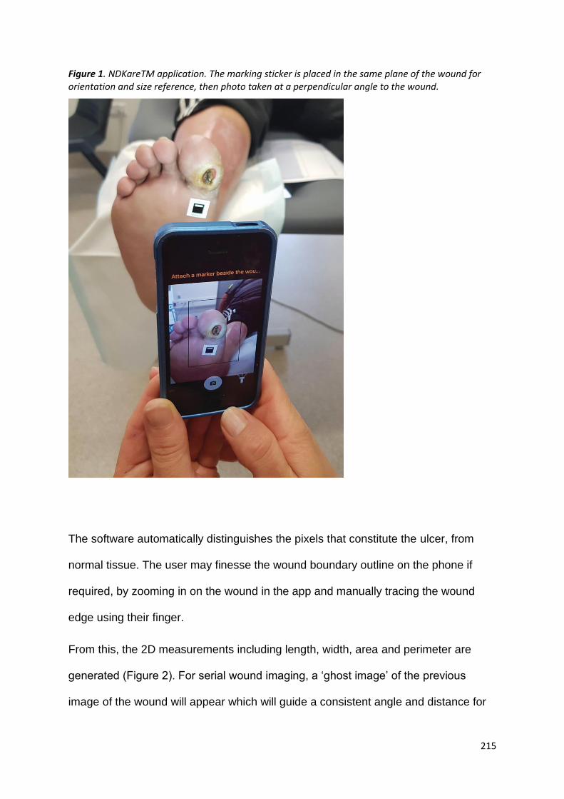
215
Figure 1. NDKareTM application. The marking sticker is placed in the same plane of the wound for orientation and size reference, then photo taken at a perpendicular angle to the wound.
The software automatically distinguishes the pixels that constitute the ulcer, from
normal tissue. The user may finesse the wound boundary outline on the phone if
required, by zooming in on the wound in the app and manually tracing the wound
edge using their finger.
From this, the 2D measurements including length, width, area and perimeter are
generated (Figure 2). For serial wound imaging, a ‘ghost image’ of the previous
image of the wound will appear which will guide a consistent angle and distance for

216
image capture. A timeline of each patient’s wounds with photographs and
measurements is generated to allow for monitoring wound progression over time.
Figure 2: NDKareTM wound assessment.
The top panel is the photographed image of the wound, bottom left panel is the generated 2D metric measurements and the bottom right panel is the wound bed composition analysis.
NDKare also requires a 20 second video panning over the wound to generate a 3D
model of the wound utilising the concept of ‘structure from motion’ (Figure 3). This
video is segmented into a smooth sequence of images of the wound taken from
different vantage points. Image processing algorithms identify a small set of highly

217
distinguishable pixels in the image set, ‘keypoints’. As keypoints are distinct, an
image processing algorithm can match corresponding keypoints across multiple
images. Triangulating the matched correspondences yields a ‘sparse 3D
reconstruction’ of the wound. More sophisticated algorithms attempt to substantially
increase the set of corresponding points beyond the initial set of matched keypoints
and triangulating the expanded set of matched points results in a ‘dense’ 3D point
cloud. To facilitate the computation of various metric properties of the wound, such
as its depth and volume, the 3D point cloud is converted into a smooth 3D ‘surface
reconstruction’. Finally, the pixel colours of the wound are combined for ‘surface
texturing’ which is mapped onto a 3D model(22). From the mobile phone, this video
is directly uploaded onto an online Health Insurance Portability and Accountability
Act (HIPAA) compliant cloud dashboard. HIPAA has established the standard for the
protection of sensitive patient data with security measures required for physical and
network data. On the dashboard, a total of 8 points are made outlining the wound,
the maximal depth and the marking sticker to generate the 3D measurements of
depth and volume.

218
Figure 3: Structure from motion pipeline. NDKareTM 3D reconstruction software processing for
converting the video of the wound to the 3D model.
115 wounds were assessed using the NDKareTM application by a single clinician
(B.K.). During the same wound review, VisitrakTM and WoundVueTM camera
measurements of the wound were also obtained. The VisitrakTM planimetry system
was used as the traditional gold standard for the 2D measurements. Our group has
previously demonstrated the validity of the WoundVueTM camera as a 3D
measurement device for diabetic foot wounds(21). We aimed to assess if the 2D
measurements on the NDKareTM application were comparable to the VisitrakTM and
WoundVueTM camera, and if the NDKareTM application 3D measurements were
consistent with the WoundVueTM camera. Subsequently, the clinician (B.K.) using a
Samsung Galaxy® S8+ smartphone (Version 9, Seoul, KR, Samsung Electronics Co.
Ltd.) and research officer (R.B.) using an Apple iPhone® 5 (Version 10.3.4,
Cupertino, CA, Apple Inc.) used the NDKareTM application to measure the same
wound for a total of 35 2D and wound measurements for inter-rater reliability. The

219
research officer had limited time availability, and as a result they were unable to
measure all 115 wounds which had been assessed by the clinician.
Statistical Analysis
Intraclass correlation coefficient (ICC) estimates and 95% confidence intervals (CIs)
were calculated. Cicchetti’s guidelines for interpretation of ICC inter-rater agreement
measures were used with ICC values ≥0.75 indicating excellent agreement, 0.60-
0.74 as good agreement and 0.40-0.59 as fair agreement(23).
A two-way mixed-effect model with consistency of agreement statistical model was
used for comparison of NDKareTM, VisitrakTM and WoundVueTM measurements, and
between different users for inter-rater reliability.
The statistical analysis was performed using Stata Statistical Software (Release
15.1, College Station, TX: StataCorp LP).
Results
115 diabetic foot wounds were assessed using the NDKareTM application, VisitrakTM
and WoundVueTM camera for 2D and 3D measurements. Wounds included in this
study were digital ulcers (49.6%), forefoot ulcers (19.1%), toe amputation sites
(10.4%), midfoot ulcers (7.8%), heel ulcers (5.2%), multiple digit ulcers (4.3%),
malleolar wounds (2.6%) and a forefoot guillotine amputation (0.9%). Diabetic foot
wounds measuring <10cm2 comprised 80.9% of the cohort, 10.4% of wounds
measured 10.1-19.9cm2 and 8.7% of wounds 20-66cm2. The median wound surface
area measurement was 2.91 cm2 (IQR: 1.05 to 8.53). The median maximum depth

220
and volume of the wounds was 0.25 cm2 (IQR: 0.04-0.86) and 1.15 cm3 (IQR: 0.07-
10.13) respectively.
There was excellent inter-rater reliability between all three measurement devices for
surface area (ICC: 0.99 [95% CI: 0.99, 0.99]). For maximum depth there was good
agreement (ICC: 0.70 [95% CI: 0.56, 0.79]) and fair agreement for volume (ICC: 0.51
[95% CI: 0.29, 0.67]) between the WoundVueTM camera and NDKareTM application
measurements.
35 diabetic foot wounds were assessed by two different assessors using the
NDKareTM on different mobile phone devices for the 2D and 3D measurements.
There was excellent inter-rater reliability for surface area (ICC: 0.98 [95% CI: 0.96,
0.99)]. For maximum depth there was good agreement (ICC: 0.63 [95% CI: 0.27,
0.81]) and good agreement for volume (ICC: 0.64 [95% CI: 0.28, 0.82]) (Figure 4).
Figure 4. The inter-rater reliability of the NDKareTM application surface area measurements between two different users and smartphones.

221
Discussion
Over the last 20 years, the use of mobile devices has significantly increased due to a
reduction in price and increase in processing and memory capabilities. Due to the
widespread availability of smartphones, the subjective use of mobile phone images
for telemedicine and serial wound imaging has become a frequent addition to
modern wound care management. Whilst this is an improvement on wound
descriptions and diagrams, we know that objective measurements of wounds are
essential in order to be able to direct patient care. The NDKareTM application utilises
the pre-existing smartphone camera hardware and the NDKareTM image processing
and interpretation software to generate the 2D and 3D wound measurements. This
design allows the NDKareTM system to be accessible to anyone owning a
smartphone without the need for specialised equipment.
From our results, the NDKareTM system accurately measures the surface area of
diabetic foot wounds in comparison to the gold standard VisitrakTM and recently
validated WoundVueTM camera. Two different smartphone platforms (Samsung
Galaxy® S8+ and Apple iPhone® 5) were used to compare the accuracy and
reproducibility of the NDKareTM application. These phones have different cameras,
flash, colour and video processing software. In addition to these variables, two
different users used each phone. Despite these multiple variables, our results
highlights that different users with different smartphones can use this software and
still obtain similar results. Although the clinician and research officer have varying
levels of wound care experience, there were no noted significant differences in the
use of the NDKareTM system to take wound photos. This is likely attributable to the
same training session both individuals attended, but it may also reflect the ease of
use of the NDKareTM interface.

222
The availability of a point-of-care, accurate, reproducible and readily available
software to measure the surface area of the wound and photographically monitor a
wound is a useful adjunct to aid clinical assessment in the multidisciplinary foot
clinic(24).
This benefit may extend to patient use as many already use mobile phone photos to
inspect and monitor their ulcers. However, patients along with health professionals
often cannot detect subtle wound healing or deterioration based on wound
photography alone. Phone applications which can provide detailed objective wound
measurement data have been shown to increase patient’s trust in health provider’s
assessment and may increase patient motivation during the wound healing
process(25). In addition, the “ghost images” may help patients with accuracy of
wound image capture as they may be elderly or have visual impairment secondary to
diabetes(26).
For patients living in remote areas who are unable to attend multidisciplinary foot
clinics, the Australian National Evidence-Based Guidelines for the Prevention,
Identification and Management of Foot Complications in Diabetes recommends
utilising remote expert consultation with digital imaging. A randomised controlled trial
showed this resulted in a reduction in ulcer size each week and a reduction in
amputation rates in comparison to local physician care with digital imaging(27).
Digital images as the sole diagnostic modality for assessing DFU has been shown to
have limited validity and reliability in assessment with the recommendation that
systems needed to develop with better diagnostic accuracy(28). Using this software,
photos of the wound and measurements can be uploaded remotely onto the cloud-
based system via the mobile phone for availability to health care professionals to aid
telemedicine consultation.
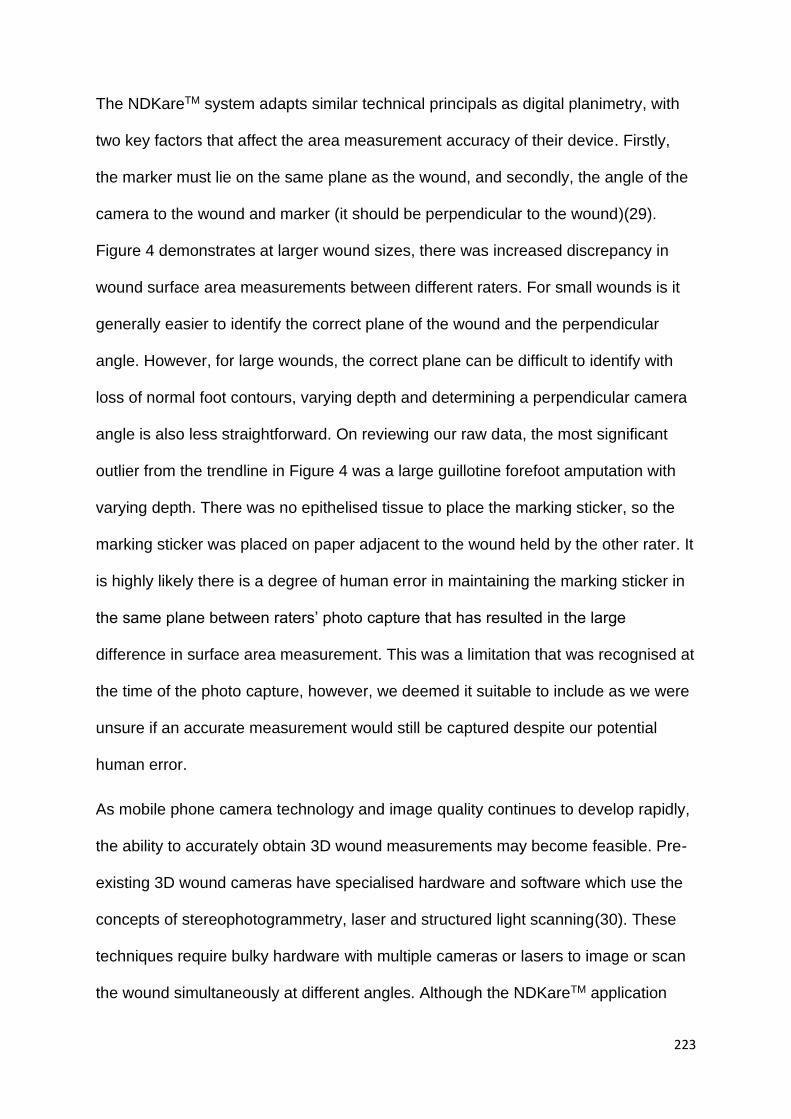
223
The NDKareTM system adapts similar technical principals as digital planimetry, with
two key factors that affect the area measurement accuracy of their device. Firstly,
the marker must lie on the same plane as the wound, and secondly, the angle of the
camera to the wound and marker (it should be perpendicular to the wound)(29).
Figure 4 demonstrates at larger wound sizes, there was increased discrepancy in
wound surface area measurements between different raters. For small wounds is it
generally easier to identify the correct plane of the wound and the perpendicular
angle. However, for large wounds, the correct plane can be difficult to identify with
loss of normal foot contours, varying depth and determining a perpendicular camera
angle is also less straightforward. On reviewing our raw data, the most significant
outlier from the trendline in Figure 4 was a large guillotine forefoot amputation with
varying depth. There was no epithelised tissue to place the marking sticker, so the
marking sticker was placed on paper adjacent to the wound held by the other rater. It
is highly likely there is a degree of human error in maintaining the marking sticker in
the same plane between raters’ photo capture that has resulted in the large
difference in surface area measurement. This was a limitation that was recognised at
the time of the photo capture, however, we deemed it suitable to include as we were
unsure if an accurate measurement would still be captured despite our potential
human error.
As mobile phone camera technology and image quality continues to develop rapidly,
the ability to accurately obtain 3D wound measurements may become feasible. Pre-
existing 3D wound cameras have specialised hardware and software which use the
concepts of stereophotogrammetry, laser and structured light scanning(30). These
techniques require bulky hardware with multiple cameras or lasers to image or scan
the wound simultaneously at different angles. Although the NDKareTM application

224
does not accurately obtain depth and volume measurements, there is already
interest amongst mobile phone companies in the development of depth estimation
hardware. Apple® acquired PrimeSense®, the company which developed the
Microsoft Kinect® structured light sensor in 2013(31). Using this technology, Apple®
has subsequently developed the iPad Pro® which has an in-built rear 3D camera and
the iPhone® X and later models which have the TrueDepth® front-facing 3D
camera(32, 33). The TrueDepth® application programming interface (API) for depth
data is available to iOS devices running iOSTM 11 or later(34) Intel® has also
developed RealSenseTM, which aims to integrate depth sensors into their mobile
phones(35)
A limitation of our study is that the feasibility of the diabetic foot ulcer patient
population to use the NDKareTM application for self-monitoring was not evaluated. A
participatory healthcare approach has been shown to increase patient engagement
with an improvement in the shared decision-making process. There is the potential to
decrease medical errors and increase staff adherence to optimal treatment practices,
particularly for chronic diseases(36). However, the adoption and consistent use of
this technology needs to be assessed. Depending on the location of the wound,
some patients will not be able to take photos of the wound and will require a carer to
take the photos for them. Future studies examining the impact on patient self-care,
compliance and limitations to patient utilisation would be useful to evaluate.
225
Acknowledgments
The authors thank LBT Innovations and Nucleus Dynamics for the provision and
support of the NDKareTM phone application. We also thank LBT Innovations for
providing us with the WoundVueTM camera. Both companies provided IT support in-
kind, but were not involved in any aspects of the study design or preparation of this
manuscript.
The authors received no financial support for the research, authorship, and/or
publication of this article.
Conflict of interest
None.
References
1. Zhang P, Lu J, Jing Y, Tang S, Zhu D, Bi Y. Global epidemiology of diabetic foot ulceration: a
systematic review and meta-analysis. Annals of Medicine. 2017;49(2):106-16.
2. Singh N, Armstrong DG, Lipsky BA. Preventing foot ulcers in patients with diabetes. Jama.
2005;293(2):217-28.
3. Driver VR, Fabbi M, Lavery LA, Gibbons G. The costs of diabetic foot: the economic case for
the limb salvage team. J Vasc Surg. 2010;52(3 Suppl):17s-22s.
4. Prompers L, Schaper N, Apelqvist J, Edmonds M, Jude E, Mauricio D, et al. Prediction of
outcome in individuals with diabetic foot ulcers: focus on the differences between individuals with and
without peripheral arterial disease. The EURODIALE Study. Diabetologia. 2008;51(5):747-55.
5. Pickwell K, Siersma V, Kars M, Apelqvist J, Bakker K, Edmonds M, et al. Predictors of Lower-
Extremity Amputation in Patients With an Infected Diabetic Foot Ulcer. Diabetes Care.
2015;38(5):852-7.
6. Walsh JW, Hoffstad OJ, Sullivan MO, Margolis DJ. Association of diabetic foot ulcer and
death in a population-based cohort from the United Kingdom. Diabet Med. 2016;33(11):1493-8.

226
7. International Best Practice Guidelines: Wound Management in Diabetic Foot Ulcers.: Wounds
International; 2013 [Available from: www.woundsinternational.com.
8. Sheehan P, Jones P, Caselli A, Giurini JM, Veves A. Percent Change in Wound Area of
Diabetic Foot Ulcers Over a 4-Week Period Is a Robust Predictor of Complete Healing in a 12-Week
Prospective Trial. Diabetes Care. 2003;26(6):1879-82.
9. Coerper S, Beckert S, Küper MA, Jekov M, Königsrainer A. Fifty percent area reduction after
4 weeks of treatment is a reliable indicator for healing—analysis of a single-center cohort of 704
diabetic patients. Journal of Diabetes and its Complications. 2009;23(1):49-53.
10. Lavery LA, Davis KE, Berriman SJ, Braun L, Nichols A, Kim PJ, et al. WHS guidelines update:
Diabetic foot ulcer treatment guidelines. Wound Repair and Regeneration. 2016;24(1):112-26.
11. Schultz G, Mozingo D, Romanelli M, Claxton K. Wound healing and TIME; new concepts and
scientific applications. Wound Repair and Regeneration. 2005;13(s4):S1-S11.
12. Rogers LC, Bevilacqua NJ, Armstrong DG, Andros G. Digital planimetry results in more
accurate wound measurements: a comparison to standard ruler measurements. Journal of diabetes
science and technology. 2010;4(4):799-802.
13. Gethin G. The importance of continuous wound measuring. Wounds UK. 2006;2(2).
14. Oien RF, Håkansson A, Hansen BU, Bjellerup M. Measuring the size of ulcers by planimetry:
a useful method in the clinical setting. J Wound Care. 2002;11(5):165-8.
15. Stockton KA, McMillan CM, Storey KJ, David MC, Kimble RM. 3D photography is as accurate
as digital planimetry tracing in determining burn wound area. Burns : journal of the International
Society for Burn Injuries. 2015;41(1):80-4.
16. Khoo R, Jansen S. The Evolving Field of Wound Measurement Techniques: A Literature
Review. Wounds : a compendium of clinical research and practice. 2016;28(6):175-81.
17. Foltynski P, Ladyzynski P, Ciechanowska A, Migalska-Musial K, Judzewicz G, Sabalinska S.
Wound Area Measurement with Digital Planimetry: Improved Accuracy and Precision with Calibration
Based on 2 Rulers. PloS one. 2015;10(8):e0134622-e.
18. Bien P, De Anda C, Prokocimer P. Comparison of Digital Planimetry and Ruler Technique To
Measure ABSSSI Lesion Sizes in the ESTABLISH–1 Study. Surgical Infections. 2014;15(2):105-10.
19. World Union of Wound Healing Societies (WUWHS), Florence Congress, Position Document.
Local management of diabetic foot ulcers. Wounds International, 2016.
20. Bills JD, Berriman SJ, Noble DL, Lavery LA, Davis KE. Pilot study to evaluate a novel three-
dimensional wound measurement device. Int Wound J. 2016;13(6):1372-7.
21. Pena G, Kuang B, Szpak Z, Cowled P, Dawson J, Fitridge R. Evaluation of a Novel Three-
Dimensional Wound Measurement Device for Assessment of Diabetic Foot Ulcers. Advances in
Wound Care. 2019.

227
22. Maiti A, Chakravarty D. Performance analysis of different surface reconstruction algorithms
for 3D reconstruction of outdoor objects from their digital images. SpringerPlus. 2016;5(1):932.
23. Cicchetti D. Guidelines, Criteria, and Rules of Thumb for Evaluating Normed and
Standardized Assessment Instrument in Psychology. Psychological Assessment. 1994;6:284-90.
24. Nair HKR. Increasing productivity with smartphone digital imagery wound measurements and
analysis. Journal of Wound Care. 2018;27(Sup9a):S12-S9.
25. Ploderer B, Brown R, Seng LSD, Lazzarini PA, van Netten JJ. Promoting Self-Care of
Diabetic Foot Ulcers Through a Mobile Phone App: User-Centered Design and Evaluation. JMIR
Diabetes. 2018;3(4):e10105-e.
26. Yap MH, Chatwin KE, Ng C-C, Abbott CA, Bowling FL, Rajbhandari S, et al. A New Mobile
Application for Standardizing Diabetic Foot Images. Journal of diabetes science and technology.
2018;12(1):169-73.
27. National Evidence-Based Guideline on Prevention, Identification and Management of Foot
Complications in Diabetes (Part of the Guidelines on Management of Type 2 Diabetes). Melbourne,
Australia. 2011.
28. van Netten JJ, Clark D, Lazzarini PA, Janda M, Reed LF. The validity and reliability of remote
diabetic foot ulcer assessment using mobile phone images. Scientific reports. 2017;7(1):9480-.
29. Rennert R, Golinko M, Kaplan D, Flattau A, Brem H. Standardization of wound photography
using the Wound Electronic Medical Record. Adv Skin Wound Care. 2009;22(1):32-38.
30. Filko D, Cupec R, Nyarko EK. Wound measurement by RGB-D camera. Machine Vision and
Applications. 2018;29(4):633-54.
31. Apple buys Israel's PrimeSense for $345 million: report: Reuters; 2013 [Available from:
https://www.reuters.com/article/us-primesense-apple/apple-buys-israels-primesense-for-345-million-
report-idUSBRE9AG03G20131117.
32. Apple unveils new iPad Pro with breakthrough LiDAR Scanner and brings trackpad support to
iPadOS: Apple Inc.; 2020 [Available from: https://www.apple.com/au/newsroom/2020/03/apple-
unveils-new-ipad-pro-with-lidar-scanner-and-trackpad-support-in-ipados/.
33. About Face ID advanced technology: Apple Inc.; 2020 [Available from:
https://support.apple.com/en-au/HT208108.
34. Streaming Depth Data from the TrueDepth Camera: Apple Inc.; 2020 [Available from:
https://developer.apple.com/documentation/avfoundation/cameras_and_media_capture/streaming_de
pth_data_from_the_truedepth_camera.
35. Grunnet-Jepsen A, Sweetser J. Intel®RealSense™ Depth Cameras for Mobile Phones: New
Technologies Group, Intel Corporation; 2018 [Available from:
https://www.intel.com.au/content/www/au/en/support/articles/000026983/emerging-technologies/intel-
realsense-technology.html.

228
36. Longtin Y, Sax H, Leape LL, Sheridan SE, Donaldson L, Pittet D. Patient participation: current
knowledge and applicability to patient safety. Mayo Clin Proc. 2010;85(1):53-62.

229


