Embed Size (px)
Citation preview

A
tApbRctcwbm©
K
1
atptotieswi
0d
Available online at www.sciencedirect.com
International Journal of Pharmaceutics 346 (2008) 133–142
Pharmaceutical Nanotechnology
Preparation and characterization of thermo-responsivealbumin nanospheres
Zhe-Yu Shen a,b,c, Guang-Hui Ma a,∗, Toshiaki Dobashi b, Yasuyuki Maki b, Zhi-Guo Su a
a State Key Laboratory of Biochemical Engineering, Institute of Process Engineering, Chinese Academy of Sciences, P.O. Box 353, Beijing 100080, Chinab Department of Biological and Chemical Engineering, Faculty of Engineering, Gunma University, 1-5-1, Tenjin-cho, Kiryu, Gunma 376-8515, Japan
c Graduate University of Chinese Academy of Sciences, 19, Yuquan Road, Shijingshan District, Beijing 100049, China
Received 18 January 2007; received in revised form 9 April 2007; accepted 6 June 2007Available online 14 June 2007
bstract
Thermo-responsive poly(N-isopropylacrylamide-co-acrylamide)-block-polyallylamine-conjugated albumin nanospheres (PAN), new thermalargeting anti-cancer drug carrier, was developed by conjugating poly(N-isopropylacrylamide-co-acrylamide)-block-polyallylamine (PNIPAM-Am-b-PAA) on the surface of albumin nanospheres (AN). PAN may selectively accumulate onto solid tumors that are maintained abovehysiological temperature due to local hyperthermia. PNIPAM-AAm-b-PAA was synthesized by radical polymerization, and AN was preparedy ultrasonic emulsification. AN with diameter below 200 nm and narrow size distribution was obtained by optimizing the preparative conditions.ose Bengal (RB) was used as model drug for entrapment into the AN and PAN during the particle preparation. The release rate of RB from PANompared with AN in trypsin solution was slower, and decreased with the increase of PNIPAM-AAm-b-PAA molecular weight, which suggestedhat the existence of a steric hydrophilic barrier on AN made digestion of AN more difficult. Moreover, the release of RB from PAN above the
loud-point temperature (Tcp) of PNIPAM-AAm-b-PAA became faster. This was because the density of temperature-responsive polymers on ANas not so high, so that the interspace between the polymer chains increased after they shrunk due to the high temperature. As a result, theiodegradable AN was attacked more easily by trypsin. The design of PAN overcame the disadvantages of temperature-responsive polymericicelles.2007 Elsevier B.V. All rights reserved.Albu
toopprodfm1
eywords: Poly(N-isopropylacrylamide-co-acrylamide)-block-polyallylamine;
. Introduction
The main problems currently associated with systemic drugdministration are: even biodistribution of pharmaceuticalshroughout the body; lack of drug specific affinity toward aathological site; the necessity of a large total dose of drugo achieve high local concentration; non-specific toxicity andther adverse side-effects due to high drug doses. Because ofhese drawbacks of systemic drug administration and the tox-city of anti-cancer drugs to both tumor and normal cells, thefficacy of cancerous chemotherapy is often limited by serious
ide-effect. A strategy could be to associate anti-cancer drugsith colloidal nanoparticles, with the aim to increase selectiv-ty of drugs towards cancer cells while reducing their toxicity
∗ Corresponding author. Tel.: +86 1082627072; fax: +86 1082627072.E-mail address: [email protected] (G.-H. Ma).
a1Ja
sw
378-5173/$ – see front matter © 2007 Elsevier B.V. All rights reserved.oi:10.1016/j.ijpharm.2007.06.004
min nanospheres; Rose Bengal; Entrapment efficiency; Controlled release
owards normal tissues (Brigger et al., 2002). The accumulationf intravenously injected nanoparticles onto tumor cells reliesn a passive diffusion or convection across the leaky and hyper-ermeable tumor vasculature (Yuan, 1998), which was calledassive targeting. Indeed, the uptake of nanoparticles can alsoesult from active targeting, a specific recognition in the casef ligand decorated nanoparticles (Moghimi et al., 2001). Toate, many distinctive active targeting nanoparticles, such asolate-conjugated albumin nanoparticles (Zhang et al., 2004),onoclonal antibody conjugated nanoparticles (Sheng et al.,
995), magnetic albumin nanoparticles (Gong et al., 2004), pH-nd temperature-responsive polymeric micelles (Kwon et al.,995; Topp et al., 1997; Inoue et al., 1998; Chung et al., 2000;eong et al., 2003), have been investigated in drug delivery
pplications.The temperature-responsive polymeric micelles were con-tructed by hydrophobic interaction, which is weak comparingith covalent bond. When it arrived at the heated cancer tissues

134 Z.-Y. Shen et al. / International Journal of
Sp
a(smif
imamchamcf
cadwt
h(mteblotfaacptaetgoawPfPn
afa
2
2
cheme 1. Schematic presentation for difference of (a) temperature-responsiveolymeric micelles and (b) PAN.
nd cells, it could accumulate on the cancer tissues and cellsKohori et al., 1999). However, the dense hydrophilic polymerequence on the surface of temperature-responsive polymericicelles would shrink and accumulate on the micelles (as shown
n Scheme 1a), resulting in the decrease of the drug release raterom the micelles and the curative efficacy.
In this study, we developed a new thermal target-ng anti-cancer drug carrier: temperature-responsive poly-er conjugated albumin nanospheres. At first, biodegrad-
ble albumin nanospheres (AN) with Rose Bengal (RB,odel drug) inside was prepared by ultrasonic emulsifi-
ation combined with chemical cross-linking (glutaralde-yde), then poly(N-isopropylacrylamide-co-acrylamide)-block-llylamine (PNIPAM-AAm-b-PAA) (thermo-responsive poly-ers) was conjugated on AN. This PNIPAM-AAm-b-PAA-
onjugated albumin nanospheres (PAN) was designed byollowing strategy.
On intravenous administration, particles are normally rapidlyoated by the adsorption of specific blood components known
s opsonins and then recognized and taken up by the reticuloen-othelial system (RES). The surface of albumin nanospheresith drug inside and thermo-responsive copolymer chains onhe surface is hydrophilic at and below 37 ◦C due to the
tTw
Scheme 2. Reaction scheme fo
Pharmaceutics 346 (2008) 133–142
ydrophilicity of the copolymer if the cloud-point temperatureTcp) of the copolymer is about 42 ◦C under local hyperther-ia. This hydrophilic copolymer layer can dramatically affect
he opsonization of particles by plasma components (Moghimit al., 1993; Lin et al., 1999). Therefore, the uptake of PANy the RES can be reduced and a significantly longer circu-ation half-life of the particles in the blood stream will bebtained (Illum and Davis, 1984). PAN will not precipitate onhe healthy tissues and cells because of the hydrophilic sur-ace, but will precipitate on the heated cancer tissues and cellsround 42 ◦C, because the copolymer will become hydrophobict higher temperature due to conformational transition of thehain. Then, the interspace between the temperature-responsiveolymer chains (as shown in Scheme 1b) will increase afterhey shrink due to the high temperature. So that, the biodegrad-ble albumin nanospheres will be attacked and degraded moreasily by proteinase, then the drug will release there to attackhe tumor cells. Furthermore, histological analysis of PNIPAM-rafted gelatin gel elucidated that PNIPAM can be biodegradedver time without excessive inflammatory reactions (Ohya etl., 2004). According to a toxicity test of the PNIPAM, whichas performed in comparison with that of the NIPAM monomer,NIPAM showed no toxicity in mice (Sheng et al., 2006). There-ore, although NIPAM, AAm and AA are toxic monomers,NIPAM-AAm-b-PAA is also biodegradable over time andon-toxic.
Consequently, PAN, which is constructed by covalent bondnd whose drug release above Tcp of PNIPAM-AAm-b-PAA isaster, can be employed as drug carrier due to their targetingbility to tumor cells by both passive and active targeting.
. Materials and methods
.1. Materials
N-isopropylacrylamide (NIPAM), 2,2′-azobis(isobutyroni-rile) (AIBN) and Rose Bengal (RB) were purchased fromokyo Chemical Industry Co., Ltd. (TCI, Japan). NIPAMas purified by recrystallization from n-hexane. Acry-
r PNIPAM-AAm-b-PAA.

al of
lpgudfmm
2
ysa2fliaa6oPwaMpoTHW
tamnw
2n
sp
3fu7tlswA(twfic
Z.-Y. Shen et al. / International Journ
amide (AAm) and sorbitan sesquioleate (Arlacel83) wereurchased from SIGMA (USA). Allylamine (AA), 50%lutaraldehyde (GA) aqueous solution, trypsin (30 USPnits/mg), N-hydroxysuccinimide (NHS) and 1-ethyl-3-(3-imethylaminopropyl) carbodiimide (EDAC) were purchasedrom Wako Pure Chemicals (Tokyo, Japan). Bovine serum albu-in (BSA), fraction V (pH 7.0), was purchased from ICN Bio-aterials.
.2. Synthesis of thermo-sensitive polymers
Poly(N-isopropylacrylamide-co-acrylamide)-block-polyall-lamine (PNIPAM-AAm-b-PAA) was synthesized from twoteps (Scheme 2) by free radical polymerization using AIBN asn initiator. First, 39.05 g of NIPAM, 2.95 g of AAm, AIBN and50 mL of ethanol were added into a three-necked round-bottleask with a magnetic stirrer. The round-bottle flask was moved
n an oil bath and stirred at 60 ◦C for 24 h under nitrogentmosphere. Second, after 24 h, 2 mL of AA was dropwisedded into the mixture. The whole mixture was stirred at0 ◦C for 3.0 h under nitrogen atmosphere. 0.20 and 0.10 gf AIBN were used to obtain PNIPAM-AAm-b-PAA1 andNIPAM-AAm-b-PAA2, respectively. The resultant polymersere separated and purified by reprecipitation into diethylether
nd then dried in vacuum. The polymers were dissolved inilliQ water for optical transmittance measurement. Gel
ermeation chromatography (GPC) measurements were made
n PNIPAM-AAm-b-PAA1 and PNIPAM-AAm-b-PAA2 inHF at 35 ◦C using a Waters GPC equipment (Waters 515PLC Pump, Waters Styragel Columns [HT2 + HT3 + HT4],aters 2410 Differential Refractometer). The flow rate and thejvto
Scheme 3. Reaction scheme for preparation of cross-linked A
Pharmaceutics 346 (2008) 133–142 135
emperature of the column oven were set to be 1 cm3 min−1
nd 35 ◦C, respectively. Elution times were converted intoolecular weights using a calibration curve constructed with
arrow polydispersity polystyrene standards, whose moleculareights range from 2500 to 600,000.
.3. Preparation of blank and RB-loaded albuminanospheres
Albumin nanospheres were prepared by an ultrasonic emul-ification method combined with chemical cross-linking. Thereparation method is as follows.
0.6 mL of 20% BSA physiological saline was added into0 mL cyclohexane containing 6.0 wt.% of Arlacel83 and stirredor 10 min. Then, the solution was emulsified for 10 min byltrasonic disruptor (TOMY UD-200) with the output power of1, 97, 127 and 165 W, respectively. After that, glutaraldehydeoluene solution (0.85 mg GA/mg BSA) was added to cross-ink the particles. The cross-linking process was performed bytirring the suspension over 24 h. 0.5 mL of 2-aminoethanolas then added to cap free aldehyde groups (Scheme 3a).fter a 1.0 h reaction time, the suspension was ultracentrifuged
44,900 × g, 20 min) at 10 ◦C (Automatic Preparative Ultracen-rifuge, HITACHI 70P-72, Japan). The harvested nanospheresere washed two times by cyclohexane to remove the emulsi-er and then acetone was dropwise added into 20 mL of ANyclohexane solution under stirring until the solution became
ust turbid. After ultracentrifuged, the samples were dried inacuum for 1.0 h. And then, the nanospheres were cleaned oneime by pure water to eliminate free albumin and the excessf the cross-linker. Between each washing, nanospheres wereN and conjugation of PNIPAM-AAm-b-PAA on AN.
136 Z.-Y. Shen et al. / International Journal of
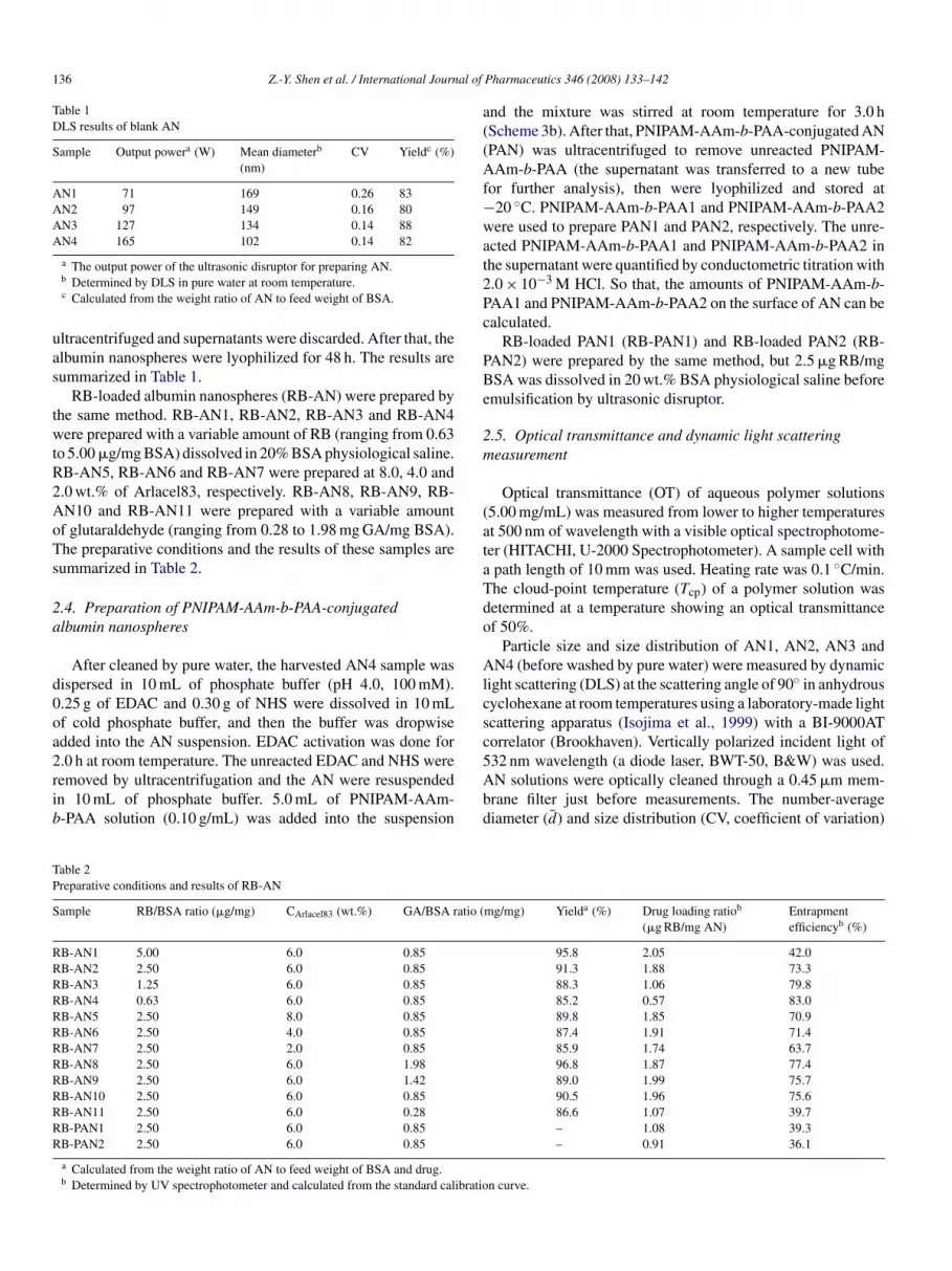
Table 1DLS results of blank AN
Sample Output powera (W) Mean diameterb
(nm)CV Yieldc (%)
AN1 71 169 0.26 83AN2 97 149 0.16 80AN3 127 134 0.14 88AN4 165 102 0.14 82
uas
twtR2AoTs
2a
d0oa2rib
a((Af−wat2Pc
PBe
2m
(ataTdo
Alcsc
TP
S
RRRRRRRRRRRRR
a The output power of the ultrasonic disruptor for preparing AN.b Determined by DLS in pure water at room temperature.c Calculated from the weight ratio of AN to feed weight of BSA.
ltracentrifuged and supernatants were discarded. After that, thelbumin nanospheres were lyophilized for 48 h. The results areummarized in Table 1.
RB-loaded albumin nanospheres (RB-AN) were prepared byhe same method. RB-AN1, RB-AN2, RB-AN3 and RB-AN4ere prepared with a variable amount of RB (ranging from 0.63
o 5.00 �g/mg BSA) dissolved in 20% BSA physiological saline.B-AN5, RB-AN6 and RB-AN7 were prepared at 8.0, 4.0 and.0 wt.% of Arlacel83, respectively. RB-AN8, RB-AN9, RB-N10 and RB-AN11 were prepared with a variable amountf glutaraldehyde (ranging from 0.28 to 1.98 mg GA/mg BSA).he preparative conditions and the results of these samples areummarized in Table 2.
.4. Preparation of PNIPAM-AAm-b-PAA-conjugatedlbumin nanospheres
After cleaned by pure water, the harvested AN4 sample wasispersed in 10 mL of phosphate buffer (pH 4.0, 100 mM)..25 g of EDAC and 0.30 g of NHS were dissolved in 10 mLf cold phosphate buffer, and then the buffer was dropwisedded into the AN suspension. EDAC activation was done for
.0 h at room temperature. The unreacted EDAC and NHS wereemoved by ultracentrifugation and the AN were resuspendedn 10 mL of phosphate buffer. 5.0 mL of PNIPAM-AAm--PAA solution (0.10 g/mL) was added into the suspension5Abd
able 2reparative conditions and results of RB-AN
ample RB/BSA ratio (�g/mg) CArlacel83 (wt.%) GA/BSA ratio (
B-AN1 5.00 6.0 0.85B-AN2 2.50 6.0 0.85B-AN3 1.25 6.0 0.85B-AN4 0.63 6.0 0.85B-AN5 2.50 8.0 0.85B-AN6 2.50 4.0 0.85B-AN7 2.50 2.0 0.85B-AN8 2.50 6.0 1.98B-AN9 2.50 6.0 1.42B-AN10 2.50 6.0 0.85B-AN11 2.50 6.0 0.28B-PAN1 2.50 6.0 0.85B-PAN2 2.50 6.0 0.85
a Calculated from the weight ratio of AN to feed weight of BSA and drug.b Determined by UV spectrophotometer and calculated from the standard calibratio
Pharmaceutics 346 (2008) 133–142
nd the mixture was stirred at room temperature for 3.0 hScheme 3b). After that, PNIPAM-AAm-b-PAA-conjugated ANPAN) was ultracentrifuged to remove unreacted PNIPAM-Am-b-PAA (the supernatant was transferred to a new tube
or further analysis), then were lyophilized and stored at20 ◦C. PNIPAM-AAm-b-PAA1 and PNIPAM-AAm-b-PAA2ere used to prepare PAN1 and PAN2, respectively. The unre-
cted PNIPAM-AAm-b-PAA1 and PNIPAM-AAm-b-PAA2 inhe supernatant were quantified by conductometric titration with.0 × 10−3 M HCl. So that, the amounts of PNIPAM-AAm-b-AA1 and PNIPAM-AAm-b-PAA2 on the surface of AN can bealculated.
RB-loaded PAN1 (RB-PAN1) and RB-loaded PAN2 (RB-AN2) were prepared by the same method, but 2.5 �g RB/mgSA was dissolved in 20 wt.% BSA physiological saline beforemulsification by ultrasonic disruptor.
.5. Optical transmittance and dynamic light scatteringeasurement
Optical transmittance (OT) of aqueous polymer solutions5.00 mg/mL) was measured from lower to higher temperaturest 500 nm of wavelength with a visible optical spectrophotome-er (HITACHI, U-2000 Spectrophotometer). A sample cell withpath length of 10 mm was used. Heating rate was 0.1 ◦C/min.he cloud-point temperature (Tcp) of a polymer solution wasetermined at a temperature showing an optical transmittancef 50%.
Particle size and size distribution of AN1, AN2, AN3 andN4 (before washed by pure water) were measured by dynamic
ight scattering (DLS) at the scattering angle of 90◦ in anhydrousyclohexane at room temperatures using a laboratory-made lightcattering apparatus (Isojima et al., 1999) with a BI-9000ATorrelator (Brookhaven). Vertically polarized incident light of
32 nm wavelength (a diode laser, BWT-50, B&W) was used.N solutions were optically cleaned through a 0.45 �m mem-rane filter just before measurements. The number-averageiameter (d̄) and size distribution (CV, coefficient of variation)mg/mg) Yielda (%) Drug loading ratiob
(�g RB/mg AN)Entrapmentefficiencyb (%)
95.8 2.05 42.091.3 1.88 73.388.3 1.06 79.885.2 0.57 83.089.8 1.85 70.987.4 1.91 71.485.9 1.74 63.796.8 1.87 77.489.0 1.99 75.790.5 1.96 75.686.6 1.07 39.7– 1.08 39.3– 0.91 36.1
n curve.

al of
w
d
C
wm
temmt
2
Jmct
2e
AiaamSoisaAf
D
E
2
2ost4tt
wRo04
0eR3
3
3
Pafb
Poobbttemperature distance �T between 5% transmittance and 95%transmittance: �T was 0.7 K for PNIPAM-AAm-b-PAA1 and0.9 K for PNIPAM-AAm-b-PAA2. Tcp was determined as 42.6and 42.4 ◦C for PNIPAM-AAm-b-PAA1 and PNIPAM-AAm-
Z.-Y. Shen et al. / International Journ
ere defined as follows:
¯ =n∑
i=1
di
N
V =(
n∑i=1
(di − d̄)2
N
)1/2
/d̄
here di is the diameter of each particle, N the total number ofeasured particles and d̄ is the number-average diameter.PAN1 and PAN2 were also measured by DLS from lower
o higher temperatures in pure water. The solutions werequilibrated at given temperatures for 10 min before eacheasurement. Diffusion coefficient was obtained by cumulantethod and the hydrodynamic diameter (Dh) was estimated from
he diffusion coefficient by using the Stokes–Einstein equation.
.6. Transmission electron microscopic observation
The AN4 and PAN1 were observed by a JEM-100cx (JEOL,apan) transmission electron microscope (TEM). Approxi-ately 2 �L of the diluted nanoparticles were mounted on
opper grids, and then dried at room temperature. After that,hey were observed by TEM (no coloration).
.7. Determination of drug loading ratio and entrapmentfficiency
The RB content was measured by digesting 24 mg of RB-N (or RB-PAN) and corresponding blank-AN (or blank-PAN)
n 6 mL of 20 mg/mL trypsin solution in the dark for 4.0 ht 37 ◦C, respectively. The digested solutions of blank-ANnd blank-PAN were used to make baseline of absorbanceeasurement by UV spectrophotometer (HITACHI, U-2000pectrophotometer). The absorbance (at 548 nm of wavelength)f the digested solutions of RB-AN (or RB-PAN) was convertednto the concentration of RB using a calibration curve con-tructed with standard RB solutions containing 4.0 mg/mL BSAnd 20 mg/mL trypsin. The drug loading ratio (DLR, �g RB/mgN) and entrapment efficiency (EE%) were calculated from the
ollowing formula:
LR = Total amount of RB loaded (�g)
Total amount of nanospheres harvested (mg)(1)
E(%) = Total amount of RB loaded
Total amount of RB added× 100 (2)
.8. ‘In vitro’ release studies
Thirty milligrams RB-AN (or RB-PAN) were dispersed in0 mL of phosphate-buffered saline (PBS, pH 7.4, 10 mM) withr without 0.20 mg/mL of trypsin. The suspensions were stirredlowly and incubated in a water bath at 37 ± 1 ◦C. At prede-
ermined time intervals, these samples were ultracentrifuged at4,900 × g for 20 min and 1.0 mL of these supernatants wereaken and analyzed for concentration of the released RB by spec-rophotometer. The release of RB from RB-AN9 and RB-AN10F(b
Pharmaceutics 346 (2008) 133–142 137
ithout trypsin, and from RB-AN8, RB-AN9, RB-AN10 andB-AN11 with 0.20 mg/mL of trypsin were determined. More-ver, the release of RB from RB-PAN1 and RB-PAN2 with.20 mg/mL of trypsin was both determined at 37 ± 1 ◦C and3 ± 1 ◦C.
RB of RB-AN10 was released for 8.0 h in PBS with.20 mg/mL of trypsin at 37 ± 1 ◦C. Then RB-AN10 was recov-red and renamed as RB-AN12. The release of RB fromB-AN12 with 0.20 mg/mL of trypsin was determined at7 ± 1 ◦C.
. Results and discussion
.1. Polymerization results
The yield determined as diethylether insoluble fraction forNIPAM-AAm-b-PAA1 and PNIPAM-AAm-b-PAA2 was 74nd 78%, respectively. The molecular weights Mw determinedrom GPC were 1.43 × 10⁴ and 1.59 × 10⁴ for PNIPAM-AAm--PAA1 and PNIPAM-AAm-b-PAA2, respectively.
Optical transmittance determined for PNIPAM-AAm-b-AA1 and PNIPAM-AAm-b-PAA2 are shown as a functionf temperature in Fig. 1. The optical transmittance of aque-us PNIPAM-AAm-b-PAA1 was almost unity at temperatureselow 42.4 ◦C, and decreased rapidly with rising temperatureetween 42.4 and 43.1 ◦C, and vanished at higher tempera-ures. The sharpness of the transition was estimated from the
ig. 1. Temperature dependence of optical transmittance of copolymers in waterc = 5.00 × 10⁻³ g cm−3) for PNIPAM-AAm-b-PAA1 (�) and PNIPAM-AAm--PAA2 (©).

138 Z.-Y. Shen et al. / International Journal of
FA
b5
ovorNTo
ctsA
catP
3n
o2woc1es
swopbd
F(
ig. 2. Size distributions of albumin nanospheres measured by DLS: AN1 (�),N2 (�), AN3 (�) and AN4 (�).
-PAA2 when the optical transmittance of the solutions crossed0% as the temperature increased.
In a previous study, we have found that the reciprocals of Tcpf aqueous solution of PNIPAM-AAm increased linearly againstolume fraction of NIPAM γ (determined from the volume ratiof NIPAM to the sum of NIPAM and AAm), where the slope was
elated to the interaction energy difference between the solvent-IPAM and the solvent-AAm (Shen et al., 2006a). Therefore,cp of PNIPAM-AAm-b-PAA can also be controlled by γ . Tcpf PNIPAM-AAm-b-PAA1 and PNIPAM-AAm-b-PAA2, whichr
6t
ig. 3. TEM photo of AN4 observed at 49,000 magnification (a), 61,000 magnificatiod).
Pharmaceutics 346 (2008) 133–142
an be decreased by decreasing the γ , are slightly higher thanhat of PNIPAM-AAm (41.0 ◦C) with the same γ . This was con-idered to result from hydration contribution from hydrophilicA in the polymer.The copolymers coherent to the current strategy need to
hange from hydrophilic type to hydrophobic type around 42 ◦C,nd the change must be very sensitive to temperature changeo increase the targeting ability. Here, both PNIPAM-AAm-b-AA1 and PNIPAM-AAm-b-PAA2 can meet these conditions.
.2. Size control and TEM observation of albuminanospheres
There are three different preparation methods for AN, basedn desolvation (Langer et al., 2003), coacervation (Lin et al.,001), or emulsion formation (Muller et al., 1996). In this paper,e prepared AN based on emulsion formation because the sizef AN prepared by desolvation and coacervation cannot beontrolled easily. In order to obtain AN with diameter around00 nm, which is required for intravenous administration, theffect of the output power of ultrasonic disruptor on the particleize and size distribution was investigated.
The preparation results of AN1, AN2, AN3 and AN4 areummarized in Table 1 and Fig. 2. The yield for each AN sampleas between 80 and 88%. d̄ and CV values both decreased as theutput power of the ultrasonic disruptor increased, by which thearticle size and size distribution of albumin nanospheres cane controlled. The appropriate output power of the ultrasonicisruptor was obtained (165 W, AN4 sample) according to the
equirement of the size of AN in drug delivery.Fig. 3(a–c) was TEM photo of AN4 observed at 49,000,1,000 and 81,000 magnification, respectively. From these pic-ures, it was found that the albumin nanospheres were dispersed
n (b) and 81,000 magnification (c) and PAN1 observed at 61,000 magnification

al of Pharmaceutics 346 (2008) 133–142 139
wcatDsab2
3l
iotpRtaoIgs0e
dnadw
R(wiaseraiwmte
tTctcilo
Fig. 4. Influence of RB/BSA ratio (a), emulsifier concentration in the oil phase(A
c(
iwfpisla
Z.-Y. Shen et al. / International Journ
ell in pure water, the coalescence and aggregation of nanoparti-les were almost not found, the particles were spherical in shapend the surfaces of the particles were smooth. From TEM pic-ures, the average diameter of AN4, smaller than the result ofLS (102 nm), was estimated to be below 100 nm due to the
welling of AN because TEM was observed at dry state of ANnd DLS was determined at wet state of AN. Therefore, AN cane achieved as a long-circulating system with a size range below00 nm in diameter for the current drug delivery scheme.
.3. Optimization of nanosphere preparation by drugoading ratio and entrapment efficiency
The ability of AN to carry a model drug RB was evaluated. RBs a water-soluble reagent (Mw = 1017.6 Da) with a high abilityf protein binding (Lillie, 1977) and widely used as a diagnos-ic agent due to its high extinction coefficient at 548 nm. In thisaper, RB was used as a model drug, and the influence of theB/BSA ratio, the emulsifier concentration in the oil phase and
he concentration of glutaraldehyde on the drug loading rationd the entrapment efficiency were investigated. The resultsf preparation of RB-loaded AN are summarized in Table 2.f not specified, the RB/BSA ratio, emulsifier concentration,lutaraldehyde concentration and output power of the ultra-onic disruptor were fixed at 2.50 �g RB/mg BSA, 6.0 wt.%,.85 mg GA/mg BSA and 165 W, respectively, in the followingxperiments.
There are three main factors which are considered to affect therug loading ratio and entrapment efficiency of RB in albuminanospheres: (1) the binding degree between the drug and thelbumin molecule; (2) coalescence and break-up of emulsionroplets, leading to leakage of drug; (3) diffusion of drug fromater phase to oil phase.Fig. 4(a) shows the capacity of albumin nanospheres for
B loading as a function of the drug/initial albumin ratioRB/BSA ratio). The high drug entrapment efficiency in ANas achieved, up to 83.0% at low RB/BSA ratio. With increas-
ng the RB/BSA ratio, the drug loading ratio increased quicklyt RB/BSA ratios lower than 2.50 �g RB/mg BSA, but increasedlowly at RB/BSA ratios higher than 2.50 �g RB/mg BSA. How-ver, the entrapment efficiency decreased gently at RB/BSAatios lower than 2.50 �g RB/mg BSA, but decreased rapidlyt higher than 2.50 �g RB/mg BSA. That was because the bind-ng degree between RB and the albumin molecule increasedith the increase of initial RB/BSA ratio, attain to maxi-um at a RB/BSA ratio and the RB leaked out easily when
he binding sites of RB on the albumin molecules have beenxhausted.
Fig. 4(b) shows the effect of oil-soluble emulsifier concen-ration on the drug loading ratio and entrapment efficiency.he drug loading ratio and the entrapment efficiency did nothange apparently when Arlacel83 concentration was higherhan 4 wt.%, but decreased with decrease of Arlacel83 con-
entration from 4 wt.%. Because oil-soluble emulsifier plays anmportant role in the maintenance of emulsion stability, the coa-escence and break-up of the emulsion droplets can be avoidedr retarded. Therefore, the drug entrapment efficiency usuallytHhB
b) and concentration of glutaraldehyde (c) on the drug loading ratio (�g RB/mgN) and the entrapment efficiency (expressed in %) in AN.
an be improved by increasing the concentration of emulsifierLiu et al., 2005).
Although RB is a water-soluble drug, it also can be dissolvedn cyclohexane appreciably. RB also may be released during theashing process by pure water or physiological saline. There-
ore, the effect of the diffusion of RB from water phase to oilhase or the loss of RB during washing process on the drug load-ng ratio and entrapment efficiency is not neglectable. Fig. 4(c)hows the effect of glutaraldehyde concentration on the drugoading ratio and entrapment efficiency. The drug loading rationd the entrapment efficiency decreased rapidly with decreasinghe glutaraldehyde concentration from 0.85 mg GA/mg BSA.
owever, they were significantly close with various glutaralde-yde concentrations when it was higher than 0.85 mg GA/mgSA. The AN with a higher cross-linking degree would be
140 Z.-Y. Shen et al. / International Journal of
Fig. 5. RB release from RB-AN9 (�) and RB-AN10 (�) in pH 7.4 PBS at 37 ◦Ca(c
dtegoh
3
Ar1AR0tRgfaaRfaci
crstter3
dt
3P
aom2mtcsc
T1wcp
Ptt
wo5ooA1osr
3P
radirp
nd RB release from RB-AN8 (�), RB-AN9 (�), RB-AN10 (�) and RB-AN11©) in pH 7.4 PBS containing 0.20 mg/mL trypsin at 37 ◦C. Error bars representalculations of standard error on the basis of triplicate determinations.
ifficult to swell and break-up, and the RB would be difficulto be released. Consequently, the drug loading ratio and drugntrapment efficiency increased by increasing the amounts oflutaraldehyde and attained to a maximum value because mostf the amino groups on the surface of the albumin nanospheresave reacted with glutaraldehyde.
.4. RB release from albumin nanospheres
Fig. 5 shows that the release of RB from RB-AN9 and RB-N10 in PBS (pH 7.4, 10 mM) at 37 ◦C was very slow and their
elease behavior was similar. After 93.0 h, only around 14.5 and3.4% of loaded drug were released from RB-AN9 and RB-N10, respectively. The release of RB from RB-AN8, RB-AN9,B-AN10 and RB-AN11 in PBS (pH 7.4, 10 mM) containing.20 mg/mL of trypsin at 37 ◦C was also shown in Fig. 5. Inhe presence of trypsin, the release of RB from RB-AN9 andB-AN10 were dramatically accelerated. The concentrations oflutaraldehyde were 1.98, 1.42, 0.85 and 0.28 mg GA/mg BSAor RB-AN8, RB-AN9, RB-AN10 and RB-AN11. After 2.0 hbout 4.2, 6.8 and 11.2 ± 1.3% of loaded RB were released andfter 93.0 h about 25.0, 45.1 and 74.4 ± 3.7% were released fromB-AN8, RB-AN9 and RB-AN10, respectively. The RB release
rom RB-AN11 was fast, after 0.5 h about 45.6% and after 23 hbout 90.1% of loaded RB was released. Therefore, it can beoncluded that release rate of RB from albumin nanospheresncreased with decrease of glutaraldehyde amount.
For the purpose of using the albumin nanospheres as anti-ancer drug carrier for tumor targeting, the slower the drugelease from the particles while during circulation in the bloodtream after intravenous injection, the weaker the side-effect ofhe anti-cancer drugs, and release rate should increase at the
arget sites (cancer cells or cancer tissues) which contain morenzymes than the blood (Muller et al., 1996). The fact that theelease of RB from the albumin nanospheres in pH 7.4 PBS at7 ◦C in the absence of the proteinase trypsin was very slow andtPta
Pharmaceutics 346 (2008) 133–142
ramatically increased in the presence of trypsin indicates thathe AN can be used as an anti-cancer drug carrier.
.5. Preparation result ofNIPAM-AAm-b-PAA-conjugated albumin nanospheres
We have tried to conjugate poly(N-isopropylacrylamide-co-crylamide)-block-polyacrylic acid (PNIPAM-AAm-b-PAAc)nto the surface of AN using the carboxyl groups of the poly-ers to react with the amino groups of the AN (Shen et al.,
006b). However, the cross-linker glutaraldehyde had consumedost of amino groups during the cross-linking process, so
hat PNIPAM-AAm-b-PAAc cannot be conjugated on AN suc-essfully. Therefore, here we used PNIPAM-AAm-b-PAA witheveral amino groups at the end of the polymer chains, whichan react with the carboxyl groups on the surface of AN.
The results of preparation of RB-PAN are summarized inable 2. The drug loading ratio of RB-PAN1 and RB-PAN2 were.08 and 0.91 �g RB/mg AN, and the entrapment efficienciesere 39.3 and 36.1%, respectively. The low entrapment effi-
iencies were considered due to the loss during the conjugationrocess.
Furthermore, the amounts of PNIPAM-AAm-b-PAA1 andNIPAM-AAm-b-PAA2 on the surface of AN were determined
o be 0.68 and 0.75 nmol/mg AN for PAN1 and PAN2, respec-ively.
Approximating
4
3π
(d̄
2
)3∼= n × 4
3π
(d0
2
)3
(3)
here d0 is the mean diameter of BSA, and n is the amountf BSA molecular in one nanosphere. d0 was determined to be.1 ± 0.1 nm by DLS at room temperature with the concentrationf 5.0 mg/mL. From Eq. (3), we can obtain that n ∼= 8000 becausef d̄ = 102 nm. So that, we can calculate that the amount ofN is 1.894 × 10⁻⁹ mol within 1.0 g AN (approximating that.0 g AN was consisted of 1.0 g BSA). Therefore, the amountsf PNIPAM-AAm-b-PAA1 and PNIPAM-AAm-b-PAA2 on theurface of AN were calculated to be 359 and 396 mol/mol AN,espectively.
.6. Hydrodynamic diameter ofNIPAM-AAm-b-PAA-conjugated albumin nanospheres
The Dh of PAN1 and PAN2 were 142.9 and 157.0 nm,espectively at 38.4 ◦C and decreased, respectively to 128.4nd 145.4 nm at 43.5 ◦C, as shown in Fig. 6. The Dh of PANecreased with increasing temperature because of the changen comformation of PNIPAM-AAm-b-PAA chains from theandom coil to compact globule, which indicated the fine tem-erature sensitivity of PAN prepared in this study.
The size distributions of PAN1 and PAN2 at room tempera-
ure were shown in Fig. 7. It is clear that the size distributions ofAN1 and PAN2 became broader than AN4 (shown in Fig. 2) dueo the molecular weight distributions of PNIPAM-AAm-b-PAA1nd PNIPAM-AAm-b-PAA2. The CV values of AN4, PAN1 and
Z.-Y. Shen et al. / International Journal of Pharmaceutics 346 (2008) 133–142 141
Fa
Piow
3a
3(cl
a4sRsfa6
Fig. 8. RB release from RB-AN12 (�), RB-PAN1 (�) and RB-PAN2 (�) in pH7.4 PBS containing 0.20 mg/mL trypsin at 37 ◦C, and RB release from RB-PAN1(�) and RB-PAN2 (�) in pH 7.4 PBS containing 0.20 mg/mL trypsin at 43 ◦Cwc
3rARrTa
iou
ig. 6. Temperature dependence of Dh of PNIPAM-AAm-b-PAA-conjugatedlbumin nanospheres in water for PAN1 (©) and PAN2 (�).
AN2 were 0.140, 0.226 and 0.234, respectively. From Fig. 3(d),t is obvious that the morphology of PAN1 was similar to thatf AN4 because the PNIPAM-AAm-AA1 on the surface of ANas too slender to be visible.
.7. RB release from PNIPAM-AAm-b-PAA-conjugatedlbumin nanospheres
After 8.0 h, 41% of RB was released from RB-AN10 at7 ± 1 ◦C. Therefore, the drug loading ratio of RB-AN12renamed sample of RN-AN10 after 8.0 h of drug release) wasalculated to be 1.06 �g RB/mg AN, which was close to the drugoading ratio of RB-PAN1 and RB-PAN2.
Fig. 8 shows the RB release from RB-AN12, RB-PAN1nd RB-PAN2 at 37 ◦C and from RB-PAN1 and RB-PAN2 at3 ◦C in pH 7.4 PBS containing 0.20 mg/mL trypsin. The resultshowed that the RB release from RB-AN12 was faster than fromB-PAN1 and RB-PAN2 at 37 ◦C, and that from RB-PAN2 was
lowest. Moreover, the RB release from RB-PAN1 was alsoaster than from RN-PAN2 at 43 ◦C. After 14.0 h about 46.7nd 39.1% of loaded RB were released and after 93.0 h about5.5 and 53.8% were released from RB-PAN1 and RB-PAN2 at
Fig. 7. Size distributions of PAN1 (�) and PAN2 (�).
tttoTftstdi
sltrcrAo
ithin 93 h (a). The RB release behavior within 16 h (b). Error bars representalculations of standard error on the basis of triplicate determinations.
7 ◦C, respectively. After 6.0 h about 40.5% of loaded RB waseleased and after 85.0 h about 65.5% was released from RB-N12 at 37 ◦C. After 14.0 h about 55.2 and 43.0% of loadedB were released and after 93.0 h about 68.6 and 55.2% were
eleased from RB-PAN1 and RB-PAN2 at 43 ◦C, respectively.he RB release from both RB-PAN1 and RB-PAN2 are fastert 43 ◦C than at 37 ◦C.
The release of RB from RB-PAN compared with RB-ANn the presence of the enzyme was slower and the release ratef RB from RB-PAN decreased with increasing the molec-lar weight of PNIPAM-AAm-b-PAA, which suggested thathe existence of a steric hydrophilic barrier on the surface ofhe AN made digestion of the AN more difficult. Moreover,he release of RB from RB-PAN at the temperature above Tcpf PNIPAM-AAm-b-PAA was faster than that below the Tcp.hat was because temperature-responsive polymers on the sur-
ace of RB-PAN were not so dense and the interspace betweenhe temperature-responsive polymer chains increased after theyhrunk due to the high temperature (as shown in Scheme 1b), sohat the biodegradable albumin nanospheres were attacked andegraded easily by trypsin and the drug release rate from the PANncreased.
These results indicated that PAN can circulate in bloodtream after intravenous injection with a longer half-life andess drug release, and release the drug fast after accumulated inumor cells. It can overcome the disadvantages of temperature-esponsive polymeric micelles, the instability because of beingonstructed by hydrophobic interaction and the decreasing drug
elease rate from the micelles after accumulated in tumor cells.nimal experiments are being carried out to prove the advantagef this new drug carrier.
1 al of
4
dosipppaPfi(aftpcec
A
StoiN2
R
B
C
G
I
I
I
J
K
K
L
L
L
L
L
M
M
M
O
S
S
S
S
T
Yuan, F., 1998. Transvascular drug delivery in solid tumors. Semin. Radiat.
42 Z.-Y. Shen et al. / International Journ
. Conclusions
Novel thermal targeting anti-cancer drug carrier PAN waseveloped by conjugating PNIPAM-AAm-b-PAA on the surfacef AN. DLS measurement showed that PAN was temperatureensitive, which indicated that they can selectively accumulaten solid tumors that are maintained above physiological tem-erature due to local hyperthermia. ‘In vitro’ release studiesroved the slow drug release from AN in the absence of theroteinase and dramatic increase in the presence of proteinase,nd fast drug release from PAN at the temperature above Tcp ofNIPAM-AAm-b-PAA, which indicated the slow drug releaserom PAN while circulating in the blood stream after intravenousnjection and the increase of drug release rate at the target sitescancer cells or cancer tissues). The release of drug from PANt the temperature above Tcp of PNIPAM-AAm-b-PAA wasaster than that at the temperature blow the Tcp, which impliedhat PAN overcame the disadvantage of temperature-responsiveolymeric micelles. From these results, it is expected that PANan be used as thermal targeting anti-cancer drug carrier andliminate undesirable side effects generated by free drugs forancerous chemotherapy.
cknowledgements
The authors thank the Ministry of Education, Culture, Sports,ciences, and Technology of Japan for providing scholarship
o the first author to pursue his research at the Departmentf Biological and Chemical Engineering, Faculty of Engineer-ng, Gunma University. Financial support from the Nationalature Science Foundation of China (contract nos. 20536050,0221603 and 20376082) is gratefully acknowledged.
eferences
rigger, I., Dubernet, C., Couvreur, P., 2002. Nanoparticles in cancer therapyand diagnosis. Adv. Drug. Deliv. Rev. 54, 631–651.
hung, J.E., Yokoyama, M., Okano, T., 2000. Inner core segment design fordrug delivery control of thermo-responsive polymeric micelles. J. ControlledRelease 65, 93–103.
ong, L.S., Zhang, Y.D., Liu, S., 2004. Target distribution of magnetic albuminnanoparticles containing adriamycin in transplanted rat liver cancer model.Hepatobiliary Pancreat. Dis. Int. 3, 365–368.
llum, L., Davis, S.S., 1984. The organ uptake of intravenously administeredcolloidal particles can be altered using a non-ionic surfactant (Poloxamer338). FEBS Lett. 167, 79–82.
noue, T., Chen, G., Nakamae, K., Hoffman, A.S., 1998. An AB block copolymerof oligo(methyl methacrylate) and poly(acrylic acid) for micellar deliveryof hydrophobic drugs. J. Controlled Release 51, 221–229.
sojima, T., Fujii, S., Kubota, K., Hamano, K., 1999. Near-critical dynamicalbehavior of an ionic micellar solution. J. Chem. Phys. 111, 9839–9846.
eong, J.H., Kim, S.W., Park, T.G., 2003. A new antisense oligonucleotide deliv-ery system based on self-assembled ODN-PEG hybrid conjugate micelles.J. Controlled Release 93, 183–191.
Z
Pharmaceutics 346 (2008) 133–142
ohori, F., Sakai, K., Aoyagi, T., Yokoyama, M., Yamato, M., Sakurai, Y.,Okano, T., 1999. Control of adriamycin cytotoxic activity using thermallyresponsive polymeric micelles composed of poly(N-isopropylacrylamide-co-N,N-dimethylacrylamide)-b-poly(d,l-lactide). Colloids Surf. B: Bioint-erfaces 16, 195–205.
won, G.S., Naito, M., Yokoyama, M., Okano, T., Sakurai, Y., Kataoka, K.,1995. Physical entrapment of adriamycin in AB block copolymer micelles.Pharm. Res. 12, 192–195.
anger, K., Balthasar, S., Vogel, V., Dinauer, N., Briesen, H.V., Schubert, D.,2003. Optimization of the preparation process for human serum albumin(HSA) nanoparticles. Int. J. Pharm. 257, 169–180.
illie, R.D., 1977. In: H.J. Conn’s Biological Stains, 9th ed., Williams & WilkinsCompany, Baltimore, MD, pp. 350–351.
in, W., Garnett, M.C., Schacht, E., Davis, S.S., Illum, L., 1999. Preparation andin vitro characterization of HSA-mPEG nanoparticles. Int. J. Pharm. 189,161–170.
in, W., Garnett, M.C., Davis, S.S., Schacht, E., Ferruti, P., Illum, L., 2001.Preparation and characterisation of rose Bengal-loaded surface-modifiedalbumin nanoparticles. J. Controlled Release 71, 117–126.
iu, R., Ma, G.H., Meng, F.T., Su, Z.G., 2005. Preparation of uniform-sized PLAmicrocapsules by combining Shirasu Porous Glass membrane emulsificationtechnique and multiple emulsion-solvent evaporation method. J. ControlledRelease 103, 31–43.
oghimi, S.M., Muir, I.S., Illum, L., Davis, S.S., Bachofen, V.K., 1993. Coatingparticles with a block co-polymer (poloxamine-908) suppresses opsoniza-tion but permits the activity of dysopsonins in the serum. Biochim. Biophys.Acta 1179, 157–165.
oghimi, S.M., Hunter, A.C., Murray, J.C., 2001. Long-circulating and target-specific nanoparticles: theory to practice. Pharmacol. Rev. 53, 283–318.
uller, B.G., Leuenberger, H., Kissel, T., 1996. Albumin nanospheres as carriersfor passive drug targeting: an optimized manufacturing technique. Pharm.Res. 13, 32–37.
hya, S., Nakayama, Y., Matsuda, T., 2004. In vivo evaluation of poly(N-isopropylacrylamide) (PNIPAM)-grafted gelatin as an in situ-formablescaffold. J. Artif. Organs 7, 181–186.
heng, J., Samten, B.K., Xie, S.S., Wei, S.L., 1995. Study on the specific killingactivity of albumin nanoparticles containing adriamycin targeted by mono-clonal antibody BDI-1 to human bladder cancer cells. Acta Pharm. Sin. 30,706–710.
heng, X.Z., Liu, Z.Q., Wu, L.B., Tang, J., Zhao, C.R., Kong, L.B., Wang,Q., Wang, C.D., 2006. Technical feasibility and histopathologic studies ofpoly(N-isopropylacrylamide) as a non-adhesive embolic agent in swine retemirabile. Chin. Med. J. 119, 391–396.
hen, Z.Y., Terao, K., Maki, Y., Dobashi, T., Ma, G.H., Yamamoto, T., 2006a.Synthesis and phase behavior of aqueous poly(N-isopropylacrylamide-co-acrylamide), poly(N-iso- propyl acrylamide-co-N, N-dimethylacrylamide)and poly (N-isopropylacrylamide-co-2- hydroxyethyl methacrylate). ColloidPolym. Sci. 284, 1001–1007.
hen, Z.Y., Ma, G.H., Dobashi, T., Maki, Y., Yoneyama, M., Yamamoto,T., 2006b. Synthesis and phase behavior of aqueous poly(N-isopropylacrylamide-co-acrylamide-co-acrylic acid). Trans. Mater.Res. Soc. Jpn. 31, 799–802.
opp, M.D.C., Dijkstra, P.J., Talsma, H., Feijen, J., 1997. Thermosensitivemicelle-forming block copolymers of poly(ethylene glycol) and poly(N-isopropylacrylamide). Macromolecules 30, 8518–8520.
Oncol. 8, 164–175.hang, L., Hou, S., Mao, S., Wei, D., Song, X., Lu, Y., 2004. Uptake of folate-
conjugated albumin nanoparticles to the SKOV3 cells. Int. J. Pharm. 287,155–162.


