Embed Size (px)
Citation preview
Preparation and evaluation of novel nano-bioglass/gelatin conduitfor peripheral nerve regeneration
Masoumeh Foroutan Koudehi • Abbas Ali Imani Fooladi •
Kourosh Mansoori • Zahra Jamalpoor •
Afsaneh Amiri • Mohammad Reza Nourani
Received: 24 July 2013 / Accepted: 20 October 2013
� Springer Science+Business Media New York 2013
Abstract Peripheral nerves are exposed to physical
injuries usually caused by trauma that may lead to a sig-
nificant loss of sensory or motor functions and is consid-
ered as a serious health problem for societies today. This
study was designed to develop a novel nano bioglass/gel-
atin conduit (BGGC) for the peripheral nerve regeneration.
The bioglass nanoparticles were prepared by sol–gel
technique and characterized using transmission electron
microscopy (TEM), Fourier transform infrared spectros-
copy (FTIR) and X-ray diffraction analysis. The interfacial
bonding interaction between the nano-bioglass and gelatin
in the developed conduits was assessed by FTIR. The
surface morphology and pore size of the nanocomposite
were investigated through scanning electron microscopy
with the pore size of the conduits being 10–40 lm. Bio-
compatibility was assessed by MTT assay which indicated
the BGGC to have good cytocompatibility. The guidance
channel was examined and used to regenerate a 10 mm gap
in the right sciatic nerve of a male Wistar rat. Twenty rats
were randomly divided into two experimental groups, one
with the BGGC and the other being normal rats. The gas-
trocnemius muscle contractility was also examined at one,
two and three months post-surgery in all groups using
electromyography (EMAP). Histological and functional
evaluation and the results obtained from electromyography
indicated that at three months, nerve regeneration of the
BGGC group was statistically equivalent to the normal
group (p [ 0.05). Our result suggests that the BGGC can
be a suitable candidate for peripheral nerve repair.
1 Introduction
Trauma injuries are the main cause of peripheral nerve
defects which are followed by disabilities. Since there are
restrictions using autografts such as limitations in avail-
ability of donor site and donor site morbidity, the need to
using tissue engineering techniques is increasing.
This condition requires the use of nerve grafts and
peripheral nerve substitutes to support the healing process.
Autografts have been considered as the gold standard for
peripheral nerve repair due to their obvious properties;
however there are restrictions in using autografts such as
limitations in availability of the donor site, the sensation
loss of donor area and donor site morbidity. Allografts, on
the other hand, are plentiful in source but in long-term
there is a high possibility of tissue rejection due to the
M. F. Koudehi
Tissue Engineering Division, Applied Biotechnology Research
Center, Baqiyatallah University of Medical Sciences, Tehran,
Iran
M. F. Koudehi � A. Amiri
Department of Chemistry, Islamic Azad University, Central
Tehran Branch, Tehran, Iran
A. A. I. Fooladi
Applied Microbiology Research Center, Baqiyatallah University
of Medical Sciences, Tehran, Iran
K. Mansoori
Physical Medicine and Rehabilitation Department, Iran
University of Medical Science, Tehran, Iran
Z. Jamalpoor
Department of Tissue Engineering, School of Advanced Medical
Technologies, Tehran University of Medical Sciences, Tehran,
Iran
M. R. Nourani (&)
Nano Biotechnology Research Center, Baqiyatallah University
of Medical Sciences, Tehran, Iran
e-mail: [email protected]
123
J Mater Sci: Mater Med
DOI 10.1007/s10856-013-5076-1
immune system, thus having a lower value in the promo-
tion of nerve regeneration as compared to autografts [1–4].
In the absence of donor tissues, conduits are required for
axons to regenerate across nerve defects.
Conduits as nerve growth structures have been devel-
oped to improve the regeneration of nerve gaps. A prom-
ising alternative seems to be the use of three-dimensional
nerve conduits of different compositions for regeneration
of the missing tissue [5].
Tissue engineering, combining biological knowledge,
materials and engineering science, is a contemporary
approach to repair and rebuild the lost tissues and organs.
In order to create a desirable environment for axonal
regeneration, it is important to choose the physical,
chemical, biological cues to use based on the attributes of
the peripheral nerves.
An ideal nerve conduit should be, (1) thin, (2) bio-
compatible, (3) biodegradable, maintain its structure for at
least 3 months in vivo, (4) controllable during nerve
growth with regards to implantation and sterilization, (5)
flexible and have the modulus of the nerve tissue to avoid
compression on the regenerating nerve, (6) sufficiently
strong when suturing and (7) with high porosity [6, 7].
Several methods have been used to produce porous
three-dimensional scaffolds, including biopolymer repli-
cation [8–10], gas foaming [11], sintering (thermal bond-
ing) a porous mass of particles, fibers [12], sol–gel [13],
solid free form fabrication [14] and freeze-drying [15, 16].
An effective method for fabrication of porous gelatin
scaffolds is freeze-drying, due to its many advantages. The
main one being that it does not require high temperatures
and leaching steps [17].
Gelatin, is a partial derivative of collagen, and has a
wide range of uses in the pharmaceutical food and cosmetic
industries [18]. The selection of gelatin as a conduit
material can decrease the concerns of immunogenicity and
pathogen transmission associated with collagen, since it
has an almost identical composition as that of collagen
[19]. For applications such as nerve tissue healing, gelatin
was used as a polymer to increase the flexibility of the
scaffold [20, 21]. However, it has poor mechanical prop-
erties [18, 22].
Bioceramics are often used in combination with biode-
gradable polymers to achieve the best possible mechanical
and biological performance [20, 21]. The characteristics of
nanobioglasses include excellent bioactivity, ability to
deliver cells and controllable biodegradability [23]. These
advantages make bioactive glasses a promising scaffold
material for tissue engineering. Bioactive glasses are also
being tested in applications where good interfaces are
needed between soft and hard tissue. The soft tissue
response may be due to their fast dissolution, which is more
rapid than that for silica based glasses. It is shown that
fibers of the bioglass 45S5 can form a biocompatible
scaffold to guide regrowing peripheral axons in vivo [24].
In another study the magnesium in the bioglass structure is
added to prevent Hydroxy-Carbonate Apatite (HCA) and is
also useful for wound healing and other soft tissue appli-
cations [25].
Considering advantages of gelatin and bioglass, we have
designed a novel conduit for peripheral nerve regeneration
for the first time. In the present study; we investigated the
suitability of a composite nerve conduit for peripheral
nerve regeneration fabricated with nano bioglass and gel-
atin utilizing the freeze drying technique.
2 Materials and methods
2.1 Materials
0.1 M nitric acid (HNO3), calcium nitrate (Ca(NO3)2�4H2O),
triethyl phosphate (TEP: C6H15O4P), magnesium nitrate
(Mg(NO3)2�6H2O) and tetraethylorthosilicate (TEOS:
C8H20O4Si) were purchased from Merck Inc. The gelatin
used in this research was purchased from Merck (microbi-
ology grade, No. 107040) at 10 % (w/v) concentration. Also,
Glutaraldehyde (C5H8O2) solution of 25 % (w/v) was pur-
chased from Merck Inc.
2.2 Synthesis of bioglass nanopowder
The sol–gel method was used to prepare 20–50 nm bio-
active glass powder. First 13.31 ml tetraethoxysilane
(TEOS) was added into 30 ml of 0.1 M nitric acid, a cat-
alyst for hydrolysis, the mixture was allowed to react for
30 min so that the acidic hydrolysis of TEOS proceeded to
near completion. The following components were added in
sequence, allowing 45 min for each component to react
completely: 0.91 ml triethylphosphate (TEP), 6.14 g cal-
cium nitrate (Ca (NO3)2�4H2O) and 1.28 g magnesium
nitrate (Mg (NO3)2�6H2O). After the final addition, mixing
was continued for 1 h to allow the hydrolysis reaction to
complete. When the gel had formed, it was dried in an oven
and heated at 120 �C to remove all the water. Subse-
quently, the powder was milled for 10 h in a planetary mill
(SVD15IG5-1, LG Company). The dry powder was heated
at 700 �C for nitrate elimination. Finally, the BG nano-
powder was prepared by ball milling (SVD15IG5-1, LG
Company, Germany) for 30 min (Table 1).
2.3 Conduits fabrication
To engineer the nanocomposite conduits, a homogeneous
aqueous solution of microbiology-grade Gelatin (GEL)
(10 % weight per volume, w/v) was prepared. The
J Mater Sci: Mater Med
123
synthesized BG nanopowder was added to obtain a GEL
(70)/BG (30) weight percent composition. After homogeni-
zation through stirring, special mandrels were dipped in the
solution several times which were subsequently frozen to
solidify at -20 �C, for 3 h. To produce porous structures, the
mandrels were transferred to a freeze-dryer (Christ Beta 2-8
LD plus) at -57 �C and 0.05 mbar for 24 h in order to
produce a 3D porous structure through sublimation to form a
gelatin network matrix on the pore walls and surface of the
conduits. The emerging conduits had an internal diameter of
1.6 mm. The external diameter was found to range between
1.8–2 mm and 12 mm length. Next, the nanocomposite was
soaked in a cross-linking bath of glutaraldehyde (GA)
solution of 0.5 % (w/v) for 24 h to reduce biodegradation
and enhance the biomechanical properties.
2.4 Characterization
2.4.1 X-ray diffraction (XRD)
Crystal structure of the nanopowder was assayed by XRD
technique with CuKa = 1.54 A wavelength (Philips, Ger-
many). The diffractometer was operated at 40 kV and
40 mA at a 2h range of 10–50� using a step size of 0.02�and a step time of 1 s. The BG nanopowder was analyzed
by X-ray fluorescence.
2.4.2 Fourier transforms infrared spectroscopy (FTIR)
The FTIR was operated in the mid-infrared range from 400
to 4,000 cm-1 in reflection mode. The FTIR spectrometer
was used to characterize the presence of specific chemical
groups in the BG nanoparticles and the functional groups
of the nanocomposite conduits. For IR analysis, 1 mg of
the powder samples was carefully mixed with 300 mg of
KBr (infrared grade) and palletized under vacuum. Then
the pellets were analyzed at the scan speed of 120 scan
min-1 with 4 cm-1 resolution.
2.4.3 Transmission electron microscopy (TEM)
and scanning electron microscopy (SEM)
The particle sizes of the bioglass nanopowder were charac-
terized using TEM (Philips, CM120, operated at 100 kV).
The scanning electron microscope photomicrographs
(SEM-Philips XL30) were used to measure the average
pore size of the modeled conduits. The nanocomposite
samples were coated with gold using a (EMITECH K450X,
England) Sputter before examination under the SEM that
operated at the acceleration voltage of 15 kV. By using
SEM, the cross-sectional pore size of the samples was
observed.
2.4.4 Porosity measurement
Conduits porosity was calculated by the following formula
[23]:
% porosity ¼ Vporosity
Vconduit
ð1Þ
VConduit and VPorosity are the conduit volume and pore
volume respectively, which were calculated according to
the following equation:
q ¼ M
Vg�cm3
� �ð2Þ
VProsity ¼ Vconduit �Mgelatin
qgelatin
�MBGpowder
qBGpowder
ð3Þ
VConduit is the conduit volume (cm3), VBG the actual vol-
ume of BG (cm3), VGel the actual volume of gelatin (cm3),
VBG and VGel were measured with the mass and density of
BG (q = 2.7) and gelatin (q = 1.35) which were used in
each sample. VConduit was determined according to the
diameter and height. The total volume of each sample was
0.048 cm3.
2.5 In vitro study
2.5.1 Cytotoxicity evaluation
Nano Bioglass/Gelatin conduits were sterilized by ethylene
oxide at 38 �C and 65 % relative humidity for 8 h. After
24 h aeration in order to remove the residual ethylene
oxide, the conduits were placed inside a standard 24-well-
plate with culture medium being added. For cytotoxicity
evaluation, culture in Dulbecco’s Modified Eagle Medium
(DMEM) supplemented with 10 % fetal bovine serum
(FBS) and streptomycin/penicillin 100 U/ml (1 %) were
added to the culture medium. Chinese hamster ovary cells
with a density of 4 9 105 cell/ml were added to the sam-
ples in PS plates and maintained in an incubator (37 �C,
CO2 5 %) for 48 h [26]. The nanocomposite conduits
crosslinked with GA 0.5 % (w/v) were studied for this
reason. The samples were kept in 100 % ethanol for
Table 1 Compositional range of the experimental glasses (wt%)
Chemical compound TEOS (SiO2) TEP(P2O5) CaO(Ca(NO3)2�4H2O) Mg(NO3)2�6H2O
Mol percent (%) 64 5 26 5
J Mater Sci: Mater Med
123
15 min, and then visualized by light microscopy (Nikon
Eclipse 50i) [27].
2.5.2 MTT detection of viable cells
The viability of the layered BG conduits in 72 h were
determined by 3-(4,5-dimethylthiazol-2-yl)-2,5-diphenyl-
tetrazolium bromide (MTT) assay [28]. Cytotoxicity
effects of the conduits were investigated on Miapaca-2 cell
lines (purchased from the Pasteur Institute, Iran). The cells
were plated in 96-well culture plates at 1.7 9 104 cell/well.
They were cultured in RPMI-1640 supplemented with
10 % FBS and 1 % PS in 5 % CO2 at 37 �C. After 72 h,
mediums were removed and 100 ll of fresh medium and
13 ll of MTT solutions (5 lg/ml, diluted with RPMI 1640
without phenol red) were added to each well. Incubation
was allowed for another 4 h in the dark at 37 �C. Mediums
were removed and 100 ll/well DMSO (dimethyl sulfoxide,
Sigma, Aldrich, Germany) was added to dissolve the for-
mazan crystals. Wells were finally read at 540 nm on an
ELISA plate reader (Tecan Sunrise TM) and the percentage
of viability was calculated.
The well without conduit was used as a negative control
and cell viability for the MTT assay control was defined as
100 %. Each test was repeated three times.
2.6 In vivo implant preparation
All animal experiments were performed in accordance with
the Ethics committee at Baqiyatallah University of medical
sciences on the protection of animals used for experimental
and other scientific purposes.
Adult male Wistar rats (200–250 g) were randomly
divided into two groups: the normal nerve group (n = 5),
and the BGGC group (n = 15).
The rats were anesthetized with a mixture of ketamine
(60 mg/kg) and xylazine (10 mg/kg) given by intraperito-
neal injection, and repeated as needed. The right sciatic
nerve was exposed after skin incision, and the muscles
around the nerve tissues were separated using blunt dis-
section. Subsequently, under a surgical microscope the
right sciatic nerve was severed into proximal and distal
segments at the center of the right thigh. Both proximal and
distal stumps were secured with 8-0 nylon to a depth of
1 mm into the conduits, leaving a 10 mm gap between the
stumps and the skin which was closed with 5-0 silk.
2.7 Electrophysiological assessment
To evaluate nerve regeneration, electromyographical mea-
surements were performed 1, 2 and 3 months after surgery in
both groups. The motor distal latency (DL) and the evoked
muscle action potential (EMAP) of the gastrocnemius
muscle were measured using an electromyographic recorder
(Biomed 3250). The sciatic nerve proximal to the site of the
cut was stimulated with an electric monophasic stimulus
using needle electrodes. To reduce any possible interference,
a ground electrode was placed inside the muscle adjacent to
the nerve. The gastrocnemius response was recorded by cap
electrodes placed on the gastrocnemius muscle. The distance
between the site of stimulation and the muscle was 2 cm
which was kept constant in the repeated studies.
2.8 Histological assessment
One, two and three months postoperatively and after the
electromyography assessment, the animals were killed with
an overdose of ketamine (200 mg/kg). Immediately after
operation, the distal segment of the sciatic nerve was har-
vested and fixed in a 2 % glutaraldehyde solution at 4 �C for
12 h. Then, the nerve was dehydrated in increased concen-
trations of ethanol, passed through propylene oxide and
embedded in Epon 812 epoxy resin. The tissue was then cut
to 0.5-lm thickness by using a microtome (Leica, Germany)
with a dry glass knife, stained with 0.1 % (w/v) toluidine
blue. Morphometry was performed with an image analysis
program Image J (http://imagej.nih.gov/ij/). Video images
were obtained with a digital camera (Nikon, Ds-Fil-L2,
Japan) attached to a light microscope (Nikon, 50i, Japan). By
using a modified version of the Etho method, a manual count
of the myelinated fibers (MFs) was performed for five ran-
domly selected square areas (total = 0.02 mm2) [29]. These
counts were then averaged to produce a mean estimate of
myelinated fibers per 1 mm2 field.
3 Results
3.1 In vitro experiment
3.1.1 XRD analysis
Figure 1a illustrates a graph obtained from XRD analysis of
bioactive glass nanopowders. The XRD pattern emphasizes
the predominant amorphous state of the internal disorder and
the glassy nature of the samples. It is worth mentioning that
the BG does not show any crystalline state.
3.1.2 FTIR analysis
The FTIR spectrum reveals the functional groups in the
conduit structure in the spectral range of 400–4,000 cm-1
recorded after synthesis of the nanocomposite conduits.
The FTIR spectra of BG showed vibration bands at
797 cm-1 and a shoulder at 1,070 cm-1, which are related
to the symmetric and asymmetric stretching Si–O–Si
J Mater Sci: Mater Med
123
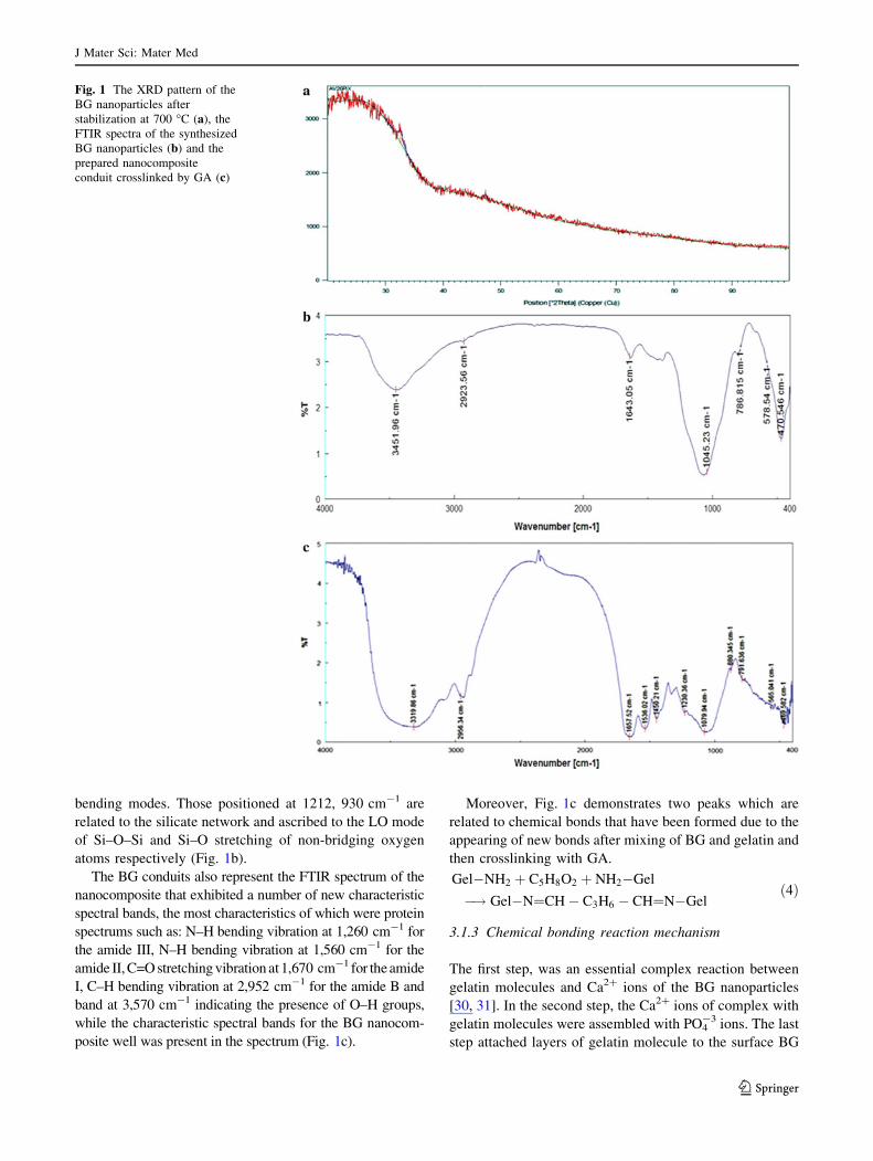
bending modes. Those positioned at 1212, 930 cm-1 are
related to the silicate network and ascribed to the LO mode
of Si–O–Si and Si–O stretching of non-bridging oxygen
atoms respectively (Fig. 1b).
The BG conduits also represent the FTIR spectrum of the
nanocomposite that exhibited a number of new characteristic
spectral bands, the most characteristics of which were protein
spectrums such as: N–H bending vibration at 1,260 cm-1 for
the amide III, N–H bending vibration at 1,560 cm-1 for the
amide II, C=O stretching vibration at 1,670 cm-1 for the amide
I, C–H bending vibration at 2,952 cm-1 for the amide B and
band at 3,570 cm-1 indicating the presence of O–H groups,
while the characteristic spectral bands for the BG nanocom-
posite well was present in the spectrum (Fig. 1c).
Moreover, Fig. 1c demonstrates two peaks which are
related to chemical bonds that have been formed due to the
appearing of new bonds after mixing of BG and gelatin and
then crosslinking with GA.
Gel�NH2 þ C5H8O2 þ NH2�Gel
�! Gel�N¼CH� C3H6 � CH¼N�Gelð4Þ
3.1.3 Chemical bonding reaction mechanism
The first step, was an essential complex reaction between
gelatin molecules and Ca2? ions of the BG nanoparticles
[30, 31]. In the second step, the Ca2? ions of complex with
gelatin molecules were assembled with PO4-3 ions. The last
step attached layers of gelatin molecule to the surface BG
Fig. 1 The XRD pattern of the
BG nanoparticles after
stabilization at 700 �C (a), the
FTIR spectra of the synthesized
BG nanoparticles (b) and the
prepared nanocomposite
conduit crosslinked by GA (c)
J Mater Sci: Mater Med
123
nanoparticles as the result of chemical bonding between the
P–O and O–H groups of BG nanoparticles with –COOH
and –NH2 groups in the gelatin molecule. There are three
main steps for chemical bonding between the gelatin
molecule and the BG nanoparticles in the nanocomposites.
Furthermore, there are two main sources of stabilization;
one is spatial stabilization and the other electrostatic sta-
bilization which avoids nanoparticle agglomeration in
these system types [32].
The spatial stabilization in this reaction system forms a
thick layer on the surface of the BG nanoparticles, mainly
due to the chemical absorption of gelatin molecules on the
BG nanoparticles.
Electrostatic stabilization is mainly due to adsorption of
Ca2? ions to the surface of the BG nanoparticles in the
reactions. In other words, the ionization of carboxyl is
enforced while that of amino is restrained, and the carboxyl
ions of gelatin are consequently against the ions in the
electrical double layer.
3.1.4 TEM and SEM observations
The BG powder particle size showed particles of less than
100 nm (data not shown). These tests confirm the Nano-
scale size of the synthesized BG nanoparticles.
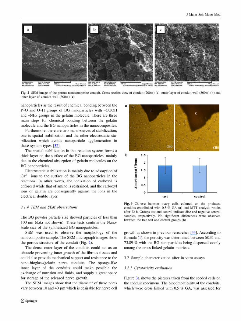
SEM was used to observe the morphology of the
nanocomposite sample. The SEM micrograph images show
the porous structure of the conduit (Fig. 2).
The dense outer layer of the conduits could act as an
obstacle preventing inner growth of the fibrous tissues and
could also provide mechanical support and resistance to the
nano-bioglass/gelatin nerve conduits. The sponge-like
inner layer of the conduits could make possible the
exchange of nutrition and fluids, and supply a great space
for storage of the released nerve growth.
The SEM images show that the diameter of these pores
vary between 10 and 40 lm which is desirable for nerve cell
growth as shown in previous researches [33]. According to
formula (1), the porosity was determined between 68.31 and
73.89 % with the BG nanoparticles being dispersed evenly
among the cross-linked gelatin matrices.
3.2 Sample characterization after in vitro assays
3.2.1 Cytotoxicity evaluation
Figure 3a shows the pictures taken from the seeded cells on
the conduit specimens. The biocompatibility of the conduits,
which were cross linked with 0.5 % GA, was assessed for
Fig. 2 SEM image of the porous nanocomposite conduit. Cross-section view of conduit (2009) (a), outer layer of conduit wall (5009) (b) and
inner layer of conduit wall (3009) (c)
Fig. 3 Chinese hamster ovary cells cultured on the produced
conduits crosslinked with 0.5 % GA (a) and MTT analysis results
after 72 h. Groups test and control indicate disc and negative control
samples, respectively. No significant differences were observed
between the two test and control groups (b)
J Mater Sci: Mater Med
123
cellular attachment, spreading and finally developing
filopodias.
After 3 days of culture, the CHO cells had attached and
proliferated in the surroundings of the conduit.
3.2.2 MTT detection of viable cells
MTT tests showed that cell viability of Miapaca-2 cultured
in the conduit extract was not significantly different from
that in the plain medium of cells during 72 h. This result
clearly suggests that the fabricated conduits were nontoxic
and posed as good candidates to be used as nerve conduits.
The results obtained from the test and control after 72 h
showed no significant cytotoxicity effects (p [ 0.05)
(Fig. 3b).
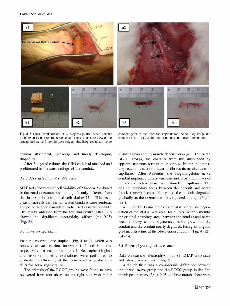
3.3 In vivo experiment
Each rat received one implant (Fig. 4 (a1)), which was
removed at various time intervals: 1, 2 and 3 months,
respectively. At each time interval, electrophysiological
and histomorphometric evaluations were performed to
evaluate the efficiency of the nano bioglass/gelatin con-
duits for nerve regeneration.
The animals of the BGGC groups were found to have
recovered from foot ulcers on the right side with minor
visible gastrocnemius muscle degeneration (n = 15). In the
BGGC groups, the conduits were not surrounded by
apparent neuroma formation or serious chronic inflamma-
tory reaction and a thin layer of fibrous tissue abundant in
capillaries. After 3 months, the bioglass/gelatin nerve
conduit implanted in rats was surrounded by a thin layer of
fibrous connective tissue with abundant capillaries. The
original boundary areas between the conduit and nerve
(black arrows) became blurry and the conduit degraded
gradually as the regenerated nerve passed through (Fig. 4
(a2)).
At 1 month during the experimental period, no degra-
dation of the BGGC was seen, for all rats. After 3 months
the original boundary areas between the conduit and nerve
became blurry as the regenerated nerve grew into the
conduit and the conduit nearly degraded, losing its original
guidance structure at the observation endpoint (Fig. 4 (a2),
(b1–3)).
3.4 Electrophysiological assessment
Data comparison electrophysiology of EMAP amplitude
and latency was shown in Fig. 5.
Although there was a considerable difference between
the normal nerve group and the BGGC group in the first
month post-surgery (*p \ 0.05), at three months there were
Fig. 4 Surgical implantation of a bioglass/gelatin nerve conduit
bridging an 10 mm sciatic nerve defect in rats (a) and the view of the
regenerated nerve 3 months post-surgery (b). Bioglass/gelatin nerve
conduits prior to and after the implantation. Nano Bioglass/gelatin
conduit (b1), 1 (b2), 2 (b3) and 3 months (b4) after implantation
J Mater Sci: Mater Med
123
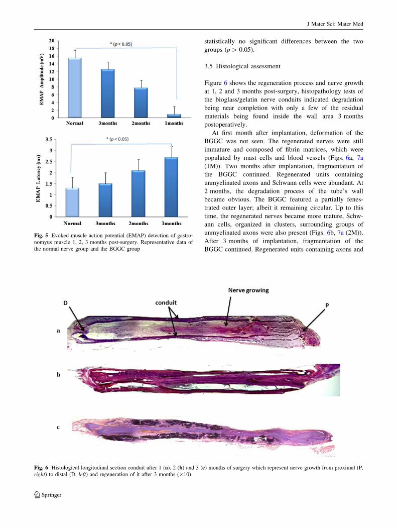
statistically no significant differences between the two
groups (p [ 0.05).
3.5 Histological assessment
Figure 6 shows the regeneration process and nerve growth
at 1, 2 and 3 months post-surgery, histopathology tests of
the bioglass/gelatin nerve conduits indicated degradation
being near completion with only a few of the residual
materials being found inside the wall area 3 months
postoperatively.
At first month after implantation, deformation of the
BGGC was not seen. The regenerated nerves were still
immature and composed of fibrin matrices, which were
populated by mast cells and blood vessels (Figs. 6a, 7a
(1M)). Two months after implantation, fragmentation of
the BGGC continued. Regenerated units containing
unmyelinated axons and Schwann cells were abundant. At
2 months, the degradation process of the tube’s wall
became obvious. The BGGC featured a partially fenes-
trated outer layer; albeit it remaining circular. Up to this
time, the regenerated nerves became more mature, Schw-
ann cells, organized in clusters, surrounding groups of
unmyelinated axons were also present (Figs. 6b, 7a (2M)).
After 3 months of implantation, fragmentation of the
BGGC continued. Regenerated units containing axons and
Fig. 5 Evoked muscle action potential (EMAP) detection of gastro-
nomyus muscle 1, 2, 3 months post-surgery. Representative data of
the normal nerve group and the BGGC group
Fig. 6 Histological longitudinal section conduit after 1 (a), 2 (b) and 3 (c) months of surgery which represent nerve growth from proximal (P,
right) to distal (D, left) and regeneration of it after 3 months (910)
J Mater Sci: Mater Med
123
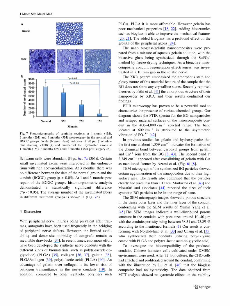
Schwann cells were abundant (Figs. 6c, 7a (3M)). Certain
small myelinated axons were interposed in the endoneu-
rium with rich neovascularization. At 3 months, there was
no difference between the data of the normal group and the
conduit (BGGC) group (p [ 0.05). At 1 and 3 months post
repair of the BGGC groups, histomorphometric analysis
demonstrated a statistically significant difference
(*p \ 0.05). The average number of the myelinated fibers
in different treatment groups is shown in (Fig. 7b).
4 Discussion
With peripheral nerve injuries being prevalent after trau-
mas, autografts have been used frequently in the bridging
of peripheral nerve defects. However, the limited avail-
ability and donor-site morbidity of autografts remain as
inevitable drawbacks [34]. In recent times, enormous effort
have been developed the synthetic nerve conduits with the
different kinds of biomaterials, such as poly(L-lactide-co-
glycolide) (PLGA) [35], collagen [36, 37], gelatin [38],
PLGA/collagen [39], poly(L-lactic acid) (PLLA) [40]. An
advantage of gelatin over collagen is its lower risk of
pathogen transmittance in the nerve conduits [19]. In
addition, compared to other Synthetic polymers such
PLGA, PLLA it is more affordable. However gelatin has
poor mechanical properties [18, 22]. Adding bioceramics
such as bioglass is able to improve the mechanical features
[20, 21]. The added Bioglass has a profound effect on the
growth of the peripheral axons [24].
The nano bioglass/gelatin nanocomposites were pre-
pared from a mixture of aqueous gelatin solution, with the
bioactive glass being synthesized through the Sol/Gel
method by freeze-drying techniques. As a bioactive nano-
composite conduit, regeneration effectiveness was inves-
tigated in a 10 mm gap in the sciatic nerve.
The XRD pattern emphasized the amorphous state and
glossy nature of this material feature of the sample that the
BG does not show any crystalline states. Recently reported
theories by Fathi et al. [41] the amorphous structure of their
nanopowder by XRD, and their results confirmed our
findings.
FTIR microscopy has proven to be a powerful tool to
characterize the presence of various chemical groups. Our
diagram shows the FTIR spectra for the BG nanoparticles
and scraped material surfaces of the nanocomposite con-
duit in the 400–4,000 cm-1 spectral range. The band
located at 609 cm-1 is attributed to the asymmetric
vibration of PO43- [42].
In previous studies for gelatin and hydroxyapatite that
the first one at about 1,359 cm-1 indicates the formation of
the chemical bond between carboxyl groups from gelatin
and Ca2? ions from the BG [8, 43]. The second band at
2,349 cm-1 appeared after crosslinking of gelatin with GA
as mentioned former by Azami et al. (Fig. 6) [8].
TEM micrograph of the synthesized BG particles showed
certain agglomeration of the nanopowders due to their high
surface area. The results also confirmed that the particles
clearly had sizes less than 100 nm. Mortazavi et al. [43] and
Mozafari and associates [44] reported the sizes of their
synthetic BG particles to be in the range of nano.
The SEM micrograph images showed a porous structure
in the dense outer layer and the inner layer of the conduit,
conforming with the SEM results of Yumin Yang et al.
[45].The SEM images indicate a well-distributed porous
structure in the conduits with pore sizes around 10–40 lm
with the conduits porosity being between 68.31 and 73.89 %
according to the mentioned formula (1) Our result is con-
forming with Nojehdehian et al. [33] and Chang et al. [35]
who synthesized their conduits utilizing poly-L-lysine
coated with PLGA and poly(DL-lactic acid-co-glycolic acid).
To investigate the biocompatibility of the produced
conduits, Chinese hamsters cells cultivated under DMEM
environment were used. After 72 h of culture, the CHO cells
had attached and proliferated around the conduit, confirming
with the illustration by Lu et al. [46] that the bioglass
composite had no cytotoxicity. The data obtained from
MTT analysis showed no cytotoxic effects on the viability
Fig. 7 Photomicrographs of semithin sections at 1 month (1M),
2 months (2M) and 3 months (3M) post-surgery in the normal and
BGGC groups. Scale (bottom right) indicates of 20 lm (Toluidine
blue staining 9100) (a) and number of the myelinated axons at
1 month (1M), 2 months (2M) and 3 months (3M) post-surgery (b)
J Mater Sci: Mater Med
123
and proliferation properties of the cells during 72 h. Sona
Jantova and colleagues reported that the bioglass scaffold
had a slight cytotoxicity and was biocompatible [47].
Therefore their data confirmed our data in this regards.
As a result, we found that successful regeneration of the
nerves across the gap occurred in all of the nano bioglass/
gelatin conduits even at the shortest experimental time
point of 1 month. The thin layer of the surrounding fibrous
tissue and the minimal inflammation also indicated that the
nano bioglass/gelatin conduit were biocompatible. These
results are not surprising since gelatin and bioglass have
both been shown to be biocompatible materials [24, 48].
Regeneration of the myelinated axons was observed in
the distal segment of all groups. The mean number of the
myelinated fibers was used as a parameter for morphom-
etry. In the BGGC group, rats presented a greater mean
number at 3 months compared to those from 1 and
2 months. At 3 months, no significant difference was
observed between the BGGC and normal group. Our result
is conforming with Yueh-Sheng Chen et al. [38].
Measurements of peak amplitude and latency both showed
an increase as a function of the experimental period, which
suggested that the transected nerve had undergone adequate
regeneration and was approximately near to the normal group.
Our result is conforming with Wenwen associates [49].
On the other hand, since rats display regeneration in the
peripheral nervous system superior to that in humans, the
results of this study can be used as a starting point for
further investigations in higher species, for example, in
dogs or cats [50, 51].
5 Conclusion
According to our study, the nano-bioglass/gelatin conduit
could be a suitable candidate for peripheral nerve regen-
eration as a biocompatible, biodegradable and novel
biomaterial.
Acknowledgments This work was supported by the grant from the
Nano biotechnology research center of Baqiyatallah University of
Medical Sciences and Iranian National Sciences foundation (INSF).
References
1. Moore MJ, Friedman JA, Lewellyn EB, Mantila SM, Krych AJ,
Ameenuddin S, et al. Multiple-channel scaffolds to promote
spinal cord axon regeneration. Biomaterials. 2006;27:419–29.
2. Prang P, Muller R, Eljaouhari A, Heckmann K, Kunz W, Weber
T, et al. The promotion of oriented axonal regrowth in the injured
spinal cord by alginate-based anisotropic capillary hydrogels.
Biomaterials. 2006;27:3560–9.
3. Amillo S, Yanez R, Barrios RH. Nerve regeneration in different
types of grafts: experimental study in rabbits. Microsurgery.
1995;16:621–30.
4. Balgude AP, Yu X, Szymanski A, Bellamkonda RV. Agarose gel
stiffness determines rate of DRG neurite extension in 3D cultures.
Biomaterials. 2001;22:1077–84.
5. Chang JY, Lin JH, Yao CH, Chen JH, Lai TY, Chen YS. In vivo
evaluation of a biodegradable EDC/NHS-cross-linked gelatin
peripheral nerve guide conduit material. Macromol Biosci.
2007;7:500–7.
6. Pfister LA, Christen T, Merkle HP, Papaloızos M, V B. Novel
biodegradable. Novel biodegradable nerve conduits for peripheral
nerve regeneration. Eur Cells and Mater. 2004;7:16–7.
7. Verreck G, Chun I, Li Y, Kataria R, Zhang Q, Rosenblatt J, et al.
Preparation and physicochemical characterization of biodegrad-
able nerve guides containing the nerve growth agent sabeluzole.
Biomaterials. 2005;26:1307–15.
8. Azami M, Moztarzadeh F, Tahriri M. Preparation characteriza-
tion and mechanical properties of controlled porous gelatin/
hydroxyapatite nanocomposite through layer solvent casting
combined with freeze-drying and lamination techniques. Porous
Mater. 2009;17:313–20.
9. Lutolf MP, Hubbell JA. Synthetic biomaterials as instructive
extracellular microenvironments for morphogenesis in tissue
engineering. Biotechnology. 2005;23:74–5.
10. Lawrencin CT, Amin SFE, Ibim SE, Willoughby DA, Attavia M,
Allcock HR, et al. A highly porous 3-dimensional polyphospha-
zene polymer matrix for skeletal tissue regeneration. J Biomed
Mater Res. 1996;30:133–8.
11. Yuan H, De Bruijn JD, Zhang X, Bitterswijk CAV, De Groot K.
Factors affecting the structure and properties of bioactive foam
scaffolds for tissue engineering. J Biomed Mater Res.
2008;68:270–6.
12. Huang ZM, Zhang YZ, Ramakrishna S, Lim CT. Electrospinning
and mechanical characterization of gelatin nanofibers. Polymer.
2004;45:5361–8.
13. Jones JR, Ahir S, Hench LL. Large-scale production of 3D bio-
active glass macroporous. Sol-Gel Sci Technol. 2004;29:179–88.
14. Taboas JM, Maddox RD, Krebsbach PH, Hollister SJ. Indirect
solid free form fabrication of local and global porous, biomimetic
and composite 3D polymer-ceramic scaffolds. Biomaterials.
2003;24:181–94.
15. Carrasquillo KG, Stanley AM, Aponte-Carro JC, De Jesus PDJ,
Costantino HR, Bosques CJ. Non-aqueous encapsulation of
excipient-stabilized spray freeze dried BSA into poly(lactide-co-
glycolide) microspheres results in release of native protein.
Control Release. 2001;76:199–208.
16. Fu H, Fu Q, Zhou N, Huang W, Rahaman MN, Wang D, et al.
In vitro evaluation of borate-based bioactive glass scaffolds
prepared by a polymer foam replication method. Eng Mater Sci
Lett. 2009;29:2079–312.
17. Boland ED, Espy P, Bowlin GL. Tissue engineering scaffolds. In:
Encyclopaedia of Biomaterials and biomedical engineering.
2004. p. 1633–5.
18. Boedtker H, Doty PA. A study of gelatin molecules aggregates
and gels. J Phys Chem. 1954;58:968–83.
19. Gardin C, Ferroni L, Favero L, Stellini E, Stomaci D, Sivolella S,
et al. Nanostructured biomaterials for tissue engineered bone
tissue reconstruction. Int J Mol Sci. 2012;13:737–57.
20. Rezwan K, Chen QZ, Blaker JJ, Boccaccini AR. Biodegradable
and bioactive porous polymer/inorganic composite scaffolds for
bone tissue engineering. Biomaterials. 2006;27:3413–31.
21. Guarino V, Causa F, Ambrosio L. Bioactive scaffolds for bone
and ligament tissue. Expert Rev Med Devices. 2007;4:405–18.
22. Liu X, Smith LA, Hu J, Ma PX. Biomimetic nanofibrous gelatin/
apatite composite scaffolds for bone tissue engineering. Bioma-
terials. 2009;30:2252–8.
23. Mozafari M, Moztarzadeh F, Rabiee M, Azami M, Maleknia S,
Tahriri M, et al. Development of macroporous nanocomposite
J Mater Sci: Mater Med
123
scaffolds of gelatin/bioactive glass prepared through layer solvent
casting combined with lamination technique for bone tissue
engineering. Ceram Int. 2010;36:2431–9.
24. Bunting S, Silvio LD, Deb S, Hall S. Bioresorbable glass fibres
facilitate peripheral nerve regeneration. Hand Surgery. 2005;30:
242–7.
25. Jones JR. Review of bioactive glass: from Hench to hybrids. Acta
Biomater. 2013;9:4457–86.
26. Picot J. Human cell culture protocol. CA, USA; 2004.
27. Fassina L, Saino E, Visai L, Avanzini MA, Cusella De Angelis MG,
Benazzo F, Van Vlierberghe S, Dubruel P, Magenes G. Use of a
gelatin cryogel as biomaterial scaffold in the differentiation process
of human bone marrow stromal cells. Conf Proc IEEE Eng Med
Biol Soc. 2010;247–50. doi:10.1109/IEMBS.2010.5627475.
28. Hafezi F, Hosseinnejad F, Fooladi AA, Mohit Mafi S, Amiri A,
Nourani MR. Transplantation of nano-bioglass/gelatin scaffold in
a non-autogenous setting for bone regeneration in a rabbit ulna.
J Mater Sci Mater Med. 2012;23:2783–92.
29. Eto M, Yoshikawa H, Fujimura H, Naba I, Sumi-Akamaru H,
Takayasu S, et al. The role of CD36 in peripheral nerve remye-
lination after crush injury. Eur J Neurosci. 2003;17:2659–66.
30. Teng S, Shi J, Peng B, Chen FL. The effect of alginate addition
on the structure and morphology of hydroxyapatite/gelatin
Nanocomposites. Compos Sci Technol. 2006;66:1532–8.
31. Chang MC, Ko CC, Douglas WH. Conformational change of
hydroxyapatite-gelatin nanocomposite by glutaraldehyde. Bio-
materials. 2003;24:3087–94.
32. Minfang C, Junjun T, Yuying L, Debao L. Preparation of gelatin
coated hydroxyapatite nanorods and the stability of its aqueous
colloidal. Appl Surf Sci. 2008;254:2730–5.
33. Nojehdehian H, Moztarzadeh F, Baharvand H, Nazarian H,
Tahriri M. Preparation and surface characterization of poly-L-
lysine-coated PLGA microsphere scaffolds containing retinoic
acid for nerve tissue engineering: in vitro study. Colloids Surf B
Biointerfaces. 2009;73:23–9.
34. Bian YZ, Wang Y, Aibaidoula G, Chen GQ, Wu Q. Evaluation of
poly(3-hydroxybutyrate-co-3-hydroxyhexanoate) conduits for
peripheral nerve regeneration. Biomaterials. 2009;30:217–25.
35. Chang C-J, Hsu S-H. The effect of high outflow permeability in
asymmetric poly(DL-lactic acid-co-glycolic acid) conduits for
peripheral nerve regeneration. Biomaterials. 2006;27:035–1042.
36. Ahmed MR, Vairamuthu S, Shafiuzama M, Basha SH, Jayakumar
R. Microwave irradiated collagen tubes as a better matrix for
peripheral nerve regeneration. Brain Res. 2005;1046:55–67.
37. Schnell E, Klinkhammer K, Balzer S, Gary B, Doris K, Paul D,
et al. Guidance of glial cell migration and axonal growth on
electrospun nanofibers of poly-epsilon-caprolactone and a col-
lagen/poly-epsilon-caprolactone blend. Biomaterials. 2007;28:
3012–25.
38. Chen YS, Chang J, Cheng CY, Tsai FJ, Yao CH, Liu BS. An
in vivo evaluation of a biodegradable genipin-cross-linked gelatin
peripheral nerve guide conduit material. Biomaterials. 2005;26:
3911–8.
39. Liu B, Cai SX, Ma KW, Xu ZL, Dai XZ, Yang L, Lin C, Fu XB,
Sung KL, Li XK. Fabrication of a PLGA-collagen peripheral
nerve scaffold and investigation of its sustained release property
in vitro. J Mater Sci Mater Med. 2008;19:1127–32.
40. Evans JR, Brandt K, Katz S, Chauvin P, Otto L, Bogle M. Bio-
active poly(L-lactic acid) conduits seeded with Schwann cells for
peripheral nerve regeneration. Biomaterials. 2002;23:841–8.
41. Fathi MH, Mortzavi VA, Doostmohammadi A. Bioactive glass
nanopowder for the treatment of oral bone defects. Dentistry.
2007;4:115–22.
42. Mami M, Lucas-Girot A, Oudadesse H, Dorbez-Sridi R, Mezahi
F, Dietrich E, et al. Investigation of the surface reactivity of a sol
gel derived glass in the ternary system SiO2-CaO-P2O5. Appl Surf
Sci. 2008;254:7386–93.
43. Mortazavi V, Nahrkhalaji MM, Fathi MH, Mousavi SB, Esfahani
BN. Antibacterial effects of sol-gel-derived bioactive glass
nanoparticle on aerobic bacteria. Biomed Mater Res A. 2010;94:
160–8.
44. Masoud M, Rabiei M, Azami M, Maleknia S. Biomimetic for-
mation of apatite on the surface of porous gelatin/bioactive glass
nanocomposite scaffolds. Appl Surf Sci. 2010;257:1740–9.
45. Yang Y, Zhao W, He J, Zhao Y, Ding F, Gu X. Nerve conduits
based on immobilization of nerve growth factor onto modified
chitosan by using genipin as a crosslinking agent. Pharm Bio-
pharmaceutics. 2011;79:519–25.
46. Lu HH, El-Amin SF, Scott KD, Laurencin CT. Three-dimen-
sional, bioactive, biodegradable, polymer-bioactive glass com-
posite scaffolds with improved mechanical properties support
collagen synthesis and mineralization of human osteoblast-like
cells in vitro. Biomed Mater Res A. 2003;64:465–74.
47. Jantova S, Theiszova M, Matejov P, Bakos D. Biocompatibility
and cytotoxicity of bioglass-ceramic composite with various
P2O5 content in Li2O-SiO2-CaO-CaF2-P2O5 system on fibroblast
cell lines. Acta Chimica Slovaca. 2011;4:15–30.
48. Mligiliche NL, Tabata Y, Ide C. Nerve regeneration through
biodegradable gelatin conduits in mice. East Afr Med J. 1999;76:
400–6.
49. Yu W, Zhau W, Zhu C, Zhang X, Ye D, Zhang W, et al. Sciatic
nerve regeneration in rats by a promising electrospun collagen/
poly(e-caprolactone) nerve conduit with tailored degradation rate.
Neuroscience. 2011;12:1471–2202.
50. Peker F, Solakoglu C, Yuksel F, Kutlay M. Effects of time lapse
on results of partial nerve injury repair. J Reconstr Microsurg.
2005;21:145–9.
51. Sinis N, Schulte-Eversum C, Doser M, Muller HW. Nerve
regeneration across a 2-cm gap in the rat median nerve using a
resorbable nerve conduit filled with Schwann cells. J Neurosurg.
2005;103:1067–76.
J Mater Sci: Mater Med
123


