Embed Size (px)
Citation preview
https://doi.org/10.5933/JKAPD.2017.44.2.210 J Korean Acad Pediatr Dent 44(2) 2017ISSN (print) 1226-8496 ISSN (online) 2288-3819
Prevalence and Clinical Features of Molar-Incisor Hypomineralization in Adolescents in Yangsan
Jonghyun Shin, Geumlang Lee, Jongsoo Kim, Jiyeon Kim, Shin Kim
Department of Pediatric Dentistry, School of Dentistry, Pusan National University
This study aimed to investigate the prevalence and distribution of enamel hypomineralization, including molar-incisor
hypomineralization (MIH), among adolescents and assess their correlation with esthetic satisfaction.
A total of 1371 adolescents between the ages of 14 and 16 years in Yangsan city were evaluated for enamel
hypomineralization, including MIH, according to the European Academy of Paediatric Dentistry (EAPD) criteria. In a
parallel survey, esthetic satisfaction about anterior teeth and its correlation with incisor enamel hypomineralization were
analyzed.
The prevalence of MIH was 13.8% (n = 189), while that of hypomineralization in any permanent tooth was 23.2% (n =
318), which was substantially greater compared to the national prevalence of MIH. Mandibular first molars exhibited the
highest prevalence of hypomineralization, followed by maxillary central incisors and mandibular second molars. Among
anterior teeth, the most frequently affected site was the incisal edge of maxillary central incisors. A high degree of
hypomineralization in anterior teeth was associated with a high demand for esthetic treatment.
Key words : Molar incisor hypomineralization, Adolescents, Prevalence, Esthetics, Anterior tooth
Abstract
210
Corresponding author : Shin KimDepartment of Pediatric Dentistry, School of Dentistry, Pusan National University, 20, Geumo-ro, Mulgeum-eup, Yangsan, 50612, KoreaTel: +82-55-360-5180 / Fax: +82-55-360-5174 / E-mail: [email protected] December 1, 2016 / Revised February 10, 2017 / Accepted January 25, 2017※This study was supported by 2015 Clinical Research Grant, Pusan National University Dental Hospital.
I. Introduction
With recent improvements in the level of oral care, the
prevalence of dental caries, a prominent oral disease, has been
decreasing[1,2]. In contrast, the prevalence of enamel hypo-
mineralization due to systemic factors has increased, which has
heightened interest regarding this condition[3,4].
Crown formation in first permanent molars and incisors oc-
curs at approximately the same time[5], during which enamel
hypomineralization is frequently observed. Enamel hypomin-
eralization in first permanent molars and anterior teeth was
first reported in 1970 in Sweden[6]. In 2001, Weekheiim et
al .[7] defined MIH as demarcated, qualitative enamel defects
of systemic origin, affecting one or more permanent molars
with or without involvement of incisor teeth. Since then, sev-
eral studies worldwide have actively investigated the etiology,
conditions, and prevalence of MIH, with most focusing on first
permanent molars and incisors.
However, the clinical categorization of hypomineralization
is broad, and clinically, all primary teeth, permanent canines,
premolars, and second permanent molars can be affected by
enamel hypomineralization[8-10]. Therefore, in a recent policy,
J Korean Acad Pediatr Dent 44(2) 2017
211
the European Academy of Paediatric Dentistry (EAPD) recom-
mended that whole dentition be included in future studies on
enamel hypomineralization[11].
Several studies have investigated the causes of MIH[12]. A
recent study reported that, while a significant correlation exist-
ed between MIH and polyvinyl chloride/dioxin discharge, MIH
was only weakly correlated with nutritional status, birth condi-
tions, childhood diseases, and fluoride intake[13]. In another
study, water fluoridation and pregnancy at an older age were
identified as risk factors for MIH[14,15]. High vitamin D con-
centration in blood has also been reported to be negatively
correlated with MIH. While it appears that MIH is caused by
various risk factors, no definite risk factors have been identified
yet[16].
Teeth affected by MIH exhibit enamel defects and rapid
progression of caries; therefore, in many cases, affected teeth
require restorative treatment right after eruption. However,
owing to the high organic content of enamel, the success rate
of adhesion of restorations is low[17,18]. If the defect is deep
and widespread, it can lead to severe loss of tooth material
and early loss of permanent teeth because of caries progres-
sion. In addition, in adolescents with active social lives and a
developing interest in appearance[19], slight defects can have
a serious impact on self-esteem, depending on the location of
the defect.
Despite the significant impact of MIH on adolescents, previ-
ous studies have only investigated MIH among elementary
school students[20], and no study till date has evaluated MIH
among a teenage population. The subjects of the present
study were middle-school students with complete eruption of
permanent dentition. The prevalence, defect patterns, and dis-
tribution of permanent tooth enamel hypomineralization, in-
cluding MIH, and their effects on tooth condition and esthetic
satisfaction were evaluated.
II. Materials and Methods
1. Subjects
An inspector performed oral examination of 1371 ado-
lescent students between the ages of 14 and 16 years at a
middle school in Yangsan province, South Korea, between May
and August 2013. Water fluoridation had not been enforced
in this area, and the fluoride concentration in water was lower
than the detection limit of 0.15 ppm. To eliminate potential
confounders, subjects with primary teeth remaining or pre-
emergent second permanent molars were excluded from the
study. Subject age and sex distribution are presented in Table 1.
2. Research Methods
1) Preliminary investigation and ethical considerations
Prior to the survey, the inspector underwent training with
clinical images and patients who visited the Pediatric Dentistry
department at Pusan Dental Hospital, in order to establish
consistency in diagnosis. With the help of an expert, the in-
spector was administered a blind test using 50 images of teeth
with MIH, enamel hypoplasia, or fluorosis, with the accuracy
set initially at 96.0%. The same images were retested a month
later, and the findings of the first and second tests exhibited a
very good agreement, with a Kappa value of 0.87.
This study was approved through review by the Bioeth-
ics Committee of Pusan University Dental Hospital (PNUDH-
2013-017).
2) Oral examination
Oral examination was performed using a head light, den-
tal mirror, and community periodontal index probe, with the
examinee seated in a chair in the health service room of the
school. Subjects were instructed to brush their teeth prior to
examination, and, when necessary, a gauze was used to re-
move food debris and dental plaques.
(1) Enamel hypomineralization
Hypomineralization in the dentition-from the central incisor
to the second permanent molar-was evaluated. Enamel hypo-
mineralization was considered present when an impervious
enamel defect with a clearly visible white or brown-colored
boundary was observed while the tooth was wet. However,
cases in which the lesion was less than 1 mm diameter, cases
Table 1. Distribution of sex and age of the subjects
Total (%) Boys (%) Girls (%)
Total 1,371 (100.0) 765 (55.8) 606 (44.2)
Age (years) 14 316 (23.0) 175 (22.9) 141 (23.3)
15 498 (36.3) 275 (35.9) 223 (36.8)
16 557 (40.6) 315 (41.2) 242 (39.9)
J Korean Acad Pediatr Dent 44(2) 2017
212
of trauma and cases of periapical inflammation in primary
teeth were excluded[21]. Teeth diagnosed with enamel hypo-
mineralization were classified according to the criteria pro-
posed by the EAPD in 2003 (Table 2)[22]. Further, the labial
surfaces of incisors, lateral incisors, and canines were divided
into nine compartments, and the presence and distribution of
enamel defects in each compartment were also recorded for
evaluation of esthetics. Subjects were assigned to one of three
groups: the normal (no defects), EH (permanent dentition with
defects in at least one tooth), or MIH (defects in first perma-
nent molars) groups.
3) Questionnaire survey
A questionnaire survey was conducted to determine the
correlation between state of enamel hypomineralization and
esthetic satisfaction about anterior teeth. The questionnaires
were distributed at the examination site, and subjects com-
pleted them under the guidance of teachers.
4) Data analysis
The collected data were analyzed using SPSS 13.0 (SPSS Inc.,
USA) for Windows. The level of significance was set at 0.05 for
all statistical data. The significance of sex and age with regard
to prevalence was verified by the chi-square test. Incidence of
tooth-specific defects was determined by frequency analysis.
The correlation between first permanent molars and the rest
of the permanent teeth in affected individuals was determined
by correlation analysis.
III. Results
1. Prevalence of enamel hypomineralization
The distribution of prevalence of enamel hypomineralization
among the subject pool is presented in Table 3. Of the total
1371 subjects, 318 (23.2%) were assigned to the EH group and
189 (13.8%) to the MIH group. In both groups, the prevalence
of enamel hypomineralization among female subjects was
greater compared to that among men, although the difference
was insignificant (p > 0.05); however, there were significant dif-
ferences in prevalence of enamel hypomineralization according
to age (p < 0.05).
Table 3. Prevalence of enamel hypomineralization
Normal EH MIHp-value
N % N % N %
Sex Male 602 78.7 163 21.3 98 12.80.06
Female 451 74.4 155 25.6 91 15.0
Age 14 238 75.3 78 24.7 50 15.8
< 0.0515 359 72.1 139 27.9 81 16.3
16 456 81.9 101 18.1 58 10.4
Total 1053 76.8 318 23.2 189 13.8
Chi-square test
Table 2. EAPD criteria for molar incisor hypomineralization
Code Definition Description
O Demarcated opacity The affected teeth show clearly demarcated opacities at the occlusal and buccal part of the crown
P Enamel disintegrationSeverely affected enamel which breaks down soon after being subjected to masticatory forces, leading to unprotected dentin and rapid caries development
R Atypical restorationFirst permanent molars and incisors with restorations revealing similar extensions as MIH are rec-ommended to be judged as affected
E Extracted teethExtracted teeth can be defined as having MIH only in cases in which there are notes in the records or demarcated opacities on the other permanent first molar
J Korean Acad Pediatr Dent 44(2) 2017
213
The average numbers of affected teeth in the EH and MIH
groups were 3.82 (boys, 3.88; girls, 3.76) and 5.18 (boys, 5.22;
girls, 5.13), respectively, with no significant differences accord-
ing to sex (p > 0.05; Table 4).
Table 4. Number of teeth affected by enamel hypomineralization and molar incisor hypomineralization
Male Female Total
EH Affected teeth 634 583 1,217
Affected children 163 155 318
Mean(teeth) 3.88 3.76 3.82
SD 3.76 3.04 3.42
p-value 0.75
MIH Affected teeth 512 467 979
Affected children 98 91 189
Mean(teeth) 5.22 5.13 5.18
SD 4.26 3.23 3.79
p-value 0.87
Frequency analysis
Table 5. Distribution of affected teeth by tooth type
Right (%) Left (%)
M2 M1 P2 P1 C LI CI CI LI C P1 P2 M1 M2
Upper 2.2 7.4 1.3 1.6 1.8 3.8 9.3 9.0 3.0 2.6 1.6 1.7 6.3 1.4
Lower 4.6 9.3 1.6 1.7 1.0 1.6 2.9 3.2 1.7 0.7 1.6 1.5 10.3 5.0
CI: central incisor, LI: lateral incisor, C: canine, P1: first premolar, P2: second premolar, M1: first molar, M2: second molar
Table 6. The correlation between first permanent molars and the rest of permanent teeth in regards to enamel hypomineralization defects
CI LI C P1 P2 M2
M1Correlation coefficient 0.50 0.33 0.25 0.35 0.37 0.44
p-value <0.05 <0.05 <0.05 <0.05 <0.05 <0.05
Correlation analysisCI: central incisor, LI: lateral incisor, C: canine, P1: first premolar, P2: second premolar, M1: first molar, M2: second molar
2. Distribution of defects according to tooth type
Among all teeth that exhibited enamel hypomineralization,
mandibular first permanent molars were the most commonly
affected, followed by maxillary central incisors, maxillary first
permanent molars, and mandibular second permanent molars,
in that order (Table 5).
Analysis of correlation between first permanent molars and
the rest of the permanent teeth with regard to enamel hypo-
mineralization defects revealed that presence of defects in first
permanent molars indicated the possibility of defects in the
rest of the permanent teeth. This correlation was the highest
among central incisors, followed by second permanent molars
(p < 0.05; Table 6).
3. Condition of affected teeth
Incisors and first and second permanent molars exhibited
high morbidity rates; Table 7 presents the results of analysis of
lesion patterns in these teeth according to the EAPD criteria.
In incisors, demarcated opacity was the most prevalent type
of defect at 99.7%, while the prevalence of enamel breakdown
Table 7. Status of affected incisors and molars
Incisor First molar Second molarp-value
N % N % N %
O 289 99.7 197 49.6 145 85.8
p < 0.05P 1 0.3 10 2.5 2 1.2
R 0 0.0 183 46.1 22 13.0
E 0 0.0 7 1.8 0 0.0
Chi-square testO: demarcated opacity, P: enamel disintegration, R: atypical restoration, E: extracted teeth
J Korean Acad Pediatr Dent 44(2) 2017
214
Table 8. The correlation between range of defects and esthetic satisfaction
No. of affected sections
No. of personsSatisfactionMean (SD)
p-value
0 1,123 2.91 (0.99)
< 0.05
1 - 10 222 2.76 (1.02)
11 - 20 16 2.63 (1.31)
21 - 30 6 2.33 (1.21)
31 - 40 2 1.00 (0.00)
41< 2 3.00 (0.00)
Chi-square test
was only 0.3%. In first permanent molars, demarcated opac-
ity was the most prevalent defect pattern at 49.6%, followed
by atypical restoration at 46.1%, and even extraction at 1.8%.
Similarly, in second permanent molars, demarcated opacity
was the most prevalent defect pattern at 85.8%, followed by
atypical restoration at 13.0%. The differences in defect patterns
according to tooth type were significant (p < 0.05).
4. Range of defects and need for treatment of labial
surfaces of incisors and canines
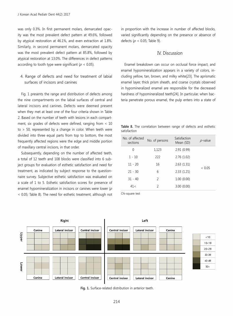
Fig. 1 presents the range and distribution of defects among
the nine compartments on the labial surfaces of central and
lateral incisors and canines. Defects were deemed present
when they met at least one of the four criteria shown in Table
2. Based on the number of teeth with lesions in each compart-
ment, six grades of defects were defined, ranging from < 10
to > 50, represented by a change in color. When teeth were
divided into three equal parts from top to bottom, the most
frequently affected regions were the edge and middle portion
of maxillary central incisors, in that order.
Subsequently, depending on the number of affected teeth,
a total of 12 teeth and 108 blocks were classified into 6 sub-
ject groups for evaluation of esthetic satisfaction and need for
treatment, as indicated by subject response to the question-
naire survey. Subjective esthetic satisfaction was evaluated on
a scale of 1 to 5. Esthetic satisfaction scores for presence of
enamel hypomineralization in incisors or canines were lower (p
< 0.05; Table 8). The need for esthetic treatment, although not
in proportion with the increase in number of affected blocks,
varied significantly depending on the presence or absence of
defects (p < 0.05; Table 9).
IV. Discussion
Enamel breakdown can occur on occlusal force impact, and
enamel hypomineralization appears in a variety of colors, in-
cluding yellow, tan, brown, and milky white[23]. The aprismatic
enamel layer, thick prism sheath, and coarse crystals observed
in hypomineralized enamel are responsible for the decreased
hardness of hypomineralized teeth[24]. In particular, when bac-
teria penetrate porous enamel, the pulp enters into a state of
Fig. 1. Surface-related distribution in anterior teeth.
J Korean Acad Pediatr Dent 44(2) 2017
215
Table 9. The correlation between range of defect and need for treatment
No. of affected sections
Esthetic treatment need
Total p-valueYes No
No. of persons % No. of persons %
0 336 29.9 787 70.1 1,123
<0.05
1 - 10 96 43.2 126 56.8 222
11 - 20 7 43.8 9 56.3 16
21 - 30 3 50.0 3 50.0 6
31 - 40 2 100.0 0 0.0 2
41< 0 0.0 2 100.0 2
Total 444 32.4 927 67.6 1,371
Chi-square test
chronic inflammation, which causes hypersensitivity and failure
of local anesthesia[24,25]. Because of the resultant constant
pain, young and adolescent patients affected by MIH often ex-
hibit behavior management issues during dental treatment.
In addition, enamel hypomineralization typically occurs at
locations other than those of dental caries, and when lesions
affect the cusp, appropriate restoration becomes very diffi-
cult[26]. Consequently, maintaining restorations is challenging,
and several claims are being raised that it is best to extract
teeth with poor prognosis in a timely manner[27]. Traditionally,
the ideal age for extraction of first permanent molars is 9 - 11
years[28]. However, recent studies are conflicted over the ap-
propriate age for extraction; some studies have proposed that
extraction should be performed at a younger age, whereas
others have stated that early extraction could increase the risk
of hypomineralization in second permanent molars[29].
Anterior teeth defects are treated with emphasis on esthetic
improvement. However, because microabrasion, resin restora-
tion, and whitening agents such as hydrogen peroxide usually
cause mineral loss[30], casein phosphopeptide-amorphous cal-
cium phosphate is now being used for active remineralization
treatment[31].
In general, the ideal age for MIH epidemiological studies is
7 - 8 years, during which the first four permanent molars will
have erupted and enamel loss due to dental caries or other
diseases would not have started yet. In addition, at this age,
children still possess a residual part of the second molar, which
makes it possible to evaluate the correlation between MIH
and second-molar hypomineralization. Elfrink et al . recently
suggested that primary molar hypomineralization and MIH
are correlated and that the former could be a predictor of
MIH[32]. In the present study, although the entire permanent
dentition was evaluated, hypomineralization of second perma-
nent molars was the focus of evaluation. Therefore, subjects
with complete dentition, between the ages of 14 and 16 years,
were enrolled as study subjects.
The most active research on prevalence of MIH is conducted
in Europe, where prevalence levels ranging from 3.6% to 37%
have been reported[10]. In Asian countries, prevalence of MIH
has been reported to be 2.8% in Hong Kong[33], 25.5% in Chi-
na[34], 20% in Thailand[35], and 6.0% in South Korea[20]. This
variation in prevalence might be attributable to environmental
or genetic factors or even differences in research methodol-
ogy and criteria among various studies[10]. In the present
study, the prevalence of MIH was 13.8%, which increased to
23.2% upon expanding the study sample to include subjects
in the stage prior to permanent dentition. This value is slightly
higher compared to that reported in a nationwide survey in
2010, probably because the previous study included children
in Pusan and larger cities as test subjects, while the present
study only included subjects from a provincial area in Yangsan.
Because the residents of this province have lower socio-eco-
nomic status and limited access to health services in compari-
son with residents of bigger cities, the risks of development
and progression of oral diseases among the former are known
to be high. Epidemiological studies on MIH have also reported
J Korean Acad Pediatr Dent 44(2) 2017
216
that children living in provincial areas exhibit higher prevalence
of MIH than those living in cities[36].
In the present study, the average number of teeth with
lesions in the EH and MH groups were 3.88 and 5.18, re-
spectively, with no significant differences according to sex.
These values were higher compared to those reported in 6 to
12-year-old children in Brazil at 3.3[37], 12-year-old children
in Hong Kong at 2.6[33], and 8-year-old children in Spain at
3.5[38].
In the EH group, mandibular first permanent molars were
the most commonly affected teeth, followed by maxillary cen-
tral incisors, maxillary first permanent molars, and mandibular
second permanent molars, in that order. These results indi-
cating high prevalence of MIH in first permanent molars and
maxillary central incisors are consistent with those of previous
studies[36]. It is noteworthy that the proportion of affected
second permanent molars, which are generally excluded from
MIH diagnosis, was relatively high. It is, therefore, necessary to
study hypomineralization in second permanent molars, which
experience a relatively high proportion of occlusal forces.
In the MIH group, where subjects exhibited defects in first
permanent molars, the results of correlation analysis dem-
onstrated that presence of defects in first permanent molars
indicated a high possibility of presence of defects in the re-
maining permanent teeth, with the correlation being especially
strong among central incisors and second permanent molars.
Given the strong correlation between MIH in first and second
permanent molars, extraction of MIH-affected first perma-
nent molars-which is one of the treatment choices-should be
planned carefully.
In the present study, the pattern of MIH defects varied ac-
cording to tooth type. Demarcated opacity was the most com-
monly observed defect at 99.7% in anterior teeth, probably
because the positional characteristics of anterior teeth renders
them unaffected by bite forces and also decreases their chanc-
es of developing crown fractures from external factors. First
permanent molars exhibited similar proportions of demarcated
opacity at 49.6% and atypical restoration at 46.1%, which indi-
cated a history of hemorrhoid damage. The highest prevalence
of demarcated opacity was observed in second permanent
molars at 85.8%. Since the present subjects had just experi-
enced eruption of second permanent molars, we believe that
they might not have yet suffered enamel loss due to occlusal
forces or dental caries. In the absence of appropriate regulated
treatment for demarcated opacity, there is a very high chance
that the condition could progress into first permanent molars
and atypical restorations.
Subjects with enamel hypomineralization in anterior teeth
frequently experienced esthetic dissatisfaction. In this study,
among the six anterior teeth, high prevalence of enamel hy-
pomineralization was observed in in the middle and 1/3 of the
incisal edge of maxillary central incisors. Hypomineralization
in these teeth becomes evident during smiling, talking, and
mastication, which might adversely affect esthetic perception.
The present results of the questionnaire survey for subjective
esthetic satisfaction revealed lower esthetic satisfaction and
greater need for esthetic treatment in presence of defects in
anterior teeth, which also reflected the effect of MIH on es-
thetic satisfaction among patients. Adolescence is a period of
transition from childhood to adulthood, which involves physi-
cal and psychological changes. Therefore, it is necessary to
consider the possibility of loss of self-esteem due to physical
defects among adolescents.
V. Conclusion
The present study aimed to investigate the prevalence and
distribution of enamel hypomineralization among adolescents.
The results indicated a 13.8% prevalence of MIH, which in-
creased to 23.2% upon inclusion of permanent teeth. Enamel
hypomineralization in first permanent molars was significantly
correlated with that in central incisors and second permanent
molars. In anterior teeth, the middle and 1/3 of the incisal
edge of maxillary central incisors were the most affected by
hypomineralization. Patients with defects in anterior teeth ex-
hibited low esthetic satisfaction and high demand for esthetic
treatment.
References
1. Marthaler TM : Changes in dental caries 1953-2003. Caries
Res , 38: 173-181, 2004.
2. Armfield JM, Spencer AJ : Quarter of century of change:
caries experience in Australian children, 1977-2002. Aust
Dent J , 53:151-159, 2008.
3. Dietrich G, Sperling S, Hetzer G : Molar Incisor Hypomin-
eralisation in a group of children and adolescents living in
Germany. Eur J Paediatr Dent , 3:133-137, 2003.
4. Crombie FA, Mandton DJ, Kilpatrick NM, et al . : Molar inci-
sor hypomineralization: a survey of members of the Aus-
J Korean Acad Pediatr Dent 44(2) 2017
217
trailian and New Zealnad Society of Paediatric Dentistry.
Aust Dent J, 53:160-166, 2008.
5. Korean Academy of Pediatric Dentistry : Textbook of Pedi-
atric Dentistry, 5th ed., Dental Wisdom, 93-104, 2014.
6. Koch G, Hallonsten AL, Ullbro C, et al . : Epidemiologic study
of idiopathic enamel hypomineralization in permanent
teeth of Swedish children. Community Dent Oral Epidemiol ,
15:279-285, 1987.
7. Weerheijm KL, Jalevik B, Alaluusua S : Molar-incisor hypo-
mineralisation. Caries Res , 35:390-391, 2001.
8. Elfrink ME, Schuller AA, Veerkamp JS, et al . : Hypominer-
alized second primary molars: prevalence data in Dutch
5-year olds. Caries Res , 42:282-285, 2008.
9. Elfrink ME, Veerkamp JS, Ten Cate JM, et al . : Validity of
scoring caries and primary molar hypomineralization (DMH)
on intraoral photographs. Eur Arch Paediatr Dent , 10:5-10,
2009.
10. Jälevik B : Prevalence and diagnosis of molar-incisor
hypomineralization(MIH) : a systematic review. Eur Arch
Paediatr Dent , 11:59-64, 2010.
11. Lygidakis NA : Best Clinical Practice Guidance for clinicians
dealing with children presenting with Molar-Incisor-Hypo-
mineralisation (MIH): An EAPD Policy Document. Eur Arch
Paediatr Dent , 11:75-81, 2010.
12. Beentjes VE, Weerheijm KL, Groen HJ : Factors involved in
the etiology of hypomineralized first permanent molars.
Ned Tijdschr Tandheelkd , 109:387-390, 2002.
13. Crombie F, Manton D, Kilpatrick N : Aetiology of molar-
incisor hypomineralization: a critical review. Int J Paediatr
Dent , 19:73-83, 2009.
14. Koch G : Prevalence of enamel mineralization disturbances
in an area with 1-1.2 ppm F in drinking water. Eur J Paedi-
atr Dent , 4:127-128, 2003.
15. Balmer R, Toumba J, Duggal M, et al . : The prevalence of
molar incisor hypomineralisation in Northern England and
its relationship to socioeconomic status and water fluorida-
tion. Int J Paediatr Dent , 22:250-257, 2012.
16. Kühnisch J, Thiering E, Heinrich J, et al . : Elevated serum
25(OH)-vitamin D levels are negatively correlated with
molar-incisor hypomineralization. J Dent Res , 94:381-387,
2015.
17. Jalevik B, Klingberg G : Dental treatment, dental fear and
behaviour management problems in children with severe
enamel hypomineralization of their permanent first molars.
Int J Paediatr Dent , 12:24-32, 2002.
18. Kotsanos N, Kaklamanos EG, Arapostathis K : Treatment
management of first molars in children with Molar-incisor
hypomineralisation. Eur J Paediatr Dent , 12:179-184, 2005.
19. Cortes MIS, Marcenes W, Sheiham A : Impact of traumatic
injuries to the permanent teeth on the oral health-related
qualithy of life in 12-14-year old children. Communit Dent
Oral Epidemiol , 30:193-198, 2002.
20. Shin JH, Ahn UJ, Jeong TS, et al . : The prevalence of molar
incisor hypomineralization and status of first molars in pri-
mary school children. J Korean Acad Pediatr Dent , 37:179-
185, 2010.
21. Federation Dentaire International(FDI) Commission on Oral
Health : Research and Epidemiology. A review of the de-
velopment defects of enamel index(DDE Index). Int Dent J ,
42:411-426, 1992.
22. Weerheijm KL, Duggal M, Hallonsten AL, et al . : Judgement
criteria for Molar Incisor Hypomineralisation (MIH) in epi-
demiologic studies: a summary of the European meeting
on MIH held in Athens, 2003. Eur J Paediatr Dent , 4:110-
113, 2003.
23. Leppaniemi A, Lukinmaa PL, Alaluusua S : Nonfluoride hy-
pomineralizations in the permanent first molars and their
impact on the treatment need. Caries Res , 35:36-40, 2001.
24. Fagrell TG, Lingstrom P, Noren JG : Bacterial invasion of
dentinal tubules beneath apparently intact but hypominer-
alized enamel in molar teeth with molar incisor hypomin-
eralization. Int J Paediatr Dent , 18:333-340, 2008.
25. Mangum J, Crombie FA, Hubbard MJ : Surface integrity
governs the proteome of hypomineralized enamel. J Dent
Res , 89:1160-1165, 2012.
26. Weerheijm KL, Groen HJ, Poorterman JH : Prevalence of
cheese molars in eleven-year-old Dutch children. ASDC
Journal of Dentistry Child , 68:259-62, 2001.
27. Jalevik B, Moller M : Evaluation of spontaneous space
closure and development of permanent dentition after
extraction of hypomineralized permanent first molars. Int J
Paediatr Dent , 17:328-335, 2007.
28. Thilander B, Jacobsen S, Skagius S : Orthodontic sequelae
of extraction of permanent first molars. A longitudinal
study. Odont Tidskr , 71:380-412, 1970.
29. Willia ms J, Gowans A : Hypomineralised first permanent
molars and the orthodontist. Eur J Paediatr Dent , 4:129-132,
2003.
30. Mastroberardino S, Campus G, Cagetti MG, et al . : An Inno-
vative Approach to Treat Incisors Hypomineralization (MIH):
J Korean Acad Pediatr Dent 44(2) 2017
218
A Combined Use of Casein Phosphopeptide-Amorphous
Calcium Phosphate and Hydrogen Peroxide-A Case Report.
Case Rep Dent , 1-5, 2012.
31. Baroni C, Marchionni S : MIH supplementation strategies:
prospective clinical and laboratory trial. J Dent Res , 90:371-
376, 2011.
32. Elfrink ME, ten Cate JM, Veerkamp JS, et al . : Deciduous
molar hypomineralization and molar incisor hypomineral-
ization. J Dent Res, 91:551-555, 2012.
33. Cho SY, Ki Y, Chu V : Molar incisor hypomineralization in
Hong Kong Chinese children. Int J Paediatr Dent, 18: 348-
352, 2008.
34. Li L, Li J : Investigation of molar-incisor hypomineraliza-
tion among children from 6 to 11 years in Lucheng district,
Wenzhou city. Shanghai Kou Qiang Yi Xue , 21:576-579,
2012.
35. Pitiphat W, Savisit R, Subarnbhesaj A : Molar incisor hypo-
mineralization and dental caries in six- to seven-year-old
Thai children. Pediatr Dent , 36:478-82, 2014.
36. da Costa-Silva CM, Jeremias F, Zuanon AC, et al . : Molar
incisor hypomineralization: prevalence, severity and clini-
cal consequences in Brazilian children. Int J Paediatr Dent,
20:426-34, 2010.
37. Jeremias F, de Souza JF, Santos-Pinto L : Dental caries
experience and Molar-Incisor Hypomineralization. Acta
Odontol Scand , 71:870-876, 2013.
38. Garcia-Margarit M, Catalá-Pizarro M, Almerich-Silla JM :
Epidemiologic study of molar-incisor hypomineralization in
8-year-old Spanish children. Int J Paediatr Dent , 24:14-22,
2013.
J Korean Acad Pediatr Dent 44(2) 2017
219
국문초록
양산시 거주 청소년의 MIH 유병률과 임상적 특성
신종현ㆍ이금랑ㆍ김종수ㆍ김지연ㆍ김 신
부산대학교 치의학전문대학원 소아치과학교실
본 연구는 영구치열이 완성된 청소년을 대상으로, MIH를 포함한 영구치 법랑질 저광화의 유병률과 분포를 조사하고, 심미적 만족도
와의 연관성을 파악할 목적으로 시도되었다.
양산시 거주 14 - 16세 청소년 1371명을 대상으로 횡단연구가 수행되었다. MIH를 포함한 법랑질 저광화는 유럽소아치과학회 기준
에 의거하여 평가하였다. 전치부 심미성에 대한 설문 조사를 병행하여 전치부 법랑질 저광화의 연관성을 분석하였다
MIH에 이환된 경우는 189명으로 13.8%의 유병률을 보였다. 모든 영구치 중 1개 이상에서 저광화 병소가 나타난 경우는 318명
(23.2%)으로 일반적인 MIH 기준 적용시에 비해 매우 높게 나타났다. 영구치에 나타난 법랑질 저광화 이환 빈도는 하악 제1대구치, 상
악 중절치, 하악 제2대구치 순이였다. 전치부에 대한 평가에서는 상악 중절치의 절단연이 가장 높은 이환 빈도를 보였다. 전치부에서
저광화 병소가 존재하는 경우 높은 심미치료 요구도가 관찰되었다.
주요어: Molar incisor hypomineralization, 청소년, 유병률, 전치부, 심미성


