Embed Size (px)
Citation preview

PDRTTLP
Mpdc
DidMa
MnHaasH
a
Journal of the American College of Cardiology Vol. 43, No. 3, 2004© 2004 by the American College of Cardiology Foundation ISSN 0735-1097/04/$30.00Published by Elsevier Inc. doi:10.1016/j.jacc.2003.07.045
Valvular Heart Disease
revention of Ischemic Mitral Regurgitationoes Not Influence the Outcome ofemodeling After Posterolateral Myocardial Infarction
. Sloane Guy IV, MD,* Sina L. Moainie, MD,* Joseph H. Gorman III, MD,* Benjamin M. Jackson, MD,*heodore Plappert, CVT,† Yoshiharu Enomoto, MD,* Martin G. St. John-Sutton, MBBS, FACC,†. Henry Edmunds, JR, MD,* Robert C. Gorman, MD*hiladelphia, Pennsylvania
OBJECTIVES This study was designed to test the hypothesis that ischemic mitral regurgitation (IMR)results from, but does not influence, the progression of left ventricular (LV) remodeling afterposterolateral infarction.
BACKGROUND Surgical correction of chronic IMR is being increasingly recommended.METHODS Three groups of sheep had coronary snares placed around the second and third obtuse
marginal coronary arteries. Occlusion of these vessels in the control group resulted inprogressive IMR over eight weeks. In a second group, Merseline mesh was fitted to cover theexposed LV before infarction. In a third group, a ring annuloplasty was placed before infarctionto prevent IMR. Remodeling and degree of IMR were assessed with echocardiography atbaseline and at 30 min and two, five, and eight weeks after infarction.
RESULTS Eight weeks after infarction, mean IMR grade was significantly higher in control animalsthan mesh and annuloplasty animals. At eight weeks, LV end-systolic volume andend-systolic muscle-to-cavity-area ratio (ESMCAR) were significantly better in mesh-treated sheep than in control sheep; also, at eight weeks, ESMCAR and akinetic segmentlength were significantly better in mesh-treated sheep than in annuloplasty sheep.Ejection fraction was significantly higher in the mesh than the annuloplasty group. Therewas no significant difference in any measure of remodeling between the annuloplasty andcontrol groups.
CONCLUSIONS Prophylactic ventricular restraint reduces infarct expansion, attenuates adverse remodeling,and reduces IMR severity. Prevention of IMR by prophylactic ring annuloplasty does notinfluence remodeling. Ischemic mitral regurgitation is a consequence, not a cause, ofpostinfarction remodeling; infarct expansion is the more important therapeutictarget. (J Am Coll Cardiol 2004;43:377–83) © 2004 by the American College ofCardiology Foundation
ws
wtafiasbrl(
otsvp
yocardial infarctions (MIs) often initiate a remodelingrocess that leads to gross ventricular distortion, contractileysfunction of normally perfused myocardium, symptomaticongestive heart failure (CHF), and premature death (1,2).
See page 384
epending on size, location, and transmurality of thenfarct, the remodeling process may be associated with theevelopment of ischemic mitral regurgitation (IMR) (1–5).ild degrees of mitral regurgitation after acute MI portendsubstantially increased risk of cardiovascular mortality
From the *Harrison Department of Surgical Research, and the †Department ofedicine, University of Pennsylvania School of Medicine, Philadelphia, Pennsylva-
ia. This work was supported by grants HL63954 and HL71137 from the Nationaleart, Lung, and Blood Institute, National Institutes of Health, Bethesda, Maryland;grant from the Mary L. Smith Charitable Trust, Newtown Square, Pennsylvania;
nd the W.W. Smith Charitable Trust, Newtown Square, Pennsylvania. Dr. Guy wasupported by a National Research Service Award (HL10498) from the Nationaleart, Lung, and Blood Institute, National Institutes of Health, Bethesda, Maryland.Manuscript received April 4, 2003; revised manuscript received July 17, 2003,
sccepted July 21, 2003.
ithin five years, even in patients who do not initially haveigns of overt CHF (6).
Increasing awareness of the poor prognosis associatedith IMR has stimulated much debate regarding the best
reatment for these patients. Valve replacement or (prefer-bly) valve repair are being increasingly recommended evenor moderate IMR, especially when coronary bypass graftings indicated. However, careful assessment of the most recentnd thoroughly analyzed surgical series leads to the conclu-ion that surgical intervention for IMR provides littleenefit compared to medical therapy for CHF (4,7). Theesults are predictable and sobering: a steady, almost lineaross of patients, culminating in a five-year survival of 50%4,7,8).
These clinical results suggest that IMR is a consequencef postinfarction remodeling and does not itself contributeo the progression of the phenomenon. Early infarct expan-ion is associated with the development of progressiveentricular dilation (1,9), a myopathic process in normallyerfused myocardium (10,11), and poor long-term progno-
is (1). We therefore hypothesized that infarct expansion is
tot
mwgv
M
S(wtUUp
mma(
tat(gFaPsomrsfnar
Biecahma(ABtefa
wemlbdEptAiepoR(mwaeslvLrpppspv2(Fdamb
378 Guy IV et al. JACC Vol. 43, No. 3, 2004Ischemic MR and Ventricular Remodeling February 4, 2004:377–83
he major stimulus for adverse remodeling and greatlyutweighs any contribution from gradually progressive mi-ral valve incompetence.
We tested the hypothesis using a well-established sheepodel of chronic IMR in which the development of IMRas prevented by prophylactic ring annuloplasty in oneroup of animals and infarct expansion was minimized byentricular restraint in another.
ETHODS
urgical protocol. Twenty-two Dorset male hybrid sheepAnimal Biotech Industries, Doylestown, Pennsylvania)eighing 35 to 40 kg were used for this study. Animals were
reated under an experimental protocol approved by theniversity of Pennsylvania’s Institutional Animal Care andse Committee (IACUC) and in compliance with NIHublication No. 85-23 as revised in 1985.Animals were induced with thiopental sodium (10 to 15g/kg intravenously [IV]) and intubated. Anesthesia wasaintained with isofluorane (1.5% to 2%) and oxygen. All
nimals received glycopyrrolate (0.4 mg IV) and enrofloxin10 mg/kg IV) on induction.
Under aseptic conditions, all animals underwent lefthoracotomy. Polypropylene snares were loosely placedround the second and third obtuse marginal branches ofhe circumflex artery supplying the posterolateral LV wall12). Group assignment was random. Ten animals (controlroup) underwent closure of the thoracotomy and recovery.ive animals (annuloplasty group) underwent placement of24-mm mitral annuloplasty ring (Carpentier-Edwards
hysio, Edwards Life Science, Irvine, California) usingtandard cardiac surgical techniques. Following terminationf cardiopulmonary bypass and decannulation, these ani-als underwent closure of the thoracotomy and were
ecovered. Seven animals (wrap group) had an appropriatelyized piece of Merseline mesh sutured in place over the LVrom base to apex and from left anterior descending coro-ary artery to posterior descending coronary artery; thesenimals, too, then underwent thoracotomy closure and
Abbreviations and AcronymsANOVA � analysis of varianceCHF � congestive heart failureEF � ejection fractionESMCAR � end-systolic muscle-to-cavity-area ratioIMR � ischemic mitral regurgitationIV � intravenousLV � left ventricleLVEDV � left ventricular end-diastolic volumeLVESV � left ventricular end-systolic volumeLVP � left ventricular pressureMI � myocardial infarctionWMA � wall motion abnormality
ecovery. i
aseline data and infarction. Fourteen days after initialnstrumentation, sheep were again anesthetized. The surfacelectrocardiogram (ECG) and arterial blood pressure wereontinuously monitored (Sonometrics Inc., London, Can-da) and recorded during all data collection procedures. Aigh-fidelity pressure transducer (SPC-350, Millar Instru-ents Inc., Houston, Texas) was inserted via a femoral
rtery into the left ventricle (LV) for continuous pressureLVP) monitoring (78534c monitor, Hewlett-Packard, Palolto, California). A pulmonary artery catheter (131-h, 7fr,axter Healthcare Corp, Deerfield, Illinois) was placed;
hermodilution cardiac output was measured in triplicate atach time point for each animal. Animals were disconnectedrom the ventilator and atrially paced at 120 beats/min forll measurements and echocardiograms.
After baseline hemodynamic and echocardiographic dataere recorded, the subcutaneous snares were exposed, tight-
ned, and tied to produce infarction. Each animal receivedagnesium sulfate (1 g IV), bretylium (10 mg/kg IV), and
idocaine (3 mg/kg IV bolus, then 2 mg/min infusion)efore infarction. Hemodynamic and echocardiographicata were collected 30 min after infarction.chocardiography. Quantitative two-dimensional subdia-hragmatic echocardiograms were obtained before infarc-ion and at 30 min and 2, 5, and 8 weeks after infarction (1)
sterile midline laparotomy (or right or left subcostalncision) was made and subdiaphragmatic two-dimensionalchocardiographic images were obtained using a 5 MHzrobe (77020A, Hewlett Packard). Images were recordedn VHS videotape at 30 Hz (Panasonic AG-6300 VHSecorder). Left ventricular short-axis images at three levels
the tips of the papillary muscles, the bases of the papillaryuscles, and the apex) and two orthogonal long-axis viewsere recorded. Previous reports validated the reproducibility
nd effectiveness of this technique for evaluating LV remod-ling in sheep (13,14). Left ventricular volumes at end-ystole (LVESV) and end-diastole (LVEDV) were calcu-ated using Simpson’s rule and normalized to preinfarctionalues (15). Ejection fraction (EF) was calculated fromVESV and LVEDV. End-systolic muscle-to-cavity-area
atio (ESMCAR) and the circumferential length of theosterolateral wall motion abnormality (WMA) at the highapillary muscle level were also determined at each timeoint. The severity of mitral regurgitation (MR) was as-essed quantitatively as the area of the regurgitant jet as aercentage of left atrial area in the apical four-chamberiew. The following grading was used: grade 1 �20%; grade� 20% to 40%, grade 3 � 40% to 60% and grade 4 �60%
16).ollow-up studies. Hemodynamic and echocardiographicata were collected at 30 min and two, five, and eight weeksfter infarction. Following the eight-week study, the ani-als were euthanized (80 mEq potassium chloride IV
olus). The heart was excised and photographed to confirm
nfarction size and location.
SdccaeupsBpbKb
WS
R
HcmbEmosm(Mi
pwedwLrnts
tade(T
T
C
A
M
Ce
F(e
379JACC Vol. 43, No. 3, 2004 Guy IV et al.February 4, 2004:377–83 Ischemic MR and Ventricular Remodeling
tatistics. Measurements are reported as means � stan-ard error of the mean. Between-group differences in allontinuous dependent variables (all but MR grade) areompared by analysis of variance for repeated measures. Ifnalysis of variance (ANOVA) revealed significant differ-nces, Student’s t test with the Bonferroni correction wassed to assess differences between groups at specific timeoints post infarction (SPSS, Chicago, Illinois). For eachignificantly different dependent variable a maximum of sixonferroni-corrected t tests were performed at a given timeoint. Differences in the degree of mitral regurgitationetween groups were assessed using the nonparametricruskal-Wallis test. To evaluate the differences in MRetween any two groups at specific time points the Mann-
able 1. Hemodynamic Data
SBP(mm Hg)
LVEDP(mm Hg)
CVP(cm H2O)
CO(1/min)
ontrolPre-infarction 106.2 � 12.1 5.0 � 4.0 12.1 � 4.7 3.5 � 1.1Postinfarction 97.9 � 15.2 7.5 � 5.7 12.7 � 9.5 3.9 � 1.22 weeks 106.1 � 15.7 5.6 � 2.1 12.6 � 5.0 3.7 � 0.75 weeks 102.6 � 17.7 6.6 � 1.4 10.8 � 4.4 3.7 � 1.08 weeks 106.0 � 19.6 5.0 � 1.3 11.0 � 4.3 3.7 � 0.9
nnuloplastyPre-infarction 101.4 � 30.0 6.0 � 6.3 13.6 � 6.8 3.7 � 1.3Postinfarction 97.4 � 15.8 6.0 � 4.6 11.0 � 5.1 3.3 � 1.02 weeks 88.8 � 5.1 6.2 � 9.0 12.0 � 8.6 2.8 � 1.45 weeks 95.2 � 9.5 7.0 � 1.9 10.0 � 1.4 3.4 � 1.08 weeks 99.2 � 11.7 6.2 � 2.3 14.6 � 5.0 3.2 � 1.3esh wrapPre-infarction 99.1 � 19.8 4.3 � 3.2 12.3 � 2.5 3.1 � 0.6Postinfarction 96.7 � 7.0 7.4 � 6.2 13.9 � 3.7 3.6 � 0.82 weeks 88.9 � 15.7 6.1 � 3.0 11.3 � 3.1 3.2 � 0.95 weeks 100.1 � 11.4 6.4 � 3.0 9.5 � 3.0 3.0 � 0.58 weeks 93.7 � 17.5 5.5 � 2.5 11.0 � 3.8 3.3 � 0.7
O � cardiac output; CVP � central venous pressure; LVEDP � left ventricularnd-diastolic pressure; SBP � systolic blood pressure.
igure 1. Degree of mitral regurgitation (MR) graded on a scale of 0 to 4circles), and ventricular wrap group (triangles). By the nonparametric
xperimental time points, the wrap, annuloplasty, and control groups are all sighitney test with the Bonferroni correction was used.tatistical significance was established at p � 0.05.
ESULTS
emodynamic data. No significant differences in LVEDP,entral venous pressure, pulmonary artery diastolic pressure,ean arterial pressure, or cardiac output were observed
etween groups at any of the time points studied (Table 1).chocardiographic data. Echocardiographic data are sum-arized in Table 1. As expected, the control group devel-
ped progressive and severe MR (3.4 � 0.3) during thetudy period. The MR in the control group was significantlyore than in either the annuloplasty (0.6 � 0.4) or the wrap
1.2 � 0.3) groups. There was no significant difference inR between the annuloplasty and wrap groups at any
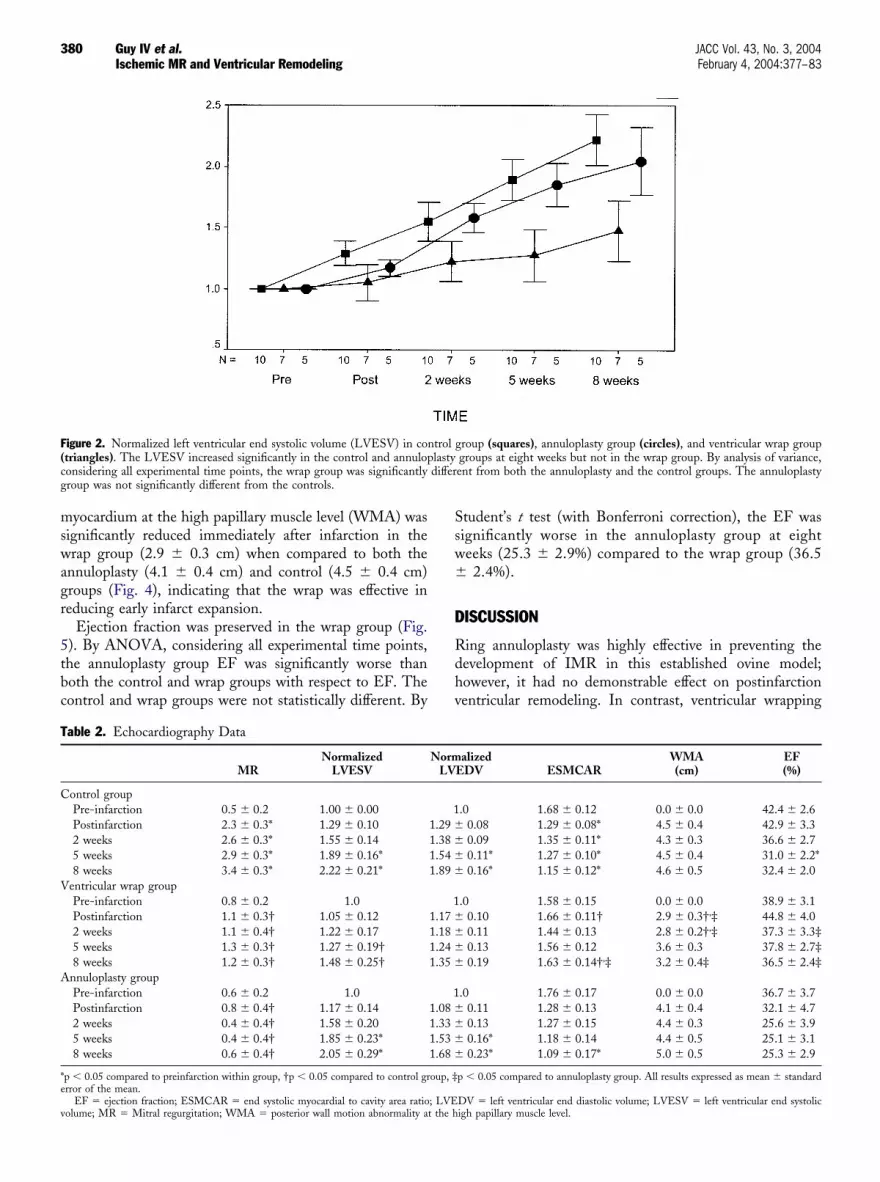
ndividual time point (Fig. 1).Normalized LVESV at eight weeks was not altered by
rophylactic annuloplasty (2.05 � 0.29) when comparedith controls (2.22 � 0.21). By ANOVA, considering all
xperimental time points, the wrap group was significantlyifferent from both the control and annuloplasty groupsith respect to LVESV (Fig. 2). Specifically, at eight weeks,VESV in the wrap group (1.48 � 0.25) was significantly
educed when compared with the control group. Changes inormalized LVEDV followed a similar pattern (Table 2),hough between-group comparisons did not reach statisticalignificance.
End-systolic muscle-to-cavity-area ratio was preserved inhe wrap group, but decreased significantly in both thennuloplasty and control groups: there was a significantifference in the wrap group ESMCAR (1.63 � 0.14) atight weeks when compared with both the annuloplasty1.09 � 0.17) and control (1.15 � 0.12) groups (Fig. 3).he circumferential length of the akinetic segment of
no MR, 4 � severe MR) in control group (squares), annuloplasty groupkal-Wallis test with MR as the dependent variable and considering all
(0 �Krus
nificantly different (p � 0.05 for all comparisons).
mswagr
5tbc
Ssw�
D
Rdhv
T
C
V
A
*e
v
F(cg
380 Guy IV et al. JACC Vol. 43, No. 3, 2004Ischemic MR and Ventricular Remodeling February 4, 2004:377–83
yocardium at the high papillary muscle level (WMA) wasignificantly reduced immediately after infarction in therap group (2.9 � 0.3 cm) when compared to both the
nnuloplasty (4.1 � 0.4 cm) and control (4.5 � 0.4 cm)roups (Fig. 4), indicating that the wrap was effective ineducing early infarct expansion.
Ejection fraction was preserved in the wrap group (Fig.). By ANOVA, considering all experimental time points,he annuloplasty group EF was significantly worse thanoth the control and wrap groups with respect to EF. Theontrol and wrap groups were not statistically different. By
able 2. Echocardiography Data
MRNormalized
LVESV
ontrol groupPre-infarction 0.5 � 0.2 1.00 � 0.00Postinfarction 2.3 � 0.3* 1.29 � 0.102 weeks 2.6 � 0.3* 1.55 � 0.145 weeks 2.9 � 0.3* 1.89 � 0.16*8 weeks 3.4 � 0.3* 2.22 � 0.21*
entricular wrap groupPre-infarction 0.8 � 0.2 1.0Postinfarction 1.1 � 0.3† 1.05 � 0.122 weeks 1.1 � 0.4† 1.22 � 0.175 weeks 1.3 � 0.3† 1.27 � 0.19†8 weeks 1.2 � 0.3† 1.48 � 0.25†
nnuloplasty groupPre-infarction 0.6 � 0.2 1.0Postinfarction 0.8 � 0.4† 1.17 � 0.142 weeks 0.4 � 0.4† 1.58 � 0.205 weeks 0.4 � 0.4† 1.85 � 0.23*8 weeks 0.6 � 0.4† 2.05 � 0.29*
p � 0.05 compared to preinfarction within group, †p � 0.05 compared to control grrror of the mean.
EF � ejection fraction; ESMCAR � end systolic myocardial to cavity area ratio
igure 2. Normalized left ventricular end systolic volume (LVESV) in contriangles). The LVESV increased significantly in the control and annuloponsidering all experimental time points, the wrap group was significantlyroup was not significantly different from the controls.
olume; MR � Mitral regurgitation; WMA � posterior wall motion abnormality at the h
tudent’s t test (with Bonferroni correction), the EF wasignificantly worse in the annuloplasty group at eighteeks (25.3 � 2.9%) compared to the wrap group (36.52.4%).
ISCUSSION
ing annuloplasty was highly effective in preventing theevelopment of IMR in this established ovine model;owever, it had no demonstrable effect on postinfarctionentricular remodeling. In contrast, ventricular wrapping
alizedEDV ESMCAR
WMA(cm)
EF(%)
.0 1.68 � 0.12 0.0 � 0.0 42.4 � 2.60.08 1.29 � 0.08* 4.5 � 0.4 42.9 � 3.30.09 1.35 � 0.11* 4.3 � 0.3 36.6 � 2.70.11* 1.27 � 0.10* 4.5 � 0.4 31.0 � 2.2*0.16* 1.15 � 0.12* 4.6 � 0.5 32.4 � 2.0
.0 1.58 � 0.15 0.0 � 0.0 38.9 � 3.10.10 1.66 � 0.11† 2.9 � 0.3†,‡ 44.8 � 4.00.11 1.44 � 0.13 2.8 � 0.2†,‡ 37.3 � 3.3‡0.13 1.56 � 0.12 3.6 � 0.3 37.8 � 2.7‡0.19 1.63 � 0.14†,‡ 3.2 � 0.4‡ 36.5 � 2.4‡
.0 1.76 � 0.17 0.0 � 0.0 36.7 � 3.70.11 1.28 � 0.13 4.1 � 0.4 32.1 � 4.70.13 1.27 � 0.15 4.4 � 0.3 25.6 � 3.90.16* 1.18 � 0.14 4.4 � 0.5 25.1 � 3.10.23* 1.09 � 0.17* 5.0 � 0.5 25.3 � 2.9
p � 0.05 compared to annuloplasty group. All results expressed as mean � standard
DV � left ventricular end diastolic volume; LVESV � left ventricular end systolic
group (squares), annuloplasty group (circles), and ventricular wrap groupgroups at eight weeks but not in the wrap group. By analysis of variance,ent from both the annuloplasty and the control groups. The annuloplasty
NormLV
11.29 �1.38 �1.54 �1.89 �
11.17 �1.18 �1.24 �1.35 �
11.08 �1.33 �1.53 �1.68 �
oup, ‡
; LVE
trollastydiffer
igh papillary muscle level.

riAa
tapwdpt
sitoe
sadir
F(pd
F(t
381JACC Vol. 43, No. 3, 2004 Guy IV et al.February 4, 2004:377–83 Ischemic MR and Ventricular Remodeling
educed acute infarct expansion, preserved EF, and significantlymproved measured parameters of postinfarction remodeling.dditionally, ventricular wrapping was as effective as ring
nnuloplasty in preventing the development of IMR.A potential limitation of this study was the fact that only
he ring annuloplasty group had open-heart surgery and thessociated need for a period of aortic cross-clamping withrotected global ischemia. We were meticulously attentiveith regard to myocardial protection during these proce-ures. The fact that there was no statistical difference in thereinfarction EF between groups indicates that intraopera-ive myocardial protection was adequate.
igure 3. End-systolic muscle-to-cavity-area ratio (ESMCAR) in contrtriangles). The ESMCAR did not change during postinfarction remodelioints, ESMCAR was significantly reduced in both the annuloplasty andifference between the control and annuloplasty groups.
igure 4. Length of the circumferential wall motion abnormality at the higcircles), and ventricular wrap group (triangles). By analysis of variance, c
he annuloplasty and control groups when compared to the wrap group. ThereClinically, IMR occurs most commonly after moderatelyized posterolateral infarctions involving the posterior pap-llary muscle (4,5,17). The degree of mitral regurgitation isypically small initially but increases, often to severe levels,ver a varying time course. The model of IMR used in thisxperiment faithfully replicates the human disease.
Recent laboratory (10,18) and clinical studies (11) havehown that expansion (stretching) of a transmural MI initiatesprogressive myopathic process in normally perfused myocar-ium. This phenomenon is initially localized to myocardium
mmediately adjacent to the infarct, but extends during theemodeling process to convert contiguous normally perfused
up (squares), annuloplasty group (circles), and ventricular wrap groupthe wrap group. By analysis of variance, considering all experimental timeol groups when compared with the wrap group. There was no significant
illary muscle level (WMA) in control group (squares), annuloplasty groupering all experimental time points, the WMA was significantly greater in
ol grong incontr
h paponsid
was no statistical difference between the control and annuloplasty groups.

mTwm(ssr
dcbeaadiLiatmffelt
gisidL
cdwm
crtippOei
iTthrersrtat
toaLl
Fcs
382 Guy IV et al. JACC Vol. 43, No. 3, 2004Ischemic MR and Ventricular Remodeling February 4, 2004:377–83
yocardium into hypocontractile remodeled myocardium.he stretch-induced myopathic process has been associatedith myocyte apoptosis (14) and disruption of the extracellularatrix secondary to activation of matrix metalloproteinases
19). The failure of surgical reshaping operations to improveurvival in ischemic cardiomyopathy patients (20–22) stronglyuggests that infarct-induced myopathy is very difficult toeverse once established.
Using contrast echocardiography, Jackson et al. (23) hasemonstrated that early postinfarction geometric changesonsistent with increased regional wall stress occur in theorderzone region adjacent to infarcts undergoing earlyxpansion and subsequent remodeling. A finite-elementnalysis by Guccione et al. (18) confirms these findings andlso demonstrates that once the myopathic process is fullyeveloped, contractile function in nonischemic myocardium
s impaired beyond what would be expected from changes inV geometry and stress distribution. Therefore, early after-
nfarction loss of contractility is due to mechanical factors;s remodeling progresses the geometric contribution (stress)o myocardial dysfunction is likely outstripped by theyopathic phenomenon that it initiates in normally per-
used myocardium. It is for this reason that most operationsor established heart failure are ineffective. The salutaryffect of ventricular wrapping demonstrated in this study isikely due to its ability to attenuate early infarct expansion,hereby reducing adverse remodeling.
Elimination of moderate to severe ischemic mitral regur-itation, either by valve replacement or valve repair, is anncreasingly recommended treatment for patients withymptomatic CHF (4,7). This trend has been driven bymprovements in mitral valvuloplasty techniques and re-uced perioperative mortality in patients with depressed
igure 5. Ejection fraction (EF) in control group (squares), annuloplasonsidering all experimental time points, the EF in the annuloplasty groutatistical difference between the control and wrap groups.
V function undergoing open-heart surgery. Recent large r
linical studies, however, fail to demonstrate that ad-ressing chronic IMR adds to patient survival beyondhat would be expected from optimal medical manage-ent (4,7,8,24).The primary intent of this study was to assess the relative
ontribution of infarct expansion and mitral regurgitation toemodeling after posterior infarctions that are predisposedo the development of progressive IMR. Our findingsndicate that although chronic IMR is a manifestation ofostinfarction LV remodeling it contributes minimally toerpetuating the phenomenon of progressive LV dilation.n the contrary, ventricular restraint that prevents infarct
xpansion dramatically influences the outcome of remodel-ng and secondarily prevents the development of MR.
The preemptive and prophylactic surgical interventions usedn this study cannot, obviously, be directly applied clinically.hey represent the best-case scenario of two very different
herapeutic strategies. The results of the study do, however,ave important clinical implications. Our findings indicate thatelief (or prevention) of IMR has very little impact on remod-ling. This helps to explain the negligible effect of mitral valveeplacement or repair on survival in patients with IMR. Thistudy confirms the results of earlier experiments in our labo-atory (13,25), and in doing so reinforces the thesis thatherapies that minimize infarct expansion early after acute MIre more likely to limit adverse postinfarction remodeling andhereby improve survival.
The threshold for surgical treatment of mitral regurgita-ion caused by primary valvular disease is declining becausef improved repair techniques, reduced operative mortality,nd proven benefits in reducing the incidence of irreversibleV dysfunction. Our data would caution against extrapo-
ating these results to patients with IMR. In IMR, mitral
rcles), and ventricular wrap group (triangles). By analysis of variance,s significantly less than both the wrap and control groups. There was no
ty (cip wa
egurgitation is a consequence rather than a cause of

pse
RDPE
R
1
1
1
1
1
1
1
1
1
1
2
2
2
2
2
2
383JACC Vol. 43, No. 3, 2004 Guy IV et al.February 4, 2004:377–83 Ischemic MR and Ventricular Remodeling
ostinfarction left ventricular remodeling. As these dataupport, the primary therapeutic target is early infarctxpansion, not late mitral regurgitation.
eprint requests and correspondence: Dr. Robert C. Gorman,epartment of Surgery, 6 Silverstein, Hospital of the University ofennsylvania, 3400 Spruce St., Philadelphia, Pennsylvania 19104.-mail: [email protected].
EFERENCES
1. St. John-Sutton M, Pfeffer MA, Moye L, et al. Cardiovascular deathand left ventricular remodeling two years after myocardial infarction:baseline predictors and impact of long-term use of captopril: informa-tion from the Survival and Ventricular Enlargement (SAVE) trial.Circulation 1997;96:3294–9.
2. Pfeffer MA, Braunwald E. Ventricular remodeling after myocardialinfarction. Circulation 1990;8:1161–72.
3. Gorman JH III, Gorman RC, Plappert T, et al. Infarct size andlocation determine development of mitral regurgitation in the sheepmodel. J Thorac Cardiovasc Surg 1998;115:615–22.
4. Gillinov AM, Wierup PN, Blackstone EH, et al. Is repair preferableto replacement for ischemic mitral regurgitation? J Thorac CardiovascSurg 2001;122:1125–41.
5. Kumanohoso T, Otsuji Y, Yoshifuku S, et al. Mechanism of higherincidence of ischemic mitral regurgitation in patients with inferiormyocardial infarction: quantitative analysis of left ventricular andmitral valve geometry in 103 patients with prior myocardial infarction.J Thorac Cardiovasc Surg 2003;125:135–43.
6. Lamas GA, Mitchell GF, Flaker GC, et al. Clinical significance ofmitral regurgitation after acute myocardial infarction. Circulation1997;96:827–33.
7. Grossi EA, Goldberg JD, LaPietra A, et al. Ischemic mitral valvereconstruction and replacement: comparison of long-term survival andcomplications. J Thorac Cardiovasc Surg 2000;122:1107–24.
8. Miller DC. Ischemic mitral regurgitation redux—to repair or toreplace? J Thorac Cardiovasc Surg 2001;122:1059–62.
9. Bolognese L, Neskovic AN, Parodi G, et al. Left ventricular remod-eling after primary coronary angioplasty: patterns of left ventriculardilation and long-term prognostic implications. Circulation 2002;106:2351–7.
0. Jackson BM, Gorman JH III, St. John-Sutton MG, et al. Progressiveborderzone extension leads to heart failure after anteroapical myocar-dial infarction. J Am Coll Cardiol 2002;40:1160–7.
1. Narula J, Dawson MS, Singh BK, et al. Noninvasive characterizationof stunned, hibernating, remodeled and nonviable myocardium inischemic cardiomyopathy. J Am Coll Cardiol 2000;36:1913–8.
2. Llaneras MR, Nance ML, Streicher JT, et al. Large animal model ofischemic mitral regurgitation. Ann Thorac Surg 1994;57:432–9.
3. Kelley ST, Malekan R, Jackson BM, et al. Restraining infarctexpansion preserves left ventricular geometry and function after acuteanteroapical infarction. Circulation 1999;99:135–42.
4. Moainie SL, Gorman JH III, Guy TS, et al. An ovine model ofpostinfarction dilated cardiomyopathy. Ann Thorac Surg 2002;74:753–60.
5. Schiller NB, Shah PM, Crawford M, et al. Recommendation for thequantification of the left ventricle by two-dimensional echocardiogra-phy: American Society of Echocardiography Committee on Standards,Subcommittee on Quantitation of Two-Dimensional Echocardio-grams. J Am Soc Echocardiogr 1989;5:358–62.
6. Miyatake K, Izumi S, Okamoto M, et al. Semiquantitative grading ofseverity of mitral regurgitation by real-time two-dimensional Dopplerflow imaging technique. J Am Coll Cardiol 1986;7:82–8.
7. Akins CW, Hilgenberg AD, Buckley MJ, et al. Mitral valve recon-struction versus replacement for degenerative or ischemic mitralregurgitation. Ann Thorac Surg 1994;58:668–75.
8. Guccione JM, Moonly SM, Moustakidis P, et al. Mechanism under-lying mechanical dysfunction in the border zone of left ventricularaneurysm: a finite element model study. Ann Thorac Surg 2001;71:654–62.
9. Wilson EM, Moainie SL, Baskin JM, et al. Region and species specificinduction of matrix metalloproteinases occurs with post-myocardialinfarction remodeling. Circulation 2003;107:2857–63.
0. Couper GS, Bunton RW, Birjiniuk V, et al. Relative risks of leftventricular aneurysmectomy in patients with akinetic scars versus truedyskinetic aneurysms. Circulation 1990;82 Suppl IV:IV248–56.
1. Batista RJV, Verde J, Nery P, et al. Partial left ventriculectomy to treatend-stage heart disease. Ann Thorac Surg 1997;64:634–8.
2. Athanasuleas CL, Stanley AW Jr, Buckberg GD, et al. Surgicalanterior ventricular endocardial restoration (SAVER) in the dilatedremodeled ventricle after anterior myocardial infarction. RESTOREgroup: Reconstructive Endoventricular Surgery, Returning TorsionOriginal Radius Elliptical Shape to the LV. J Am Coll Cardiol2001;37:1199–209.
3. Jackson BM, Gorman JH III, Salgo IS, et al. Increased wall stress dueto altered borderzone geometry as assessed by perfusion echocardiog-raphy. Am J Physiol Heart Circ Physiol 2003;284:H475–9.
4. Levy D, Kenchaiah S, Larson MG, et al. Long-term trends in theincidence of and survival with heart failure. N Engl J Med 2002;347:1397–402.
5. Moainie SL, Guy TS, Gorman JH III, et al. Infarct restraintattenuates remodeling and reduces chronic ischemic mitral regurgita-tion after postero-lateral infarction. Ann Thorac Surg 2002;74:444–9.


