Embed Size (px)
Citation preview

Maltais et al Acquired Cardiovascular Disease
ACD
Mitral regurgitation surgery in patients with ischemiccardiomyopathy and ischemic mitral regurgitation: Factors thatinfluence survival
Simon Maltais, MD, PhD,a Hartzell V. Schaff, MD,a Richard C. Daly, MD,a Rakesh M. Suri, MD, PhD,a
Joseph A. Dearani, MD,a Thoralf M. Sundt III, MD,a Maurice Enriquez-Sarano, MD,b
Yan Topilsky, MD,b and Soon J. Park, MD, MSca
From th
Clini
Disclos
Read at
gery,
Receive
publi
Address
Mayo
(E-m
0022-52
Copyrig
doi:10.1
Objective: The treatment of patients with ischemic cardiomyopathy and concomitant mitral regurgitation canbe challenging and is associated with reduced long-term survival. It is unclear how mitral valve repair versusreplacement affects subsequent outcome. Therefore, we conducted this study to understand the predictors ofmortality and to delineate the role of mitral valve repair versus replacement in this high-risk population.
Methods: From 1993 to 2007, 431 patients (mean age, 70 � 9 years) with ischemic cardiomyopathy (leftventricular ejection fraction � 45%) and significant ischemic mitral regurgitation (>2) were identified.Patients (44) with concomitant mitral stenosis were excluded from the analysis. A homogeneous group of387 patients underwent combined coronary artery bypass grafting and mitral valve surgery, mitral valve re-pair in 302 (78%) and mitral valve replacement in 85 (22%). Uni- and multivariate analyses were performedon the entire cohort, and the predictors of mortality were identified in 2 distinct risk phases. Furthermore, wespecifically examined the impact of mitral valve repair versus replacement by comparing 2 propensity-matched subgroups.
Results: Follow-up was 100% complete (median, 3.6 years; range, 0–15 years). Overall 1-, 5-, and 10-year sur-vivals were 82.7%, 55.2%, and 24.3%, respectively, for the entire group. The risk factors for an increased mor-tality within the first year of surgery included previous coronary artery bypass grafting (hazard ratio ¼ 3.39;P<.001), emergency/urgent status (hazard ratio ¼ 2.08; P ¼ .007), age (hazard ratio ¼ 1.5; P ¼ .03), andlow left ventricular ejection fraction (hazard ratio ¼ 1.31; P ¼ .026). Thereafter, only age (hazardratio ¼ 1.58; P<.001), diabetes (hazard ratio ¼ 2.5; P ¼ .001), and preoperative renal insufficiency (hazardratio ¼ 1.72; P ¼ .025) were predictive. The status of mitral valve repair versus replacement did not influencesurvival, and this was confirmed by comparable survival in propensity-matched analyses.
Conclusions: Survival after combined coronary artery bypass grafting and mitral valve surgery in patients withischemic cardiomyopathy (left ventricular ejection fraction � 45%) and mitral regurgitation is compromisedand mostly influenced by factors related to the patient’s condition at the time of surgery. The specifics of mitralvalve repair versus replacement did not seem to affect survival. (J Thorac Cardiovasc Surg 2011;142:995-1001)
Earn CME credits athttp://cme.ctsnetjournals.org
Patients with ischemic heart disease and mitral regurgita-tion (MR) comprise one of the more perplexing and
e Divisions of Cardiovascular Surgerya and Cardiovascular Medicine,b Mayo
c College of Medicine, Rochester, Minn.
ures: Authors have nothing to disclose with regard to commercial support.
the 91st Annual Meeting of The American Association for Thoracic Sur-
Philadelphia, Pennsylvania, May 7–11, 2011.
d for publication April 8, 2011; revisions received June 21, 2011; accepted for
cation July 20, 2011; available ahead of print Aug 22, 2011.
for reprints: Soon J. Park, MD, MSc, Division of Cardiovascular Surgery,
Clinic College of Medicine, 200 First Street SW, Rochester, MN 55905
ail: [email protected]).
23/$36.00
ht � 2011 by The American Association for Thoracic Surgery
016/j.jtcvs.2011.07.044
The Journal of Thoracic and Ca
challenging groups to treat in cardiac surgery. Some of thesepatients have 2 independent disease processes of coronaryischemia and myxomatous MR, and they tend to havea favorable survival outcome after surgical correction.1-3
However, the outcome seems to be more guarded in thosewho have coronary ischemia and functional MR, that is,ischemic MR (IMR).4 In this population, IMR can be dueto various pathophysiologic processes ranging from acutepapillary muscle ischemia/rupture to chronic left ventricu-lar (LV) remodeling after myocardial infarction, resultingin tethered and incompetent mitral leaflets unable to coapt.Accordingly, their clinical presentation varies, ranging fromacute pulmonary edema to recurrent bouts of congestiveheart failure. This has been shown to influence the surgeon’sdecision to perform mitral valve repair (MVP) or mitralvalve replacement (MVR).5,6 All these factors contributeto the difficulty in understanding the comparative efficacyof MVP versus MVR, and it is not surprising to have
rdiovascular Surgery c Volume 142, Number 5 995
Abbreviations and AcronymsCABG ¼ coronary artery bypass graftingHR ¼ hazard ratioICM ¼ ischemic cardiomyopathyIMR ¼ ischemic mitral regurgitationLAD ¼ left anterior descendingLITA ¼ left internal thoracic arteryLV ¼ left ventricular, left ventricleLVEF ¼ left ventricular ejection fractionMR ¼ mitral regurgitationMV ¼ mitral valveMVP ¼ mitral valve repairMVR ¼ mitral valve replacement
Acquired Cardiovascular Disease Maltais et alACD
conflicting literature reports supporting MVP or MVR.7
Furthermore, many of the reported experiences have short-comings of small sample size, experiences accumulatedover many decades of evolving practices, and a heteroge-neous patient population.8 Nonetheless, the current generalconsensus seems to favor MVP over MVR in patients withIMR even though a significant rate of MR recurrence isa well-recognized fact.9,10 The rate of MR recurrenceafter MVP seems to be particularly high in patients withpoor left ventricular ejection fraction (LVEF), and thismay adversely affect survival. We conducted this studyspecifically to examine these perplexing issues ina currently relevant cohort of patients with significantischemic cardiomyopathy (ICM) and IMR. The primaryobjective was to understand the risk factors associatedpoor survival after combined coronary artery bypassgrafting (CABG) and MVP/MVR in patients with anLVEF 45% or less. The secondary objective was tounderstand the impact of MVP versus MVR on survival.
PATIENTS AND METHODSStudy Design
This is a retrospective analysis of prospectively gathered data over more
than a 10-year period (median, 3.6 years; range, 0–15 years). The cardiac sur-
gery database (1993–2007) at Mayo Clinic (Rochester, Minn) was used to
create a homogeneous study cohort of patients who underwent cardiac sur-
gery for ischemic heart disease with significant IMR. We identified patients
who underwent a combined CABG and MVP or MVR first. Then, we ex-
cluded thosewho have had any one of the following conditions: LVEFgreater
than 45%, infective endocarditis, congenital valvular heart disease, rheu-
matic valvular disease, or any degree of mitral stenosis. All patients with
mixed pathologies were thoroughly assessed, as echocardiographic data
and operative findings were reviewed to confirm the ischemic cause of the
MR. The Mayo Foundation Institutional Review Board approved this study,
and individual consent was obtained for all patients included in this study.
We explored themortality hazards for the entire cohort in 2 different risk
phases (early up to 1 year after surgery and late thereafter). Statistically sig-
nificant risk factors for mortality were identified for each of these 2 risk
phases. Furthermore, we examined the specific impact related to the type
of mitral valve (MV) procedure by comparing 2 subgroups based on
996 The Journal of Thoracic and Cardiovascular Surg
MVP andMVR, propensitymatched on 14 baseline characteristic variables
(age, gender, hypertension, diabetes, history of smoking, body surface area,
preoperative New York Heart Association class, chronic renal failure, pre-
operative dialysis, previous CABG surgery, previous valve procedure, his-
tory of congestive heart failure, LVEF, and emergency/urgent status of the
procedure).
DefinitionsThe cause of MR was presumed to be ischemic. All patients had a sig-
nificant degree of mitral annular dilation and LV dysfunction due to prior
myocardial infarction. In patients who required more complex repair for
mixed valvular pathology, operative and echocardiography reports were re-
viewed to confirm that myocardial ischemia was the primary mechanism
for MR. The operative and echocardiographic findings were reviewed in
detail in these patients, and they were deemed to have IMR on the basis
of leaflet tethering, prior myocardial infarction, and leaflet tethering. All
patients included in this study had an undersized ring/band implanted
when applicable. In patients with repairs, the ring was chosen according
to the undersized intercommissural distance when applicable. In patients
with a 63- to 65-mm posterior band, the band was cut and undersized to
the appropriate length. The exact nature of chordal preservation in each
patient undergoing MVR was not established. However, our institutional
policy has been to preserve the posterior leaflet whenever possible with
an increasing recent tendency toward preserving as much of the anterior
leaflet and by transposing it to the posterior annulus.
Follow-upPatients were followed systematically by using mailed questionnaires,
telephone interview, or examination at the Mayo Clinic. Clinical follow-
up for both patients with MVR and MVP was 100% complete. Mean
follow-up among survivors was 4.2 years (range, 0–15.7 years).
Statistical AnalysisDescriptive statistics for categoric variables are reported as frequency
and percentage, and continuous variables are reported as mean (standard
deviation) or median (range) as appropriate. Categoric variables were
compared between MVP and MVR groups using the chi-square test, and
continuous variables were compared using 2-sample t test or Wilcoxon
rank-sum test when appropriate. The Kaplan–Meier method was used to
draw survival curves and calculate 1-, 5-, and 10-year survival statistics.
Cox regression models were used to find the univariate and multivariate
predictors of early (1-year) and long-term (late or constant) survivals.
The multivariable model considered univariate significant variables
(P<.05) with model selection using the stepwise method (backward and
forward methods resulted in the same model). All statistical tests were
2-sided with the alpha level set at .05 for statistical significance.
RESULTSPatient Characteristics
From 1993 to 2007, 431 patients were identified and re-quired a combined CABG and MVP/MVR procedure. Ofthese, 44 patients were excluded because they had mitralstenosis, yielding 387 patients for this study. The meanage at the time of surgery was 70.1 � 9.1 years (range43–91 years), and 261 patients were male (67%). CABGsurgery was performed in all patients. MVP was performedin 302 patients (78%), and MVR was performed in 85 pa-tients (22%). All patients had LV dysfunction (LVEF �45%), and the mean LVEF by preoperative transthoracicechocardiography was 33.6% � 8.4% (range, 9–45).
ery c November 2011
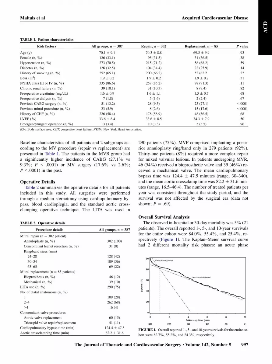
TABLE 1. Patient characteristics
Risk factors All groups, n ¼ 387 Repair, n ¼ 302 Replacement, n ¼ 85 P value
Age (y) 70.1 � 9.1 70.3 � 8.8 69.5 � 9.9 .93
Female (n, %) 126 (33,1) 95 (31.5) 31 (36.5) .38
Hypertension (n, %) 273 (70.5) 215 (71.2) 58 (68.2) .59
Diabetes (n, %) 126 (32.5) 104 (34.4) 22 (25.9) .14
History of smoking (n, %) 252 (65.1) 200 (66.2) 52 (62.2 .22
BSA (m2) 1.9 � 0.2 1.9 � 0.2 1.9 � 0.2 .31
NYHA class III or IV (n, %) 335 (86.6) 257 (85.2) 78 (91.3) .11
Chronic renal failure (n, %) 39 (10.1) 31 (10.3) 8 (9.4) .82
Preoperative creatinine (mg/dL) 1.6 � 0.9 1.6 � 1.1 1.5 � 0.7 .68
Preoperative dialysis (n, %) 7 (1.8) 5 (1.6) 2 (2.4) .67
Previous CABG surgery (n, %) 51 (13.2) 28 (9.3) 23 (27.1) <.0001
Previous mitral procedure (n, %) 23 (5.9) 8 (2.6) 15 (17.6) <.0001
History of CHF (n, %) 226 (58.4) 178 (58.9) 48 (56.5) .68
LVEF (%) 33.6 � 8.4 33.6 � 8.5 34.3 � 7.9 .50
Emergency/urgent operation (n,%) 13 (3.4) 10 (3.3) 3 (3.5) .96
BSA, Body surface area; CHF, congestive heart failure; NYHA, New York Heart Association.
Maltais et al Acquired Cardiovascular Disease
ACD
Baseline characteristics of all patients and 2 subgroups ac-cording to the MV procedure (repair vs replacement) arepresented in Table 1. The patients in the MVR group hada significantly higher incidence of CABG (27.1% vs9.3%; P < .0001) or MV surgery (17.6% vs 2.6%;P<.0001) in the past.
Operative DetailsTable 2 summarizes the operative details for all patients
included in this study. All surgeries were performedthrough a median sternotomy using cardiopulmonary by-pass, blood cardioplegia, and the standard aortic cross-clamping operative technique. The LITA was used in
TABLE 2. Operative details
Procedure details All groups, n ¼ 387
Mitral repair (n ¼ 302 patient)
Annuloplasty (n, %) 302 (100)
Concomitant leaflet resection (n, %) 31 (8)
Ring/band sizes (mm)
24–28 128 (42)
30–34 109 (36)
63–65 69 (22)
Mitral replacement (n ¼ 85 patients)
Bioprosthesis (n, %) 46 (12)
Mechanical (n, %) 39 (10)
LITA use (n, %) 290 (75)
No. of distal anatomosis (n, %)
1 109 (28)
2–4 262 (68)
>4 16 (4)
Concomitant valve procedures
Aortic valve replacement 60 (15)
Tricuspid valve repair/replacement 41 (11)
Cardiopulmonary bypass time (min) 124.4 � 47.5
Aortic crossclamping time (min) 82.2 � 31.6
The Journal of Thoracic and Ca
290 patients (75%). MVP comprised implanting a poste-rior annuloplasty ring/band only in 279 patients (92%).Thirty-one patients (8%) required a more complex repairfor mixed valvular lesions. In patients undergoing MVR,46 (54%) received a bioprosthetic valve and 39 (46%) re-ceived a mechanical valve. The mean cardiopulmonarybypass time was 124.4 � 47.5 minutes (range, 30–340),and the mean aortic crossclamp time was 82.2 � 31.6 min-utes (range, 16.5–46.4). The number of treated patients peryear was consistent throughout the study period, and thesurvival was not affected by the surgical era (data notshown; P ¼ .69).
Overall Survival AnalysisThe observed in-hospital or 30-day mortality was 5% (21
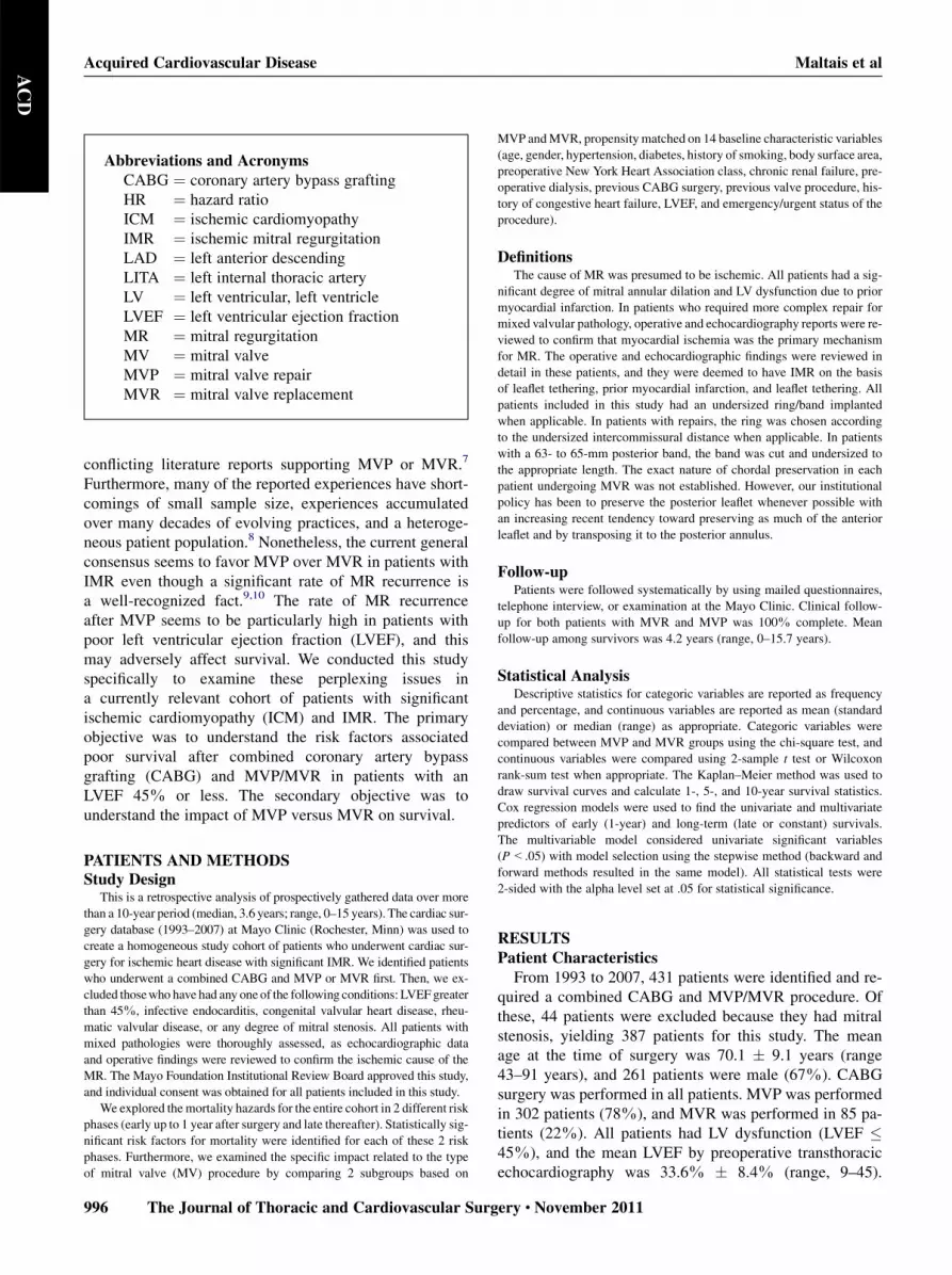
patients). The overall reported 1-, 5-, and 10-year survivalsfor the entire cohort were 84.0%, 55.4%, and 25.4%, re-spectively (Figure 1). The Kaplan–Meier survival curvehad 2 different mortality risk phases: an acute phase
FIGURE1. Overall reported 1-, 5-, and 10-year survivals for the entire co-
hort were 82.7%, 55.2%, and 24.3%, respectively.
rdiovascular Surgery c Volume 142, Number 5 997

TABLE 3. Multivariate risk factor analysis for death in patients
undergoing concomitant coronary artery bypass grafting and mitral
valve surgery
Early (1-y)
hazard
Late
(constant hazard)
Risk factors HR (P value) HR (P value)
Previous CABG surgery 3.39 (<.001)
Emergency/urgent status 2.08 (.007)
LVEF (per 10% decrease) 1.31 (.02)
Age 1.50 (.026) 1.58 (<.001)
Renal insufficiency 1.72 (.025)
Diabetes 2.50 (<.001)
Multivariate Cox regression analysis model for early (1-year) and late (constant)
survivals.
Acquired Cardiovascular Disease Maltais et alACD
corresponding to the early (1-year) period and a second late(constant) phase.
Predictors of Early (1-Year) and Late (Constant)Mortality
Table 3 summarizes identified risk factors based on mul-tivariate Cox’s regression analyses. Previous CABG surgery(hazard ratio [HR]¼ 3.39; P<.001), emergency/urgent sta-tus of the procedure (HR¼ 2.08; P¼ .007), age (HR¼ 1.5;P¼ .03), and low LVEF (HR¼ 1.31;P¼ .026) were predic-tive of earlymortality. The factors found to influence the late(constant) mortality were age (HR¼ 1.58; P<.001), diabe-tes (HR ¼ 2.5; P ¼ .001), and preoperative renal insuffi-ciency (HR ¼ 1.72; P ¼ .025). The type of MV procedureperformed (MVP or MVR) did not influence early or latemortality.
Propensity-Matched Comparisons ComparingMitral Valve Procedures
We noted comparable survival between 2 propensity-matched subgroups composed of 76 patients in each typeof MV procedure. There were no significant differences be-tween the MVP and MVR groups; 1-year (81.9% vs79.6%; P ¼ .72) and 5-year (44.0% vs 54.2%; P ¼ .72)survivals were comparable.
DISCUSSIONThe resurgence of interest in MVP over the past decade
has provided long-term data supporting the superiority ofrepair over replacement in patients with normal LV functionor degenerative/myxomatous valve disease.11 With valve-related advantages aside, it is generally accepted that inthis population, MVP has demonstrated improvementsover MVR in terms of heart function recovery and long-term survival.12 However, in patients with ICM and IMR,the impact of MVP over MVR remains controversial. Fewretrospective studies have focused on the predictors of sur-vival in patients with severe IMR and ICM.7,8,13 The presentstudy included patients who underwent a combined CABG
998 The Journal of Thoracic and Cardiovascular Surg
and MVP/MVR for significant ICM (LVEF � 45%) andsignificant IMR over the past 15 years.
Key FindingsThe survival curve of our entire cohort demonstrated 2
different mortality risk phases as shown in Figure 1. Ac-cordingly, we divided the mortality risk analyses into theearly phase (up to 1 year after surgery) and the late phase(beyond the first year). This is concordant with previouslypublished studies by Gillinov and associates7 describingdistinct phases of instantaneous risk of death in these pa-tients. In our study, factors influencing mortality in the earlyphase included a history of CABG surgery (HR ¼ 3.39;P < .001), emergency/urgent status of the surgery(HR ¼ 2.08; P ¼ .007), age (HR ¼ 1.5; P ¼ .03), andlow LVEF (HR ¼ 1.31; P ¼ .026), as shown in Table 3.These risk factors were reflective of the associated technicalchallenges of a redo CABG surgery, patient’s medical statusrequiring nonelective surgery, and poor cardiac reserve. Thefinding of these risk factors is consistent with our clinicalimpression that an expected mortality within the first yearwould be high, for example, in a patient with an LVEF of30% who would require a technically challenging redoCABG and MVP/MVR on an emergency/urgent basis.4
For this patient, the expected operative mortality (30-dayor in-hospital) also would be high, but we were unable toperformmeaningful multivariate analyses for this end point,because the operative mortality observed in this study waslow (5%).
Once patients survived the first year, their subsequentmortality was mostly based on their noncardiac comorbid-ities of diabetes (HR ¼ 2.5; P ¼ .001), renal insufficiency(HR ¼ 1.72; P ¼ .025), and age (HR ¼ 1.58; P< .001).One could speculate many potential reasons for the findingthat none of the cardiac specific risk factors played a role onsurvival during the late phase. One of the major reasons maybe that because all patients in the study started with poorcardiac reserve (LVEF � 45% with significant MR) beforesurgery, such significant underlying cardiac dysfunctionhad a uniform and overwhelming influence on survival.The specifics of mitral surgery (MVP vs MVR) had noinfluence on survival in the early or late risk phase in thisstudy cohort. Furthermore, in small subgroup analysis, weconfirmed that the type of repair (rigid/flexible, complete/partial, concomitant leaflet procedure) did not influencesurvival in patients with repairs (P ¼ .24).
We examined the issue of MVP versus MVR in this spe-cific population by comparing 2 propensity subgroups. Weidentified a matching pair of patients undergoing MVP for76 of 85 patients undergoingMVR on the basis of 14 impor-tant clinical variables, as mentioned in the ‘‘Materials andMethods’’ section. As shown in Figure 2, survival in thesepatients with an LVEF 45% or less was comparable be-tween patients with MVR and MVP. This is comparable
ery c November 2011

FIGURE 2. Survival results for propensity-matched patients. There were
no differences between patients undergoing MVR or MVP. Survival was
comparable between propensity-matched groups of patients (P ¼ .72).
MV, Mitral valve.
Maltais et al Acquired Cardiovascular Disease
ACD
to previously published studies suggesting that survival islimited after MV operation and CABG, with no advantageof either strategy for managing the MV.4,7,14 Gillinov andassociates7 also attempted to address the question of MVPversus MVR. Their surgical practice of MVP versus MVRwas significantly confounded with the patient’s condition,and their matched pair yielded ‘‘only one end of spectrumof IMR (the most complex with the sickest patients).’’They exercised sophisticated and elaborate statistical anal-yses to compare different quintile risk subgroups based onpropensity-adjusted for MVP versus MVR selectionfactors. The experience reported by Grossi and associates8
further demonstrated a high degree of confounding relation-ship between the type of MV procedure and the preopera-tive patient characteristics of angina and New York HeartAssociation class. In our cohort, we also noted some differ-ences in baseline characteristics between the MVP andMVR groups, but such differences were only related to priorcardiac surgery (Table 1). By selecting a relatively homoge-neous study population with an LVEF of 45% or less withinthe recent surgical era, we were able to compare a meaning-ful number of propensity-matched patients and examine thesurvival impact of MVP versus MVR specifically.
Other Important ConsiderationsSeveral groups have shown that survival after coronary
revascularization is strongly influenced by the use of theleft internal thoracic artery (LITA) to the left anterior de-scending (LAD) artery.15,16 In this study, 75% of patientswith an MV procedure and concomitant CABG had theLITA to the LAD used. We strongly believe thatpreviously reported differences in survival observed inMVP compared with MVR in patients with ICM may berelated to the low incidence of using the LITA to theLAD, especially in patients with MVR (15%).7 When there
The Journal of Thoracic and Ca
is an associated LAD pathology, the LITA should always beused in this population with ischemic LV function to opti-mize LV recovery and possibly survival. Furthermore, inpatients with MVP, residual MR can be present and nega-tively influence the degree of LV recovery and remodeling.In a recent study performed in 111 patients undergoingMV surgery, De Bonis and colleagues9 showed that the pro-gression of LV remodeling paralleled the recurrence of MR.At 3 years, freedom from recurrence of MR of 2þor greaterwas 74% in those who showed a reverse remodelingprocess. In a multicenter study, Shiota and associates10
reported a surprisingly high 33% recurrence of MR6 months after mitral annuloplasty in patients with ICM.In their study, recurrent MR was associated with increasedLV size and decreased LVEF. Hypothetically, in terms of re-currence of MR, MVR could represent a more reproducibleand durable surgical treatment in these patients. Of course,preservation of as much MV apparatus as possible at thetime of MVR would be important.17,18
LimitationsOne of the major limitations of our study is that it is a ret-
rospective review of patients who underwent a combinedsurgery of CABG and MVP or MVR, in which a selectionbias plays a role, and it is hard to account for all of its impacton outcome. We also do not have a comparison group withno concomitant MV procedures. Therefore, the added bene-fit of MVP/MVR is still speculative on the presumption thatreducing MR renders a therapeutic benefit on outcome. Theproposed National Institutes of Health-sponsored cardiacsurgery network studies of prospective randomized trialsinvolving patients with IMR would be critically importantin providing further answers in treating these perplexingand challenging patients. The end points of this study werepredictors of death, and we did not analyze other complica-tions because we have already described valve procedure-related differences in durability in the literature.19
CONCLUSIONSWe evaluated the predictors of survival in patients with
ICM and IMR. We identified 2 distinct risk phases basedon the survival curve kinetics. The risk predictors of mortal-ity during the early phase (1-year) were factors associatedwith the patient’s presenting condition and projected techni-cal difficulty at the time of the intervention (previousCABG surgery, emergency intervention, low LVEF). Inthe late (after the first year) phase, patients’ comorbidities(age, diabetes, and renal insufficiency) were the determi-nants of mortality. In patients with ICM and IMR, the natureof the MV intervention (MVP or MVR) had no influence onsurvival.
We would like express our gratitude to Judy Lenoch and ZhuoLi for their help with the database and statistical analyses.
rdiovascular Surgery c Volume 142, Number 5 999

Acquired Cardiovascular Disease Maltais et alACD
References1. Kuwaki K, Kiyofumi M, Tsukamoto M, Abe T. Early and late results of mitral
valve repair for mitral valve regurgitation. Significant risk factors of reoperation.
J Cardiovasc Surg (Torino). 2000;41:187-92.
2. Enriquez-Sarano M, Schaff HV, Orszulak TA, Tajik AJ, Bailey KR, Frye RL.
Valve repair improves the outcome of surgery for mitral regurgitation. A multi-
variate analysis. Circulation. 1995;91:1022-8.
3. Ling LH, Enriquez-SaranoM, Seward JB, et al. Clinical outcome of mitral regur-
gitation due to flail leaflet. N Engl J Med. 1996;335:1417-23.
4. Dahlberg PS, Orszulak TA, Mullany CJ, Daly RC, Enriquez-Sarano M,
Schaff HV. Late outcome of mitral valve surgery for patients with coronary artery
disease. Ann Thorac Surg. 2003;76:1539-487; discussion 1547-8.
5. Orszulak TA, Schaff HV, Danielson GK, et al. Mitral regurgitation due to rup-
tured chordae tendineae. Early and late results of valve repair. J Thorac Cardio-
vasc Surg. 1985;89:491-8.
6. Tribouilloy CM, Enriquez-Sarano M, Schaff HV, et al. Impact of preopera-
tive symptoms on survival after surgical correction of organic mitral regur-
gitation: rationale for optimizing surgical indications. Circulation. 1999;99:
400-5.
7. Gillinov AM, Faber C, Houghtaling PL, et al. Repair versus replacement for de-
generative mitral valve disease with coexisting ischemic heart disease. J Thorac
Cardiovasc Surg. 2003;125:1350-62.
8. Grossi EA, Goldberg JD, LaPietra A, et al. Ischemic mitral valve reconstruction
and replacement: comparison of long-term survival and complications. J Thorac
Cardiovasc Surg. 2001;122:1107-24.
9. De Bonis M, Lapenna E, Verzini A, et al. Recurrence of mitral regurgitation par-
allels the absence of left ventricular reverse remodeling after mitral repair in ad-
vanced dilated cardiomyopathy. Ann Thorac Surg. 2008;85:932-9.
10. Shiota M, Gillinov AM, Takasaki K, Fukuda S, Shiota T. Recurrent mitral regur-
gitation late after annuloplasty for ischemic mitral regurgitation. Echocardiogra-
phy. 2011;28:161-6.
11. Lawrie GM. Mitral valve repair vs replacement. Current recommendations and
long-term results. Cardiol Clin. 1998;16:437-48.
12. Nishimura RA, Carabello BA, Faxon DP, et al. ACC/AHA 2008 Guideline up-
date on valvular heart disease: focused update on infective endocarditis: a report
of the American College of Cardiology/American Heart Association Task Force
on Practice Guidelines endorsed by the Society of Cardiovascular Anesthesiolo-
gists, Society for Cardiovascular Angiography and Interventions, and Society of
Thoracic Surgeons. J Am Coll Cardiol. 2008;52:676-85.
13. Cohn LH, Rizzo RJ, Adams DH, et al. The effect of pathophysiology on the sur-
gical treatment of ischemic mitral regurgitation: operative and late risks of repair
versus replacement. Eur J Cardiothorac Surg. 1995;9:568-74.
14. Lee EM, Shapiro LM, Wells FC. Superiority of mitral valve repair in surgery for
degenerative mitral regurgitation. Eur Heart J. 1997;18:655-63.
15. Pick AW, Orszulak TA, Anderson BJ, Schaff HV. Single versus bilateral internal
mammary artery grafts: 10-year outcome analysis. Ann Thorac Surg. 1997;64:
599-605.
16. Boylan MJ, Lytle BW, Loop FD, et al. Surgical treatment of isolated left anterior
descending coronary stenosis. Comparison of left internal mammary artery and
venous autograft at 18 to 20 years of follow-up. J Thorac Cardiovasc Surg.
1994;107:657-62.
17. Borger MA, Yau TM, Rao V, Scully HE, David TE. Reoperative mitral valve re-
placement: importance of preservation of the subvalvular apparatus. Ann Thorac
Surg. 2002;74:1482-7.
18. Okita Y, Miki S, Ueda Y, Tahata T, Sakai T. Left ventricular function after mitral
valve replacement with or without chordal preservation. J Heart Valve Dis. 1995;
4(Suppl 2):S181-93.
19. Mohty D, Orszulak TA, Schaff HV, Avierinos JF, Tajik JA, Enriquez-Sarano M.
Very long-term survival and durability of mitral valve repair for mitral valve pro-
lapse. Circulation. 2001;104(12 Suppl. 1):I1-7.
DiscussionDr Patrick McCarthy (Chicago, Ill). By disclosure, I am
a coinventor of the IMR ETlogix ring (Edwards Lifesciences,Irvine, Calif).
Congratulations, Dr Maltais, on an excellent and timely presen-tation. We look forward to the results of the ongoing National
1000 The Journal of Thoracic and Cardiovascular Sur
Institutes of Health prospective randomized trial that will give fur-ther information on this important subject.
As you note, patient factors were important to survival, but wecan’t modify those. As surgeons, we can decide, however, betweenMVR and MVP. So I have questions about how that decision wasmade and late outcomes.
First, can you provide insight into why MVR was sometimeschosen?Was it influenced by the degree of leaflet tethering or otherfactors seen on echocardiography? Was MVR used for worse ven-tricles or more complex and extensive MR? Others have found nodifference in survival between MVP and MVR in single-centerstudies, but did you look at other outcomes, such as need for trans-plant or ventricular assist device, late New York Heart functionalclass, and rehospitalization for cardiac cause, and do you haveany data on late MR recurrence? Finally, after this study, whatdo you do now, repair or replace?
DrMaltais.Regarding your first question, this is a retrospectivestudy with all the biases that it implies. I would say that in our cen-ter, the most important thing is to achieve a complete repair, and allthe patients are leaving the operating room with trivial MR. Thatbeing said, one could argue that when a patient presents witha heavy ischemic burden, one will be more inclined to repair thevalve, compared with a patient who presents with an extremelydilated LV or lots of tethering, where surgeons might be moreinclined to replace the valve. This is, however, certainly a goodquestion, and it warrants prospective randomized studies toanswer.
In regard to your second question, as you pointed out, lots ofstudies have shown that these patients are really sick, especiallythe subgroup with low ejection fractions. We don’t have the inci-dence of ventricular assist device or heart transplantation in thispopulation because this is a retrospective study. Furthermore, wedon’t have the incidence of late recurrence of MR or readmissionfor symptomatic heart failure.
However, in regard to ventricular assist device or heart trans-plantation, we are certainly looking at this population of sickpatients, and we believe there might be a subgroup of patientswith poor targets for bypasses who could be candidates forthese advanced heart failure therapies even as a first option oftreatment.
In regard to MR recurrence, although all those patients are leav-ing the operating room with trivial MR, we know the amount ofrecurrence of MR is approximately 20% to 30%. This can influ-ence a surgeon’s decision to replace or repair the valve initially ac-cording to the patient’s symptoms. If the patient presents withsevere angina symptoms, the most important thing is to performCABG, but if the patient presents with long-term congestive heartfailure symptoms, the most important thing is to have no recur-rence of MR. This might influence the surgeon’s decision.
Dr McCarthy. What would you do now?Dr Maltais. This is a simple study that has shown us that when
we look at the overall population, the type of procedure didn’tseem to influence the overall survival. This study was intendedto be simple, to look at what is really important in this population.From these results, the most important aspect seems to be patientselection. If the patient has anMVP, it has to be quick and efficient.Only the underlying patients’ condition and presentation at thetime of surgery seem to influence survival.
gery c November 2011

Maltais et al Acquired Cardiovascular Disease
ACD
Dr Ottavio Alfieri (Milan, Italy). May I ask which kind ofrepair you carried out?
DrMaltais. This is it. A retrospective study, but more than 90%of those patients had an undersized ring. Patients with mixed dis-ease are a little harder to analyze, but all those patients had IMR,and neochordae and leaflet resection were the most frequent con-comitant techniques used.
Dr Robert Dion (Genk, Belgium). I would like to return to thetechnique. When you repair the valve, do you use a complete,pliable, or rigid ring? If you undersize, when and how much doyou undersize?
Dr Maltais. With regard to the type of repair, the numbers aresmall when we looked at subgroups, but the type of ring did notchange results. Most of the surgeons try to undersize 2 sizes.
Dr Dion. If you replace the valve, do you systematically usea bioprosthesis, and if you use a bioprosthesis, what sizing areyou applying? Are you aiming at the largest possible size or tryingto somewhat shrink the base of the heart by using some undersiz-ing? What is the policy?
Dr Maltais. We don’t have any policy to look at those resultsspecifically. In patients with replacement, all had preservation ofthe posterior mitral apparatus, and we believe this is the mostimportant thing.
Dr Paul Kurlansky (Miami, Fla). Very interesting and provoc-ativework. As you probably know, there are emerging data on IMRthat although the patients with MR have a worse prognosis, itdoesn’t necessarily seem to matter whether or not you repair/replace the valve in terms of their long-term survival. So in viewof that, I was just wondering what exactly are your indicationsfor surgically addressing the MV?
Dr Maltais. Patients with moderate IMR are another topic. Butthese are all patients who needed to have a procedure performed onthe MV, and therefore all patients had more than moderate MR. Ifrepair is possible, this is the procedure of choice.
Dr Thierry Mesana (Ottawa, Ontario, Canada). A greatstudy, Simon, and great results. We actually presented thesame study with propensity case matching at the Society ofThoracic Surgeons. First, in your population with MVP, youhad a high number of patients with redos, were these patientswith previous MVR? Second, did you look at the LV functionin both groups? Did you see a difference in recovery of LV func-tion with one or the other group and eventually LV size changesafter the LV repair versus replacement?
Dr Maltais. In terms of MVR, most of the patients with a pre-vious MVP had MVR. This was a retrospective study, again, and
The Journal of Thoracic and Car
the hard thing to point out is how many patients had replacementbecause they had 1 or 2 failed attempts to repair.
In terms of recurrence of MR and LV remodeling, this is some-thing we are looking into. Approximately 20% to 30% of those pa-tients will have recurrence of MR, which reflects the underlyingdisease and ventricular problem. In further studies, we will belooking into LV dimension reduction or recuperation of LV func-tion as a potential marker of survival in patients with recurrentMR.
Dr Mesana. A great study, and actually we had presented sim-ilar results, including the recurrence of MR, which did not affectlong-term survival.
Dr Maltais. Thank you.Dr Steven Bolling (Ann Arbor, Mich). Simon, a very nice study.
You don’t have long-term echocardiography follow-up on thesepatients, is that correct?
Dr Maltais. We do have echocardiography follow-up on thosepatients but with midterm follow-up.
Dr Bolling. So then, I will ask you in a different way. Is it pos-sible that on your curve you actually have 3 groups, good mitralrepair, mitral replacement, and bad mitral repair, meaning a bunchof patients who had recurrence. Could it be you are not really look-ing at MVP versus MVR, but whether the patients have recurrentMR in one group and not in another and whether they were able toremodel their ventricles. Do you have enough data and follow-upechocardiography to say that?
Dr Maltais. At this point, I would say we don’t. We are cur-rently looking at those data and trying to assess this more in detail.Themain advantage ofMVR compared withMVP is obviously thefact that all those patients don’t have recurrentMR. As you pointedout, there might be a subgroup of patients with recurrent MR witha higher mortality compared with patients with perfect repairs andMVR.
Dr Soon Park. I think we need to separate a few things out fordiscussion. The National Heart, Lung, and Blood Institute Cardio-thoracic Surgical Trial Network study may inform us about whatmight happen to the LV geometry after repair versus replacement,but it probably will not tell us much about survival. The currentstudy is a retrospective review, and its primary focus is on survival.We really do not know how recurrent MR might affect survival orthe impact of MVR, whether it has a significant adverse impact ornot. Clearly, we need to study these issues further in the future.Meanwhile, this study seems to illustrate important factors that in-fluence the outcome after surgery in this high-risk group, and itseems that MVR versus MVP does not make a difference.
diovascular Surgery c Volume 142, Number 5 1001


