Embed Size (px)
Citation preview

lable at ScienceDirect
Neurobiology of Aging 35 (2014) 1901e1912
Contents lists avai
Neurobiology of Aging
journal homepage: www.elsevier .com/locate/neuaging
Primary cultured astrocytes from old rats are capable to activate theNrf2 response against MPPþ toxicity after tBHQ pretreatment
Adriana Alarcón-Aguilar a,1, Armando Luna-López b, José L. Ventura-Gallegos c,d,Roberto Lazzarini a, Sonia Galván-Arzate e, Viridiana Y. González-Puertos a, Julio Morán f,Abel Santamaría g, Mina Königsberg a,*
aDepartamento de Ciencias de la Salud, DCBS, Universidad Autónoma Metropolitana Iztapalapa, ciudad de México, MéxicobArea de Ciencia Básica, Instituto Nacional de Geriatría, SSA, ciudad de México, MexicocDepartamento de Medicina Genómica y Toxicología Ambiental, IIB, UNAM, ciudad de México, MéxicodDepartamento de Bioquímica, INCMNZS, ciudad de México, MéxicoeDepartamento de Neuroquímica, Instituto Nacional de Neurología y Neurocirugía, SSA, ciudad de México, MéxicofDivisión de Neurociencias, Instituto de Fisiología Celular, Universidad Nacional Autónoma de México, ciudad de México, Méxicog Laboratorio de Aminoácidos Excitadores, Instituto Nacional de Neurología y Neurocirugía, SSA, México, México
a r t i c l e i n f o
Article history:Received 3 May 2013Received in revised form 18 December 2013Accepted 30 January 2014Available online 7 February 2014
Keywords:AstrocytesPrimary culturesAgingNrf2tBHQMPPþOxidative stressAntioxidant enzymesGSH
* Corresponding author at: Departamento de CienCiencias Biológicas y de la Salud, Universidad AutónomA.P. 55-535, C.P. 09340, México D.F., México. Tel.: þ55804 4727.
E-mail address: [email protected] (M. Königsbe1 Posgrado en Biología Experimental, UAMI.
0197-4580/$ e see front matter � 2014 Elsevier Inc. Ahttp://dx.doi.org/10.1016/j.neurobiolaging.2014.01.143
a b s t r a c t
Astrocytes are key players for brain physiology, protecting neurons by releasing antioxidant enzymes;however, they are also susceptible to damage by neurotoxins. Nuclear factor erythroid-derived 2-like 2(Nrf2) is a central regulator of the antioxidant response, and therefore, pharmacologic inducers are oftenused to activate this transcription factor to induce cellular protection. To date, it still remains unknown ifcells from aged animals are capable of developing this response. Therefore, the purpose of this work wasto determine if cortical astrocytes derived from old rats are able to respond to tertbuthyl-hydroquinene(tBHQ) pretreatment and stimulate the Nrf2-antioxidant response pathway to induce an antioxidantstrategy against MPPþ toxicity, one of the most used molecules to model Parkinson’s disease. Our resultsshow that, although astrocytes from adult and old rats were more susceptible to MPPþ toxicity thanastrocytes from newborn rats, when pretreated with tertbuthyl-hydroquinene, they were able totransactivate Nrf2, increasing antioxidant enzymes and developing cellular protection. These results arediscussed in terms of the doses used to create protective responses.
� 2014 Elsevier Inc. All rights reserved.
1. Introduction
Aging is the main risk factor for numerous neurodegenerativedisorders, and even though their accurate etiology is largely un-known, oxidative stress has been proposed as one of the primarycauses that links the aging process with the establishment of mostneuropathies (Simonian and Coyle, 1996; Reynolds et al., 2007), notonly through the structural and functional alterations that reactiveoxygen species (ROS) produce to cell biomolecules, but also becausethey are potential mediators of cell death by either necrosis orapoptosis (Friedlander, 2003).
cias de la Salud, División dea Metropolitana-Iztapalapa,255 5804 4732; fax: þ5255
rg).
ll rights reserved.
Astrocytes are the most abundant glial cell type, representingmore than 50% of the total cortical cells (Dringen, 2000). They areknown to be important modulators of brain physiology, particularlyduring regenerative or protective processes, by producing andreleasing several antioxidant enzymes like superoxide dismutaseand glutathione precursors, which in turn support neuronal sur-vival and stability (Kahlert and Reiser, 2004; Takuma et al., 2004).Additionally, astrocytes regulate the synaptic transmission as partof the tripartite synapse; maintain the bloodebrain barrier integ-rity, brain cholesterol levels, and copper homeostasis (Scheiber andDringen, 2013; Kim and De Vellis, 2005). Moreover, it is known thatthese cells decrease their neuroprotective capacity during aging,thereby playing critical roles in neurodegenerative diseases,because astrocytes are involved in responses to damage and stressin a multifactorial manner, by synthesizing and secreting cytokinesand chemokines (Sofroniew and Vinters, 2010). This response iscalled reactive astrogliosis (Ting et al., 2009) and may be either

A. Alarcón-Aguilar et al. / Neurobiology of Aging 35 (2014) 1901e19121902
harmful or beneficial because reactive astrocytes can exert bothpro- and anti-inflammatory effects. Under pathologic conditions,the development of the pro-inflammatory phenotype mightexplain the relevance of astroglial cells in the genesis of degener-ative processes in the brain (Zhang et al., 2010).
Modifications in redox state are known to modulate transcrip-tion factors (Forman et al., 2004; Jones, 2008), such as the nuclearfactor erythroid-derived 2-like 2 (Nrf2). Nrf2 is a central regulatorof antioxidant and phase II detoxifying enzymes. This transcriptionfactor is an ubiquitous cytosolic protein that is continuouslydegraded during cellular homeostasis; however, in response tomodifications in cellular redox state, Nrf2 is released from itsrepressor (Keap-1), phosphorylated and translocated into the nu-cleus, where it binds to the antioxidant response element (ARE) andinduces the expression of enzymes such as gGCS and GST, which inturn are related to glutathione (GSH) metabolism (Kraft et al., 2004;Lee et al., 2003). GSH is one of the most intensively studied intra-cellular nonprotein-thiols because of the critical role it plays in cellbiochemistry and physiology. Through maintenance of proteinsulfhydryls in the appropriate redox state, GSH regulates importantdeath and/or survival pathways. Redox changes, induced by analtered GSH and/or GSSG balance, also modulate Nrf2 release fromKeap-1, and changes in GSH homeostasis have been implicated inthe etiology and progression of a number of human diseases(Fernández-Checa and García-Ruiz 2008; Darlington, 2005).
Phenols like curcumin, resveratrol, and tertbuthyl-hydroquinene(tBHQ) are well-known Nrf2 inducers in neurons and astrocytes,and have been widely used to activate the antioxidant response inboth cell types (Erlank et al., 2011; Thimmulappa et al., 2002).However, it remains unknown if cells from aged animals are stillcapable of developing an antioxidant response in reply to such Nrf2inducers, as it is expected for astrocytes fromnewborn and adult rats.Hence, the purpose of this study was to determine if astrocytesderived from old rats are able to recruit Nrf2-associated responsesand evoke an antioxidant protection against an acute toxic insult.One of the most used molecules to model neurodegenerative dis-eases, in particular Parkinson’s disease, is MPTP (1-methyl-4-phenyl-1,2,3,6-tetrahydropyridine). MPTP is oxidized in glial cells, mainly inastrocytes, throughout the action of monoamine oxidase B intoMPPþ (1-methyl-4-phenylpyridinium), which is further incorpo-rated to the dopaminergic neurons where it impairs mitochondrialfunction, induces ROS generation and cell demise (Przedborski andVila, 2003; Zhang et al., 2010).
Although its is generally accepted that neurons are vulnerable tothe toxic actions of MPTP because of their ability to accumulate andretain MPPþ, there are also some reports where MPPþ has beenshown to directly exert damage on cultured astrocytes from rats.For example, it has been shown that MPPþ causes impaired energyin astrocytes by affecting mitochondrial function (Di Monte et al.,1992; Chen et al., 2008), MPPþ is concentrated by the mitochon-dria, where it inhibits complex I activity at the same site as therespiratory inhibitor rotenone (Krueger et al., 1990; Schapira, 2008).In addition, it has been suggested that MPPþ toxicity in culturedastrocytes depends on oxidative and nitrergic stress (Schapira,2008; Tsai and Lee, 1998). This evidence was preceded bycomparative studies demonstrating a differential ability to accu-mulate MPPþ and express toxicity between rats and mice, sug-gesting that cultured astrocytes from the first species accumulateless MPPþwhile express toxicity at higher concentrations (Tsai andLee, 1994). Previous data from our group (Alarcón-Aguilar et al.,2014) showed that astrocytes isolated from 24-month-old ratswere more susceptible to MPPþ toxicity that astrocytes fromnewborn and adult (9-month-old) rats. tBHQ is a known Nrf2inductor, which has been proved in several cellular models and inyoung animals but not much is known of its effect on old animals.
Hence, it was interesting to find out if cells derived from old ani-mals, would still be competent to activate the Nrf2 pathway whenpretreated with an inductor such as tBHQ. Therefore, in this studyastrocytes derived from old animals were pretreated with tBHQbefore the MPPþ insult to determine if old cells are capable toactivate Nrf2 protective responses. Our data indicate that whenastrocytes derived from old rats were pretreated with tBHQ, theywere able to transactivate Nrf2, increasing the content of antioxi-dant enzymes, improving redox homesotasis measured by GSH/GSSG ratio, and developing protection against mild MPPþ toxicity,supporting the protective character of this pathway for cell survival.
2. Methods
2.1. Chemicals
All chemicals and reagents were purchased from Sigma Chem-ical Co (St Louis, MO, USA). The reagents obtained from othersources are detailed throughout the text.
2.2. Animals
Astrocytes were isolated from the frontal cortex of neonatal (1-to 3-day-old), adult (9-month-old), and old (24-month-old) albinoWistar rat brains (Rattus norvegicus), provided by the closedbreeding colony at the Universidad Autónoma Metropolitana-Iztapalapa. A total of 40 neonatal, 80 adult, and 80 old rats wereused throughout the study. Before they were assigned to the ex-periments, adult animals were housed 5-per-cage in polycarbonatecages and provided with standard commercial rat diet (Harlan2018S, USA) and water ad libitum. All procedures with animals werestrictly carried out according to the National Institutes of HealthGuide for the Care and Use of Laboratory Animals, and the Princi-ples of the Mexican Official Ethics Standard 062-ZOO-1999.
2.3. Cortical astrocytes isolation and culture
Astrocyte primary cultures were obtained according to a protocolestablished inprevious reports (Alarcón-Aguilar et al., 2014; Lin et al.,2007;McCarthy andDeVilles,1980). Pools of 3 animalswere used forneonatal astrocytes, while 4 animals were pooled tougher for adultand old cultures. Cells were maintained routinely in MEM mediumsupplemented with 10% fetal bovine serum, 5% glutamine, 10%glucose, and 10% penicillin-streptomycin. The mediumwas replacedevery 2e3 days. Cells were grown at 37 �C in 60mm-diameter plates(Corning, Acton,MA, USA) in an atmosphere of 95% air and 5% CO2. Toensure that the isolated cells were indeed astrocytes, cells wereimmunostained using polyclonal antibodies against glial fibrillaryacidic protein (GFAP). Under these conditions, cultures wereconfirmed to contain more than 90% cells positive to GFAP. Forimmunofluorescence experiments, cells were washed with PBS andfixed with 4% paraformaldehyde for 30 minutes. Immediately there-after, cells were incubated in blocking buffer (2% bovine serumalbumin [BSA], 0.2% nonfat milk, 0.4% Triton X100 in phosphate-buffered saline [PBS]) for 1 hourat room temperature. Cellswere thenwashed and incubated for one more hour with the primary antibodyanti-GFAP (Santa CruzBiotechnology, SantaCruz, CA,USA). Cellswerewashed 3 times with PBS-Tween 0.2% and incubated with the sec-ondary antibody (ALEXA FLUOR 594 anti-rabbit dilution 1:200). After4 more washes, cells were incubated with HOECHST (1 mg/mL) for5minutes to stain DNA andmark the nuclei. Cells werewashed againtwice and mounted with fluorescent mounting medium (DakoCyto-mation, Glostrup, Denmark). Images were obtained with a confocalmicroscope OLYMPUS BX-51W1 imaging at 30� with the MercuryLamp, and 2 filters: U-MWU2 330e385 nm excitation and 420 nm

A. Alarcón-Aguilar et al. / Neurobiology of Aging 35 (2014) 1901e1912 1903
emission for HOECHST, and U-MRFPHQ 535e555 excitation and570e625 nm emission for ALEXA FLUOR 594.
2.4. Cell viability
After preserved for 2 weeks under in vitro conditions, astrocyteswere reseeded at a cell density of 3� 105 cells per well on a 24-wellmultichamber (Corning, Acton, MA, USA). Cells were treated with10, 25, 50, 75, or 100-mM tBHQ for 24 hours. To determine cellularviability, astrocytes were trypsinized and a 20 mL aliquot wasstained with an equal volume of a 0.4% trypan blue physiologicalsolution (trypan blue exclusion). The number of living cells in 10 mLof this suspension was scored using 5 fields of a hemocytometer,under a phase-contrast optical microscope, as described elsewhere(López-Diazguerrero et al., 2006).
2.5. Western blot analysis
Treated and untreated astrocytes were trypsinized and resus-pended in lysis buffer M-PER (Pierce Chemical, Rockford, IL, USA)supplemented with proteases inhibitor (Complete; Roche AppliedScience, Indianapolis, IN,USA),1mMphenylmethyl sulfonylfluoride(PMSF) and 0.1 mM dithiothreitol (DTT). Cell homogenates wereincubatedat4 �C for5e10minutes, andcentrifugedat14,000�g, 4 �Cfor 20 minutes. Protein concentration was determined in the su-pernatants using a commercial Bradford reagent (BioRad, HerculesCA, USA) (Bradford,1976). Cell lysateswere separated on13% sodiumdodecyl sulfate-polyacrylamide gel electrophoresis (SDS-PAGE) andtransferred to polyvinylidene difluoride membranes (Invitrogen),and probed with anti-GFAP (A-21282) (Life technologies, Carlsbad,CA, USA), anti-Nrf2 (sc-722), anti-GST (sc-138), or anti-gGCS (sc-22755) (Santa Cruz Biotechnology, Santa Cruz, CA, USA).Membraneswere washed 3 times with TBS-Tween and incubated with a horse-radish peroxidase-conjugated a-mouse IgG secondary antibody(Pierce, Rockford, IL, USA) for 1 hour. After the 3 washes, the blotswere developed using a commercial chemiluminescent reagent(Supersignal Pierce, Rockford, IL, USA).
2.6. Immunofluorescence experiments
Astrocytes were washed with PBS and fixed with 4% para-formaldehyde for 30 minutes. Immediately thereafter, cells wereincubated in blocking buffer (2% BSA, 0.2% nonfat milk, 0.4% TritonX100 in PBS) for 1 hour at room temperature. Cells were washedand incubated for one more hour with the primary antibody anti-Nrf2 (sc-722), (Santa Cruz Biotechnology, Santa Cruz, CA, USA).Cells were washed 3 times with PBS-Tween 0.2% and were incu-bated with the secondary antibody (ALEXA 488 dilution 1:200).After 4 morewashes, cells were further incubated with DAPI (10 mg/mL) for 10 minutes to stain the DNA and mark the nucleus. Cellswere washed twice again and mounted with fluorescent mountingmedium (DakoCytomation, Glostrup, Denmark). Single plane im-ages were obtained with a confocal microscope LSM-META-ZeissAxioplan 2 imaging at 30� with the Diodo Laser 405 nm for DAPIand Ar/ML 458/488/514 nm for ALEXA. A co-localization analysisbetween Nrf2 and the DNA staining (DAPI) was specially performedusing the ZEN 2010 program version 6.0 (Carl Zeiss).
2.7. Electrophoretic mobility shift assay
Nuclear extracts were prepared with Igepal CA-630 accordingto (Gómez-Quiroz et al., 2008). Protein concentration was deter-mined in the supernatant using a commercial Bradford reagent(BioRad, Hercules, CA, USA) (Bradford, 1976). Nrf2-DNA bindingactivity was assayed using the Nrf2 consensus oligonucleotide:
50-TTTTCTGCTGACTCAAGGTCCG-30 (Kweon et al., 2006) (Promega,Madison,WI, USA). Probewas labeledwith T4 polinucleotide kinase(USB, Cleveland, OH, USA) and (g-32P) ATP (3000 Ci/mmol, MPBiomedical, Irving, CA, USA) and purified using Bio-spin 30 chro-matography columns (BioRad, Hercules, CA, USA). The reactionmixture contained nuclear protein extract (20 mg) in 5 mL of incu-bation buffer (50 mM Tris-HCl, pH 7.5, 200 mM NaCl, 5 mMethylenediamine-tetraacetic acid (EDTA), 5 mM b-mercaptoetha-nol, 20% glycerol, 1 mg dI-dC, and 32P-labeled probe. In competitionexperiments, 100-fold molar excess of non-labeled oligonucleotidewas included in the reaction mixture 5 minutes before addition ofthe labeled probe. The reactions were electrophoresed on 6%polyacrylamide native gels. Gels were exposed in a Storage Phos-phor Screen (Amersham Bioscience, Arlington, IL, USA) and wereanalyzed after 24 hours in a variable-mode imager (Typhon 9400;Amersham Bioscience), using the software Image Quant TLversion 6.0 (Amersham Bioscience).
2.8. GSH and GSSG determination
The fluorometric assay previously described by Galván-Arzateet al. (2005) was used. To determine GSH, cells were homoge-nized in 3.75 mL EDTA and/or phosphate buffer (pH 8.0) plus 1 mLof H3PO3 (25%). Homogenates were centrifuged at 3000�g for15 minutes and supernatants were separated. Five hundred mL ofsupernatant were added to 4.5 mL of phosphate buffer plus 100 mLof o-phthalaldehyde. To quantify GSSG, the same method was used,except that the 500 mL aliquots were resuspended in 200 mL of N-ethylmaleimide (0.04 M) for 30 minutes, and then 100 mL of thehomogenates were mixed with 4.3 mL of NaOH (0.01 N) plus 1.8 mLof phosphate buffer and 100 mL of o-phthalaldehyde. All mixtureswere incubated at room temperature for 15 minutes, and theirfluorescent signals were recorded in the luminescence spectrom-eter at 420 nm of emission and 350 nm of excitation wavelengths.Final results were expressed as mg of GSH or GSSG permg of protein.
2.9. Statistical analysis
All experiments minimally represent 3 independent experi-ments per group performed by triplicate. Data are reported asmeanvalues � standard deviation, and results were analyzed using aparametric 1-way analysis of variance followed by TukeyeKramertest. Multiple levels of confidence (p< 0.05, p< 0.01, and p< 0.001)were considered as statistically significant.
3. Results
3.1. Astrocytes isolated from rats of different ages have differentialsusceptibility to tBHQ
First, to confirm that the primary cultures obtained were indeedastrocytes, GFAP expression was observed in astrocytes derivedfrom newborn, adult, and old rats. The confocal photomicrographsshown in Fig. 1A confirm that more than 90% of cultured cells werecertainly astrocytes, a result that has been previously demonstratedby our group (Alarcón-Aguilar et al., 2014). To simplify terminology,from this point on, astrocytes derived from old rats will be abbre-viated as OA, whereas astrocytes from adult and newborn rats willbe termed as AA and NA, respectively.
Cells derived from old animals are known to bemore susceptibleto redox alterations and toxicants (Klamt et al., 2002; Gottfried et al.,2002), and because tBHQ is a redox state modifier that is known tomodify the redox state by generating H2O2 (Erlank et al., 2011), atBHQ dose-response curve was performed, to assure that the redoxmodification were maintained at the physiological and not at the

Fig. 1. Representative newborn, adult, and old primary astrocyte staining, and differential susceptibility to tBHQ treatment. (A) Confocal microscopic representative images ofastrocyte primary cultures from newborn (NA) (a, b, c), 9-month-old adult rats (AA) (d, e, f), and 24-month-old aged rats (OA) (g, h, i). In a, d and g, hoescht staining (blue); in b, e,and h, GFAP immunostaining (red); in c, f, and i, merge. (B) Primary astrocytes derived from newborn (NA, white bars), adult (AA, shredded bars), and old rats (OA, black bars) weretreated with increasing tBHQ concentrations (10, 25, 50, 75, and 100 mM) for 24 hours. Cell viability was assessed using trypan blue as described in section 2, and was normalizedagainst NA as control, which was considered 100%. Each bar represents the mean � SD of 9 determinations performed in 3 independent experiments. Statistical significance withrespect to untreated cells: * p < 0.05, ** p < 0.01, *** p < 0.001 were considered; that is, treated NA, AA, and OA versus untreated NA. Statistical difference between AA and OA versusNA treated with the same tBHQ concentration was designated as & p < 0.05. (C) Primary NA, AA, and OAwere treated with different tBHQ concentrations (10, 25, 50 mM) for 24 hoursand Nrf2 expressionwas determined from total protein lysates byWestern blot as described in section 2. (D) Densitometric analysis was normalized against actin house keeping, andthen against NA as control, which was considered 100%. (E) Each point represents the mean � SD of 3 determinations performed in independent experiments. NT, nontreated, NA(white bars), AA (shredded bars), and OA (black bars). Statistical significance with respect to untreated cells: * p < 0.05, ** p < 0.01, *** p < 0.001 were considered; that is, treated NA,AA, and OA versus untreated NA. Statistical difference between AA and OA versus NA treated with the same tBHQ concentration was designated as & p < 0.05. Abbreviations: GFAP,glial fibrillary acidic protein; Nrf2, nuclear factor erythroid-derived 2-like 2; SD, standard deviation; tBHQ, tertbuthyl-hydroquinene.
A. Alarcón-Aguilar et al. / Neurobiology of Aging 35 (2014) 1901e19121904
pathologic level. NA, AA, and OA entirely survived to 10 and 25 mMtBHQ 24 hour-treatments (Fig. 1B), while approximately 50% of OAandAAdiedafter the50-mMtreatment. Interestingly, cellulardemisewith 75 mMwas 44% for NA, 65% for AA, and 89% for OA, whereas at100 mM, more than 80%e90% of the cells from all age groups died.
Along with cellular resistance to this agent, it was important todetermine if astrocytes derived from the 3 age groups were able toinduce Nrf2 expression. The results presented in Fig. 1C show thatall cell cultures increased their Nrf2 content at 24 hours after the 10and 25 mM tBHQ treatment but only the NA resisted the 50-mM

Fig. 1. (continued).
A. Alarcón-Aguilar et al. / Neurobiology of Aging 35 (2014) 1901e1912 1905
treatment and increased Nrf2. No significant differences werefound between OA and AA groups at the lower concentrations,indicating that both adult and aged animals might be able torespond to Nrf2 inducers in the same manner than young rats do inthat induction range; however, when the tBHQ concentrationincreased, OA and AA died more rapidly than NA. No astrocytesderived from the different age groups were able to resist the 100-mM tBHQ concentration, all of them died before 24 hours, therebysuggesting that there is a window of efficacy for the pre-conditioning treatment. Based on these results, 25-mM tBHQ wassubsequently used to effectively induce the Nrf2-antioxidantresponse in AA and OA, while 25 or 50 mM tBHQ was used in NA.
3.2. tBHQ induces Nrf2 nuclear translocation
Nrf2 nuclear translocation was assessed by immunofluorescenceina timelinecourse fromthefirst30minutes to24hours for theentiredurationof tBHQtreatment (0.5,1, 2, 3, 6, 9,12, and24hours). PanelsA(NA), B (AA), and C (OA) in Fig. 2 show some representative imagesfrom selected time points to illustrate Nrf2 localization during tBHQtreatment (the remaining time points are not shown). It can beobserved that cytosolic Nrf2 levels tend to increase since the beggingof the treatment andupto24hours, but there is alsoahighNrf2 signalin thenucleus since thefirsthour,which is sustainedduring thewholetreatment, especially in the NA and AA. To better analyze nucleartranslocation, Nrf2 co-localization with the DNA marker was deter-mined using the ZEN 2010 program version 6.0; Fig. 2D presents theco-localization in a graphical representation. Interestingly, Nrf2 levelsare higher in all time points, when compared with the non-treatedcells. The figures also show a dynamic behavior, where Nrf2 trans-locates in and out of the nucleus. However, it is surprising that thistranscription factor remains in the nucleus longer thanexpected. Still,when the immunofluorescence images are observed, it is also evidentthat Nrf2 is in the cytosol too, suggesting that besides Nrf2 nucleartranslocation, there might also be de novo synthesis, corroboratingthe Western blots for total Nrf2 in Fig. 1C.
3.3. Nrf2 binds to its DNA response element (ARE) only at early timepoints
Because Nrf2 was observed in the nucleus for a long time, it wasimportant to determine if it was actually binding to its DNA
consensus sequence, the ARE, an electrophoreticmobility shift assayduring the same timeline for the tBHQduration treatment (0.5,1, 2, 3,6, 9, 12, and 24 hours) was performed as a functional marker of Nrf2signaling. The results in Fig. 3 indicate that Nrf2mightmigrate to thenucleus as part of a primary response to the change in steady stateROS levels but it only binds to the DNA at early time points. Inparticular in NA, Nrf2 is attached to the ARE sequence from 30 mi-nutes to2hours,with amaximal peak at 1hour (Fig. 3A),whereasAAhave a larger activity window, from 30 minutes to 3 hours (Fig. 3B),and in OA, Nrf2 is apparently only active for a very short time periodat 30 minutes (Fig. 3C). So, even if Nrf2 remains in the nucleus for24 hours (a surprisingly long time), the antioxidant response is onlybeing activated early on time during tBHQ treatment.
As part of the antioxidant defense system commonly activatedby Nrf2 through the Nrf2/ARE pathway, GST and gGCS enzymeswere evaluated (Fig. 4). Both protein levels increased after tBHQtreatment, although their behavior was quite different. Forinstance, GST increased between 2.5 and 3.5 times in all agegroups, and these results were significantly different from theuntreated astrocytes (p < 0.01) but no differences were foundamong the tBHQ concentrations used, nor among the age group. Inregard to gGCS, only AA increased their enzymatic content (100%)with 10-mM tBHQ in a significant manner (p < 0.01), whereas at25-mM tBHQ, both AA and OA augmented their gGCS levels (180%and 150%, respectively) (p < 0.01). Interestingly, an increase in NAgGCS was only observed at the higher tBHQ concentration, 50 mM(110%) (p < 0.01).
3.4. tBHQ pretreatment increases GSH levels and protects oldastrocytes against 25 mMPPþ toxicity
Nrf2 induction by tBHQ was found to enhance proteins relatedto the antioxidant response, even in OA. Therefore the next stepwasto determine if tBHQ pretreatment protects OA astrocytes againstMPPþ toxicity. Previous reports from our group have shown asignificant decrease in cellular functionality and viability after1 hours MPPþ exposure (25 mM and 50 mM) in OA > AA > NA, andthis effect was associated to a considerable increase in protein andlipid oxidation. Moreover, OA were found to be significantly moresusceptible to MPPþ toxicity (Alarcón-Aguilar et al., 2014). There-fore, those same conditions were used here to challenge tBHQpretreated astrocytes.

Fig. 2. Nrf2 nuclear translocation after tBHQ treatment. Primary astrocytes were treated with tBHQ for 24 hours. Immunofluorescence was assessed in a timeline course from 0.5, 1,2, 3, 6, 9 12, and 24 hours during tBHQ treatment. The figure shows 3 representative time points obtained with a confocal microscope LSM-META-Zeiss Axioplan 2 imaging at 30�with the Diodo Laser 405 nm for DAPI (nucleus) and Ar/ML 458/488/514 nm for ALEXA (Nrf2), as described in section 2. (A) Confocal microscopic representative images of astrocyteprimary cultures from newborn (NA) at 30 minutes, 1 hour and 24 hours. (B) Confocal microscopic representative images of astrocyte primary cultures from 9-month-old adult rats(AA) at 30 minutes, 3 hours and 24 hours. (C) Confocal microscopic representative images of astrocyte primary cultures from 24-month-old aged rats (OA) at 30 minutes, 3 hours,and 24 hours. (D) Analysis of Nrf2 in the nucleus. The co-immunolocalization of Nrf2 (green) with the DNA marker (DAPI) was done throughout the complete timeline course (0.5, 1,2, 3, 6, 9 12, and 24 hours) during tBHQ treatment. Co-localization was determined using Zen 2010 program version 6.0. Each point represents the mean � SD of 3 determinationsperformed in independent experiments. NA (with bars), AA (shredded bars), and OA (black bars). Statistical significance with respect to each untreated cell type control (NT): * p <
0.05), ** p < 0.01), *** p < 0.001) were considered. Abbreviations: Nrf2, nuclear factor erythroid-derived 2-like 2; SD, standard deviation; tBHQ, tertbuthyl-hydroquinene.
A. Alarcón-Aguilar et al. / Neurobiology of Aging 35 (2014) 1901e19121906
Redox state measured as GSH and/or GSSG was also deter-mined (Fig. 5AeC). When the cells were pretreated with 25 mMtBHQ, GSH increased 2.5 times in OA and 3.5 times in AA, whereasonly 10% was observed in NA (Fig. 5A), however a 2.5 increase wasobserved in NA with 50 mM tBHQ (inset in Fig. 5A). Once the cellswere subjected to 25 mM MPPþ toxicity, all cell types changedtheir redox state to a more oxidized one (Fig. 5C), mainly becauseof an increase in GSSG (Fig. 5B). Interestingly, OA dramaticallyaugmented their GSSG content (almost 2 times) (Fig. 5B blackbars). To determine redox changes induced by tBHQ, cells pre-treated with this drug for 24 hours were subjected to an oxidativeinsult with either 25 mM MPPþ for 1 hour. tBHQ protection againstan oxidative shift because of MPPþ toxicity was observed for NA at50 mM tBHQ, where GSH to GSSG ratio remained similar to thecontrol or untreated astrocytes (Fig. 5C). Noteworthy, AA highlyincreased GSH levels (shredded bars in Fig. 5A), leading the redoxstate to a reduced status, even more reduced than the control
astrocytes (shredded bars in Fig. 5C) (p < 0.001). Finally, asmentioned previously, MPPþ severely increased GSSG levels in OA(black bars in Fig. 5B), thus the increase in GSH brought on bytBHQ stimulation (black bar in Fig. 5A) was effective to sustainnormal redox levels. To verify the physiological effect and actualtBHQ/Nrf2/ARE pathway activation for cell protection against anoxidative insult, cellular viability was determined in 25 mM tBHQpretreated astrocytes and then exposed to 25 mM MPPþ. Fig. 6shows the percentage of viable cells after these conditions. AApresented a significant survival rate of 45% when compared withnon-tBHQ pretreated astrocytes, while the protection against celldeath in OA was around 60% (p < 0.05). Consistently with theprevious data, NA pretreated with 25 mM tBHQ did not show animportant protective effect, but when pretreated with 50 mM tBHQa 30% protection was observed (insert in Fig. 6). These results showthat even so OA are more susceptible to toxins (cell death observedat 25 mM MPPþ: 30% NA, 38% AA, 52% OA), tBHQ pretreatment was

Fig. 3. Nrf2 DNA binding to the ARE sequence was determined by EMSA assay as described in section 2. The images show representative EMSA for: (A) representative EMSA forprimary NA treated with tBHQ (50 mM) for 0.5, 1, 2, 3, 6, 9, 12, and 24 hours. (B) Representative EMSA for primary AA treated with tBHQ (25 mM) for 0.5, 1, 2, 3, 6, 9, 12, and 24 hours.(C) Representative EMSA for primary OA treated with tBHQ (25 mM) for 0.5, 1, 2, 3, 6, 9, 12, and 24 hours. NT stands for non-treated cells. The cold probe (CP) was performed using90% of unlabeled probe and 10% of labeled probe. The figure shows representative gels performed in 3 independent experiments. Abbreviations: ARE, antioxidant response element;EMSA, electrophoretic mobility shift assay; NA, Nrf2, nuclear factor erythroid-derived 2-like 2; tBHQ, tertbuthyl-hydroquinene.
A. Alarcón-Aguilar et al. / Neurobiology of Aging 35 (2014) 1901e1912 1907
able to activate Nrf2 pathway and protect those cells against amoderate oxidative dare.
4. Discussion
The increase in glial reactivity that has been reported duringaging, in combination with an increase in ROS production and adecrease in antioxidant content in the brain (Kanwar and Nehru,2007) are primary features that trigger the development ofneurodegenerative disorders associated with age. Noteworthy, as-trocytes have been demonstrated to be susceptible to diverse toxinsin particular to MPPþ (Tsai and Lee, 1994, 1998). Previous data fromour group (Alarcón-Aguilar et al., 2014) showed that when a pro-oxidant condition induced with MPPþ takes place, astrocytesviability and functionality diminish significantly in direct propor-tion to the age group tested. Moreover, oxidative damage (lipidperoxidation and protein oxidation) also increased with age. Theseresults may be explained according to Harman free radicals theoryof aging (Harman, 2003), which postulates that the decrease in thebiochemical and physiological functions associated to aging arebecause of an increase in accumulated oxidative damage. However,
this explanation does not clarify at all one of the controversies stilldebated in the field: is damage accumulation during aging becauseof a real increase in ROS production or conversely, is it related to adecrease in antioxidant and repair systems? For example, a declinein antioxidant levels and activity has been reported in the brain ofold Fisher rats, especially a drop of g-GCS, which was accompaniedby the expected decrease in GSH (Liu, 2002). In a similar study Zhuet al. (2006) reported a decrease in GSH and g-GCS associated to anelevation in GSSG and lipid peroxidation, along with an increase inenzymes like GST and GGT, pointing toward a decline in the anti-oxidant system associated to aging.
On the other hand, an increase in superoxide anion and free ioncoupled to a higher lipid and protein oxidative damage wasobserved in astrocytes derived from old animals, which did notpresent variations in the antioxidant enzymes, thus suggesting thatastrocytes from senile animals are more susceptible to oxidativedamage (Klamt et al., 2002; Papadopoulos et al., 1998). Other ex-periments carried out with primary astrocytes obtained fromSAMP8 mice (senescent accelerated prone mice), confirmed theincrease in ROS production and macromolecular damage (García-Matas et al., 2008). Similar results were recently obtained in

Fig. 4. Antioxidant response in astrocytes pre-treated with tBHQ. (A) Primary astrocytes were pretreated with different concentrations of tBHQ for 24 hours and GST and gGCS weredetermined by Western blot as described previously. (B) Densitometric analysis was normalized against actin house keeping and then against NA as control, which was considered100%. Each point represents the mean � SD of 3 determinations performed in independent experiments. NA (white bars), AA (shredded bars), and OA (black bars). Statisticalsignificance with respect to untreated cells: * p < 0.05, ** p < 0.01 were considered; that is, treated NA, AA, and OA versus untreated NA. Statistical difference between AA and OAversus NA treated with the same tBHQ concentration was designated as & p < 0.05. Abbreviations: SD, standard deviation; tBHQ, tertbuthyl-hydroquinene.
A. Alarcón-Aguilar et al. / Neurobiology of Aging 35 (2014) 1901e19121908
astrocytes derived from newborn (1-day-old), mature (12-month-old), old (25-month-old), and senescent (31-month-old) mice,showing that the antioxidant capability and GSH metabolism wasnot altered in old and senescent astrocytes, thus proposing that theoxidative stress might be associated to extrinsic factors (Liddellet al., 2010).
There is a vast literature demonstrating that the phenolic com-pound tBHQ, which has been used as an antioxidant and preser-vative in the food industry, is a well-known Nrf2 inducer (Kraftet al., 2004; Li et al., 2005), which protects cells against oxidativestress (Li et al., 2005; Yan et al., 2010). Nrf2 is known to activate thecellular antioxidant defense system (Motohashi and Yamamoto,2004; Osburn et al., 2006). Lee et al. (2003) showed that Nrf2 in-duction by tBHQ treatment increased gGCS, GST, and NQO1 ex-pressions, which protected primary astrocytes against H2O2 andPFG oxidative insults.
Nrf2 is a ubiquitous cytosolic protein that is being continuouslydegraded during cellular homeostasis; however, in response to anincrease in oxidative stress, Nrf2 is released from its repressor tobe translocated into the nucleus. Our results agree with most ofthe reports where Nrf2 translocation occurs during the first 2hours (Niture et al., 2010), even though we found a differentialtranslocation rate among the diverse age groups. However, ourdata differ from other studies in 2 aspects: first, Nrf2 translocationis usually accompanied by a decrease in cytosolic Nrf2, which wasnot the case of our model. Herein, Nrf2 total levels augmented incomparison to untreated astrocytes and along with Nrf2 nucleartranslocation, there was still a high Nrf2 signal observed in thecytosol. Second, Nrf2 is believed to leave the nucleus rapidly to bedegraded via porteasome (Niture et al., 2010) after performing its
role; however, through our findings we demonstrated that inprimary astrocytes Nrf2 just binds to the ARE for a short period,albeit it is not released from the nucleus during the 24 hoursscreened. These results are of major relevance because most Nrf2studies have been carried out in cell lines and not in primarycultures; therefore, signal transduction in our model might evi-dence dissimilar mechanisms, maybe even closer to what ishappening in the animal. Some possible explanations, whichwould be worthy to further explore, are related to Nrf2 degrada-tion processes. These events hold numerous regulatory mecha-nisms, which are still on debate. It has been proposed that somemembers of Src A kinase sub family, such as Fyn, Src, Yes, and Fgr,negatively regulate Nrf2 nuclear levels by phophorylating Tyr568.This modification allows Nrf2 to be exported to the cytosol for itsfurther degradation (Lee et al., 2007). Nevertheless, it is knownthat GSK3b, which is sensitive to redox changes, controls Src ki-nases nuclear localization, so the redox state modificationsbrought on by tBHQ might as well be interfering with this process,leading to Nrf2 nuclear accumulation. In addition, Akt, which isalso activated by redox changes during Nrf2 activation pathway(Luna-López et al., 2013), is known to phosphorylate GSK3b,entailing its inactivation (Niture et al., 2011) and thus, causing Nrf2nuclear accumulation.
As previously mentioned, the principal degradation process forNrf2 is mediated by Keap-1/Cul3/Rbx1 in the cytosol and in thenucleus (Niture and Jaiswal, 2009; Niture et al., 2009). Because Nrf2regulates Keap-1 (Lee et al., 2007) and Cul3/Rbx1 expression levels(Kaspar and Jaiswal, 2010), this systemmight also be impaired, bothin the cytosol and the nucleus. Finally, even if there is an excess ofNrf2 in the cytosol, either because of its de novo synthesis, its
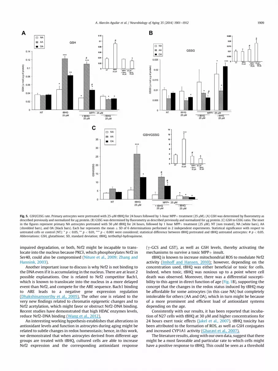
Fig. 5. GSH/GSSG rate. Primary astrocytes were pretreated with 25-mM tBHQ for 24 hours followed by 1-hour MPPþ treatment (25 mM). (A) GSH was determined by fluorometry asdescribed previously and normalized for mg protein. (B) GSSG was determined by fluorometry as described previously and normalized for mg protein. (C) GSH to GSSG ratio. The insetin the figures represent primary NA astrocytes pretreated with 50 mM tBHQ for 24 hours, followed by 1 hour MPPþ treatment (25 mM). NT (non treated), NA (white bars), AA(shredded bars), and OA (black bars). Each bar represents the mean � SD of 6 determinations performed in 2 independent experiments. Statistical significance with respect tountreated cells or control (NT): * p < 0.05, ** p < 0.01, *** p < 0.001 were considered; statistical difference between tBHQ pretreated and tBHQ untreated astrocytes: # p < 0.05.Abbreviations: GSH, glutathione; SD, standard deviation; tBHQ, tertbuthyl-hydroquinene.
A. Alarcón-Aguilar et al. / Neurobiology of Aging 35 (2014) 1901e1912 1909
impaired degradation, or both, Nrf2 might be incapable to trans-locate into the nucleus because PKCd, which phosphorylates Nrf2 inSer40, could also be compromised (Niture et al., 2009; Zhang andHannink, 2003).
Another important issue to discuss is why Nrf2 is not binding tothe DNA even if it is accumulating in the nucleus. There are at least 2possible explanations. One is related to Nrf2 competitor Bach1,which is known to translocate into the nucleus in a more delayedevent than Nrf2, and compete for the ARE sequence. Bach1 bindingto ARE leads to a negative gene expression regulation(Dhakshinamoorthy et al., 2005). The other one is related to thevery new findings related to chromatin epigenetic changes and toNrf2 acetylation, which might favor or obstruct Nrf2-DNA binding.Recent studies have demonstrated that high HDAC enzymes levels,reduce Nrf2-DNA binding (Wang et al., 2012).
An interesting working hypothesis establishes that alterations inantioxidant levels and function in astrocytes during aging might berelated to subtle changes in redox homeostasis; hence, in this work,we demonstrated that when astrocytes derived from different agegroups are treated with tBHQ, cultured cells are able to increaseNrf2 expression and the corresponding antioxidant response
(g-GCS and GST), as well as GSH levels, thereby activating themechanisms to survive a toxic MPPþ insult.
tBHQ is known to increase mitochondrial ROS to modulate Nrf2activity (Imhoff and Hansen, 2010); however, depending on theconcentration used, tBHQ was either beneficial or toxic for cells.Indeed, when toxic, tBHQ was noxious up to a point where celldeath was observed. Moreover, there was a differential suscepti-bility to this agent in direct function of age (Fig. 1B), supporting theconcept that the changes in the redox status induced by tBHQ maybe affordable for some astrocytes (in this case NA) but completelyintolerable for others (AA and OA), which in turn might be becauseof a more prominent and efficient load of antioxidant systemsdepending on the age.
Consistently with our results, it has been reported that incuba-tion of N27 cells with tBHQ at 30 mM and higher concentrations for24 hours exert toxic effects (Jakel et al., 2007). tBHQ toxicity hasbeen attributed to the formation of ROS, as well as GSH conjugatesand increased CYP1A1 activity (Gharavi et al., 2007).
The literature results, alongwith our owndata, suggest that theremight be a most favorable and particular rate to which cells mighthave a positive response to tBHQ. This could be seen as a threshold

#
* ** *
**
#
**#
Fig. 6. Cell survival after MPPþ toxicity in astrocytes pretreated with tBHQ Primary astrocytes were pretreated with 25 mM tBHQ for 24 hours, followed by 1 hours MPPþ treatment(25 mM). The inset in the figure represent primary NA astrocytes pretreated with 50-mM tBHQ for 24 hours, followed by 1 hour MPPþ treatment (25 mM). NT (nontreated), NA (whitebars), AA (shredded bars), and OA (black bars). Cell viability was assessed by trypan blue as described before. Each bar represents the mean � SD of 3 determinations performed inindependent experiments. Statistical significance * p < 0.05, was considered. Statistical significance with respect to untreated or control cells (NT): * p < 0.05, ** p < 0.01 wereconsidered; statistical difference between tBHQ pretreated and tBHQ untreated astrocytes: # p < 0.05. Abbreviations: SD, standard deviation; tBHQ, tertbuthyl-hydroquinene.
A. Alarcón-Aguilar et al. / Neurobiology of Aging 35 (2014) 1901e19121910
that might be dependant on the amount of ROS generated, incontrast to the cellular capacity to modulate oxidative stress and torepair oxidative damage. Moreover, Erlank et al. (2011) have shownthat tBHQ in cell growth medium generates H2O2 in an equimolaramount to its concentration, which penetrates into the cells, acti-vating Nrf2 signaling. Therefore higher tBHQ concentrations maygenerate greater H2O2 concentrations, which could be cytotoxic. Inour case, AA and OAwere more susceptible to this effect and did notsurvive to tBHQ high concentrations. However, it is also known thatcells preconditioning with low H2O2 concentrations, induces anadaptive response, which activates Nrf2, protecting the cells fromfurther oxidative cytotoxicity (Luna-López et al., 2010). This wasprobably the scenario for tBHQ lower concentrations in AA and OA,whereasNA requiredhigher tBHQconcentrations to shift their redoxstate and activateNrf2, probably because of their antioxidant systemproficiency. Furthermore, not only tBHQ or other inductors con-centrations are important but as we showed here, the cell type, aswell as the age and cellular conditionsmight be crucial to determineif the same tBHQ concentration can trigger a beneficial or a harmfuleffect.
The idea of an antioxidant response-threshold is consistent withthe physiological and evolutionary processes present in living be-ings, and in some papers it has been defined as hormesis. Hormesisis a consented terminology that unifies the main mechanism thatpreconditioning and adaptive responses have in common: theexposure to low levels of stress will activate existing cellular andmolecular pathways that will enhance the ability of the cell andorganism to withstand to more severe stress (Calabrese, 2008;Mattson, 2008). So, the preconditioning model used in the pre-sent work shows that the redox status can be readdress to a morereduced state, as evaluated as the GSH and/or GSSG rate, whichcorrelates with the resistance against MPPþ toxicity. Interestingly,tBHQ preconditioning was successful in protecting AA (45%) and OA
(60%) in correlation with the GSH and/or GSSG rate recovery at alow MPPþ concentration (25 mM), but it was not able to recoverredox state in OA at a higher dose (50 mM), thereby leading to celldamage (data not shown). These data suggest that cells of old an-imals might be able to activate the Nrf2 pathway in response toinducers but only in a very narrow concentration range; that is, atBHQ concentration that is suitable for cell of young animals mightharm and even kill cells of old animals. There are, however, still a lotof questions to answer, in particular in regard to the transductionalmechanism that induces Nrf2. It is therefore important to under-stand the oxidative degree and threshold necessary to activate Nrf2without damaging the cells, as well as its relationship with therepair and cell death initiation.
Most importantly, our results show for the first time that as-trocytes derived from adult and aged animals are capable to evokean antioxidant response, if they are appropriately pretreated witha molecule proficient in modulating redox status. This is ofparamount significance while developing therapies related tocounteract or protect against age-related pathologies, principallythose in which oxidative stress is involved, such as Alzheimer’sdisease, Parkinson’s disease, and other neurodegenerativedisorders.
Disclosure statement
The authors have no conflicts of interest to disclose.
Acknowledgements
The authors thank Dr Rocío González-Vieira from UAM-I foranimal supply and Dr A. Hernández from CINVESTAV for generouslydonating us the actin antibody; the authors also thank the CBS-UAMI Confocal Core for confocal images acquisition and analysis.

A. Alarcón-Aguilar et al. / Neurobiology of Aging 35 (2014) 1901e1912 1911
This work was supported by CONACyT CB-2006-1-59659 and CB2012-1-178349. As well as the “Red Temática de Investigación enSalud y Desarrollo Social” from CONACyTand INGER DI-PI004/2012.Alarcón-Aguilar is a CONACyT scholarship holder.
References
Alarcón-Aguilar, A., González-Puertos, V.Y., LunaeLópez, A., Morán, J.,Santamaría, A., Königsberg, M., 2014. Comparing the effects of two neurotoxinsin cortical astrocytes from newborn and adult rats: involvement of oxidativedamage. J. Appl. Toxicol. 34, 127e138.
Bradford, M.M., 1976. A rapid and sensitive method for the quantitation of micro-gram quantities of protein utilizing the principle of protein-dye binding. Anal.Biochem. 72, 248e254.
Calabrese, E.J., 2008. Converging concepts: adaptive response, preconditioning, andthe YerkeseDodson Law are manifestations of hormesis. J. Ageing Res. Rev. 7,8e20.
Chen, H., Wang, S., Ding, J.H., Hu, G., 2008. Edaravone protects against MPPþ-induced cytotoxicity in rat primary cultured astrocytes via inhibition ofmitochondrial apoptotic pathway. J. Neurochem. 106, 2345e2352.
Darlington, C.L., 2005. Astrocytes as targets for neuroprotective drugs. Curr. Opin.Investig. Drugs 6, 700e703.
Dhakshinamoorthy, S., Jain, A.K., Bloom, D.A., Jaiswal, A.K., 2005. Bach1 competeswith Nrf2 leading to negative regulation of the antioxidant response element(ARE)-mediated NAD(P)H:quinone oxidoreductase 1 gene expression and in-duction in response to antioxidants. J. Biol. Chem. 280, 16891e16900.
Di Monte, D.A., Wu, E.Y., Langston, J.W., 1992. Role of astrocytes in MPTP metabolismand toxicity. Ann. N.Y. Acad. Sci. 11, 219e228.
Dringen, R., 2000. Metabolism and functions of glutathione in brain. Prog. Neuro-biol. 62, 649e671.
Erlank, H., Elmann, A., Kohen, R., Kanner, J., 2011. Polyphenols activate Nrf2 in as-trocytes via H2O2, semiquinones, and quinones. Free Radic. Biol. Med. 51,2319e2327.
Fernández-Checa, J.C., García-Ruiz, C., 2008. Glutation. In: Konigsberg, M. (Ed.),Radicales Libres Y Estrés Oxidativo: Aplicaciones Médicas. El Manual Moderno,Mexico City, pp. 253e267.
Forman, H.J., Fukuto, J.M., Torres, M., 2004. Redox signaling: thiol chemistry defineswhich reactive oxygen and nitrogen species can act as second messengers. Am.J. Physiol. Cell.Physiol. 287, 246e256.
Friedlander, R.M., 2003. Apoptosis and caspases in neurodegenerative diseases.N. Engl. J. Med. 348, 1365e1375.
Galván-Arzate, S., Pedraza-Chaverrí, J., Medina-Campos, O.N., Maldonado, P.D.,Vázquez-Román, B., Ríos, C., Santamaría, A., 2005. Delayed effects of thallium inthe rat brain: regional changes in lipid peroxidation and behavioral markers,but moderate alterations in antioxidants, after a single administration. FoodChem. Toxicol. 43, 1037e1045.
García-Matas, S., Gutierrez-Cuesta, J., Coto-Montes, A., Rubio-Acero, R., Díez-Vives, C., Camins, A., Pallàs, M., Sanfeliu, C., Cristòfol, R., 2008. Dysfunction ofastrocytes in senescence-accelerated mice SAMP8 reduces their neuro-protective capacity. Aging Cell 7, 630e640.
Gharavi, N., Haggarty, S., El-Kadi, A.O., 2007. Chemoprotective and carcinogeniceffects of tert-butylhydroquinone and its metabolites. Curr. Drug Metab. 8, 1e7.
Gómez-Quiroz, L.E., Factor, V.M., Kaposi-Novak, P., Coulouarn, C., Conner, E.A.,Thorgeirsson, S.S., 2008. Hepatocyte-specific c-Met deletion disrupts redoxhomeostasis and sensitizes to Fas-mediated apoptosis. J. Biol. Chem. 283,14581e14589.
Gottfried, C., Tramontina, F., Goncalves, D., Goncalves, C.A., Moriguchi, E., Dias, R.D.,Wofchuck, S.T., Souza, D.O., 2002. Glutamate uptake in cultured astrocytes de-pends on age: a study about the effect of guanosine and the sensitivity tooxidative stress induced by H2O2. Mech. Ageing Dev. 123, 1333e1340.
Harman, D., 2003. The free radical theory of aging. Antioxid. Redox Signal. 5,557e561.
Imhoff, B.R., Hansen, J.M., 2010. Tert-butylhydroquinone induces mitochondrialoxidative stress causing Nrf2 activation. Cell. Biol. Toxicol. 26, 541e551.
Jakel, R.J., Townsend, J.A., Kraft, A.D., Johnson, J.A., 2007. Nrf2-mediated protectionagainst 6-hydroxydopamine. Brain Res. 1144, 192e201.
Jones, P.D., 2008. Radical-free biology of oxidative stress. Am. J. Physiol. Cell. Physiol.295, 849e868.
Kahlert, S., Reiser, G., 2004. Glial perspectives of metabolic states during cerebralhypoxiaecalcium regulation and metabolic energy. Cell Calcium 36, 295e302.
Kanwar, S.S., Nehru, B., 2007. Modulatory effects of N-acetylcysteine on cerebralcortex and cerebellum regions of ageing rat brain. Nutr. Hosp. 22, 95e100.
Kaspar, J.W., Jaiswal, A.K., 2010. An auto regulatory loop between Nrf2 andCul3eRbx1 controls their cellular abundance. J. Biol. Chem. 285, 21349e21358.
Kim, S.U., De Vellis, J., 2005. Microglia in health and disease. J. Neurosci. Res. 81,302e313.
Klamt, F., Gottfried, C., Tramontina, F., Dal-Pizzol, F., Conte da Frota, M.L., FonsecaMoreira, J.C., Dias, R.D., Moriguchi, E., Wofchuck, S., Souza, D.O., 2002. Time-related increase in mitochondrial superoxide production, biomolecule damageand antioxidant enzyme activities in cortical astrocyte cultures. Neuroreport 13,1515e1518.
Kraft, A.D., Johnson, D.A., Johnson, J.A., 2004. Nuclear factor E2-related factor 2-dependent antioxidant response element activation by tert-butylhydroquinone
and sulforaphane occurring preferentially in astrocytes conditions neuronsagainst oxidative insult. J. Neurosci. 24, 1101e1112.
Krueger, M.J., Singer, T.P., Casida, J.E., Ramsay, R.R., 1990. Evidence that the blockadeof mitochondrial respiration by the neurotoxin 1-methyl-4-phenylpyridinium(MPP+) involves binding at the same site as the respiratory inhibitor, rotenone.Biochem. Biophys. Res. Commun. 169, 123e128.
Kweon, M.H., In-Park, Y., Sung, H.C., Mukhtar, H., 2006. The novel antioxidant 3-O-caffeoyl-1- methylquinic acid induces Nrf2-dependent phase II detoxifyinggenes and alters intracellular glutathione redox. Free Radic. Biol. Med. 40,1349e1361.
Lee, J.M., Calkins, M.J., Chan, K., Kan, Y.W., Johnson, J.A., 2003. Identification of theNF-E2-related factor-2-dependent genes conferring protection against oxidativestress in primary cortical astrocytes using oligonucleotide microarray analysis.J. Biol. Chem. 278, 12029e12038.
Lee, O.H., Jain, A.K., Papusha, V., Jaiswal, A.K., 2007. An auto-regulatory loop be-tween stress sensors Inrf2 and Nrf2 controls their cellular abundance. J. Biol.Chem. 282, 36412e36420.
Li, J., Johnson, D., Calkins, M., Wright, L., Svendsen, C., Johnson, J., 2005. Stabilizationof Nrf2 by tBHQ confers protection against oxidative stress-induced cell deathin human neural stem cells. Toxicol. Sci. 8, 313e328.
Liddell, J.R., Robinson, S.R., Dringen, R., Bishop, G.M., 2010. Astrocytes retain theirantioxidant capacity into advanced old age. Glia 58, 1500e1509.
Lin, D.T., Wu, J., Holstein, D., Upadhyay, G., Rourk, W., Muller, E., Lechleiter, J.D., 2007.Ca2þ signaling, mitochondria and sensitivity to oxidative stress in aging as-trocytes. Neurobiol. Aging 28, 99e111.
Liu, R.M., 2002. Down-regulation of gamma-glutamylcysteine synthetase regulatorysubunit gene expression in rat brain tissue during aging. J. Neurosci. Res. 68,344e351.
López-Diazguerrero, N.E., López-Araiza, H., Conde-Pérezprina, J.C., Bucio, L.,Cárdenas, M.C., Ventura, J.L., Covarrubias, L., Gutiérrez-Ruiz, M.C., Zentella, A.,Königsberg, M., 2006. Bcl-2 protects against oxidative stress while inducingpremature senescence. Free Radic. Biol. Med. 40, 1161e1169.
Luna-López, A., Triana-Martínez, F., López-Diazguerrero, N.E., Ventura-Gallegos, J.L.,Gutiérrez-Ruiz, M.C., Damián-Matsumura, P., Zentella, A., Gómez-Quiroz, L.E.,Königsberg, M., 2010. Bcl-2 sustains hormetic response by inducing Nrf2 nucleartranslocation in L929 mouse fibroblasts. Free Radic. Biol. Med. 49, 1192e1204.
Luna-López, A., González-Puertos, V.Y., Romero-Ontiveros, J., Ventura-Gallegos, J.L.,Zentella, A., Gomez-Quiroz, L.E., Königsberg, M., 2013. A noncanonical NF-kBpathway through the p50 subunit regulates Bcl-2 overexpression during anoxidative conditioning hormesis response. Free Radic. Biol. Med. 63, 41e50.
Mattson, M.P., 2008. Hormesis defined. Ageing Res. Rev. 7, 1e7.McCarthy, K.F., De Villes, J., 1980. Preparation of separate astroglial and oligoden-
droglial cell cultures from rat cerebral tissue. J. Cell. Biol. 85, 890e902.Motohashi, H., Yamamoto, M., 2004. Nrf2-Keap1 defines a physiologically important
stress response mechanism. Trends Mol. Med. 10, 549e557.Niture, S.K., Kaspar, J.W., Shen, J., Jaiswal, A.K., 2010. Nrf2 signaling and cell survival.
Toxicol. Appl. Pharmacol. 244, 37e42.Niture, S.K., Jain, A.K., Jaiswal, A.K., 2009. Antioxidant induced modification of INrf2-
cysteine 151 and PKCd- mediated phosphorylation of Nrf2 serine 40 are bothrequired for stabilization and nuclear translocation of Nrf2 and increased drugresistance. J. Cell Sci. 122, 4452e4464.
Niture, S.K., Jaiswal, A.K., 2009. Prothymosin-alpha mediates nuclear import of theInrf2/Cul3/Rbx1 complex to degradenuclearNrf2. J. Biol. Chem. 284,13856e13868.
Niture, S.K., Jain, A.K., Shelton, P., Jaiswal, A.K., 2011. Src subfamily kinases regulatenuclear export and degradation of transcription factor Nrf2 to switch off Nrf2-mediated antioxidant activation of cytoprotective gene expression. J. Biol. Chem.286, 28821e28832.
Osburn, W.O., Wakabayashi, N., Misra, V., 2006. Nrf2 regulates an adaptive responseprotecting against oxidative damage following diquat-mediated formation ofsuperoxide anion. Arch. Biochem. Biophys. 454, 7e15.
Papadopoulos, M.C., Koumenis, I.L., Yuan, T.Y., Giffard, R.G., 1998. Increasingvulnerability of astrocytes to oxidative injury with age despite constant anti-oxidant defenses. Neuroscience 82, 915e925.
Przedborski, S., Vila, M., 2003. The 1-methyl-4-phenyl-1,2,3,6-tetrahydropyridinemouse model: a tool to explore the pathogenesis of Parkinson’s disease. Ann.N.Y. Acad. Sci. 991, 189e198.
Reynolds, A., Laurie, C., Mosley, R.L., Gendelman, H.E., 2007. Oxidative stress and thepathogenesis of neurodegenerative disorders. Int. Rev. Neurobiol. 82, 297e325.
Schapira, A.H., Olanow, C.W., 2008. Drug selection and timing of initiation oftreatment in early Parkinson’s disease. Ann. Neurol. 2 (64 Suppl), S47eS55.
Scheiber, I.F., Dringen, R., 2013. Astrocyte functions in the copper homeostasis of thebrain. Neuroch. Int. 62, 556e565.
Simonian, N.A., Coyle, J.T., 1996. Oxidative stress in neurodegenerative diseases.Ann. Rev. Pharmacol. Toxicol. 36, 83e106.
Sofroniew, M.V., Vinters, H.V., 2010. Astrocytes: biology and pathology. Acta Neu-ropathol. 119, 7e35.
Takuma, K., Baba, A., Matsuda, T., 2004. Astrocyte apoptosis: implications for neu-roprotection. Prog. Neurobiol. 72, 111e127.
Thimmulappa, R.K., Mai, K.H., Srisuma, S., Kensler, T.W., Yamamoto, M., Biswal, S.,2002. Identification of Nrf2-regulated genes induced by the chemo-preventive agent sulforaphane by oligonucleotide microarray. Cancer Res. 62,5196e5203.
Ting, K.K., Brew, B.J., Guillemin, G.J., 2009. Effect of quinolinic acid on human as-trocytes morphology and functions: implications in Alzheimer’s disease.J. Neuroinflammation 6, 36.

A. Alarcón-Aguilar et al. / Neurobiology of Aging 35 (2014) 1901e19121912
Tsai, M.J., Lee, E.H., 1994. Differences in the disposition and toxicity of1-methyl-4-phenylpyridinium in cultured rat and mouse astrocytes. Glia 12,329e335.
Tsai, M.J., Lee, E.H., 1998. Nitric oxide donors protect cultured rat astrocytes from 1-methyl-4-phenylpyridinium-induced toxicity. Free Radic. Biol. Med. 23,705e713.
Wang, B., Zhu, X., Kim, Y.T., Li, J., Huang, S., Saleem, S., Li, R., Xu, Y., Dore, S.,Cao, W., 2012. Histone deacetylase inhibition activates transcription factorNrf2 and protects against cerebral ischemic damage. Free Radic. Biol. Med. 52,928e936.
Yan, D., Dong, J., Sulik, K.K., Chen, S.Y., 2010. Induction of the Nrf2-driven antioxi-dant response by tert-butylhydroquinone prevents ethanol-induced apoptosisin cranial neural crest cells. Biochem. Pharmacol. 80, 144e149.
Zhang, D., Hu, X., Qian, L., O’Callaghan, J.P., Hong, J.S., 2010. Astrogliosis in CNS isthere a role for microglia? Mol. Neurobiol. 41, 232e241.
Zhang, D.D., Hannink, M., 2003. Distinct cysteine residues in Keap1 are required forKeap1-dependent ubiquitination of Nrf2 and for stabilization of Nrf2 by che-mopreventive agents and oxidative stress. Mol. Cell. Biol. 23, 8137e8151.
Zhu, Y., Carvey, P.M., Ling, Z., 2006. Age-related changes in glutathione andglutathione-related enzymes in rat brain. Brain Res. 1090, 35e44.


