Embed Size (px)
Citation preview

Eur. J. Biochem. 228, 297-304 (1995) 0 FEBS 1995
Primary structure of the neuronal clathrin-associated protein auxilin and its expression in bacteria Stephan SCHRODER', Stephen A. MORRIS', Ruth KNORR3, Uwe PLESSMANN', Klaus WEBER', Nguyen G. VINH' and Ernst UNGEWICKELL'
' Max-Planck-Institute for Biochemistry, Martinsried, Germany * Max-Planck-Institute for Biophysical Chemistry, Department of Biochemistry, Gottingen, Germany ' Center for Immunology, Department of Pathology, Washington University School of Medicine, St. Louis, MO, USA
(Received 19 October 1994) - EJB 94 1597/3
The protein auxilin is a coat component of brain clathrin-coated vesicles. It interacts directly with the heavy chain of clathrin and supports its assembly into regular cages [Ahle, S. & Ungewickell, E. (1990) J. Cell Biol. 111, 19-29]. The combined open reading frames of three cow brain cDNA clones with a total of 4531 nucleotides predict a molecular mass of 99504 Da for auxilin. The coding region is followed by a very long untranslated region of at least 1670 nucleotides. By Northern analysis, auxilin transcripts are found only in brain tissue. Auxilin is not related to any of the previously sequenced clathrin-binding proteins, but the region of positions 50-350 is 29% identical (similarity 56%) to the corresponding region of the actin-binding protein tensin from chicken fibroblasts. Recombinant auxilin expressed in and purified from bacteria by affinity chromatography is functional with respect to clathrin binding.
Keywords. Auxilin ; cDNA ; clathrin ; microsequencing ; tensin.
Clathrin-coated vesicles transport selective membrane com- ponents from the plasma membrane and the trans-Golgi network to the endosomal system (Brodsky, 1988; Goldstein et al., 1985). To elucidate the molecular basis for this process, biochemical and functional studies have been directed towards the identifica- tion and characterization of coat components and other factors involved in the assembly of coat proteins on membranes and their disassembly at later stages in the transport cycle (Pearse, 1987 ; Schmid, 1993). Clathrin-coated vesicles have been puri- fied and characterized from different mammalian tissues includ- ing brain. In all cases the coat consists of clathrin, which forms the characteristic polyhedral surface lattice of clathrin-coated vesicles and of adaptor proteins, which connect clathrin to pro- teins in the membrane (Pearse and Robinson, 1990).
Two types of adaptor molecules have been identified, re- ferred to as APl and AP2, respectively (reviewed in Keen, -1993). AP1 localizes to the trans-Golgi network and Golgi-de- rived coated vesicles, while AP2 is found at the plasma mem- brane and on plasma membrane-derived clathrin-coated vesicles. AP2 is very abundant in brain, reflecting the high endocytic ac- tivity of nervous tissue. For nerve-cell-specific functions, such as the retrieval of synaptic vesicle membrane after neuro- transmitter release (Heuser, 1989; Maycox et al., 1992), clathrin- coated vesicles are equipped with adaptor and clathrin light- chain isoforms (Jackson et al., 1987; Kirchhausen et al., 1987, 1989; Robinson, 1989; Ponnambalam et al., 1990) and two addi- tional proteins. These are AP180 and auxilin (Keen and Black, 1986; Ahle and Ungewickell, 1986; Kohtz and Puszkin, 1988; Ahle and Ungewickell, 1990; Su et al., 1991 ; Perry et al., 1992).
Correspondence to E. Ungewickell, Washington University School of Medicine, Department of Pathology, 660 S. Euclid Avenue, St. Louis, MO 63110, USA
Abbreviation. AP, adaptor protein. Note. The nucleotide sequence reported in this paper has been sub-
mitted to the GenBankm Data Base with the accession number U09237.
Both proteins are monomers of about 90 kDa. Binding studies using purified AP180 and auxilin showed that they interact inde- pendently with clathrin and promote its assembly into polyhedral cages (Ahle and Ungewickell, 1986, 1990). The binding sites of AP180 and auxilin on triskelia have not been mapped, but differences in binding stoichiometries suggest distinct sites for them (Ahle and Ungewickell, 1986, 1990; Prasad and Lippoldt, 1988). The primary structures of the adaptor subunits and AP180 are known from cDNA cloning (Thurieau et al., 1988; Kirch- hausen et al., 1989, 1991 ; Robinson, 1989; Ponnambalam et al., 1990; Zhou et al., 1992; Morris et al., 1993).
Here we report the primary structure of auxilin. While no sequence similarities between auxilin and other coat proteins was detected, we found significant similarities between auxilin and the actin binding protein tensin. Recombinant auxilin, capa- ble of binding to clathrin cages, was produced in bacteria and obtained from lysates by affinity chromatography.
MATERIALS AND METHODS
Materials. Fresh bovine brains were obtained from the local abattoir. T7 sequencing kit was from Pharmacia. Trypsin (treated with tosylphenylalanine chloromethane) was from Worthington Biochemical Corporation. Endoproteinase Asp-N, restriction enzymes, Klenow fragment and kinases were from Boehringer Mannheim. DNA-grade agarose was from Bio-Rad Laborato- ries. Reagents for SDSPAGE and urea were from LKB Instru- ments GmbH. Horseradish-peroxidase-conjugated IgGs to mouse immunoglobulins were from Dakopatts GmbH. Nitro- cellulose transfer membranes (BA83, 0.2 pm) were from Schleicher & Schiill or from MSI. Radiochemicals and Hybond- N nylon membraneswere from Amersham International. The oli- go(dT)-primed bovine brain jlgtll cDNA expression library and the random primed AgtlO cDNA library were from Clonetech

298 Schroder et al. ( E m J. Biochem. 228)
(Palo Alto, CA). pBluescript SK+ and SK- vector were from Stratagene. Nitrilotriacetic acid affinity resin was from Qiagen (Chatworth, USA) as were the plasmids pQE and pREP4. Escherichia coli BL21 was from Novagen (Madison, USA). All chemicals and reagents were of analytical grade.
Protein chemistry. Auxilin was affinity-purified from either a 0.5 M Tris extract of bovine brain clathrin-coated vesicles (Keen et al., 1979) or from 1 1 bovine brain cytosol (the su- pemant obtained from the first ultracentrifugation step in stan- dard coated vesicle preparations (Campbell et al., 1984)) using 3 mg mAb 100/4 attached to 1.5 ml Sepharose 4B (Ahle and Ungewickell, 1990). Auxilin was eluted from the column with 0.1 % SDS at 60°C for 5 min. For sequencing, auxilin was further purified by SDSPAGE on linear 7.5-19% gradient acrylamide mini gels (Hoefer Mighty Small I1 apparatus; dimen- sion: 8 x 7 cm) using the buffer system of Laemmli (1970). The gels were briefly stained and destained, after which the auxilin band was excised from the gel. To concentrate the protein, ap- propriate gel slices were combined into a single slot and electro- phoresed again as described (Vandekerckhove et al., 1993). The resulting gel was blotted on to a polyvinylidene difluonde mem- brane by electrophoretic transfer (Bauw et al., 1993). Blotted polypeptides were visualized by staining with amido black solu- tion (0.1 % in 9% acetic acid) for 1-2 min. Blots were subjected to direct microsequencing using an Applied Biosystems 470A sequenator or a Knauer model 810 sequenator. Both instruments operated with on-line phenylthiohydantoin analysis. To obtain internal sequences the blots were treated for 3 h at 37°C with trypsin and in some cases also with endoproteinase. The eluted peptides were subjected to HPLC (Applied Biosystems, Aqua- pore C8 column). Elution was with 7-42% acetonitrile in 0.1 % trifluoroacetic acid. We also made use of auxilin eluted electro- phoretically from preparative gels into dialysis tubes. The pro- tein present in 6 ml Laemmli sample buffer was recovered by methanoVCHC1, precipitation (Wessel and Flugge, 1984) and subjected to treatment with CNBr in 50% trifluoroacetic acid for 20 h at 22°C. Fragments were subjected to HPLC using an Aquapore C8 column (100X2.1 mm, Applied Biosystems). Se- quencing was as described above. The material present in two fractions, which comprised a mixture of fragments, was treated with endoproteinase Lys-C for 20 h at 37°C in 50 mM TridHCl pH 8.5. Peptides were separated by HPLC using a Spheri-SRP18 column (220X2.1 mm; Applied Biosystem). Sequence analysis was as above. The total amount of auxilin used for sequencing was about 50 pg.
Isolation and sequence analysis of cDNA. 300 000 plaque- forming units from an oligo(dT)-primed bovine brain Agtll cDNA expression library were screened with mAb 100/4. To confirm that positve clones were related to auxilin, the sequence YEQYAK (peptide BrCN-K-5) was used for the synthesis of an oligonucleotide mixture of the following sequence : 5’-TA(C, T)GA(A, G)CA(A, G)TA(C, T)GC(A, C, G, T)AA-3’. The oligonucleotide was radiolabeled by phosphorylation with T4 polynucleotide kinase (Sambrook et al., 1989). Aux 11 was sub- cloned into pBluescript SK+ and sequenced by the dideoxy- chain-termination method (Sanger et al., 1977). Synthetic 18- residue oligonucleotides were used as internal primers. Restric- tion fragments used for re-screening were labeled by comple- mentary strand synthesis primed with random hexanucleotide mixtures (Sambrook et al., 1989). Sequence data were analyzed using programs of the University of Wisconsin Genetics Com- puter Group (Devereux et al., 1984) and MacVector software (Kodak). Protein and DNA data bases used for sequence analysis were: Genbank, release 81; EMBL, release 37.0; PIR 39.0; SWISS protein, release 28.0 and ProSite, release 8.1.
Electrophoresis and transfer to nitrocellulose membrane were performed as described (Sambrook et al., 1989). Genomic DNA for Southern hybridizations was prepared from 3 X lo7 MDBK cells according to protocol 1 of Sambrook et al. (1989); 10 pg digested genomic DNA was loaded/lane of a 0.7% aga- rose gel. Electrophoresis and transfer to nitrocellulose membrane were performed as described by Sambrook et al. (Sambrook et al., 1989). For Northern blot analysis, total RNA from mouse and rat brains was prepared by acidic guanidinium thiocyanate/ phenokhloroform extraction (Chomczynski and Sacchi, 1987) ; 20 pg total RNA fractionated in a 1.2 % agarose gel containing 1.1 M formaldehyde and then transferred in 1.5 M NaCl, 0.15 M sodium citrate, pH 7.0 (1OX NaCVCit) to a nylon-reinforced nitrocellulose-membrane (Nitroplus) and crosslinked by expo- sure to ultraviolet light. The 1.9-kb HindIII-EcoRI fragment of clone Aux F was radiolabeled by random priming (Feinberg and Vogelstein, 1984). Hybridization was performed at 42°C in 50% formamide, 5 X NaCl/Cit, 1 X Denhardt’s solution (Sambrook et al., 1989), 20 mM sodium phosphate pH 7.0, 10% dextran sul- fate, 0.1 % SDS and 100 pg/ml sonicated denatured salmon sperm DNA. Hybridized blots were washed at 60°C under low (2X NaCVCit, 0.1 % SDS) or high (0.1 X NaCVCit, 0.1 % SDS) stringent conditions. The actin probe was a gift from Dr S. Speck (Washington University, St Louis).
Expression of auxilin in E. coli. cDNA clone Aux 3 was cleaved with ThaI and HindIII to generate a 286-bp fragment containing the 5’-end of the auxilin DNA. The fragment was ligated into the SmaI-Hind111 site of pQE30. The central por- tion of the auxilin DNA was obtained from Aux F using HindIII and EcoRV. The 3‘-end of the coding region was excised from Aux B with EcoRV and PstI. Both fragments were ligated into pBluescript KS- utilizing the HindIII-PstI site. The vector was subsequently cleaved with HindIII and PstI and ligated with a 3 12-bp BamHI-Hind111 auxilin fragment that was excised from the pQE30. The resultant full-length auxilin DNA was inserted into the BamHI-PstI site of pQE30.
For expression of auxilin we used E. coli BL21. The bacteria were grown for 3 h before the expression of auxilin was induced with 1 mM isopropyl P-D-thiogalactopyranoside for another 3 h. Cells were harvested and lysed by sonification in 0.5 M Tris/ HC1 pH 8.0, 0.5% Triton X-100 in the presence of 0.1 mM phenylmethylsulfonyl fluoride, 0.01 mM leupeptin, 1 mM benz- amidine and 1 pg/ml E64. The lysate was clarified by ultracen- trifugation at 1OOOOOXg for 30 min and then applied to nitrilo- triacetic acid affinity resin. Typically 40-70 ml lysate was applied to 10 ml resin at a flow rate of 20 mlh. The column was washed with at least 2 vol. lysis buffer and 3 vol. 0.5 M Tris/ HC1 pH 7.0. Auxilin was eluted with 0.25 M imidazol in 0.5 M Tris/HCl pH 7.0. Auxilin was transferred into 10 mM Tris/HCl pH 8.0, 0.02% NaN, by gel filtration using a PDlO column and then concentrated in a Centricon 30. Clathrin binding experi- ments in 0.1 M potassium tartrate, 1 mM EGTA, 0.5 mM MgCl,, 10 mM Hepes (buffer T), using pre-formed cages were per- formed as described previously (Lindner and Ungewickell, 1991).
RESULTS
Molecular cloning of auxilin cDNA. Probably due to the presence of a blocking group, direct sequencing of bovine brain auxilin failed to provide an amino-terminal sequence. By in- ternal cleavage with cyanogen bromide and endoproteinases fol- lowed by HPLC, 12 pure fragments were obtained (BrCN1- BrCN4, BrCN-K-1-BrCN-K-5, D1, D2 and K1) and sequenced
Schroder et al. ( E m J. Biochem. 228) 299
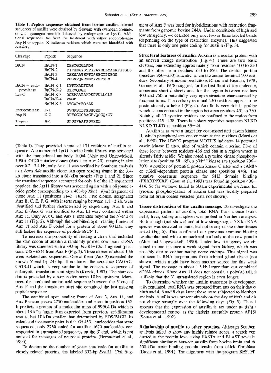
Table 1. Peptide sequences obtained from bovine auxilin. Internal sequences of auxilin were obtained by cleavage with cyanogen bromide, or with cyanogen bromide followed by endoproteinase Lys-C. Addi- tional sequences are from the treatment with either endoproteinase Asp-N or trypsin. X indicates residues which were not identified with certainty.
Cleavage Peptide Sequence
BrCN BrCN-1 BrCN-2 BKN-3 BrCN-4
BrCN + endo- BrCN-K-1 proteinase BrCN-K-2
BrCN-K-4 BrCN-K-5
Lys-c BrCN-K-3
Endoproteinase D-1 Asp-N D-2
Trypsin K- 1
EPSYGGGLFDM FIFXNLYSTPGPAVRLLYAKRPGIGLS GSKSAATSPTGSSHGTPXHQN PHSSPQNRPNYXVSFSSM
IYTTXADFERM EYRVQDGK QQEPAAPAPPEDVDLLGLE PGGF ATGQPYEQYAK
DVNPSILFSSHQEH DLFGGGGAAGPVQSGQXGV
NFSSPAAPPSNXEL
(Table 1). They provided a total of 171 residues of auxilin se- quence. A commercial l g t l l bovine brain library was screened with the monoclonal antibody 100/4 (Ahle and Ungewickell, 1990). Of 20 putative clones (Aux 1 to Aux 20), ranging in size over 0.2-3.4 kb, only Aux 11 was verified by Southern analysis as a bonajde auxilin clone. An open reading frame in the 3.4- kb clone translated into a 61-kDa protein (Figs 1 and 2). Since the translated sequence accounted for only 8 of the 12 sequenced peptides, the l g t l l library was screened again with a oligonucle- otide probe corresponding to a 483-bp XbaI-KpnI fragment of clone Aux 11 (positions 1142- 1625). Five clones, designated Aux B, C, E, F, G, with inserts ranging between 1.1-2 kb, were identified and further characterized by sequencing. Aux B and Aux E (Aux G was identical to Aux E) were contained within Aux 11. Only Aux C and Aux F extended beyond the 5‘-end of Aux 11 (Fig. 2). Although the combined open reading frames of Aux 11 and Aux F coded for a protein of about 90 kDa, they still lacked the sequence of peptide BrCN-1.
To increase the probability of finding a clone that included the start codon of auxilin a randomly primed cow brain cDNA library was screened with a 392-bp EcoRI- CluI fragment (posi- tions 245-636) from Aux F (Fig. 2). Three clones of about 1 kb were isolated and sequenced. One of them (Aux 3) extended the known 5’-end by 245 bp. It contained the sequence CAGAC- CATGG which is very similar to the consensus sequence of eukaryotic translation start signals (Kozak, 1987). The start co- don is preceded by a stop codon some 10 bp upstream. More- over, the predicted amino acid sequence between the 5’-end of Aux F and the translation start site contained the last missing peptide sequence.
The combined open reading frame of Aux 3, Aux 11, and Aux F encompasses 2730 nucleotides and starts in position 132. It predicts a protein of a molecular mass of 99504 Da which is about 13 kDa larger than expected from previous gel-filtration results, but 10 kDa smaller than determined by SDSPAGE. Its calculated isoelectric point is 6.9. Of 4531 nucleotides that were sequenced, only 2730 coded for auxilin; 1670 nucleotides cor- responded to untranslated sequences on the 3’ end, which is not unusual for messages of neuronal proteins (Bernasconi et al., 1990).
To determine the number of genes that code for auxilin or closely related proteins, the labeled 392-bp EcoRI- CluI frag-
ment of Aux F was used for hybridizations with restriction frag- ments from genomic bovine DNA. Under conditions of high and low stringency, we detected only one, two or three labeled bands (depending on the type of restriction enzyme). This suggested that there is only one gene coding for auxilin (Fig. 3).
Structural features of auxilin. Auxilin is a neutral protein with an uneven charge distribution (Fig. 4.) There are two basic clusters, one extending approximately from residues 100 to 250 and the other from residues 550 to 850. The central portion (residues 350-550) is acidic, as are the amino-terminal 100 resi- dues. Secondary structure predictions (Chou and Fasman, 1978 ; Gamier et al., 1978) suggest, for the first third of the molecule, numerous short ,B sheets and, for the region between residues 400 and 750, a potentially very open structure characterized by frequent turns. The carboxy-terminal 150 residues appear to be predominantly a-helical (Fig. 4). Auxilin is very rich in proline which is concentrated in the region between residues 451 to 750. Notably, all 13 cysteine residues are confined to the region from positions 125-438. There is a short repetitive sequence NLKD NLKD TLKD at position 33-44.
Auxilin is in vitro a target for coat-associated casein kinase 11, which phosphorylates one or more serine residues (Morris et al., 1990). The UWCG program MOTIFS indicates 14 potential casein kinase I1 sites, nine of which contain a serine. Five of these locate between residues 436 and 588 in a region which is already fairly acidic. We also noted a tyrosine kinase phosphory- lation site (position 58-65), a p3pdcZ kinase site (position 706- 709), a number of potential protein kinase C sites and a CAMP- or cGMP-dependent protein kinase site (position 476). The putative consensus sequence for SH3 domain binding (PXXXPPXXP) (Gout et al., 1993) was found in position 406- 414. So far we have failed to obtain experimental evidence for tyrosine phosphorylation of auxilin that was freshly prepared from rat brain coated vesicles (data not shown).
Tissue distribution of the auxilin message. To investigate the expression pattern of auxilin, total RNA from mouse brain, heart, liver, kidney and spleen was probed in Northern analysis. Both at high (not shown) and at low stringency, a 5.8-kb RNA species was detected in brain, but not in any of the other tissues tested (Fig. 5). This confirmed our previous immuno-blotting results obtained with a monoclonal antibody to the cow protein (Ahle and Ungewickell, 1990). Under low stringency we ob- tained in one instance a weak signal from kidney, which was possibly due to contaminating nerve endings. This signal was not seen in RNA preparations from adrenal gland tissue (no1 shown) which might have been another source for this weak signal. The message is about 1.3 kb larger than our combined cDNA clones. Since Aux 11 does not contain a poly(A) tail, ii is likely that the 3’-untranslated region is even longer.
To determine whether the auxilin transcript is developmen tally regulated, total RNA was prepared from rats on their day o birth and 4, 6 and 8 days later; these were subjected to Norther1 analysis. Auxilin was present already on the day of birth and dic not change strongly over the following days (Fig. 5) . Thus i appears that the expression of auxilin is not under as tight ;
developmental control as the clathrin assembly protein AP18 (Sousa et al., 1992).
Relationship of auxilin to other proteins. Although Souther] analysis failed to show any highly related genes, a search con ducted at the protein level using FASTA and BLAST revealel significant similarity between auxilin from bovine brain and th 200-kDa actin binding protein tensin from chick fibroblast (Davis et al., 1991). The alignment with the program BESTFI

300 Schroder et al. ( E m J. Biochem. 228)
m----?Gax M D S S G A S S P D M E P S Y G G
Bra-1
*m G L F D M V K G G A G R L F S N L K D N
m- AT-m L K D T L K D T S S R V I Q S V T S Y T
C- K G D L D F T Y V T S R I I V M S F P L
P - W T A T K - D S V D I G F R N Q V D D I R S F L D S
O m - - R H L D H Y T V Y N L S P K S Y R T A K
F H S R V S E C S W P I R Q A P S L H N
K- L F A V C R N M Y N W L L O N P K N V C
G K X l t X C C - A - V V H C L D G R A A S S I L V G A M F I
T G b c F C N L Y S T P G P A V R L L Y A K R P
B l a - 2
C r n S G I G L S P S H R R Y L G Y M C D L L A
U - T T m - D K P Y R P H F K P L T I K S I T V S P
m- m m ~ m m m m m m V P F F N K Q R N G C R P Y C D V L I G
- T A T A T - m n T - E T K I Y T T C A D F E R M K E Y R V Q
BtUi-K-1 BrCN-K-2
~ T C G W A h % T C l W A T T C C l T T G A G Y . ~ T ~ ~ - D G K I F I P L S I T V Q G D V V V S M
m-n--- Y H L R S T I G S R L Q A K V T N T Q I
T A - T A c P X A P APAGPTCA F Q L Q F H T G F I P L D T T V L K F T
~ T K A T G K % ~ T A T C C K P E L D A C D V P E K Y P Q L F Q V T
C T c r w G r ~ m ~ m - m m - L D V E L Q P H D K V M E L T P P W E H
A T T A c r G c K A w x 4 m U V I C O C U I O C A T C C I ' C I " I Y C T K D V N P S I L F S S H Q E H Q D
D-1
A T W E K G X T C ~ W T A G % T A P Z X T C C A X T W K C X X X Z T I T L V L G G Q A P I D I P P D N P R H F
TT- lCKXb4TC-T G Q G G F F S T L C W Q D Q K S E K S F
T L T C C - r n - p TGMC C E E D H A A L V N Q E S E Q S D D E L
TF2T-T-m-m L T L S S P H G N A N G D K P H A A R K
xcc- ( T G h X A m r n P S K K Q Q E P A A P A P P E D V D L L
BrCN-K-3
? i x a x z r ~ ~ C T m ~ m G L E G S A V S K N F S S P A A P P S N
K-1
M) 120 180
240
300
3M)
420
480
540
600
660
720
7 80
840
900
960
1020
1080
1140
1200
1260
1320
1380
1440
1500
1560
1620
1680
S E L L S D L F G C G G A A G P V Q S G
D-2 -- AcATccK- Q S G V D D V F H P S G P T S T Q S T P
X-- __ c A c G O x 4 - P GGTcav\ R R S A T S T S A S P T L R V G E G A T
arlxXxa--- F D P F G A P S K P S G Q D L L G S F L
-m- CITcrrCpA N T A S A S S D P F L Q P T R S P S P T
l X m a x T ~ r n ~ CTGGG V H A S S T P A V N I O P D V S G A W D
W T C f Z A T h C U m - W H T K P G G F G M G S K S A A T S P T
BrcN-K-4 m - 3
CWX%DXKCATGT--- G S S H G T P T H Q N K P Q T L D P F A x
- T F - T l T P - T D L G T L G G S S F A S K P S T P T G L
~~~ TCftCCCAGCMTGCXG G G G F P P L S S P Q K A S P Q P M G G
~~~~~
G W Q Q G G G Y N W Q Q T Q S K P Q S S
C A T G C C C C C R X E K Z a X 3 L 4 X ~ ~ T X 4 l t X H P H S S P Q N R P N Y N V S F S S M P
B r a - 4
m - p m- G G Q N E R G K A A A N L E G K Q K A A
C r m - AxTOCICACAAR09CARRAAGGtPC D F E D L L S G Q G F N A H K D K K G P
C R X U X A T A G C T G h G R T U G I U U O W \ G C U V \ T O ; C T A A R T I A E M R K E E M A K E M D P E K L
T W A C A T F 2 T K W C l 3 2 A ~ T A T C 4 G W X C T R X C T C C A C A 4 K I L E W I E G K E R N I R A L L S T M
T G m m c I w - p H T V L W A G E T K W K P V G M A D L V
T a ~ T A T P E Q V K K V Y R K A V L V V H P D K
A A I Z C A ~ ~ ~ A T G M C W T A T Q X P & ~ ~ G ~ T T I T C A T G G W X C M T G A T G A T G Q P Y E Q Y A K M I F M E L N D A
EZCW-K-5
C C T G G F C W A A l 7 T G W \ A R T U \ R O ; C C A R R K ; C C C P T G W S E F E N Q G Q K P L Y
1740
1800
1860
1920
1980
2040
2100
2160
2220
2280
2340
2400
2460
2520
2580
2640
2700
2760
2820
2880
2940 3000 3060 3120 3180 3240 3300 3360 3420 3480 3540 3600 3660 3720 3780 3840 3900 3960 4020 4080 4140 4200 4260 4320 4380 4440 4500
~ T A l T C T T T I V W + V A T ~ 4531
Fig. 1. Complete nucleotide and deduced amino acid sequence of bovine auxilin. Beneath the nucleotide sequence is the deduced amino acid sequence. The broken lines indicate the positions of peptides obtained by direct sequencing of bovine auxilin (X = unidentified amino acid).
indicated an overall similarity of 46% with 22% identical resi- dues. When the regions between residues 47 and 350 of both proteins are compared the similarity increases to 56% and the identity to 28 %. Preliminary experiments failed to demonstrate an interaction between auxilin and F-actin (data not shown). Considerable similarity was also seen between auxilin and an expressed sequence tag of 111 residues designating an unknown
human brain protein (GenBank accession: M78861) (Adams et al., 1992). The tagged protein may correspond to human auxilin (see Discussion). For an optimal alignment we had to introduce a frameshift at nucleotide position 133 in the published sequence (Fig. 6). It is possible that the published sequence misses a thymidine in this position. With this manipulation, auxilin from cow and man are about 60% identical.

Schroder et al. ( E m J. Biochem. 228) 301
PstI Sac1 C1a1 ’ *4531 AUX 11 1142 T&J * - - 4
3511 AUX B
1344 e --t
1282 2362 c e
769 2132 - t
AUX E
AUX C
AUX F
AUX 3
I 1 NUCLEOTIDE
1 4531
Fig. 2. Alignment of auxilin clones. Thick lines designate coding and thin lines non-coding sequences. Clone Aux 11 was obtained from a Lgtll bovine brain expression library by screening with mAb 100/4. The hatched region in Aux 11 corresponds to a 483-bp XbaI-KpnI fragment which was labeled by random hexanucleotide priming and used as a probe for further screening of the Igtl 1 bovine brain expression library. Aux B to Aux F were obtained in this way. The hatched region in Aux F corresponds to a 392-bp fragment generated by digestion with EcoRI and CluI. This was used to obtain Aux 3 by screening a randomly primed cow brain cDNA library. The probe was also employed in Southern blotting experiments. Arrows indicate the direction and extent of the sequence analysis. Asterisks mark the positions of potential polyadenyla- tion signals.
Fig. 3. Autoradiograph of a DNA blot according to Southern (1975). Genoinic DNA from MDBK cells was digested with the restriction endo- nucleases as indicated above each lane and fractionated in a 0.7% aga- rose gel. DNA size markers (values in kb) are on the left. After hybrid- ization the blot was washed twice for 10 min at room temperature and 42°C with 250 ml each of 2X NaCUCit, 0.1 % SDS. The final wash was for 10 min at 65°C with 0.1 X NaClICit.
Expression of functional auxilin in bacteria. Auxilin is rather difficult to purify from mammalian tissue because it is not an abundant protein and, moreover, it is very sensitive to proteoly- sis. We therefore attempted to express the recombinant protein, which was tagged at its amino-terminal with six histidine resi- dues in E. coli. Although the yields of the expressed auxilin (He- auxilin) were also low, we were able to obtain enough protein (up to 300 pg from a 2-1 culture) for preliminary characteriza- tions. H,-auxilin which was eluted from the resin was heavily contaminated with a series of polypeptides between 60-90 kDa and a 30-kDa moiety (Fig. 7). Direct sequencing indicated that the 30-kDa polypeptide corresponds to a histidine-rich E. coli protein (PIR database accession number: S33444). The other major two contaminants between 60 kDa and 90 kDa are also of bacterial origin. One corresponds to the products of groEL and
A
BETA SHEET
BETA TURN
ALPHA HELIX
. . . , . . . . .
B
200 400 600 800 Residue
200 400 600 800 PROLINE
CYSTETNE Residue
Fig. 4. Structural organization of the amino acid sequence of auxilin. (A) The combined predicted secondary structure of auxilin based on the algorithms of Chou and Fassman (1978) and Gamier et al. (1978). (B) A plot of the distribution of proline and cysteine residues. Each block covers 50 residues. Asterisks indicate the positions of putative casein kinase I1 sites. The positions of the putative tyrosine phosphorylation site and the putative SH3 domain binding site are indicated by arrows.
Fig. 5. Northern blot analysis. (A) A blot of total RNA (== 20 pg) from rat brain (b), heart (h), liver (l), kidney (k) and spleen (s) was incubated with the 1.9-kb HindIII-EcoRI fragment of clone Aux F which was labeled by random priming (Sambrook et al., 1989). The positions of 28s and 18s ribosomal RNA are shown as markers. In brain a strong diffuse signal at 5.8 kb is seen (arrow). mRNA for auxilin was detected reproducibly only in brain, even at low loadings (track 2). The gel below shows labeling of actin mRNA. (B) Analysis of total RNA extracted from neonatal rats on the days indicated on top of the blot. Note that there is no significant change in the amount of auxilin mRNA between day of birth and eight days later. The gel below shows labeling of actin mRNA.
the other to glutamine-fructose-6-phosphate transaminase (EC 2.6.1.16).
The mobility of H,-auxilin in SDSPAGE was very similar to that of the cow protein. However, the band appears to be sharper which might reflect the lack of post-translational modifi- cations. From our previous studies it is already known that auxilin is, at least in vitro, the target of coated-vesicle-associated protein kinases. The Stokes radius of the H,-auxilin as deter- mined by gel filtration on Superose 6 was indistinguishable from that of its cow counterpart (not shown). Next we tested if the

302 Schroder et al. (Eu,: J. Biochem. 228)
111 A (h) . - - - . . . - . - - - - _ - _ - _ _ _ _ - _ _ _ _ _ _ $(b) fib? y ym+J;;m:; 1 ;;;;;; $J$Pgl;; ;; F&$p 350 350
Fig. 6. Alignment of auxilin from cow brain with chick fibroblast tensin (T) and with the partial sequence of a protein from buman brain (GenBank accession: M78861) (Adams et al., 1992). A (b), Bovine brain auxilin; A (h), human auxilin-like protein; T, chick tensin. Boxed sequences denote similar sequences.
Fig. 7. Expression of recombinant auxilin in bacteria. Auxilin carry ing an amino-terminal histidine tag (H,) was expressed in bacteria and purified from the lysate by metal chelate affinity chromatography. Eluted protein is shown in track 1. Auxilin corresponds to the uppermost band that migrates in the position of a 116-kDa polypeptide. The auxilin- specific monoclonal antibody mAb 100/4 reacts predominantly with a band corresponding to intact auxilin. For comparison, an immunoblot of total brain coated vesicles is shown in track 3. The polypeptides between the 66-97 kDa and at about 30 kDa were shown by direct sequencing to be of bacterial origin.
recombinant protein was capable of binding to clathrin. To this end we incubated H,-auxilin with preassembled clathrin cages in buffer T. Binding was analyzed by ultracentrifugation (Fig. 8). Only intact H,-auxilin co-sedimented quantitatively with the clathrin cage in the gradient, suggesting that it is correctly folded.
DISCUSSION
The clathrin-associated protein auxilin is the last of the pres- ently known coat proteins from bovine brain coated vesicles to be characterized by sequence analysis. It is expected that this information will help to obtain a more complete view of the function of auxilin in synaptic membrane trafficking. Auxilin has not received much attention in the past because it is rather difficult to purify and to handle. This is due to its extraordinarily high sensitivity to proteolysis and its tendency to aggregate at pH 6.5, the pH traditionally used to study assembly of clathrin (Pearse, 1989). Both properties can be rationalized on the basis of auxilin’s sequence. Its isoelectric point ( ~ 7 ) might explain
Fig. 8. Binding of H,-auxilin to clathrin cages. H,-auxilin was incu- bated in the absence and presence of clathrin cages in buffer T and the extent of binding was analyzed by ultracentrifugation. Supernatant (track 1) and pellet (track 2) of incubations of H,-auxilin without clathrin; supernatant (track 3) and pellet (track 4) of incubations of H,- auxilin with clathrin cages. Note that all of the intact recombinant auxilin bound to clathrin.
its low solubility at and below pH 7. Secondary structure predic- tions suggest a folding into three domains : the amino-terminal 400 residues are likely to form &sheets which are linked to the carboxy-terminal a-helical domain via a = 300-residue region dominated by (Fig. 4). It is possible that the middle re- gion, with its perhaps more open structure, makes auxilin vul- nerable to proteolytic attack. The molecular mass of auxilin (99.5 kDa) is =13 kDa higher than expected from gel filtration of the unfolded protein in 6 M guanidinium hydrochloride (Ahle and Ungewickell, 1990). The reasons for this discrepancy are not clear but it is possible that proteolysis might have interfered with our previous measurements. Auxilin contains numerous potential protein kinase sites and is indeed a target for coat- associated casein kinase I1 (Morris et al., 1990).
Among the more exotic motifs present in auxilin are a puta- tive consensus sequence for a SH3 binding domain (position 406-414). The consensus sequence PXXXPPXXP is based on the alignment of putative SH3 domain binding sites in dynamin and murine 3BP1 (Gout et al., 1993). Although the motif in auxilin matches the SH3 binding motif in dynamin perfectly, the variable positions of the known SH3 binding motifs are occu- pied by either neutral or basic residues, while auxilin has two acidic residues in these positions.
Auxilin is unrelated to any of the known clathrin-coated vesicle proteins. It shares, however, significant sequence simi-

Schroder et al. (Eur J. Biochem. 228) 303
larity with the = 200-kDa protein tensin from chicken fibroblasts (Davis e t al., 1991). Tensin localizes to adherens junctions in a large variety of cells (Bockholt et al., 1992). It has received considerable attention because it is tyrosine-phosphorylated, contains a SH2 domain and is a putative actin binding protein with capping and bundling properties (Lo et al., 1994). Taken together, these properties suggest that tensin might play a crucial role in transducing signals to the cytoskeleton which might regulate cell adhesion. The similarity between auxilin and tensin covers a region of tensin which has been implicated in actin binding (Lo et al., 1994). Our preliminary binding studies did not reveal any affinity of auxilin for F-actin. We have not yet investigated the possibility of an interaction between auxilin and the ends of actin filaments.
The role of microfilaments in endocytosis is complex. There is evidence that in non-polarized cells, the integrity of the micro- filament system is not required for clathrin-mediated endocyto- sis, while phagocytosis and clathrin-independent endocytosis are strongly inhibited by microfilament-disrupting drugs (Axline and Reaven, 1974; Sandvig and van Deurs, 1990). In polarized MDCK cells, receptor-mediated endocytosis at the basolateral surface appears to be unaffected by the microfilament-disrupting drug cytochalasin D, while the generation of coated vesicles on the apical surface is inhibited by the drug (Gottlieb et al., 1993). Since the axon of a neuron corresponds to the apical surface of a MDCK cell, it is conceivable that clathrin-mediated endocyto- sis in nerve endings may also require the participation of micro- filaments and hence the need for an actin binding protein. There has been a report on the interaction of actin with two coat-asso- ciated proteins of 40 kDa and 80 kDa (Kohtz et al., 1990). We think it unlikely that the two proteins are related to auxilin because we have never seen any stable degradation products of auxilin in this molecular mass range.
Auxilin is 60% identical (similarity 80%) to a short cDNA clone from human brain which encodes 111 residues (Adams et al., 1992). The human protein sequence is as closely related to the chicken tensin sequence as is the corresponding region from cow brain auxilin. This suggests that the expressed sequence tag may correspond to an auxilin-like protein. If so, auxilin would be the least conserved of the coat proteins in vertebrates. In contrast, the sequence of the p2 subunit of AP2 is 100% con- served between man and rodents. Even human and fly j3-type sequences (GenBank accession: X75910) share more than 7 0 % identical residues. However, it is also possible that the sequence similarity between chicken tensin, cow auxilin and the human brain protein identify a sequence module which is shared by a number of otherwise unrelated proteins and which undergoes considerable evolutionary drift.
The finding that auxilin mRNA is expressed in significant amounts only in brain confirms and extends our previous results which showed by immuno-blotting that the expression of auxilin might be restricted to neuronal cells (Ahle and Ungewickell, 1990). Previous biochemical and cell fractionation studies have suggested that auxilin might be localized to nerve endings (Maycox et al., 1992). Ultimate proof for this notion should emerge from immuno-histochemical studies.
The successful expression of recombinant auxilin in bacteria will set the stage for exploring functional domains of the protein. Hitherto, this has been difficult because controlled digestion with protease did not produce stable intermediates.
This work was supported in part by a project grant from the Deutsche Forschungsgemeinschaft to E. U. (Un 43/3-5) and by start-up funds from the Department of Pathology (Washington University, St Louis, MO). Dr Woodford-Thomas is thanked for helpful discussions.
REFERENCES Adams, M. D., Dubnick, M., Kerlavage, A. R., Moreno, R., Kelley, 3.
M., Utterback, T. R., Nagle, J. W., Fields, C. & Venter, J. C. (1992) Sequence identification of 2,375 human brain genes [see comments], Nature 355, 632-634.
Ahle, S . & Ungewickell, E. (1986) Purification and properties of a new clathrin assembly protein, EMBO J. 5, 3143-3149.
Ahle, S. & Ungewickell, E. (1990) Auxilin, a newly identified clathrin- associated protein in coated vesicles from bovine brain, J. Cell Biol. 111, 19-29.
Axline, S. G. & Reaven, E. P. (1974) Inhibition of phagocytosis and plasma membrane mobility of the cultivated macrophage by cytocha- lasin B. Role of subplasmalemmal microfilaments, J. Cell Biol. 62, 647-659.
Bauw, G., Van den Bulcke, M., Van Damme, J., Pupye, M., Van Mon- tagu, M. & Vandekerckhove, 3. (1988) Protein-electroblotting on polybase coated glass-fibre and poly(viny1idene difluoride) mem- branes: an evaluation, J. Protein Chem. 7, 194-196.
Bernasconi, P., Rausch, T., Struve, I., Morgan, L. & Taiz, L. (1990) An mRNA from human brain encodes an isoform of the B subunit of the vacuolar H( +)-ATPase, J. Biol. Chem. 265, 17 428- 17 431.
Bockholt, S. M., Otey, C. A., Glenney, J. R. Jr & Burridge, K. (1992) Localization of a 215-kDa tyrosine-phosphorylated protein that cross-reacts with tensin antibodies, Exp. Cell Rex 203, 39-46.
Brodsky, F. M. (1988) Living with clathrin: its role in intracellular mem- brane traffic, Science 242, 1396-1402.
Campbell, C., Squicciarini, J., Shia, M., Pilch, P. & Fine, R. (1984) Identification of a protein kinase as an intrinsic component of rat liver coated vesicles, Biochemistry 23, 4420-4426.
Chomczynski, P. & Sacchi, N. (1987) Single-step method of RNA isola- tion by acid guanidinium thiocyanate-phenol-chloroform extraction, Anal. Biochem. 162, 156-159.
Chou, P. Y. & Fasman, G. D. (1978) Empirical predictions of protein conformation, Annu. Rev. Biochem. 47, 251 -276.
Davis, S., Lu, M. L., Lo, S. H., Lin, S., Butler, J. A,, Druker, B. J., Roberts, T. M., An, Q. & Chen, L. B. (1991) Presence of an SH2 domain in the actin-binding protein tensin, Science 252, 712-715.
Devereux, J., Haeberli, P. & Smithies, 0. (1984) Nucleic Acids Res. 12, 387 -395.
Feinberg, A. P. & Vogelstein, B. (1984) “A technique for radiolabeling DNA restriction endonuclease fragments to high specific activity”. Addendum, Anal. Biochem. 137, 266-267.
Gamier, J., Osguthorpe, D. J. & Robson, B. (1978) Analysis of the accu- racy and implications of simple methods for predicting the secondary structure of globular proteins, J. Mol. Biol. 120, 97-120.
Goldstein, J. L., Brown, M. S., Anderson, R. G., Russell, D. W. & Schneider, W. J. (1 985) Receptor-mediated endocytosis : concepts emerging from the LDL receptor system, Annu. Rev. Cell Biol. I , 1-39.
Gottlieb, T. A,, Ivanov, I. E., Adesnik, M. & Sabatini, D. D. (1993) Actin microfilaments play a critical role in endocytosis at the apical but not the basolateral surface of polarized epithelial cells, J. Cell Biol. 120, 695-710.
Gout, I . , Dhand, R., Hiles, I. D., Fry, M. J., Panayotou, G., Das, P., Truong, O., Totty, N. F., Hsuan, J., Booker, G. W., Campbell, I. D. & Waterfield, M. D. (1993) The GTPase dynamin binds to and is activated by a subset of SH3 domains, Cell 75, 25-36.
Heuser, J . (1989) The role of coated vesicles in recycling of synaptic vesicle membrane, Cell. Biol. Int. Rep. 13, 1063-1076.
Jackson, A. P., Seow, H. F., Holmes, N., Drickamer, K. & Parham, P. (1 987) Clathrin light chains contain brain-specific insertion sequences and a region of homology with intermediate filaments, Nature 326, 154-159.
Keen, J . H., Willingham, M. C. & Pastan, I. (1979) Clathrin-coated vesicles : Isolation, dissociation and factor-dependent reassociation of clathnn baskets, Cell 16, 303-312.
Keen, J. H. & Black, M. M. (1986) The phosphorylation of coated membrane proteins in intact neurons, J. Cell Biol. 102, 1325-1333.
Keen, J . H. (1993) Clathrin and associated assembly and disassembly proteins, Annu. Rev. Biochem. 59, 415-438.
Kirchhausen, T., Scarmato, l?, Harrison, S. C., Monroe, J. J., Chow, E. P., Mattaliano, R. J., Ramachandran, K. L., Smart, J. E., Ahn, A.

304 Schroder et al. (Eu,: J. Biochem. 228)
H. & Brosius, J. (1987) Clathrin light chains LCA and LCB are similar, polymorphic, and share repeated heptad motifs, Science 236,
Kirchhausen, T., Nathanson, K. L., Matsui, W., vdisberg, A., Chow, E. P., Burne, C., Keen, J. H. & Davis, A. E. (1989) Structural and func- tional division into two domains of the large (100- to 115-kDa) chains of the clathrin- associated protein complex AP-2, Proc. Nut1 Acad. Sci. USA 86, 2612-2616.
Kirchhausen, T., Davis, A. C., Frucht, S . , Greco, B. O., Payne, G. S. & Tubb, B. (1991) AP17 and AP19, the mammalian small chains of the clathrin-associated protein complexes show homology to Yap1 7p, their putative homolog in yeast, J. Biol. Chem. 266, 11 153-11 157.
Kohtz, D. S . & Puszkin, S. (1988) A neuronal protein (NP185) associ- ated with clathrin-coated vesicles. Characterization of NP185 with monoclonal antibodies, J. Biol. Chem. 263, 741 8-7425.
Kohtz, D. S., Hanson, V. & Puszkin, S. (1990) Novel proteins mediate an interaction between clathrin-coated vesicles and polymerizing actin filaments, Eu,: J. Biochem. 192, 291-298.
Kozak, M. (1987) An analysis of 5’-noncoding sequences from 699 vertebrate messenger RNAs, Nucleic Acids Res. 15, 8125-8148.
Laemmli, U. K. (1970) Cleavage of structural proteins during the assembly of the head of bacteriophage T4, Nature 227, 680-685.
Lindner, R. & Ungewickell, E. (1 991) Light-chain-independent binding of adaptors, API 80, and auxilin to clathrin, Biochemistry 30, 9097 - 9101.
Lo, S. H., Janmey, P. A,, Hartwig, J. H. & Chen, L. B. (1994) Inter- actions of tensin with actin and identification of its three distinct actin-binding domains, J. Cell Biol. 125, 1067- 1075.
Maycox, P. R., Link, E., Reetz, A,, Morris, S. A. & Jahn, R. (1992) Clathrin-coated vesicles in nervous tissue are involved primarily in synaptic vesicle recycling, J. Cell. Bid. 118, 1379-1388.
Morris, S. A,. Mann, A. & Ungewickell, E. (1990) Analysis of 100- 180-kDa phosphoproteins in clathrin-coated vesicles from bovine brain, J. Biol. Chem. 265, 3354-3357.
Morris, S. A., Schroder, S . , Plessmann, U., Weber, K. & Ungewickell, E. (1993) Clathrin assembly protein AP180: primary structure, domain organization and identification of a clathrin binding site, EMBO J .
Pearse, B. M. (1987) Clathrin and coated vesicles, EMBO J. 6, 2507- 2512.
Pearse, B. M. (1989) Characterization of coated-vesicle adaptors: their reassembly with clathrin and with recycling receptors, Methods Cell Biol. 31, 229-246.
Pearse, B. M. & Robinson, M. S . (1990) Clathrin, adaptors, and sorting, Annu. Rev. Cell Biol. 6. 151 - 171.
320-324.
12, 667-675.
Perry, D. G., Li, S., Hanson, V. & Puszkin, S. (1992) Neuromuscular junctions contain NP185 : the multifunctional protein is located at the presynaptic site, J. Neurosci. Res. 33, 408-417.
Ponnambalam, S . , Robinson, M. S., Jackson, A. P., Peiperl, L. & Parham, P. (1990) Conservation and diversity in families of coated vesicle adaptins, J. Biol. Chem. 265, 4814-4820.
Prasad, K. & Lippoldt, R. E. (1988) Molecular characterization of the AP180 coated vesicle assembly protein, Biochemistry 27, 6098- 6104.
Robinson, M. S. (1989) Cloning of cDNAs encoding two related 100-kD coated vesicle proteins (alpha-adaptins), J. Cell Biol. 108, 833-842.
Sambrook, J., Fritsch, E. F. & Maniatis, T. (1989) Molecular cloning, Cold Spring Harbor Laboratory Press, Cold Spring Harbor, NY.
Sandvig, K. & van Deurs, B. (1990) Selective modulation of the endo- cytic uptake of ricin and fluid phase markers without alteration in transferrin endocytosis, J. Biol. Chem. 265, 6382-6388.
Sanger, F., Nicklen, S . & Coulson, A. R. (1977) Proc. Natl Acad. Sci.
Schmid, S. L. (1993) Biochemical requirements for the formation of clathrin- and COP-coated vesicles, Cur,: Opin. Cell Biol., 5621 - 5627.
Sousa, R., Tannery, N. H., Zhou, S. & Lafer, E. M. (1992) Characteriza- tion of a novel synapse-specific protein. I. Developmental expression and cellular localization of the F1-20 protein and nlRNA, J. Neu- rosci. 12, 2130-2143.
Southern, E. M. (1975) Detection of specific sequences among DNA fragments separated by gel electrophoresis, J. Mol. Bid. 98, 503- 517.
Su, B., Hanson, V., Perry, D. & Puszkin, S. (1991) Neuronal specific protein NPI 85 is enriched in nerve endings : binding characteristics for clathrin light chains, synaptic vesicles, and synaptosomal plasma membrane, J. Neurosci. Rex 29, 461 -473.
Thurieau, C., Brosius, J., Burne, C., Jolles, P., Keen, J. H., Mattaliano, R. J., Chow, E. P., Ramachandran, K. L. & Kirchhausen, T. (1988) Molecular cloning and complete amino acid sequence of AP50, an assembly protein associated with clathrin-coated vesicles, DNA 7, 663 - 669.
Vandekerckhove, J., Rider, M., Rasmussen, H. H., De Boeck, S., Puype, M., Van Damme, J., Gesser, B. & Celis, J. (1993) Methods in protein sequence analysis, Plenum Press, New York.
USA 74, 5463-5467.
Wessel, D. & Fliigge, U. I. (1984) Anal. Biochem. 138, 141 -143. Zhou, S., Sousa, R., Tannery, N. H. & Lafer, E. M. (1992) Characteriza-
tion of a novel synapse-specific protein. 11. cDNA cloning and sequence analysis of the F1-20 protein, J. Neurosci. 12, 2144- 2155.


