Embed Size (px)
Citation preview

Proangiogenic Contribution of Adiponectin toward MammaryTumor Growth In vivo
Shira Landskroner-Eiger1, Binzhi Qian2, Eric S. Muise4, Andrea R. Nawrocki1, Joel P.Berger5, Eugene J. Fine3, Wade Koba3, Yingfeng Deng6, Jeffrey W. Pollard2, and Philipp E.Scherer6
1Department of Cell Biology, Center of Reproductive Biology and Womens’ Health, AlbertEinstein Cancer Center, Bronx, New York2Department of Developmental and Molecular Biology, Center of Reproductive Biology andWomens’ Health, Albert Einstein Cancer Center, Bronx, New York3Department of Nuclear Medicine, M. Donald Blaufox Laboratory for Molecular Imaging, AlbertEinstein College of Medicine, Bronx, New York4Department of Molecular Profiling, Merck Research Laboratories, Rahway, New Jersey5Department of Metabolic Disorders, Merck Research Laboratories, Rahway, New Jersey6Touchstone Diabetes Center, Departments of Internal Medicine, Cell Biology and SimmonsCancer, University of Texas Southwestern Medical Center, Dallas, Texas
AbstractPurpose—Adipocytes represent one of the most abundant constituents of the mammary gland.They are essential for mammary tumor growth and survival. Metabolically, one of the moreimportant fat-derived factors (“adipokines”) is adiponectin (APN). Serum concentrations of APNnegatively correlate with body mass index and insulin resistance. To explore the association ofAPN with breast cancer and tumor angiogenesis, we took an in vivo approach aiming to study itsrole in the mouse mammary tumor virus (MMTV)-polyoma middle T antigen (PyMT) mammarytumor model.
Experimental Design—We compared the rates of tumor growth in MMTV-PyMT mice inwild-type and APN-null backgrounds.
Results—Histology and micro-positron emission tomography imaging show that the rate oftumor growth is significantly reduced in the absence of APN at early stages. PyMT/APN knockoutmice exhibit a reduction in their angiogenic profile resulting in nutrient deprivation of the tumorsand tumor-associated cell death. Surprisingly, in more advanced malignant stages of the disease,tumor growth develops more aggressively in mice lacking APN, giving rise to a larger tumorburden, an increase in the mobilization of circulating endothelial progenitor cells, and a geneexpression fingerprint indicative of more aggressive tumor cells.
Copyright © 2009 American Association for Cancer ResearchRequests for reprints: Philipp E. Scherer, Touchstone Diabetes Center, The University of Texas Southwestern Medical Center, 5323Harry Hines Boulevard, Dallas, TX 75390-8549. Phone: 214-648-8715; Fax: 214-648-8720; [email protected] address for A.R. Nawrocki: Department of Metabolic Disorders, Merck Research Laboratories, Rahway, NJ 07065.Note: Supplementary data for this article are available at Clinical Cancer Research Online (http://clincancerres.aacrjournals.org/).Disclosure of Potential Conflicts of InterestNo potential conflicts of interest were disclosed.
NIH Public AccessAuthor ManuscriptClin Cancer Res. Author manuscript; available in PMC 2011 December 14.
Published in final edited form as:Clin Cancer Res. 2009 May 15; 15(10): 3265–3276. doi:10.1158/1078-0432.CCR-08-2649.
NIH
-PA Author Manuscript
NIH
-PA Author Manuscript
NIH
-PA Author Manuscript

Conclusions—These observations highlight a novel important contribution of APN inmammary tumor development and angiogenesis, indicating that APN has potent angio-mimeticproperties in tumor vascularization. However, in tumors deprived of APN, this antiangiogenicstress results in an adaptive response that fuels tumor growth through mobilization of circulatingendothelial progenitor cells and the development of mechanisms enabling massive cellproliferation despite a chronically hypoxic micro-environment.
The physiologic role of adipose tissue as a dynamic organ has shown its crucial role inmaintaining normal systemic energy balance, glucose homeostasis, and the immuneresponse (1, 2). In the context of the mammary gland, the adipocytes are an abundant celltype in the stroma and are vital for ductal development and survival. This is due largely totheir secretory profile of adipokines such as leptin, adiponectin, hepatocyte growth factor,collagen VI, interleukin 6, and tumor necrosis factor α (3). Mammary tumor growth isdetermined by cell-autonomous effects of epithelial cancer cells as well as by contributionsof the stromal compartment (4–6). Here, we focus on the adipocyte microenvironment of themammary gland. Our previous work characterized a broad spectrum of effects mediated bysoluble adipokines on the proliferative, invasive, and angiogenic capacity of ductal epithelialcells (5, 7), suggestive of major contributions of adipokines to the malignant progression ofbreast cancer.
A widely studied adipokine in the area of metabolism is adiponectin (APN; ref. 8). APN isan adipocyte-specific secretory protein that enhances hepatic insulin sensitivity bysuppressing hepatic glucose output from gluconeogenesis (9, 10) and also affects glucoseuptake in the muscle (11). In addition, it has potent protective effects against inflammation,adverse lipid profiles, and atherosclerosis. As a result, it is thought to be potentlycardioprotective (11, 12).
Recently, a great deal of attention has been given to the study of the epidemiologicassociation between APN levels in circulation and breast cancer incidence. It is generallyaccepted that obesity is a risk factor for breast cancer in postmenopausal but notpremenopausal women (13). Because APN levels are inversely correlated with obesity (11),it has been suggested that the decreased levels of APN may explain the increased risk ofbreast cancer in obesity (13, 14). The epidemiologic association between APN levels andbreast cancer incidence suggested an inverse correlation between APN and breast cancerrisk, an association that seems to be stronger for postmenopausal women (14, 15). In vitroassays have studied the APN-mammary cancer axis, suggesting an inhibitory role for APNin mammary tumor growth (16, 17). Similar results were obtained after intratumoralinjection of APN into fibrosarcoma tumors (18). In the majority of these cases, bacteriallyproduced forms of APN were used either in vitro or in the xenograft models, conditions withlimited relevance for the physiologic action of endogenous, full-length APN, andautochthonous tumors. Due to its highly complex tertiary and quaternary structure, APN hasto be synthesized in a mammalian production system to recapitulate the complex nature ofits endogenous counterpart (9). In addition, xenograft models may not accurately predict theantiangiogenic and antitumor responses in human tumors (19, 20).
Thus, we took a direct in vivo approach, using our previously generated APN knockout(KO) mice (21), to decipher the role of APN and its effect on mammary tumor growth withthe widely used mouse mammary tumor virus (MMTV)-polyoma middle T antigen (PyMT)model, a spontaneous mammary tumor model (22). This tumor model has been shown torecapitulate many processes found in human breast cancer progression both morphologicallyand in the pattern of expression of biomarkers associated with poor prognosis (23). TheAPN KO mice develop diet- and obesity-induced insulin resistance (21, 24) and an impairedangiogenic response to ischemia (25), but have, in an unchallenged state, minimal
Landskroner-Eiger et al. Page 2
Clin Cancer Res. Author manuscript; available in PMC 2011 December 14.
NIH
-PA Author Manuscript
NIH
-PA Author Manuscript
NIH
-PA Author Manuscript

phenotypic manifestations. We observed that in early stages of tumor progression, lack ofAPN delays tumor growth by inhibiting angiogenesis in a paracrine and/or endocrinefashion.
Despite the defects in angiogenesis in early stages of tumorigenesis in APN KO mice, tumorgrowth persisted, leading to rapid tumor growth at later stages of carcinoma. This involvedincreased levels of vascular endothelial growth factor (VEGF)-A in the tumor, themobilization of circulating endothelial progenitor cells (CEP), and the emergence of avasculature with normal appearance as opposed to the dilated and distorted shapecharacteristic of tumor vessels. We hypothesized that in mice lacking APN, theantiangiogenic stress at early stages leads to an adaptation mechanism during which CEPsare mobilized to contribute to tumor growth, similar to other tumor models undergoingantiangiogenic stress (26–28).
Combined, our observations indicate that APN has potent angio-mimetic properties in theearly steps of tumor vascularization where mice lacking APN have a lagging angiogenicresponse. However, this antiangiogenic stress triggers an adaptive reaction that fuels tumorgrowth at later stages. The present data suggest a possible mechanism for the increased riskand poorer prognosis seen in breast cancer patients with low serum APN levels (such asobese women) due to acquired adaptation of tumors to antiangiogenic stress.
Materials and MethodsAnimals
All animal experimental protocols were approved by the Institute for Animal Studies of theAlbert Einstein College of Medicine and by the Institutional Animal Care and UseCommittee of University of Texas Southwestern Medical Center at Dallas. Mice weremaintained as described previously (29). Experiments presented were done on a mixed C57/Bl6 and FVB background or a pure FVB background (an excess of nine backcrosses; Figs.1D, 2, 3, 5, and 6; Supplementary Figs. S1C–D and S2). The majority of experiments weredone on both genetic backgrounds with similar results. Double heterozygous PyMT/ APNmales were crossed with APN heterozygous females to obtain littermates that are deficientfor APN or wild type (WT). Further experimental cohorts were generated by crossing theheterozygous PyMT/APN KO males to APN KO females, and male heterozygous PyMT toWT females (21). Intermittently, animals were bred as male PyMT/APN heterozygous atboth loci crossed to APN heterozygous females. Mammary tumor development wascompared between age-matched females of PyMT and PyMT/APN null mice. For detectionof mammary tumor volume, mice were monitored biweekly. Tumor diameter was measuredwith a caliper, and tumor volume calculated as (H × W2)/2. APN-over-expressing mice werepreviously obtained and characterized (29).
Whole-mount staining and histology of mammary glandFor histology, inguinal mammary glands were fixed for 24 h in 10% normal bufferedformalin (Fisher) and embedded in paraffin. Five-micrometer sections were stained withH&E; two to three sections per mouse were examined for tumor staging in a blind fashionand staged as described previously (23). Whole-mount staining was done as described,7imaged on a Stemi SV11 stereo dissection microscope, and analyzed for lesion area usingImage J software.
7http://mammary.nih.gov/
Landskroner-Eiger et al. Page 3
Clin Cancer Res. Author manuscript; available in PMC 2011 December 14.
NIH
-PA Author Manuscript
NIH
-PA Author Manuscript
NIH
-PA Author Manuscript
Morphologic analysis of mammary gland developmentTo determine ductal lengths, mammary gland whole-mount preparations were analyzed bymeasuring the distance of the three longest ducts from the edge of the 4th mammary gland atthe interface with the 5th mammary gland to the leading edge of the mammary gland.Terminal end buds were counted in the whole mammary gland.
Peroxisome proliferator–activated receptor-γ agonist regimenThe peroxisome proliferator–activated receptor-γ (PPARγ) agonist 2-(2-(4-phenoxy-2-propylphenoxy)ethyl)indole-5-acetic acid (COOH) was a kind gift from Merck ResearchLaboratories. The drug was mixed with a standard chow diet powder at 10 mg/kg bodyweight/d and mice were treated from 4 until 9 wk of age. The mice were subjected to tailbleeding before and during the course of the treatment.
Immunohistochemistry analysis—CD31 and terminal deoxyribonucleotidyl transferase–mediated dUTP nick end labeling
Inguinal mammary glands were evaluated for the presence of microvessels on sections fixed24 h in Zinc Fixative (BD Pharmingen) and stained with antimouse platelet endothelial celladhesion molecule/CD31 antibody (BD Pharmingen) overnight at 4°C (1:30). Biotinylatedsecondary antibodies were detected with a DAB Substrate Chromagen System(DakoCytomation). The CD31-positive blood vessels in PyMT and PyMT/APN KO derivedspecimens were counted in four fields having the maximal number of CD31-positivestaining per unit area of a section [modification of Weidner et al. (30)]. These were averagedto calculate the micro-vessel density. Vessel imaging on frozen sections was carried out witha 1:50 dilution of antimouse platelet endothelial cell adhesion molecule/CD31 antibody (BDPharmingen) and a secondary antibody labeled with Alexa Fluor 488 at 1:200 (MolecularProbes). Nuclei were detected with 4′,6-diamidino-2-phenylindole. Terminaldeoxyribonucleotidyl transferase–mediated dUTP nick end labeling (TUNEL) staining wasdone according to the manufacturer’s protocol (Chemicon), and 10 random fields from areasof lesions were imaged per mouse for further analysis. Sections were imaged using the ZeissAxioskop Plus with the AxioCam MRc camera or Olympus IX81 microscope.
In vivo vessel labelingTexas red–conjugated dextran (MW 70,000; Molecular Probes) was prepared at 6.2 mg/mLin PBS. One hundred microliters were injected i.v., and 5 min after the injection, mice weresacrificed and inguinal mammary glands were fixed in 10% buffered formalin to beprocessed by standard procedures (6). For analysis, samples were further stained with 4′,6-diamidino-2-phenylindole and imaged using the Olympus IX81 microscope. Every secondfield of tissue section was imaged, of which 10 random images per mouse were furtherquantified using Image J software.
APN, leptin, insulin, and glucose measurementsAPN was measured from tail blood with a mouse adiponectin RIA kit (LINCO Research) orby Western blot analysis with rabbit polyclonal anti-mouse APN antibody. The bands weredetected by the Odyssey Infrared Imaging System (LI-COR) and band intensity wasquantified with Odyssey v1.2 software (LI-COR Biotechnology). Leptin levels weremeasured using the mouse leptin Elisa kit (LINCO Research). Insulin, glucose, and oralglucose tolerance tests were done as described (29).
APN neutralization and APN injectionsFor APN neutralization experiments, 5-wk-old PyMT females were injected i.p. for a 2-wkperiod with either antimouse APN monoclonal antibodies (clones 14 and 45) or purified
Landskroner-Eiger et al. Page 4
Clin Cancer Res. Author manuscript; available in PMC 2011 December 14.
NIH
-PA Author Manuscript
NIH
-PA Author Manuscript
NIH
-PA Author Manuscript

mouse IgGs (Sigma) at 50 μg per mouse every 3rd day (31). APN protein levels weremonitored from tail blood by Western blot analysis.
For APN injections, APN KO and WT male littermates were injected with a preparation ofAPN enriched in high molecular weight form (~70% of high molecular weight). Mice wereanesthetized briefly with isoflurane, and APN was delivered through retro-orbital injectionsat a concentration of 1 μg/g body weight. Injections were done at 10 a.m. and 6 p.m. on day1 and at 10 a.m. on day 2. After the last injection, food was removed and animals werefasted for 4 h before isolation of tissues. Epididymal fat was collected and flash-frozen inliquid nitrogen.
In vivo Matrigel plug assaysFemale mice (10–12 wk of age) were briefly anesthetized and injected s.c. at the abdominalupper midline with 0.5 mL of Matrigel (BD Biosciences) either supplemented with 100 ng/mL of basic fibroblast growth factor (FGF; PeproTech, Inc.) or without basic FGF as anegative control. Mice were sacrificed 10 d after injection and Matrigel plugs were excisedand fixed in Zinc Fixative for 24 h at room temperature. For histologic analysis, paraffin-embedded sections were stained for H&E. A total of nine sections per Matrigel were imagedin regions of highest population. Cell density per Matrigel area was quantified using Image Jsoftware.
[18F]Fluorodeoxyglucose positron emission tomography imagingPyMT and PyMT/APN KO mice were fasted 2 to 3 h before a tail vein injection of[18F]fluorodeoxyglucose ([18F]FDG; 300 μCi). One hour after injection, mice weresubjected to positron emission tomography (PET) scanning with the ConcordeMicrosystems R4 microPET Scanner. Animals were imaged while anesthetized throughinhalation with isoflurane. Image acquisition was done using the MicroPET Manager withthe ASPIRO dedicated software. For analysis, manual regions of interest were definedaround areas of visually identified mammary gland activity. Successive two-dimensionalslices (each 1.2 mm thick in the axial images), after stacking, derived a volume fitted to eachmammary gland. The counts per cubic centimeter within this volume multiplied by the fittedvolume provided total percent of injected dose against the calibrated injection activity. Theglucose uptake was measured for all 10 mammary glands by calculating the percentage ofuptake in the mammary gland relative to the injected dose of the [18F]FDG normalized tobody weight.
RNA isolation and analysisMice were sacrificed, and tissue was immediately harvested and frozen in liquid nitrogen.RNA was isolated from frozen mammary gland tissue by using the Qiagen RNeasy Lipidtissue kit following the manufacturer’s protocol. cDNA was synthesized from 5 μg RNAusing Superscript III and oligo dT (Invitrogen). All PCR reactions were normalized to actinmRNA. Quantitative PCR, using SYBR Green I master mix, was done in the RocheLightcycler 480. Primer sequences are listed in Supplementary Fig. S4.
Flow cytometryBlood was obtained from anesthetized mice through cardiac puncture and was followed byred blood lysis. Cells were stained with FITC-, allophycocyanin-, phycoerythrin-, or biotin-conjugated antibodies against murine CD45 (e-Biosciences), CD31 (BioLegend), VEGFreceptor (VEGFR)-1 (R&D), and VEGFR-2 (R&D). CEPs were identified by doublepositivity for VEGFR-2 and CD31 and double negativity for VEGFR-1 and CD45. Viable
Landskroner-Eiger et al. Page 5
Clin Cancer Res. Author manuscript; available in PMC 2011 December 14.
NIH
-PA Author Manuscript
NIH
-PA Author Manuscript
NIH
-PA Author Manuscript

cells were distinguished from dead cells by using 4′,6-diamidino-2-phenylindole staining.Cells were measured by LSR II flow cytometer and analyzed using FlowJo software.
Body composition analysisBody composition was measured using an Echo magnetic resonance imaging system (EchoMedical Systems).
Gene expression profiling for animals injected with APNMale animals were euthanized by CO2 asphyxiation 4 h after the final treatment dose, andepididymal white adipose tissue was harvested and flash-frozen in liquid nitrogen. TotalRNA was isolated after homogenizing the frozen tissues in Trizol reagent (Invitrogen) andprocessed using RNeasy kits (Qiagen) according to the manufacturers’ instructions.Microarray processing was done as previously described (32). Briefly, labeled cRNA washybridized for 48 h onto Agilent 60-mer two-color spotted micro-arrays. Individual strain orcompound treatment samples (including individual control or vehicle treatment samples)were hybridized against a control or vehicle treatment pool. Fold change and P values weregenerated by averaging replicates (two to three replicates per strain or treatment) and usingthe Rosetta Resolver v6.0 software (Rosetta Inpharmatics LLC, a wholly owned subsidiaryof Merck & Co., Inc.). Fold change values represent the difference in regulation of thetreatment samples versus the control samples, with a positive value signifying up-regulationfollowing treatment and vice versa.
Gene expression profiling for late-stage tumorscDNA synthesis was done on pooled RNA from two to three animals per group. Threeindependent groups per genotype were hybridized to an Illumina mouse whole-genomeexpression beadchip MouseWG-6. Microarray data were analyzed using Partek GenomicSuite Software and Ingenuity Pathways Analysis. For the Ingenuity Pathways Analysis,genes were filtered using the criteria P < 0.03.
Statistical analysisResults are shown as mean ± SE of all experiments combined. Experiments were done thriceindependently (except the Matrigel in vivo assays and PET imaging, which were repeatedtwice independently). Statistical analysis was done by Student’s t test or two-way ANOVAanalysis and subsequent Tukey test using Prism 4.0 or SigmaStat version 2.03. Significancewas accepted at P ≤ 0.05.
ResultsMammary tumorigenesis in PyMT/APN KO mice is delayed
We sought to test whether MMTV-PyMT–driven mammary tumor growth is affected by theabsence of APN. Having shown that lack of APN is dispensable for mammary development(Supplementary Fig. S1A and B), in contrast to the altered ductal architecture in miceoverexpressing APN (Supplementary Fig. S1C and D), we decided to focus on thephenotypic analysis of the loss of function of APN rather than the gain-of-function model.Whole-mount preparations were analyzed for mammary tumor growth at 5 and 7 weeks ofage. Lesion areas in PyMT and PyMT/APN KO mice were comparable at 5 weeks of age,yet mice lacking APN showed a 50% reduction in lesion area compared with PyMT by 7weeks of age (14.1 ± 2.3 versus 7 ± 0.7 mm2; Fig. 1A, top and bottom). Similarly, ahistologic analysis of mammary glands examined at 7 weeks of age revealed that 38% of thePyMT have transitioned into the malignant stage (i.e., early carcinoma or late carcinomastage), in contrast to only 15% of the PyMT/APN KO mice (Fig. 1B). In addition, PyMT/
Landskroner-Eiger et al. Page 6
Clin Cancer Res. Author manuscript; available in PMC 2011 December 14.
NIH
-PA Author Manuscript
NIH
-PA Author Manuscript
NIH
-PA Author Manuscript
APN KO had a delayed onset of palpable tumors compared with PyMT mice (data notshown). This time course analysis of the inguinal glands suggests that mammarytumorigenesis may critically depend on APN during the early stages of tumor progression.
APN neutralization mimics PyMT/APN KO phenotypeWhereas the genetic model with a disruption of the APN gene has great advantages, wecannot formally rule out the possibility that the absence of APN during development causessecondary effects that escaped our analysis. To address this issue in a more acute setting, wetook advantage of two monoclonal antibodies that we have generated against mammalian-produced recombinant mouse APN (31). This injection protocol effectively triggers theclearance of circulating APN, thereby lowering its concentrations in serum sharply by 75%to 80%. As a control, mice were injected with nonimmune mouse IgGs, a process that didnot alter APN levels in circulation (Fig. 1C, top). Because the APN-null model suggested acritical involvement of APN around 7 weeks of age in PyMT mice, we started the antibodytreatment at week 5 and maintained the systemic lowering of APN for 2 weeks with repeatedinjections of antibodies every 3 days. Consistent with the chronic depletion of APN,lowering of APN levels with an antibody-mediated approach showed similar trends with a 2-fold reduction in lesion area, suggesting that APN plays an important role in maintainingmammary tumor progression at this stage (Fig. 1C, bottom).
PPARγ agonists accelerate tumorigenesis—a gain-of-function analysisPPARγ, a member of the nuclear hormone receptor superfamily, is an integral regulator ofenergy homeostasis and has a central role in the differentiation of adipocytes (33). It has alsobeen shown to regulate cell growth and differentiation of a variety of cancer cells, yet itsinvolvement in the different cancer models is controversial (34). Clinically, APN circulatinglevels are highly up-regulated in response to treatment with PPARγ agonists, such as thethiazolindinediones (35). This up-regulation of APN is an integral component of theantidiabetic activity of these compounds. In light of the pluripotent effects of PPARγagonists that include up-regulation of APN, this is an effective tool to test for the effects ofelevated systemic APN levels on mammary gland tumor development. Starting at 4 weeksof age, PyMT mice were fed a standard chow diet or a standard chow diet supplementedwith the PPARγ agonist COOH for a duration of 5 weeks. Measurements of circulating APNshowed a 3-fold increase in APN serum levels by day 6 of treatment (data not shown). Anintegrated assessment of whole-mount analysis shows a 35% increase in lesion area inPyMT mice treated with the PPARγ agonist COOH compared with control PyMT. This is incontrast to a more modest increase of 21% in lesion area in PyMT/APN KO mice treatedwith PPARγ agonist compared with control PyMT/APN KO (Fig. 1D). These observationsare consistent with the notion that PPARγ activators may promote mammary tumor growthat early stages partially through induction of APN. This may be generally relevant for alltumor types invading stromal compartments enriched in adipocytes.
Loss of APN predisposes to reduced tumor glucose uptake and increased tumor-associated death
PET imaging is a valuable method that is widely used clinically and in animal models ofcancers, allowing to monitor the presence and progression of tumors using a positronemitting tracer. To further monitor the mammary tumor progression, PET imaging wasapplied using [18F]FDG as a measurement of tumor glucose uptake (36). At 9 weeks of age,in accordance with the dramatic increase of metabolism that occurs during tumordevelopment (36), [18F]FDG uptake was elevated in the mammary glands of PyMT mice. Incontrast, only modest levels of [18F]FDG were accumulated in the mammary glands of age-matched PyMT/ APN KO animals. The glucose uptake was measured for all 10 mammary
Landskroner-Eiger et al. Page 7
Clin Cancer Res. Author manuscript; available in PMC 2011 December 14.
NIH
-PA Author Manuscript
NIH
-PA Author Manuscript
NIH
-PA Author Manuscript

glands, showing an 11-fold increase in the [18F]FDG uptake in PyMT mammary glandscompared with age-matched PyMT/APN KO mammary glands (Fig. 2A).
A histologic examination at this age group revealed necrotic regions in 20% of PyMT/APNKO mice. In contrast, none of the age-matched PyMT presented such characteristics (Fig.2B). TUNEL staining revealed a 43% increase in TUNEL-positive cells in mammary lesionsof PyMT/APN KO compared with PyMT mice (Fig. 2C). This suggests that lack of APN atthis time frame of tumor progression limits the availability of glucose and other nutrients forthe tumor environment, leading to a nutrient-deprived cell mass susceptible to cell death andlimiting tumor progression.
APN as a positive regulator of angiogenesisPrevious work has proposed a role for APN in vessel remodeling and promotion ofangiogenesis in an in vivo hind limb model of ischemia (25, 37). To obtain a comprehensiveoverview of the angiogenic potential of APN, independent of tumor cells, WT mice wereexposed to a series of APN injections for 2 days, followed by microarray gene expressionprofiling of white adipose tissue. The mRNA expression profile points to an induction ofkey proangiogenic markers, the most robust effect being on VEGF-A (1.6-fold), VEGF-B(1.5-fold), VEGFR2 (1.65-fold), and VEGFR1 (1.4-fold; Fig. 3A).
To functionally validate whether APN can mediate its proangiogenic effect independent ofthe tumor cells, basic FGF–impregnated Matrigel Matrix preparation was s.c. injected intoeither WT or APN KO mice to assess recruitment of vascular endothelial cells. A histologicexamination showed that Matrigel explants from APN KO mice had a significantly lowercellular content (2.2-fold) with fewer infiltrating cells compared with those from WT controlmice (Fig. 3B). As a negative control, Matrigel devoid of basic FGF was injected, resultingin very few infiltrating cells in both groups (data not shown). The presence of vascularchannels was examined by immunostaining with anti-CD31 antibody. Figure 3C revealselevated blood vessel content in WT compared with the APN KO derived Matrigel.Combined, these observations lend support for a systemic angiogenic and promigratoryenvironment in the presence of APN by promoting recruitment of endothelial cells andpossibly additional stromal subpopulations, in addition to shaping the angiogenic profile bycontrolling the expression of key factors that are of great potential as targets forantiangiogenic therapy.
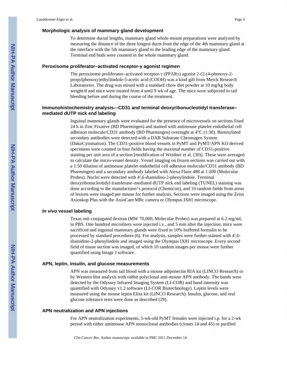
Tumor vasculature in PyMT/APN KO mice is impairedGiven the proangiogenic role of APN in the in vivo model, we reasoned that the lack of APNmay prominently lead to deficiencies under conditions when vascularization is rate-limiting.We sought to examine the contribution of APN on tumor-associated angiogenesis at timeswhen lesions have progressed from a pre-malignant to a malignant stage. Total VEGF-AmRNA levels measured in the mammary gland showed a 2.3-fold increase in PyMTcompared with PyMT/APN KO mice (Fig. 4A). Similarly, mRNA levels of additionalangiogenic markers such as CD31 (1.8-fold), von Willebrand factor (2.3-fold), and VEGFR1(2.1-fold) were detected at increased levels in PyMT compared with PyMT/APN KO mice(Fig. 4A). Consistent with these results, immunostaining with anti-CD31 antibody revealeda 2-fold increase in microvessel density in the mammary glands of PyMT mice relative toPyMT/APN KO mice (Fig. 4B).
The density of functional vascularization was further assessed by in vivo labeling of theblood vessels using an i.v. injection of Texas red–conjugated dextran (6). We show that inthe absence of APN, tumors are likely to have diminished functional vascular density (Fig.
Landskroner-Eiger et al. Page 8
Clin Cancer Res. Author manuscript; available in PMC 2011 December 14.
NIH
-PA Author Manuscript
NIH
-PA Author Manuscript
NIH
-PA Author Manuscript

4C and D). This strongly suggests that APN promotes tumor growth, at least in part, bypromoting an angiogenic response during lesion expansion.
Changes in APN levels during tumor expansionWe examined the fluctuations in circulating plasma APN levels over the course of tumorprogression in the PyMT model. By 12 weeks of age, a time point when tumorscharacteristically advance to late carcinoma, circulating APN levels were reduced by 44% intumor-bearing PyMT mice compared with their WT littermates [Supplementary Fig.S2A(a)]. Other circulating adipokines such as leptin were down-regulated in both modelsindependently of the presence of tumors [Supplementary Fig. S2A(b)]. These results showthat APN levels at late tumor stages may not be indicative of the pivotal role of APN at theearly stage of tumorigenesis when its circulating levels are more prevalent, and highlight thecomplex dynamic relationship of the tumor with its microenvironment.
In spite of the dramatic reduction in APN levels, fasting glucose and insulin levels wereunaltered as was the glucose excursion during an oral glucose tolerance test done at bothearly and late stages of tumor progression (Supplementary Fig. S3). The decline in APNoccurs despite the reduction in fat mass as assessed by whole-body Echo magneticresonance imaging analysis (Supplementary Fig. S2B). Loss in fat mass is generallyassociated with an increase in plasma APN levels and a positive effect on insulin sensitivity.In our tumor model, the loss of fat mass does not trigger an increase in APN. This may becaused by increased local inflammation in the mammary fat pads or may simply be areflection of adipocyte loss in the context of increased tumor burden in the mammary fatpad.
Bypass of the antiangiogenic stress in late-stage mammary tumors of APN KO miceWe next probed for the effects of APN at later stages of tumor progression by measuringtumor volume. Surprisingly, tumor growth in PyMT/APN KO mice not only caught up buteven far exceeded tumor growth in age-matched PyMT mice at advanced stages oftumorigenesis, reaching a significant 2.2-fold difference in tumor volume by 14 weeks ofage (Fig. 5A). Differences were also shown histologically: Large solid tumors with somenecrosis in central local regions were present in the PyMT/APN KO tumors, whereas thePyMT-derived tumors revealed significantly smaller solid tumor regions and no necroticregions (data not shown).
PET was applied at this late stage. In contrast to the modest levels of [18F]FDGaccumulation in 9-week-old PyMT/APN KO animals [Fig. 2A(a–b)], 12-week-old PyMT/APN KO mice showed a massive increase in uptake of [18F]FDG into the mammary glands(Fig. 5B). This is in agreement with our advanced malignancy phenotype and correlates withthe epidemiologic association of an inverse association between APN and breast cancer risk(14, 15). Our data suggest that these late-stage tumors arising from a long-term nutrient-deprived environment in the absence of APN adapt to the anaerobic conditions and emergeas very aggressive and metabolically highly active cells at later stages in spite of thepresence of an initially deficient vasculature. This coincides with a provocative concept thathas emerged involving the hypothesis that tumors can acclimatize to the presence ofantiangiogenic stress by adaptive mechanisms that may reinitiate and induce tumor growth(26).
Induced mobilization of CEPs in acclimatized PyMT/APN KO miceAntiangiogenic treatment can cause recruitment of various bone marrow–derived cells thathave the capacity to fuel tumor growth (26, 28, 38). These bone marrow–derived cellsconsist of vascular progenitors and vascular modulatory cells (26). We hypothesized that
Landskroner-Eiger et al. Page 9
Clin Cancer Res. Author manuscript; available in PMC 2011 December 14.
NIH
-PA Author Manuscript
NIH
-PA Author Manuscript
NIH
-PA Author Manuscript

following a period of angiogenic and nutrient deprivation in mice lacking APN, themobilization of bone marrow–derived cell subpopulations would be unique to the PyMT/APN KO mice at late-stage tumorigenesis and may contribute to the rapid surge in growth oftumors, similar to the situation in tumors that are exposed to pharmacologic antiangiogenictreatments.
We thus studied mice of both early-stage and late-stage tumors (9 and 12 weeks,respectively) and assessed circulating bone marrow–derived cell subpopulations in bothPyMT and PyMT/APN KO mice using fluorescence-activated cell sorting. Differences wereobserved for CEPs defined as CD45−/CD31+/VEGFR2+/VEGFR1− (27). Differences wereobserved only for late-stage tumor–bearing mice, with PyMT/ APN KO showing a 6-foldincrease in circulating levels of CEP subpopulations compared with the age-matched PyMTanimals (Fig. 6A).
Several secreted factors such as stromal-derived factor (SDF-1) and VEGF-A have beenimplicated in the recruitment of CEPs into the tumor mass through an endocrine pathway (4,27). There is also evidence that adipose tissue capillary endothelial cells may secrete SDF-1(39). We thus measured serum levels of SDF-1 in PyMT and PyMT/APN KO mice. In theadvanced tumor stage, SDF-1 circulating levels were increased by 24% in the PyMT/APNKO compared with PyMT mice. However, this was not statistically significant (data notshown). Thus, SDF-1 may provide, at best, only a partial mechanism for CEP recruitment inour model. We further measured total VEGF-A mRNA levels in tumors, showing a 2.4-foldup-regulation in PyMT/APN KO compared with PyMT-derived tumors (Fig. 6B). Thiscorrelates with work by others showing increased VEGF-A expression in mice treated withantiangiogenic inhibitors and was even proposed as a predictive biomarker of tumorresponse (26).
We further examined the angiogenic milieu of these late-stage tumors. In contrast to theelevated VEGF-A mRNA levels measured at this stage (Fig. 6B), other angiogenic markersthat had shown to be decreased in the PyMT/APN KO at the early stage, such as CD31 (1.3-fold), VEGFR1, and VEGFR2 (Fig. 4), were still either down-regulated or unaltered,respectively, in the PyMT/APN KO compared with PyMT tumors. This strongly suggests analternative pathway of inducing tumor growth when the tumors are deprived of APN for anextended period of time.
We further examined the tumor vessels to evaluate whether the presence of high levels ofVEGF-A in the tumor is indicative of tumor revascularization, implicating a mechanism toeffectively cope with the absence of APN. No difference was observed in total blood vesseldensity. However, qualitatively, the majority of vessels in the PyMT tumors exhibiteddilated and distorted tumor vessels, indicative of vessel remodeling. In contrast, tumorvessels derived from mice lacking APN showed vessels that were slim, elongated, and withfew tortuous structures, all reflective of normal vasculature [ref. 40; Fig. 6C(a–b)]. Theseelongated blood vessels were seen in many cases as major vessels rather than the small,scattered blood vessels observed in the PyMT tumors [Fig. 6C(c–d)]. This is consistent withour initial observation that APN is an angio-mimetic adipokine, and it is evident that theselate-stage tumors still lag behind in their angiogenic response.
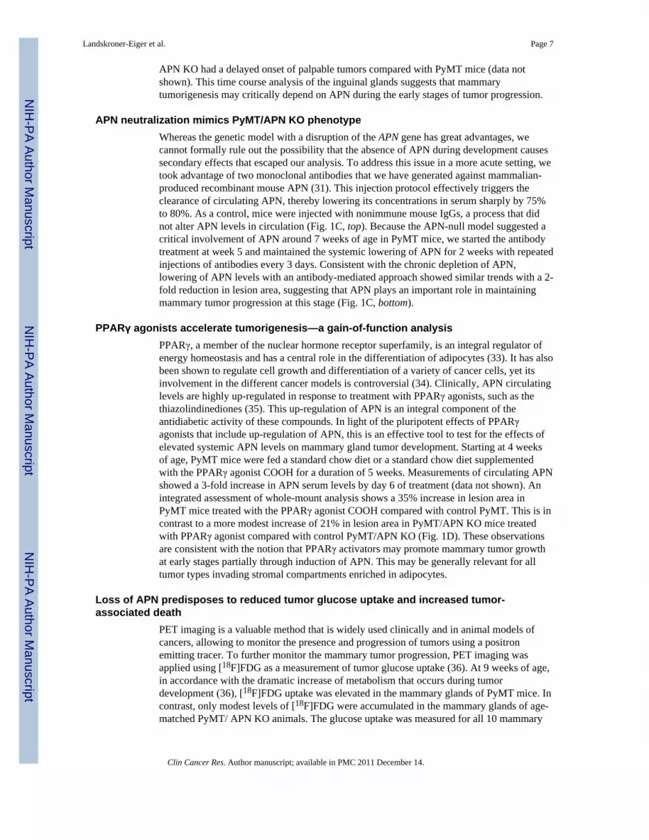
Transcriptional signature of late-stage APN KO mammary tumorsGiven the clinical importance of identifying key factors that are turned on at these advancedstages of the disease, we decided to use DNA microarrays to more globally asses thedifferences in gene expression of late-stage PyMT/APN KO and PyMT tumor samples.Cluster analysis depicted in Table 1 shows that the most robust differences were observed inthe cancer cluster. A general down-regulation of tumor suppressors such as caveolin 1 (1.4-
Landskroner-Eiger et al. Page 10
Clin Cancer Res. Author manuscript; available in PMC 2011 December 14.
NIH
-PA Author Manuscript
NIH
-PA Author Manuscript
NIH
-PA Author Manuscript

fold) and Rb (1.3-fold) and up-regulation of antiapoptotic genes such as BFAR (4.5-fold)and DDIT4 (1.6-fold) was observed, in line with our observation of a tumor growth surge inthe absence of APN at this stage of the disease. Importantly, central regulators ofangiogenesis such as matrix metalloproteinase 2 (1.6-fold) and FGF1 (2.9-fold) remainedclearly diminished despite the up-regulation of hypoxia-inducible factor 1α (1.5-fold) andCXCR4 (1.3-fold). A further reflection of the continued hypoxic environment in thesetumors is the up-regulation of glycolytic genes such as phosphoglycerate kinase 1 andtriosephosphate isomerase 1. These late-stage tumors arising under chronic hypoxicconditions in the PyMT/APN KO mice have a distinct transcriptional fingerprint that is fullyconsistent with a gene expression signature associated with a dramatically more aggressivetumor phenotype.
DiscussionAPN has been implicated in attenuating mammary tumor progression in studies using invitro or xenografts models, partially attributable to a postulated role as a negative regulatorof angiogenesis (16–18). However, because there are important differences between themechanism of tumor growth and angio-genesis in transplanted and autochthonous tumors(19, 20, 28), we have analyzed, for the first time, the role of APN in mammary tumorprogression in vivo using the spontaneously occurring mammary tumor model of MMTV-PyMT. We did this through the use of APN KO mice and by taking advantage ofneutralizing monoclonal antibodies that effectively clear APN from circulation. Contrary tothe published data (16–18), we show that APN can accelerate mammary tumor growth invivo at early stages when tumor lesions start to critically depend on angiogenesis forcontinued oxygen and nutrient supply. Such a role was also implied in an elegant recentarticle that was published in the course of the preparation of this article (41). These authorssuggested the existence of a molecular cross talk between the putative APN receptor T-cadherin and mammary tumor growth.
Previous in vivo work found that APN promotes angiogenesis in response to tissue ischemiathrough the activation of an AMP protein kinase AMPK–dependent signaling pathway (25),and that APN promotes endothelial cell survival, migration, and differentiation intocapillary-like structures (37, 42). We therefore sought to determine the contribution of APNto tumor-associated angiogenesis. Our experiments suggest that APN supports the release ofangiogenic factors such as VEGF-A and VEGF-B and can promote blood vessel formationas judged by an increase in endothelial cell markers such as CD31 and von Willebrandfactor in the PyMT mammary tumors relative to PyMT/ APN KO–derived tumors. Ourexperiments using Matrigel implants suggest potent effects of APN that can lead toextensive reprogramming and induction of blood vessel formation. Importantly, theseexperiments, together with the data obtained from our microarray analysis on WT animalsinjected with APN, suggest a direct role for APN in the angiogenic response by paracrineand/or endocrine effects on cell types such as endothelial cells and inflammatory cells,rather than mediating its effects exclusively through action on the cancer cells.
It is generally accepted that obesity is a risk factor for breast cancer in postmenopausal, butnot premenopausal women (13). Because APN levels are inversely correlated with obesity(11), it has been suggested that the decreased levels of APN may explain the increased riskof breast cancer with obesity (14, 15). In fact, numerous retrospective case-controls studiesand a recent prospective study have shown that APN is inversely associated with breastcancer risk, an association that might be stronger for postmenopausal women (14, 15).Several observations in the PyMT model are consistent with this inverse association: (a) arobust decline in circulating APN levels that is observed as the tumor progresses and (b) anacceleration of mammary tumorigenesis in PyMT/APN KO compared with PyMT in
Landskroner-Eiger et al. Page 11
Clin Cancer Res. Author manuscript; available in PMC 2011 December 14.
NIH
-PA Author Manuscript
NIH
-PA Author Manuscript
NIH
-PA Author Manuscript

advanced malignant stages of the disease. However, similar to the differential effect ofmenopausal status in women, a thorough analysis of the PyMT tumor model indicates acomplex role for APN, suggesting a possibly biphasic effect of APN on tumor progressionor, alternatively, the development of an adaptive mechanism at late stages to bypass thedependence of APN-driven angiogenesis. The switch from a lagging to a more aggressivetumor growth rate may be a reflection of a Darwinian dynamic similar to what is observedunder conditions of exposure to angiogenesis inhibitors, during which tumors acquire theability to survive and proliferate within the suboptimal tumor microenvironment byactivating alternative ways to sustain tumor growth and angiogenesis (26, 36, 43). Similarly,our data imply that following a prolonged antiangiogenic stress due to the absence of APN,an adaptive resistant mechanism takes place. A detailed gene expression analysis of thetumors arising in the PyMT/APN KO mice at late stages reveals easily interpretable changesin critical genes involved in cancer growth, survival, and enhanced glycolysis. In line withdata that VEGF-A and SDF-1 can induce recruitment of endothelial progenitor cells (4, 44,45), we show an increase in local VEGF-A in late-stage APN KO tumor–bearing mice thatpotentially play a role in the observed robust mobilization of CEPs. This subpopulation ofCEPs has the capacity to enhance tumor growth and has been postulated to be crucial insituations such as relapse after tumor shrinkage brought about by surgery or antiangiogenictherapies (27, 28, 38, 46). These results raise the possibility that the recruitment of thissubpopulation contributes functionally to tumor growth in the PyMT/APN KO animals atlate stage, rescuing the cells from their original nutrient-deprived status.
APN critically modulates insulin resistance, atherogenesis, and cardiac remodeling (11). Ourdata presented here indicate that it also functions as an angiogenic mediator in mammarytumors. Our work suggests that vessel development is impaired in mammary tumors lackingsystemic APN, leading to a nutrient-deprived tumor at the early malignant stages. Yet,alternative pathways are activated at later stages of tumorigenesis to bypass the dependenceon APN and allow for further fueling of tumor growth. Compounds that modulate itscirculating levels such as the PPARγ agonists thiazolindinediones (34, 35) may have aprofound effect on mammary tumorigenesis through the angiogenesis axis (34).Interestingly, PPARγ agonists have been shown to induce VEGF-A in adipocytes (47) and,importantly, may be involved in adipose tissue–associated angiogenesis (48). PPARγ is mostabundantly expressed in adipocytes, yet it is also expressed in endothelial and tumor cells,macrophages, and a number of epithelial tissues including epithelia in the mammary gland(34). Thus, this class of drugs clearly induces pleiotropic affects in multiple tissues. Takentogether, the effects of PPARγ agonists on the enhancement of mammary tumorigenesis, asjudged by the analysis of these models, may be mediated in part by the cross talk betweenAPN and the vasculature axis, stimulating tumor growth. In contrast, PPARγ may act as anantimitogenic factor in tumor cells and as an anti-inflammatory factor in macrophages (34).The net result of PPARγ stimulation on the transformed ductal epithelium of the mammarygland raises the distinct possibility that these agonists may enhance neovascularization ofemerging tumors and should be used with caution in a diabetic population with newlydiagnosed malignancies of the mammary gland. On the other hand, tumors arising in anenvironment with low APN (such as in an obese and/or diabetic population) may assume amore aggressive phenotype as suggested by the increased tumor growth at later stages in ourAPN KO mice.
Given that elevated levels of CEPs were observed in APN KO mice independent of thepresence or absence of a tumor (data not shown), the proangiogenic role of APN may notonly be relevant under tumor-associated conditions but may also contribute to its potentantidiabetic effects in our previously described mouse models with moderately elevatedlevels of APN achieved through transgenic overexpression. APN enables adipose tissue padsto massively expand during times of excess caloric intake, allowing a redistribution of
Landskroner-Eiger et al. Page 12
Clin Cancer Res. Author manuscript; available in PMC 2011 December 14.
NIH
-PA Author Manuscript
NIH
-PA Author Manuscript
NIH
-PA Author Manuscript

excess ectopic lipid deposits. Despite the massive hyperplasia of the fat pads in these miceoverexpressing APN, the fat pads remain minimally inflamed and fully insulin sensitive anddo not display the classic hallmarks of expansion under suboptimal conditions, such ashypoxia and macrophage infiltration (29).
Collectively, our detailed analysis suggests several possible strategies to delay an acquiredadaptation to antiangiogenic stress under conditions when APN abundance is low, such as inthe context of obesity. This includes treatment regimens targeting CEPs, an approach thathas previously been shown to enhance treatment efficacy of cytotoxic chemotherapy (49).These patients may potentially also show higher sensitivity to chemotherapy given theirnormalized vasculature (46, 50). A molecular understanding of the APN-mediatedproangiogenic activity combined with the identification of key stimulating factors that areturned on at advanced stages of the disease has the potential to reveal novel therapeuticapproaches for breast cancer in blocking both the angiogenic switch and the specificadaptive pathways that are triggered as a result of chronic angiogenic stress and nutrientdeprivation.
Supplementary MaterialRefer to Web version on PubMed Central for supplementary material.
AcknowledgmentsWe thank the members of the Scherer laboratory for discussions; Michael Cammer and Yan Deng from theAnalytical Imaging Facility at Albert Einstein College of Medicine, the Albert Einstein College of MedicineDiabetes Research and Training Center and Radioimmunoprecipitation Assay Core Facility (Robin Sgueglia); theUniversity of Texas Southwestern Metabolic Core for phenotyping efforts; Dr. Quan Li from the University ofTexas Southwestern Microarray Core Facility; Bei B. Zhang and John Thompson for microarray processing; theRosetta Gene Expression Laboratory; Dr. Rani Sellers from the Histopathology Facility at Albert Einstein Collegeof Medicine for expert help; Nils Halberg for statistical analysis; and Jiufeng Li, Aisha Cordero, and Yuan Xin fortechnical assistance.
Grant support: NIH grant R01-CA112023 (P.E. Scherer) and Training Program in Cellular and Molecular Biologyand Genetics Grant T32-GM04791 (S. Landskroner-Eiger).
References1. Scherer PE. Adipose tissue: from lipid storage compartment to endocrine organ. Diabetes. 2006;
55:1537–45. [PubMed: 16731815]2. Rosen ED, Spiegelman BM. Adipocytes as regulators of energy balance and glucose homeostasis.
Nature. 2006; 444:847–53. [PubMed: 17167472]3. Trujillo ME, Scherer PE. Adipose tissue-derived factors: impact on health and disease. Endocr Rev.
2006; 27:762–78. [PubMed: 17056740]4. Orimo A, Gupta PB, Sgroi DC, et al. Cell. 2005; 121:335–48. [PubMed: 15882617]5. Iyengar P, Combs TP, Shah SJ, et al. Adipocyte-secreted factors synergistically promote mammary
tumorigenesis through induction of anti-apoptotic transcriptional programs and proto-oncogenestabilization. Oncogene. 2003; 22:6408–23. [PubMed: 14508521]
6. Lin EY, Li JF, Gnatovskiy L, et al. Macrophages regulate the angiogenic switch in a mouse modelof breast cancer. Cancer Res. 2006; 66:11238–46. [PubMed: 17114237]
7. Iyengar P, Espina V, Williams TW, et al. Adipocyte-derived collagen VI affects early mammarytumor progression in vivo, demonstrating a critical interaction in the tumor/stromamicroenvironment. J Clin Invest. 2005; 115:1163–76. [PubMed: 15841211]
8. Scherer PE, Williams S, Fogliano M, Baldini G, Lodish HF. A novel serum protein similar to C1q,produced exclusively in adipocytes. J Biol Chem. 1995; 270:26746–9. [PubMed: 7592907]
9. Berg AH, Combs TP, Du X, Brownlee M, Scherer PE. The adipocyte-secreted protein Acrp30enhances hepatic insulin action. Nat Med. 2001; 7:947–53. [PubMed: 11479628]
Landskroner-Eiger et al. Page 13
Clin Cancer Res. Author manuscript; available in PMC 2011 December 14.
NIH
-PA Author Manuscript
NIH
-PA Author Manuscript
NIH
-PA Author Manuscript

10. Combs TP, Berg AH, Obici S, Scherer PE, Rossetti L. Endogenous glucose production is inhibitedby the adipose-derived protein Acrp30. J Clin Invest. 2001; 108:1875–81. [PubMed: 11748271]
11. Trujillo ME, Scherer PE. Adiponectin—journey from an adipocyte secretory protein to biomarkerof the metabolic syndrome. J Intern Med. 2005; 257:167–75. [PubMed: 15656875]
12. Shibata R, Sato K, Pimentel DR, et al. Adiponectin protects against myocardial ischemia-reperfusion injury through AMPK- and COX-2-dependent mechanisms. Nat Med. 2005; 11:1096–103. [PubMed: 16155579]
13. Lorincz AM, Sukumar S. Molecular links between obesity and breast cancer. Endocr Relat Cancer.2006; 13:279–92. [PubMed: 16728564]
14. Mantzoros C, Petridou E, Dessypris N, et al. Adiponectin and breast cancer risk. J Clin EndocrinolMetab. 2004; 89:1102–7. [PubMed: 15001594]
15. Tworoger SS, Eliassen AH, Kelesidis T, et al. Plasma adiponectin concentrations and risk ofincident breast cancer. J Clin Endocrinol Metab. 2007; 92:1510–6. [PubMed: 17213279]
16. Wang Y, Lam JB, Lam KS, et al. Adiponectin modulates the glycogen synthase kinase-3β/β-catenin signaling pathway and attenuates mammary tumorigenesis of MDA-MB-231 cells in nudemice. Cancer Res. 2006; 66:11462–70. [PubMed: 17145894]
17. Dieudonne MN, Bussiere M, Dos Santos E, et al. Adiponectin mediates antiproliferative andapoptotic responses in human MCF7 breast cancer cells. Biochem Biophys Res Commun. 2006;345:271–9. [PubMed: 16678125]
18. Brakenhielm E, Veitonmaki N, Cao R, et al. Adiponectin-induced antiangiogenesis and anti-tumoractivity involve caspase-mediated endothelial cell apoptosis. Proc Natl Acad Sci U S A. 2004;101:2476–81. [PubMed: 14983034]
19. Talmadge JE, Singh RK, Fidler IJ, Raz A. Murine models to evaluate novel and conventionaltherapeutic strategies for cancer. Am J Pathol. 2007; 170:793–804. [PubMed: 17322365]
20. Sikder H, Huso DL, Zhang H, et al. Disruption of Id1 reveals major differences in angiogenesisbetween transplanted and autochthonous tumors. Cancer Cell. 2003; 4:291–9. [PubMed:14585356]
21. Nawrocki AR, Rajala MW, Tomas E, et al. Mice lacking adiponectin show decreased hepaticinsulin sensitivity and reduced responsiveness to peroxisome proliferator–activated receptor γagonists. J Biol Chem. 2006; 281:2654–60. [PubMed: 16326714]
22. Guy CT, Cardiff RD, Muller WJ. Induction of mammary tumors by expression of polyoma-virusmiddle T oncogene: a transgenic mouse model for metastatic disease. Mol Cell Biol. 1992;12:954–61. [PubMed: 1312220]
23. Lin EY, Jones JG, Li P, et al. Progression to malignancy in the polyoma middle T onco-proteinmouse breast cancer model provides a reliable model for human diseases. Am J Pathol. 2003;163:2113–26. [PubMed: 14578209]
24. Maeda N, Shimomura I, Kishida K, et al. Diet-induced insulin resistance in mice lackingadiponectin/ACRP30. Nat Med. 2002; 8:731–7. [PubMed: 12068289]
25. Shibata R, Ouchi N, Kihara S, et al. Adiponectin stimulates angiogenesis in response to tissueischemia through stimulation of amp-activated protein kinase signaling. J Biol Chem. 2004;279:28670–4. [PubMed: 15123726]
26. Bergers G, Hanahan D. Modes of resistance to anti-angiogenic therapy. Nat Rev Cancer. 2008;8:592–603. [PubMed: 18650835]
27. Bertolini F, Shaked Y, Mancuso P, Kerbel RS. The multifaceted circulating endothelial cell incancer: towards marker and target identification. Nat Rev Cancer. 2006; 6:835–45. [PubMed:17036040]
28. Ruzinova MB, Schoer RA, Gerald W, et al. Effect of angiogenesis inhibition by Id loss and thecontribution of bone-marrow-derived endothelial cells in spontaneous murine tumors. Cancer Cell.2003; 4:277–89. [PubMed: 14585355]
29. Kim JY, van de Wall E, Laplante M, et al. Obesity-associated improvements in metabolic profilethrough expansion of adipose tissue. J Clin Invest. 2007; 117:2621–37. [PubMed: 17717599]
30. Weidner N, Semple JP, Welch WR, Folkman J. Tumor angiogenesis and metastasis-correlation ininvasive breast carcinoma. N Engl J Med. 1991; 324:1–8. [PubMed: 1701519]
Landskroner-Eiger et al. Page 14
Clin Cancer Res. Author manuscript; available in PMC 2011 December 14.
NIH
-PA Author Manuscript
NIH
-PA Author Manuscript
NIH
-PA Author Manuscript

31. Wang ZV, Schraw TD, Kim JY, et al. Secretion of the adipocyte-specific secretory proteinadiponectin critically depends on thiol-mediated protein retention. Mol Cell Biol. 2007; 27:3716–31. [PubMed: 17353260]
32. Hughes TR, Mao M, Jones AR, et al. Expression profiling using microarrays fabricated by an ink-jet oligonucleotide synthesizer. Nat Biotechnol. 2001; 19:342–7. [PubMed: 11283592]
33. Rosen ED, Spiegelman BM. PPARγ: a nuclear regulator of metabolism, differentiation, and cellgrowth. J Biol Chem. 2001; 276:37731–4. [PubMed: 11459852]
34. Michalik L, Desvergne B, Wahli W. Peroxisome-proliferator-activated receptors and cancers:complex stories. Nat Rev Cancer. 2004; 4:61–70. [PubMed: 14708026]
35. Combs TP, Wagner JA, Berger J, et al. Induction of adipocyte complement-related protein of 30kilodaltons by PPARγ agonists: a potential mechanism of insulin sensitization. Endocrinology.2002; 143:998–1007. [PubMed: 11861525]
36. Gatenby RA, Gillies RJ. Why do cancers have high aerobic glycolysis? Nat Rev Cancer. 2004;4:891–9. [PubMed: 15516961]
37. Ouchi N, Kobayashi H, Kihara S, et al. Adiponectin stimulates angiogenesis by promoting cross-talk between AMP-activated protein kinase and Akt signaling in endothelial cells. J Biol Chem.2004; 279:1304–9. [PubMed: 14557259]
38. Lyden D, Hattori K, Dias S, et al. Impaired recruitment of bone-marrow-derived endothelial andhematopoietic precursor cells blocks tumor angiogenesis and growth. Nat Med. 2001; 7:1194–201.[PubMed: 11689883]
39. Sengenes C, Miranville A, Maumus M, et al. Chemotaxis and differentiation of human adiposetissue CD34+/CD31− progenitor cells: role of stromal derived factor-1 released by adipose tissuecapillary endothelial cells. Stem Cells. 2007; 25:2269–76. [PubMed: 17525234]
40. Du R, Lu KV, Petritsch C, et al. HIF1α induces the recruitment of bone marrow-derived vascularmodulatory cells to regulate tumor angiogenesis and invasion. Cancer Cell. 2008; 13:206–20.[PubMed: 18328425]
41. Hebbard LW, Garlatti M, Young LJ, et al. T-cadherin supports angiogenesis and adiponectinassociation with the vasculature in a mouse mammary tumor model. Cancer Res. 2008; 68:1407–16. [PubMed: 18316604]
42. Kobayashi H, Ouchi N, Kihara S, et al. Selective suppression of endothelial cell apoptosis by thehigh molecular weight form of adiponectin. Circ Res. 2004; 94:e27–31. [PubMed: 14752031]
43. Gatenby RA, Gillies RJ. A microenvironmental model of carcinogenesis. Nat Rev Cancer. 2008;8:56–61. [PubMed: 18059462]
44. Hattori K, Dias S, Heissig B, et al. Vascular endothelial growth factor and angiopoietin-1 stimulatepostnatal hematopoiesis by recruitment of vasculogenic and hematopoietic stem cells. J Exp Med.2001; 193:1005–14. [PubMed: 11342585]
45. Asahara T, Takahashi T, Masuda H, et al. VEGF contributes to postnatal neovascularization bymobilizing bone marrow-derived endothelial progenitor cells. EMBO J. 1999; 18:3964–72.[PubMed: 10406801]
46. Batchelor TT, Sorensen AG, di Tomaso E, et al. AZD2171, a pan-VEGF receptor tyrosine kinaseinhibitor, normalizes tumor vasculature and alleviates edema in glioblastoma patients. Cancer Cell.2007; 11:83–95. [PubMed: 17222792]
47. Emoto M, Anno T, Sato Y, et al. Troglitazone treatment increases plasma vascular endothelialgrowth factor in diabetic patients and its mRNA in 3T3-1 adipocytes. Diabetes. 2001; 50:1166–70.[PubMed: 11334422]
48. Fukumura D, Ushiyama A, Duda DG, et al. Paracrine regulation of angiogenesis and adipocytedifferentiation during in vivo adipogenesis. Circ Res. 2003; 93:e88–97. [PubMed: 14525808]
49. Shaked Y, Henke E, Roodhart JM, et al. Rapid chemotherapy-induced acute endothelial progenitorcell mobilization: implications for anti-angiogenic drugs as chemosensitizing agents. Cancer Cell.2008; 14:263–73. [PubMed: 18772115]
50. Stockmann C, Doedens A, Weidemann A, et al. Deletion of vascular endothelial growth factor inmyeloid cells accelerates tumorigenesis. Nature. 2008; 456:814–8. [PubMed: 18997773]
Landskroner-Eiger et al. Page 15
Clin Cancer Res. Author manuscript; available in PMC 2011 December 14.
NIH
-PA Author Manuscript
NIH
-PA Author Manuscript
NIH
-PA Author Manuscript

Translational Relevance
The contributions of stromal adipocytes toward mammary tumor growth have beenappreciated for decades. The effects of adiponectin (APN) on tumor cells are not wellunderstood, but a number of cancer epidemiologic studies have focused on APN as apredictive marker. APN levels are becoming a popular and widely measured clinicalparameter, including in the context of breast cancer studies. Here, we present the first invivo data on the role of APN on mammary tumor growth and highlight its function as apotent proangiogenic factor. APN is a key mediator of the antidiabetic actions of thewidely used peroxisome proliferator–activated receptor γ agonists (thiazolindinediones).These compounds lead to an increased tumor mass in the present studies. Our datasuggest a possible mechanism for the increased risk and poorer prognosis seen in breastcancer patients with low serum APN levels (such as obese women) due to acquiredadaptation of tumors to antiangiogenic stress.
Landskroner-Eiger et al. Page 16
Clin Cancer Res. Author manuscript; available in PMC 2011 December 14.
NIH
-PA Author Manuscript
NIH
-PA Author Manuscript
NIH
-PA Author Manuscript

Fig. 1.PyMT/APN KO mice display delayed mammary tumor progression. A, top, representativewhole-mount preparations of inguinal mammary gland derived from age-matched 7-wk-oldmice. Bar, 1 mm. Bottom, total lesion area of PyMT and PyMT/APN KO at 5 wk (n = 4–6per group) and 7 wk (n = 11–12 per group; bars, SE; *, P < 0.05). B, summary of tumorstaging distribution derived from H&E of 7-wk-old inguinal mammary glands sections (n =13 per group). C, a representative experiment is shown at the top. Western blotquantification of circulating APN levels in mice treated with monoclonal antibodiesneutralizing APN (bars, SE; *, P < 0.001, two-way ANOVA). Bottom, APN neutralizingantibody–treated mice (n = 6) show a trend of a 2-fold decrease in lesion area comparedwith IgG-treated mice (n = 3) as measured in whole mounts of inguinal mammary glands(bars, SE; P = 0.2). D, mice were treated with either vehicle or the PPARγ agonist COOHfrom 4 to 9 wk of age. Total lesion area from whole mounts was measured. PyMT micetreated with the PPARγ agonist have a 35% increase in lesion area compared with vehicle-treated PyMT, whereas PyMT/APN KO mice treated with the PPARγ agonist show a moremodest increase in lesion area compared with the untreated PyMT/APN KO group (n = 9–10per group; bars, SE; *, P < 0.05).
Landskroner-Eiger et al. Page 17
Clin Cancer Res. Author manuscript; available in PMC 2011 December 14.
NIH
-PA Author Manuscript
NIH
-PA Author Manuscript
NIH
-PA Author Manuscript

Fig. 2.Decreased accumulation of [18F]FDG as assessed by microPET and increased tumor-associated death in PyMT/APN KO tumor–bearing mice. A, coronal images showing themammary tumor spread (arrows) in addition to bladder (*) and heart (**) that have a highglucose uptake. By 9 wk of age, PyMT mice (n = 4–5) displayed an 11-fold increase in[18F]FDG uptake in the mammary glands compared with age-matched PyMT/APN KO mice(a and b; n = 4; bars, SE; *, P < 0.05). B, H&E staining showing necrotic regions (N) inmammary tumors (T) lacking APN; no necrotic regions were observed in age-matchedmammary tumors of PyMT mice (n = 21 per group). C, quantitative analysis of TUNELstaining in 9-wk-old PyMT mice and PyMT/APN KO mice (n = 6 per group; bars, SE; *, P< 0.05). Bar, 100 μm (B), 20 μm (C).
Landskroner-Eiger et al. Page 18
Clin Cancer Res. Author manuscript; available in PMC 2011 December 14.
NIH
-PA Author Manuscript
NIH
-PA Author Manuscript
NIH
-PA Author Manuscript

Fig. 3.APN as a positive regulator of angiogenesis independent of tumor cells. A, gene expressionprofiling of the epididymal fat tissue was determined by microarray analysis. Results areexpressed as the fold change in gene expression in WT mice treated with APN injectionscompared with WT mice treated with vehicle (n = 3 per treated group, n = 10 per controlgroup). *, P < 0.005; **, P < 0.01. B and C, mice were s.c. injected with Matrigel + basicFGF. B, a representative H&E staining of the Matrigel matrix (a and b). Image Jquantification shows a >2-fold difference between the WT and APN KO Matrigel cellularcontent (c; bars, SE; *, P < 0.05; n = 6 per group). C, a representative immunohistochemicalstaining for CD31 endothelium (n = 4 per group). Dashed lines, Matrigel region. Red arrow,enlarged section. Bar, 100 μm [C (a and c)]; 20 μm [B (a and b) and C (b and d)].
Landskroner-Eiger et al. Page 19
Clin Cancer Res. Author manuscript; available in PMC 2011 December 14.
NIH
-PA Author Manuscript
NIH
-PA Author Manuscript
NIH
-PA Author Manuscript
Fig. 4.PyMT/APN KO show a decrease in the angiogenic profile. A, mammary glands wereexcised and mRNA levels were quantified by real-time PCR; β-actin was used as an internalcontrol (*, P < 0.05; n = 5–8 per group). B, immunohistochemistry analysis of CD31endothelium (bars, SE; *, P < 0.05; n = 5 per group). C, vessel density evaluation in tumorsections from Texas red-dextran–injected mice counterstained with 4′,6-diamidino-2-phenylindole. Bar, 50 μm. D, inguinal mammary gland was imaged for vessel densitydistribution and compared between PyMT (n = 7) and PyMT/APN KO mice (n = 5; bars,SE; P = 0.15).
Landskroner-Eiger et al. Page 20
Clin Cancer Res. Author manuscript; available in PMC 2011 December 14.
NIH
-PA Author Manuscript
NIH
-PA Author Manuscript
NIH
-PA Author Manuscript

Fig. 5.Late-stage tumorigenesis: PyMT/APN KO mice display an adaptive phenotype. A, averagevalues are presented for the longitudinal measurements of the first palpable tumors. PyMT, n= 28; PyMT/APN KO, n = 21 (bars, SE; *, P < 0.05, two-way ANOVA). B, coronal imagesshowing the mammary tumor spread (arrows) in addition to bladder (*) and heart (**) thathave a high glucose uptake as measured by microPET. Twelve-week-old PyMT/APN KOmice (n = 4) showed comparable glucose uptake to PyMT mice (n = 7).
Landskroner-Eiger et al. Page 21
Clin Cancer Res. Author manuscript; available in PMC 2011 December 14.
NIH
-PA Author Manuscript
NIH
-PA Author Manuscript
NIH
-PA Author Manuscript

Fig. 6.PyMT/APN KO mice initiate mobilization of circulating endothelial progenitor cells duringlate-stage tumorigenesis. A, fluorescence-activated cell sorting analysis of CEPs levels incirculation, showing a 6-fold increase in PyMT/APN KO compared with PyMT mice (bars,SE; *, P < 0.05; n = 5–7 mice per group). B, inguinal mammary tumors were excised andmRNA levels were quantified by real-time PCR (**, P = 0.07; *, P < 0.05; n = 5–7 pergroup). C, tumors were imaged for blood vessel morphology using CD31 antibody. PyMTtumors exhibited dilated and torturous tumor vessels (a, c). In contrast, PyMT/APN KOblood vessels showed slim and elongated characteristics (b, d; n = 4 per group). Bar, 50 μm.
Landskroner-Eiger et al. Page 22
Clin Cancer Res. Author manuscript; available in PMC 2011 December 14.
NIH
-PA Author Manuscript
NIH
-PA Author Manuscript
NIH
-PA Author Manuscript
NIH
-PA Author Manuscript
NIH
-PA Author Manuscript
NIH
-PA Author Manuscript
Landskroner-Eiger et al. Page 23
Table 1
Differential gene expression in PyMT/APN KO versus PyMT in late-stage tumors
Genes up-regulated in PyMT/APN KO tumors
Cluster Gene name Accession no. Fold change
Cancer ST6GAL1 NM_1459332.2 1.6
HIF1a NM_010431.1 1.5
MAPKS1P1 NM_011162.2 1.5
PGK1 NM_008828.2 1.5
PTGS1 NM_008969.1 1.3
CXCR4 NM_009911.2 1.3
HDAC5 NM_010412.2 1.3
CDKN1B NM_009875.2 1.2
Cell death BFAR NM_025976 4.5
DD1T4 NM_029083.1 1.6
LGALS7 NM_008496.4 1.6
MAPK81P1 NM_011162.2 1.5
MMP13 NM_008607.1 1.3
Glycolysis PGK1 NM_008828.2 1.5
TP11 NM_009415.1 1.4
Genes down-regulated in PyMT/APN KO tumors
Cancer FGF1 NM_010197.2 2.9
MMP2 NM_008610.1 1.6
AXL A1323647 1.5
CAV1 NM_007616.2 1.4
RB1 NM_009029.1 1.3
CCR5 NM_9919.2 1.3
MMP1 NM_032006.2 1.2
TLR4 NM_021297.1 1.2
Cell death TNFRSF10 NM_020275.3 1.6
IL15RA NM_133836.1 1.4
Clin Cancer Res. Author manuscript; available in PMC 2011 December 14.


