Embed Size (px)
Citation preview
431
Original Paper
Cell Physiol Biochem 2009;23:431-440 Accepted: March 13, 2009Cellular PhysiologyCellular PhysiologyCellular PhysiologyCellular PhysiologyCellular Physiologyand Biochemistrand Biochemistrand Biochemistrand Biochemistrand Biochemistryyyyy
Copyright © 2009 S. Karger AG, Basel
Fax +41 61 306 12 34E-Mail [email protected]
© 2009 S. Karger AG, Basel1015-8987/09/0236-0431$26.00/0
Accessible online at:www.karger.com/cpb
Production of Functionally Active Palytoxin-likeCompounds by Mediterranean Ostreopsis cf.siamensisEva Cagide1, M. Carmen Louzao1, Begoña Espiña1, Mercedes R.Vieytes2, David Jaen3, Luz Maman3, Takeshi Yasumoto4 and Luis M.Botana1
1Departamento de Farmacología, Facultad de Veterinaria, Universidad de Santiago de Compostela,2Departamento de Fisiología Animal, Facultad de Veterinaria, Universidad de Santiago de Compostela,3Laboratorio de Control de Calidad de los Recursos Pesqueros, Consejería de Agricultura y Pesca, ElRompido, 4Japan Food Research Laboratories, Tama Laboratory, Tokyo
Luis M. BotanaDepartamento de Farmacología, Facultad de VeterinariaUniversidad de Santiago de Compostela, Campus de Lugo27002 Lugo (Spain)Tel. 34 982 252 242, E-Mail [email protected]
Key WordsOstreopsis • Palytoxin analogues • Metabolic activity• Depolarization • Cytosolic calcium • Ouabain
AbstractBackground and purpose: Dinoflagellates from thegenus Ostreopsis have been related to the produc-tion of palytoxin and analogues. Based on that, thispaper describes functional studies of crude extractsfrom Ostreopsis cf. siamensis collected in the Medi-terranean Sea in order to biochemically characterizetheir toxic compounds. Methods: We compared theeffects of 5 crude dinoflagellates extracts with acommercially available palytoxin and a purifiedOstreopsis ovata extract on metabolic activity, mem-brane potential, and cytosolic calcium levels byusing fluorescent dyes. Results: All the extractsresulted to be neurotoxic. In addition, all of theminduced a membrane depolarization and a calciumincrement that were abolished when preincubatingwith ouabain, an inhibitor of the Na+/K+ pump.Conclusion: The effects observed were quite closeto those induced by palytoxin and the Ostreopsisovata extract as well, suggesting that Ostreopsis cf.
siamensis is actually producing palytoxin-like com-pounds that are highly toxic and functionally active.
Introduction
Palytoxin is one of the largest and highly potentmarine toxins first isolated from zooanthids of the genusPalythoa [1-3]. It has been also found in sea anemones,polychaete worms, crabs and herbivorous fishes [4-6].However, algae from the genus Ostreopsis have beenproposed as the possible biogenetic origin of this toxin aswell as some potent analogues, e.g. ostreocin-D [7-10].
Ostreopsis species are distributed worldwide, andeven though are more common in tropical and subtropicalwaters, since 1994 these dinoflagellates started to appearin the Mediterranean Sea [11-14]. Several species suchas O. siamensis [13, 15, 16] and O. ovata [17-19], whichhave been postulated as producers of palytoxin analogues,have been also involved in damaging effects in seafoodor human toxic outbreaks.
Seafood poisoning in humans due to palytoxins, ischaracterized by abdominal cramps, nausea, diarrhea,
432
paresthesia, severe muscle spasms, and respiratorydistress [20, 21]. Palytoxin-like compounds also causehuman sufferings because of the exposure to the marineaerosols, with symptoms that include fever associated toserious respiratory disturbs, such as bronchoconstriction,mild dyspnea, wheezes, and in some cases conjunctivitis[17, 22].At cellular level, it is widely accepted that palytoxin actson the Na+/K+-ATPase [23-25]. This pump hydrolyzesone molecule of ATP and transports 3 Na+ ions out for 2K+ ions into the cell. Palytoxin seems to bind to the Na+/K+-ATPase from the extracellular side and thereforeinhibit the active transport of Na+ and K+ across the cellmembrane by transforming the pump into a permanentlyopen ion channel [24, 26-28]. The pump is also the specifictarget of heart glycosides, such as ouabain, and thereported data suggest that there is a competitive inhibitionof the palytoxin actions by ouabain [3, 29-32].
One of the first effects induced by palytoxin is amembrane depolarization by increasing the sodium entry,potassium efflux, and altering the cationic permeabilityof the cells [33-37]. Another reported effect of this toxinis the induction of calcium influx [36, 38-40]. In excitablecells, at least three Ca2+ entry pathways are involved inthis effect, though are not completely characterized: (i)voltage-dependent Ca2+ channels, which are activated bydepolarization of the membrane; (ii) Na+/Ca2+ exchangerin reversed mode, which causes a Ca2+ accumulation inresponse to the increase in intracellular Na+ andmembrane depolarization, and (iii) an unknown pathwayindependent on its depolarizing action or on its action onintracellular pH [41, 42].
The rise in cytoplasmatic Na+, the Ca2+ increase,and, in general, the ionic disorder induced by palytoxintriggers a series of cellular events that lead to its cytotoxiceffect [27, 43-49]. Palytoxin and its analogues exhibitextreme toxicity in mammals, with a LD50 value of 450ng/kg and 750 ng/kg after intraperitoneal injection ofpalytoxin and ostreocin-D respectively [50].
Toxic compounds produced by Ostreopsis spp. arestill not well known. Actually, a new palytoxin analogue,ovatoxin-A, has been described as produced by O. ovatacollected in the Mediterranean coast [19], but almostnothing has been published with regard to their functionalbehavior. The aim of this work was to pharmacologicallycharacterize the toxins from O. cf. siamensis collectedin the Mediterranean Sea. Based on that, we comparedtheir effects on viability, membrane potential andintracellular calcium, with the ones induced by acommercially available palytoxin and a purified extract
of O. ovata. Results allowed us to provide a deeperknowledge about the presence of palytoxin-likecompounds in O. cf. siamensis produced by themselves.
Materials and Methods
MaterialsFluorescent dyes bis-(1,3-dibutylbarbituric acid)
trimethine oxonol (bis-oxonol), and fura-2 acetoxymethyl ester(fura-2) were from Molecular Probes (Leiden, The Netherlands).Alamar Blue (AB) was from Biosource International (Nivelles,Belgium). Eagle’s Minimum Essential Medium (EMEM) andHam’s F12 supplemented with glutamine, non-essential aminoacids, gentamycin, and amphotericin B were purchased fromBiochrom AG (Berlin, Germany). Fetal bovine serum (FBS) forneuroblastoma cell culture, trypsin-EDTA, Nutrient mixture F-12 Ham Kaighn’s modification, streptomycin sulfate salt,penicillin G potassium salt, bovine serum albumine (BSA), andouabain were from Sigma (Madrid, Spain). Fetal bovine serumfor hepatocytes cell culture was from Gibco (Barcelona, Spain).Palytoxin from Palythoa tuberculosa was purchased from WakoPure Chemical Industries, Ltd (Osaka, Japan). Ostreopsis ovataextract was purified by Dr. T. Yasumoto (Tama Laboratories,Japan). All other chemicals were reagent grade and purchasedfrom Sigma (Madrid, Spain). 25 cm2 tissue culture flasks and 60mm tissue culture plates were from Nunc (Roskilde, Denmark)and 96 well plates were from Corning (Schiphol-Rijk, TheNetherlands).
Experimental standard salt solution used for theexperiments contained (in mmol/L): NaCl 137, KCl 5, CaCl2 1,Glucose 10, MgCl2 1.2, KH2PO4 0.44, NaHCO3 4.2, pH 7.4.
Ostreopsis cf. siamensis Crude ExtractsOstreopsis cf siamensis were collected as epiphytic
macroalgae at several locations along andalusian coast.Seawater containing the macroalgae sample was shakenvigorously to dislodge the epiphytic organisms. Clonal isolateswere obtained by transferring individual cells through sterileseawater washes with a capillary pipette. After the washes,every single cell was similarly transferred into a microtittercontaining 300 µL of sterile L1 medium.
Clonal cultures of Ostreopsis cf siamensis were establishedin L1 media [51]. The cultures were grown at 18 ± 1°C and a14:10 h light:dark photoperiod. Illumination was provided bycool white fluorescent lamps. The cultures were shaken twice aday.
Ostreopsis cells were collected from the culture using a20 µm mesh plankton net. The extracts were newly concentratedby centrifugation. The supernatants were removed and pelletswere resuspended with methanol [methanol:pellet 3:1 (v/v)].These extracts were stored at -14ºC.
Cell Line CultureHuman neuroblastoma cell line BE(2)-M17 used as
excitable cellular model was cultured in EMEM:Ham´s F12 (1:1)supplemented with 2 mM glutamine, 1% non-essential amino
Cagide/Louzao/Espiña/Vieytes/Jaen/Maman/Yasumoto/BotanaCell Physiol Biochem 2009;23:431-440
433
acids, 50 mg/L gentamycin, 50 µg/L amphotericin B and 10%heat-inactivated FBS. Cells were grown in 25 cm2 tissue cultureflasks at 37°C in a humidified atmosphere containing 5% CO2,and were subcultured by transferring cells released by theapplication of 0.1% trypsin-EDTA [52, 53].
Comparatively, rat hepatocytes from the cell line Clone 9were used as non-excitable cellular model. Cells were culturedin a nutrient mixture F-12 Ham Kaighn’s modificationsupplemented with 2.5 g/L NaHCO3, 28 mg/L streptomycinsulfate salt, 17 mg/L penicillin G potassium salt and 10% FBS.Cells were grown on 60 mm tissue culture plates in a humidifiedatmosphere with 5% CO2 at 37°C, and subcultured bytransferring cells released by the application of 0.25% trypsin-EDTA [54].
For the fluorescence microscope assays, neuroblastomacells were seeded on 22 mm glass coverslips at a density of 1.5-5 x 10⁴ cells/well and used after 4-7 days. For the metabolicactivity assay, neuroblastoma and hepatocytes were seededon 96 well plates at a density of 4 x 10⁴ cells/well, and werecultured for 24 h prior to treatment in order to allow them toattach to the bottom of the microplate.
Metabolic Activity AssayCell metabolic inhibition was tested as cytotoxic effect
directly in the cell culture wells using the fluorescent probeAlamar Blue (AB). This redox indicator is a widely used nontoxicreagent that exhibits both fluorimetric and colorimetricproperties in response to metabolic activity. The reduction-induced color change varies proportionately with cell numberand time, changing from a nonfluorescent color blue to areduced pink fluorescent form [55-60].
Toxins, extracts and controls were added in the wells byduplicate. Then, a 1:10 dilution of AB was aseptically added tothe culture wells. AB fluorescence was measured using a FL600fluorescence plate reader (Bio-Tek, Vermont, U.S.A.) at theexcitation wavelength of 530 nm and the emission wavelengthof 590 nm. Control cells were considered as the 100% viability,and results are expressed as the percentage of the fluorescenceof treated cells versus the fluorescence in controls ones.
Measurement of Plasma Membrane PotentialPlasma membrane potential was measured with the slow
potential-sensitive fluorescent dye bis-oxonol. Neuroblastomacells were incubated in a thermostated chamber with thestandard salt solution containing 5 nM bis-oxonol for 10 min at37ºC. Fluorescence recordings were performed with a NikonDiaphot microscope with epifluorescence optics (Nikon 40ximmersion fluor objective) at the wavelengths 490 nm excitationand 530 nm emission [61, 62]. Results are expressed as relativefluorescence as in Louzao et al. [63].
[Ca2+]i MeasurementsNeuroblastoma cells were incubated for 10 min at 37ºC in
2 mL standard salt solution with 0.1% BSA and containing 0.5µM fura-2. Then, the cells were washed three times and placedinto the thermostated chamber at 37ºC. Calcium measurementswere made with the same microscope as above. Fluorescentimages were collected by dual excitation wavelengths at 340
and 380 nm, and a wavelength emission at 530 nm with a Lambda10-2 Sutter Instrument Co equipment [61, 62]. Data are expressedas normalized ratio by dividing the fura-2 340/380 ratio at time twith respect to the 340/380 ratio at the baseline.
Statistical AnalysisAll the experiments were carried out three times. Results
were analyzed using the Student’s t-test for paired data. Aprobability level lower than 0.05 (p<0.05) was used for statisticalsignificance. Results are expressed as the mean ± S.E.M.
Results
In this work we evaluate the functional behavior offive Ostreopsis cf. siamensis crude extracts comparedwith a commercially available palytoxin used as reference.Crude extracts are original from cultured cells collectedfrom the South of Spain.
We first checked the cytotoxic effect at 24h ofpalytoxin and the Ostreopsis extracts in two cell lines, anexcitable model (neuroblastoma BE(2)-M17) and ahepatic model (Clone-9) by using the metabolic activitydye Alamar Blue. 24h of incubation with palytoxin caused
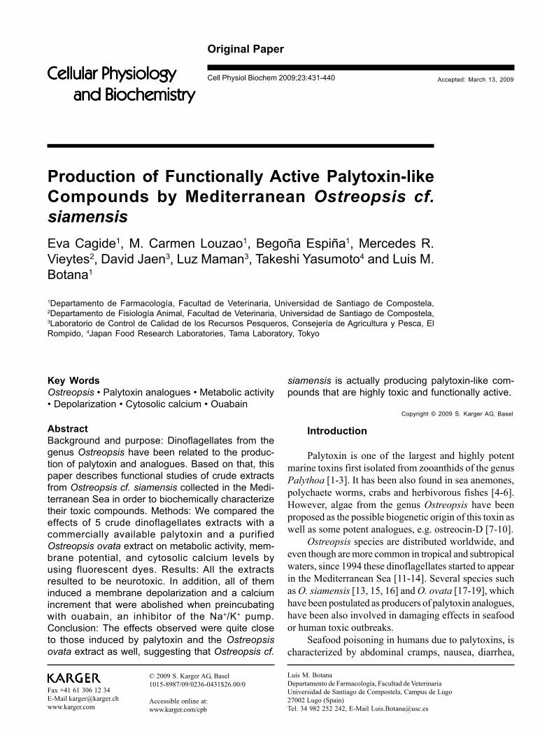
Fig. 1. Dose-dependent cell metabolism inhibition induced bypalytoxin in neuroblastoma BE(2)-M17 cells (black) and Clone9 hepatocytes (white). The cells were incubated for 24h in thepresence of the toxin. Results are presented as % fluorescencevs control, which is equivalent to cell viability.
Table 1. Cell meta-bolism inhibition ofneuroblastoma BE(2)-M17 cells after 24hexposure to severalOstreopsis cf. sia-mensis extracts.
Toxic Compounds in Ostreopsis cf. siamensis Cell Physiol Biochem 2009;23:431-440
434
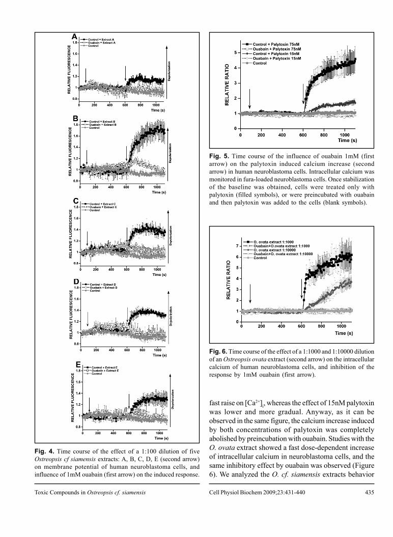
a concentration-dependent decrease of fluorescenceindicating a reduction of cell viability in both cellular models(Figure 1). Since an excitable model results a better modelfor our study, in Table 1 we present the effect of 1:100dilutions of O. cf. siamensis crude extracts inneuroblastoma cells viability (Table 1). The samplesinduced a decrease in the cellular viability, showing thatall the extracts contained toxic compounds, even thoughextract A was the most cytotoxic and E the less one.
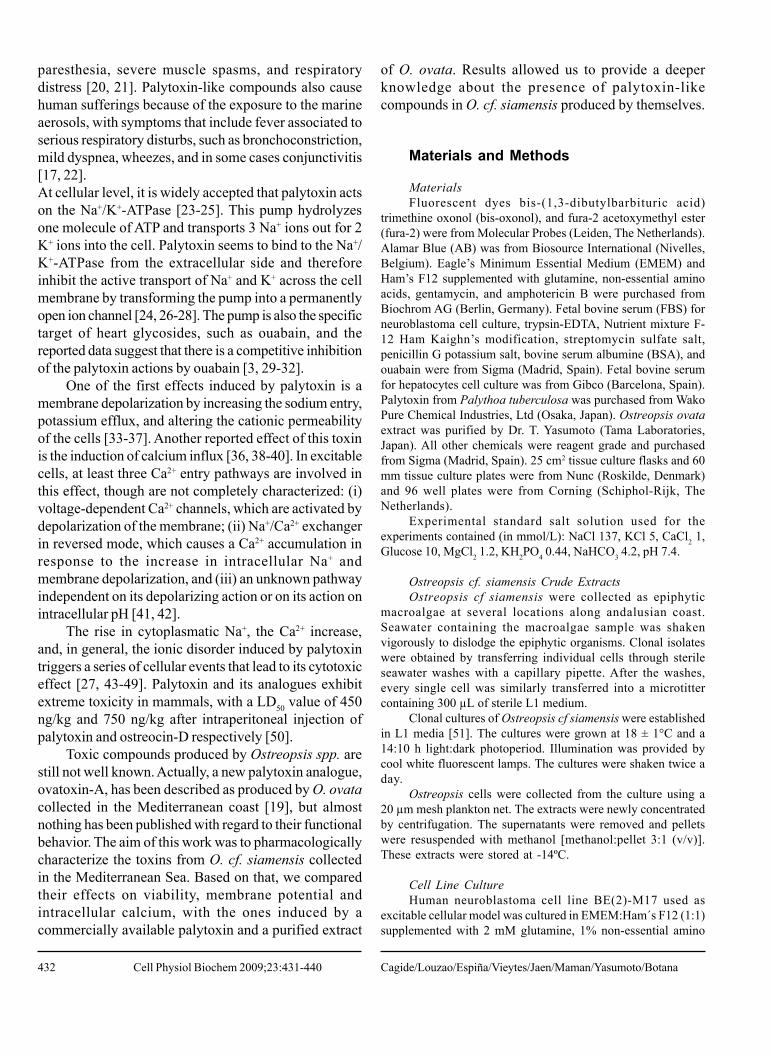
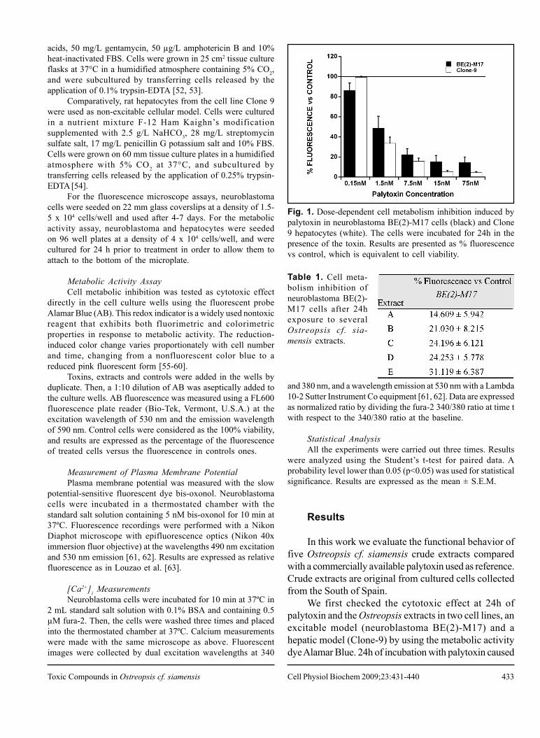
It is widely accepted that palytoxin turns the sodiumpump into a nonselective cation channel, resulting in amembrane depolarization. We used a previouslydeveloped assay to determine the effects of O. cf.siamensis crude extracts and palytoxin in neuroblastomacells membrane potential, testing palytoxin as a positivecontrol [63]. Results are presented by normalizing thefluorescence values obtained in the imaging microscopewith respect to the baseline. Figure 2 shows that palytoxininduced a significant increase in bis-oxonol fluorescence,indicating a membrane depolarization of neuroblastomacells. In addition, preincubation with 1mM ouabain fornearly 10 min, resulted in the complete abolishment ofthe depolarizing effect evoked by the toxin. This confirmsthe interaction between palytoxin and the specific inhibitorof the Na+/K+-ATPase ouabain. We also evaluated theeffect of a 1:1000 and 1:10000 dilution of an Ostreopsisovata extract, finding a high membrane depolarization ofneuroblastoma cells that was also inhibited by ouabain(Figure 3).
Next, we tested a dilution 1:100 of the crude extractsof O. cf. siamensis. All of them evoked a significantincrement of the fluorescence of bis-oxonol loaded cells,which pointed out a membrane depolarization (Figure 4).Furthermore, in order to determine if those extracts-evoked depolarizations were due to the presence ofpalytoxin-like compounds, cells were incubated with 1mMouabain for nearly 10min before addition of each extract.As in the case of the palytoxin used as reference, themembrane depolarizing effect was inhibited. All thefractions were found to induce a similar behavior, speciallyextracts C (Figure 4C) and D (Figure 4D), while extractA (Figure 4A) and E (Figure 4E) were the lessdepolarizing ones, and extract B (Figure 4B) seemed tobe the most depolarizing dilution. But, interestingly, thislast one was the only extract where ouabain did not inhibitall the increment in bis-oxonol fluorescence induced bythe toxic compounds.
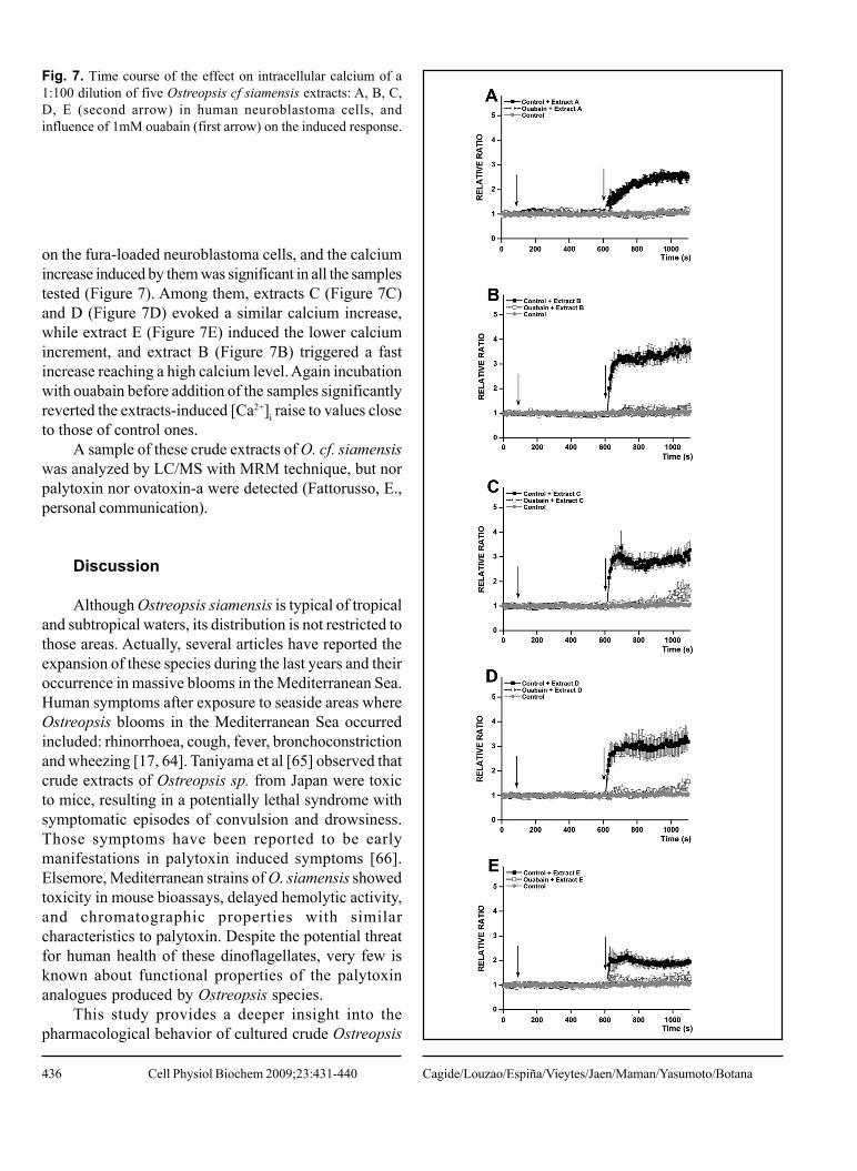
A secondary action of palytoxin in excitable cellshas been reported to be the activation of voltage-dependentCa2+ channels and the Na+/Ca2+ exchanger in the reversemode, inducing an increase in intracellular calcium([Ca2+]i). Again as a positive control we first studied theeffect of palytoxin on the basal state of intracellularcalcium of neuroblastoma cells by using the ratiometricdye fura-2. Figure 5 shows that palytoxin induced asignificant fluorescence increment in fura-loadedneuroblastoma cells, which was indicative of a cytosoliccalcium influx. 75nM palytoxin stimulated a great and
Fig. 2. Time course of the influence of ouabain 1mM (firstarrow) on the palytoxin induced depolarization (second arrow)in human neuroblastoma cells. Neuroblastoma cells wereequilibrated with bis-oxonol. Once stabilization of the baselinewas obtained, cells were treated only with palytoxin (filledsymbols), or were preincubated with ouabain and then palytoxinwas added to the cells (blank symbols).
Fig. 3. Time course of the effect of a 1:1000 and 1:10000 dilutionof an Ostreopsis ovata extract (second arrow) on the membranepotential of human neuroblastoma cells, and inhibition of theresponse by 1mM ouabain (first arrow).
Cagide/Louzao/Espiña/Vieytes/Jaen/Maman/Yasumoto/BotanaCell Physiol Biochem 2009;23:431-440
435
Fig. 4. Time course of the effect of a 1:100 dilution of fiveOstreopsis cf siamensis extracts: A, B, C, D, E (second arrow)on membrane potential of human neuroblastoma cells, andinfluence of 1mM ouabain (first arrow) on the induced response.
Fig. 5. Time course of the influence of ouabain 1mM (firstarrow) on the palytoxin induced calcium increase (secondarrow) in human neuroblastoma cells. Intracellular calcium wasmonitored in fura-loaded neuroblastoma cells. Once stabilizationof the baseline was obtained, cells were treated only withpalytoxin (filled symbols), or were preincubated with ouabainand then palytoxin was added to the cells (blank symbols).
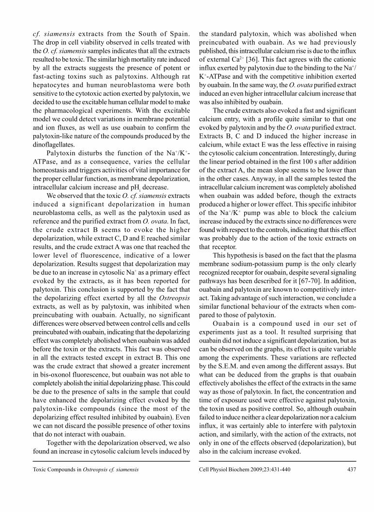
fast raise on [Ca2+]i, whereas the effect of 15nM palytoxinwas lower and more gradual. Anyway, as it can beobserved in the same figure, the calcium increase inducedby both concentrations of palytoxin was completelyabolished by preincubation with ouabain. Studies with theO. ovata extract showed a fast dose-dependent increaseof intracellular calcium in neuroblastoma cells, and thesame inhibitory effect by ouabain was observed (Figure6). We analyzed the O. cf. siamensis extracts behavior
Fig. 6. Time course of the effect of a 1:1000 and 1:10000 dilutionof an Ostreopsis ovata extract (second arrow) on the intracellularcalcium of human neuroblastoma cells, and inhibition of theresponse by 1mM ouabain (first arrow).
Toxic Compounds in Ostreopsis cf. siamensis Cell Physiol Biochem 2009;23:431-440
436
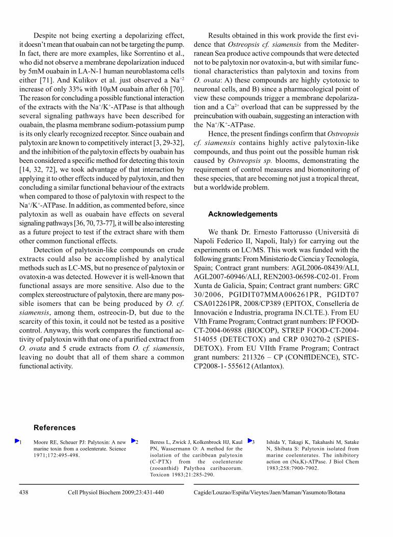
on the fura-loaded neuroblastoma cells, and the calciumincrease induced by them was significant in all the samplestested (Figure 7). Among them, extracts C (Figure 7C)and D (Figure 7D) evoked a similar calcium increase,while extract E (Figure 7E) induced the lower calciumincrement, and extract B (Figure 7B) triggered a fastincrease reaching a high calcium level. Again incubationwith ouabain before addition of the samples significantlyreverted the extracts-induced [Ca2+]i raise to values closeto those of control ones.
A sample of these crude extracts of O. cf. siamensiswas analyzed by LC/MS with MRM technique, but norpalytoxin nor ovatoxin-a were detected (Fattorusso, E.,personal communication).
Discussion
Although Ostreopsis siamensis is typical of tropicaland subtropical waters, its distribution is not restricted tothose areas. Actually, several articles have reported theexpansion of these species during the last years and theiroccurrence in massive blooms in the Mediterranean Sea.Human symptoms after exposure to seaside areas whereOstreopsis blooms in the Mediterranean Sea occurredincluded: rhinorrhoea, cough, fever, bronchoconstrictionand wheezing [17, 64]. Taniyama et al [65] observed thatcrude extracts of Ostreopsis sp. from Japan were toxicto mice, resulting in a potentially lethal syndrome withsymptomatic episodes of convulsion and drowsiness.Those symptoms have been reported to be earlymanifestations in palytoxin induced symptoms [66].Elsemore, Mediterranean strains of O. siamensis showedtoxicity in mouse bioassays, delayed hemolytic activity,and chromatographic properties with similarcharacteristics to palytoxin. Despite the potential threatfor human health of these dinoflagellates, very few isknown about functional properties of the palytoxinanalogues produced by Ostreopsis species.
This study provides a deeper insight into thepharmacological behavior of cultured crude Ostreopsis
Fig. 7. Time course of the effect on intracellular calcium of a1:100 dilution of five Ostreopsis cf siamensis extracts: A, B, C,D, E (second arrow) in human neuroblastoma cells, andinfluence of 1mM ouabain (first arrow) on the induced response.
Cagide/Louzao/Espiña/Vieytes/Jaen/Maman/Yasumoto/BotanaCell Physiol Biochem 2009;23:431-440
437
cf. siamensis extracts from the South of Spain.The drop in cell viability observed in cells treated withthe O. cf. siamensis samples indicates that all the extractsresulted to be toxic. The similar high mortality rate inducedby all the extracts suggests the presence of potent orfast-acting toxins such as palytoxins. Although rathepatocytes and human neuroblastoma were bothsensitive to the cytotoxic action exerted by palytoxin, wedecided to use the excitable human cellular model to makethe pharmacological experiments. With the excitablemodel we could detect variations in membrane potentialand ion fluxes, as well as use ouabain to confirm thepalytoxin-like nature of the compounds produced by thedinoflagellates.
Palytoxin disturbs the function of the Na+/K+-ATPase, and as a consequence, varies the cellularhomeostasis and triggers activities of vital importance forthe proper cellular function, as membrane depolarization,intracellular calcium increase and pHi decrease.
We observed that the toxic O. cf. siamensis extractsinduced a significant depolarization in humanneuroblastoma cells, as well as the palytoxin used asreference and the purified extract from O. ovata. In fact,the crude extract B seems to evoke the higherdepolarization, while extract C, D and E reached similarresults, and the crude extract A was one that reached thelower level of fluorescence, indicative of a lowerdepolarization. Results suggest that depolarization maybe due to an increase in cytosolic Na+ as a primary effectevoked by the extracts, as it has been reported forpalytoxin. This conclusion is supported by the fact thatthe depolarizing effect exerted by all the Ostreopsisextracts, as well as by palytoxin, was inhibited whenpreincubating with ouabain. Actually, no significantdifferences were observed between control cells and cellspreincubated with ouabain, indicating that the depolarizingeffect was completely abolished when ouabain was addedbefore the toxin or the extracts. This fact was observedin all the extracts tested except in extract B. This onewas the crude extract that showed a greater incrementin bis-oxonol fluorescence, but ouabain was not able tocompletely abolish the initial depolarizing phase. This couldbe due to the presence of salts in the sample that couldhave enhanced the depolarizing effect evoked by thepalytoxin-like compounds (since the most of thedepolarizing effect resulted inhibited by ouabain). Evenwe can not discard the possible presence of other toxinsthat do not interact with ouabain.
Together with the depolarization observed, we alsofound an increase in cytosolic calcium levels induced by
the standard palytoxin, which was abolished whenpreincubated with ouabain. As we had previouslypublished, this intracellular calcium rise is due to the influxof external Ca2+ [36]. This fact agrees with the cationicinflux exerted by palytoxin due to the binding to the Na+/K+-ATPase and with the competitive inhibition exertedby ouabain. In the same way, the O. ovata purified extractinduced an even higher intracellular calcium increase thatwas also inhibited by ouabain.
The crude extracts also evoked a fast and significantcalcium entry, with a profile quite similar to that oneevoked by palytoxin and by the O. ovata purified extract.Extracts B, C and D induced the higher increase incalcium, while extact E was the less effective in raisingthe cytosolic calcium concentration. Interestingly, duringthe linear period obtained in the first 100 s after additionof the extract A, the mean slope seems to be lower thanin the other cases. Anyway, in all the samples tested theintracellular calcium increment was completely abolishedwhen ouabain was added before, though the extractsproduced a higher or lower effect. This specific inhibitorof the Na+/K+ pump was able to block the calciumincrease induced by the extracts since no differences werefound with respect to the controls, indicating that this effectwas probably due to the action of the toxic extracts onthat receptor.
This hypothesis is based on the fact that the plasmamembrane sodium-potassium pump is the only clearlyrecognized receptor for ouabain, despite several signalingpathways has been described for it [67-70]. In addition,ouabain and palytoxin are known to competitively inter-act. Taking advantage of such interaction, we conclude asimilar functional behaviour of the extracts when com-pared to those of palytoxin.
Ouabain is a compound used in our set ofexperiments just as a tool. It resulted surprising thatouabain did not induce a significant depolarization, but ascan be observed on the graphs, its effect is quite variableamong the experiments. These variations are reflectedby the S.E.M. and even among the different assays. Butwhat can be deduced from the graphs is that ouabaineffectively abolishes the effect of the extracts in the sameway as those of palytoxin. In fact, the concentration andtime of exposure used were effective against palytoxin,the toxin used as positive control. So, although ouabainfailed to induce neither a clear depolarization nor a calciuminflux, it was certainly able to interfere with palytoxinaction, and similarly, with the action of the extracts, notonly in one of the effects observed (depolarization), butalso in the calcium increase evoked.
Toxic Compounds in Ostreopsis cf. siamensis Cell Physiol Biochem 2009;23:431-440
438
Despite not being exerting a depolarizing effect,it doesn’t mean that ouabain can not be targeting the pump.In fact, there are more examples, like Sorrentino et al.,who did not observe a membrane depolarization inducedby 5mM ouabain in LA-N-1 human neuroblastoma cellseither [71]. And Kulikov et al. just observed a Na+2
increase of only 33% with 10µM ouabain after 6h [70].The reason for concluding a possible functional interactionof the extracts with the Na+/K+-ATPase is that althoughseveral signaling pathways have been described forouabain, the plasma membrane sodium-potassium pumpis its only clearly recognized receptor. Since ouabain andpalytoxin are known to competitively interact [3, 29-32],and the inhibition of the palytoxin effects by ouabain hasbeen considered a specific method for detecting this toxin[14, 32, 72], we took advantage of that interaction byapplying it to other effects induced by palytoxin, and thenconcluding a similar functional behaviour of the extractswhen compared to those of palytoxin with respect to theNa+/K+-ATPase. In addition, as commented before, sincepalytoxin as well as ouabain have effects on severalsignaling pathways [36, 70, 73-77], it will be also interestingas a future project to test if the extract share with themother common functional effects.
Detection of palytoxin-like compounds on crudeextracts could also be accomplished by analyticalmethods such as LC-MS, but no presence of palytoxin orovatoxin-a was detected. However it is well-known thatfunctional assays are more sensitive. Also due to thecomplex stereostructure of palytoxin, there are many pos-sible isomers that can be being produced by O. cf.siamensis, among them, ostreocin-D, but due to thescarcity of this toxin, it could not be tested as a positivecontrol. Anyway, this work compares the functional ac-tivity of palytoxin with that one of a purified extract fromO. ovata and 5 crude extracts from O. cf. siamensis,leaving no doubt that all of them share a commonfunctional activity.
Results obtained in this work provide the first evi-dence that Ostreopsis cf. siamensis from the Mediter-ranean Sea produce active compounds that were detectednot to be palytoxin nor ovatoxin-a, but with similar func-tional characteristics than palytoxin and toxins fromO. ovata: A) these compounds are highly cytotoxic toneuronal cells, and B) since a pharmacological point ofview these compounds trigger a membrane depolariza-tion and a Ca2+ overload that can be suppressed by thepreincubation with ouabain, suggesting an interaction withthe Na+/K+-ATPase.
Hence, the present findings confirm that Ostreopsiscf. siamensis contains highly active palytoxin-likecompounds, and thus point out the possible human riskcaused by Ostreopsis sp. blooms, demonstrating therequirement of control measures and biomonitoring ofthese species, that are becoming not just a tropical threat,but a worldwide problem.
Acknowledgements
We thank Dr. Ernesto Fattorusso (Università diNapoli Federico II, Napoli, Italy) for carrying out theexperiments on LC/MS. This work was funded with thefollowing grants: From Ministerio de Ciencia y Tecnología,Spain; Contract grant numbers: AGL2006-08439/ALI,AGL2007-60946/ALI, REN2003-06598-C02-01. FromXunta de Galicia, Spain; Contract grant numbers: GRC30/2006, PGIDIT07MMA006261PR, PGIDT07CSA012261PR, 2008/CP389 (EPITOX, Consellería deInnovación e Industria, programa IN.CI.TE.). From EUVIth Frame Program; Contract grant numbers: IP FOOD-CT-2004-06988 (BIOCOP), STREP FOOD-CT-2004-514055 (DETECTOX) and CRP 030270-2 (SPIES-DETOX). From EU VIIth Frame Program; Contractgrant numbers: 211326 – CP (CONffIDENCE), STC-CP2008-1- 555612 (Atlantox).
References
1 Moore RE, Scheuer PJ: Palytoxin: A newmarine toxin from a coelenterate. Science1971;172:495-498.
2 Beress L, Zwick J, Kolkenbrock HJ, KaulPN, Wassermann O: A method for theisolation of the caribbean palytoxin(C-PTX) from the coelenterate(zooanthid) Palythoa caribaeorum.Toxicon 1983;21:285-290.
3 Ishida Y, Takagi K, Takahashi M, SatakeN, Shibata S: Palytoxin isolated frommarine coelenterates. The inhibitoryaction on (Na,K)-ATPase. J Biol Chem1983;258:7900-7902.
Cagide/Louzao/Espiña/Vieytes/Jaen/Maman/Yasumoto/BotanaCell Physiol Biochem 2009;23:431-440
439
4 Gleibs S, Mebs D: Distribution andsequestration of palytoxin in coral reefanimals. Toxicon 1999;37:1521-1527.
5 Mebs D: Occurrence and sequestrationof toxins in food chains. Toxicon1998;36:1519-1522.
6 Yasumoto T: Fish poisoning due to toxinsof microalgal origins in the pacific.Toxicon 1998;36:1515-1518.
7 Ukena T, Satake M, Usami M, OshimaY, Fujita T, Naoki H, Yasumoto T:Structural confirmation of ostreocin-Dby application of negative-ion fast-atombombardment collision-induceddissociation tandem mass spectrometricmethods. Rapid Commun Mass Spectrom2002;16:2387-2393.
8 Ukena T, Satake M, Usami M, OshimaY, Naoki H, Fujita T, Kan Y, YasumotoT: Structure elucidation of ostreocin d, apalytoxin analog isolated from thedinoflagellate Ostreopsis siamensis.Biosci Biotechnol Biochem2001;65:2585-2588.
9 Usami M, Satake M, Ishida S, Inoue A,Kan Y, Yasumoto T: Palytoxin analogsfrom the dinoflagellate Ostreopsissiamensis. J Am Chem Soc1995;117:5389-5390.
10 Rhodes L, Towers N, Briggs L, MundayR, Adamson J: Uptake of palytoxin-likecompounds by shellfish fed Ostreopsissiamensis (Dinophyceae). NZ J MarFreshwat Res 2002;36:631-636.
11 Tognetto L, Bellato S, Moroi I, AndreoliC: Occurrence of Ostreopsis ovata(Dinophyceae) in the thyrrhenian seaduring summer 1994. Bot Mar1995;38:291-295.
12 Riobó P, Paz B, Franco JM: Analysis ofpalytoxin-like in Ostreopsis cultures byliquid chromatography with precolumnderivatization and fluorescencedetection. Anal Chim Acta2006;566:217-223.
13 Vila M, Camp J, Garcés E, Masò M,Delgado M: High resolution spatio-temporal detection of potentiallyharmful dinoflagellate in confined watersof the NW Mediterranean. J PlanktonRes 2001;23:497-514.
14 Aligizaki K, Katikou P, Nikolaidis G,Panou A: First episode of shellfishcontamination by palytoxin-likecompounds from Ostreopsis species(Aegean Sea, Greece). Toxicon 2007
15 Penna A, Vila M, Fraga S, Giacobbe MG,Andreoni F, Riobo P, Vernesi C:Characterization of Ostreopsis andCoolia (Dinophyceae) isolates in thewestern Mediterranean Sea based onmorphology, toxicity and internaltranscribed spacer 5.8S rDNA sequences.J Phycol 2005;41:212-225.
16 Vila M, Garcés E, Masò M: Potentiallytoxic epiphytic dinoflagellateassemblages on macroalgae in the nwMediterranean. Aquat Microbiol Ecol2001;26:51-60.
17 Ciminiello P, Dell’Aversano C, FattorussoE, Forino M, Magno GS, Tartaglione L,Grillo C, Melchiorre N: The Genoa 2005outbreak. Determination of putativepalytoxin in mediterranean Ostreopsisovata by a new liquid chromatographytandem mass spectrometry method. AnalChem 2006;78:6153-6159.
18 Monti M, Minocci M, Beran A, Ivesa L:First record of Ostreopsis cfr. ovata onmacroalgae in the Northern Adriatic Sea.Mar Pollut Bull 2007;54:598-601.
19 Ciminiello P, Dell’aversano C, FattorussoE, Forino M, Tartaglione L, Grillo C,Melchiorre N: Putative palytoxin and itsnew analogue, ovatoxin-a, in Ostreopsisovata collected along the Ligurian Coastsduring the 2006 toxic outbreak. J AmSoc Mass Spectrom 2007
20 Alcala AC, Alcala LC, Garth JS, YasumuraD, Yasumoto T: Human fatality due toingestion of the crab Demania reynaudiithat contained a palytoxin-like toxin.Toxicon 1988;26:105-107.
21 Yasumoto T, Yasumura D, Ohizumi Y,Takahashi M, Alcala AC, Alcala L:Palytoxin in two species of xanthid crabfrom the Philippines. Agric Biol Chem1986;50:163-167.
22 Gallitelli M, Ungaro N, Addante LM,Procacci V, Silver NG, Sabbà C:Respiratory illness as a reaction totropical algal blooms occurring in atemperate climate. JAMA2005;293:2599-2600.
23 Terao KK, Ito E, Yasumoto T: Light andelectron microscopic observation ofexperimental palytoxin poisoning inmice. Bull Soc Pathol Exot 1992;85:494-496.
24 Habermann E: Palytoxin acts throughNa+,K+-ATPase. Toxicon 1989;27:1171-1187.
25 Hirsh JK, Wu CH: Palytoxin-inducedsingle-channel currents from the sodiumpump synthesized by in vitro expression.Toxicon 1997;35:169-176.
26 Artigas P, Gadsby DC: Na+/K+-pumpligands modulate gating of palytoxin-induced ion channels. Proc Natl Acad SciU S A 2003;100:501-505.
27 Hilgemann DW: From a pump to a pore:How palytoxin opens the gates. ProcNatl Acad Sci U S A 2003;100:386-388.
28 Scheiner-Bobis G: Ion-transportingATPases as ion channels. NaunynSchmiedebergs Arch Pharmacol1998;357:477-482.
29 Artigas P, Gadsby DC: Large diameter ofpalytoxin-induced na/k pump channelsand modulation of palytoxin interactionby Na/K pump ligands. J Gen Physiol2004;123:357-376.
30 Ishida Y, Satake N, Habon J, Kitano H,Shibata S: Inhibitory effect of ouabainon the palytoxin-induced contraction ofhuman umbilical artery. J Pharmacol ExpTher 1985;232:557-560.
31 Bottinger H, Habermann E: Palytoxinbinds to and inhibits kidney anderythrocyte Na+, K+-ATPase. NaunynSchmiedebergs Arch Pharmacol1984;325:85-87.
32 Artigas P, Gadsby DC: Ouabain affinitydetermining residues lie close to the Na/K pump ion pathway. Proc Natl Acad SciU S A 2006;103:12613-12618.
33 Ito K, Karaki H, Ishida Y, Urakawa N,Deguchi T: Effects of palytoxin onisolated intestinal and vascular smoothmuscles. Jpn J Pharmacol 1976;26:683-692.
34 Habermann E: Action and binding ofpalytoxin, as studied with brainmembranes. Naunyn Schmiedebergs ArchPharmacol 1983;323:269-275.
35 Castle NA, Strichartz GR: Palytoxininduces a relatively non-selective cationpermeability in frog sciatic nerve whichcan be inhibited by cardiac glycosides.Toxicon 1988;26:941-951.
36 Louzao MC, Ares IR, Vieytes MR,Valverde I, Vieites JM, Yasumoto T,Botana LM: The cytoskeleton, a structurethat is susceptible to the toxic mechanismactivated by palytoxins in humanexcitable cells. Febs J 2007;274:1991-2004.
37 Rakowski RF, Artigas P, Palma F,Holmgren M, De Weer P, Gadsby DC:Sodium flux ratio in Na/K pump-channelsopened by palytoxin. J Gen Physiol2007;130:41-54.
38 Satoh E, Ishii T, Nishimura M: Palytoxin-induced increase in cytosolic-free Ca(2+)
in mouse spleen cells. Eur J Pharmacol2003;465:9-13.
39 Ito K, Karaki H, Urakawa N: [increased45Ca uptake by the vascular smoothmuscles caused by palytoxin]. NipponHeikatsukin Gakkai Zasshi 1976;12:225-226.
40 Amano K, Sato K, Hori M, Ozaki H,Karaki H: Palytoxin-induced increase inendothelial Ca2+ concentration in therabbit aortic valve. NaunynSchmiedebergs Arch Pharmacol1997;355:751-758.
41 Frelin C, Van Renterghem C: Palytoxin.Recent electrophysiological andpharmacological evidence for severalmechanisms of action. Gen Pharmacol1995;26:33-37.
42 Ishii K, Ikeda M, Ito K: Characteristicsof palytoxin-induced cation currents andCa2+ mobilization in smooth muscle cellsof rabbit portal vein. NaunynSchmiedebergs Arch Pharmacol1997;355:103-110.
Toxic Compounds in Ostreopsis cf. siamensis Cell Physiol Biochem 2009;23:431-440
440
43 Sheridan RE, Deshpande SS, Adler M:Cytotoxic actions of palytoxin on aorticsmooth muscle cells in culture. J ApplToxicol 2005;25:365-373.
4 4 Rouzaire-Dubois B, Dubois JM:Characterization of palytoxin-inducedchannels in mouse neuroblastoma cells.Toxicon 1990;28:1147-1158.
4 5 Yoshizumi M, Nakanishi A, Houchi H,Morita K, Katoh I, Oka M:Characterization of palytoxin-inducedcatecholamine secretion from culturedbovine adrenal chromaffin cells. Effectsof Na+- and Ca2+-channel blockers.Biochem Pharmacol 1991;42:17-23.
46 Yoshizumi M, Ishimura Y, Masuda Y,Houchi H, Kato I, Oka M: Inhibition byouabain of palytoxin-inducedcatecholamine secretion and calciuminflux into cultured bovine adrenalchromaffin cells. Biochem Pharmacol1994;48:1047-1049.
47 Nakanishi A, Yoshizumi M, Morita K,Murakumo Y, Houchi H, Oka M:Palytoxin: A potent stimulator ofcatecholamine release from culturedbovine adrenal chromaffin cells. NeurosciLett 1991;121:163-165.
48 Tosteson MT, Scriven DR, Bharadwaj AK,Kishi Y, Tosteson DC: Interaction ofpalytoxin with red cells: Structure-function studies. Toxicon 1995;33:799-807.
49 Schilling WP, Snyder D, Sinkins WG,Estacion M: Palytoxin-induced cell deathcascade in bovine aortic endothelial cells.Am J Physiol Cell Physiol2006;291:C657-667.
50 Onuma Y, Satake M, Ukena T, Roux J,Chanteau S, Rasolofonirina N,Ratsimaloto M, Naoki H, Yasumoto T:Identification of putative palytoxin asthe cause of clupeotoxism. Toxicon1999;37:55-65.
51 Guillard RRL, Hargraves PE:Stichochrysis immobilis is a diatom, nota chrysophyte. Phycologia 1993;32:234-236.
52 Vilarino N, Nicolaou KC, Frederick MO,Cagide E, Ares IR, Louzao MC, VieytesMR, Botana LM: Cell growth inhibitionand actin cytoskeleton disorganizationinduced by azaspiracid-1 structure-activity studies. Chem Res Toxicol2006;19:1459-1466.
53 Ares IR, Louzao MC, Espina B, VieytesMR, Miles CO, Yasumoto T, Botana LM:Lactone ring of pectenotoxins: A keyfactor for their activity on cytoskeletaldynamics. Cell Physiol Biochem2007;19:283-292.
54 Louzao MC, Espina B, Vieytes MR, VegaFV, Rubiolo JA, Baba O, Terashima T,Botana LM: “Fluorescent glycogen”Formation with sensibility for in vivo andin vitro detection. Glycoconj J2008;25(6):503-10.
55 O’Brien J, Wilson I, Orton T, Pognan F:Investigation of the alamar blue(resazurin) fluorescent dye for theassessment of mammalian cellcytotoxicity. Eur J Biochem2000;267:5421-5426.
56 Larson EM, Doughman DJ, GregersonDS, Obritsch WF: A new, simple,nonradioactive, nontoxic in vitro assayto monitor corneal endothelial cellviability. Invest Ophthalmol Vis Sci1997;38:1929-1933.
57 Abe T, Takahashi S, Fukuuchi Y:Reduction of alamar blue, a novel redoxindicator, is dependent on both theglycolytic and oxidative metabolism ofglucose in rat cultured neurons. NeurosciLett 2002;326:179-182.
58 Vanitha JD, Paramasivan CN: Evaluationof microplate alamar blue assay for drugsusceptibility testing of Mycobacteriumavium complex isolates. Diagn MicrobiolInfect Dis 2004;49:179-182.
59 Hamid R, Rotshteyn Y, Rabadi L, ParikhR, Bullock P: Comparison of alamar blueand MTT assays for high through-putscreening. Toxicol In Vitro 2004;18:703-710.
60 Schreer A, Tinson C, Sherry JP, SchirmerK: Application of alamar blue/5-carboxyfluorescein diacetateacetoxymethyl ester as a noninvasive cellviability assay in primary hepatocytesfrom rainbow trout. Anal Biochem2005;344:76-85.
61 Cagide E, Louzao MC, Ares IR, VieytesMR, Yotsu-Yamashita M, Paquette LA,Yasumoto T, Botana LM: Effects of asynthetic analog of polycavernoside Aon human neuroblastoma cells. CellPhysiol Biochem 2007;19:185-194.
62 Louzao MC, Cagide E, Vieytes MR, SasakiM, Fuwa H, Yasumoto T, Botana LM:The sodium channel of human excitablecells is a target for gambierol. Cell PhysiolBiochem 2006;17:257-268.
63 Louzao MC, Vieytes MR, Yasumoto T,Yotsu-Yamashita M, Botana LM:Changes in membrane potential: An earlysignal triggered by neurologically activephycotoxins. Chem Res Toxicol2006;19:788-793.
64 Brescianini C, Grillo C, Melchiorre N,Bertolotto R, Ferrari A, Vivaldi B, IcardiG, Gramaccioni L, Funari E, Scardala S:Ostreopsis ovata algal blooms affectinghuman health in Genova, Italy, 2005 and2006. Euro Surveill 2006;11:E060907060903.
65 Taniyama S, Arakawa O, Terada M,Nishio S, Takatani T, Mahmud Y,Noguchi T: Ostreopsis sp., a possibleorigin of palytoxin (PTX) in parrotfishScarus ovifrons. Toxicon 2003;42:29-33.
66 Vick JA, Wiles JS: The mechanism ofaction and treatment of palytoxinpoisoning. Toxicol Appl Pharmacol1975;34:214-223.
67 Ferrandi M, Manunta P, Ferrari P, BianchiG: The endogenous ouabain: Molecularbasis of its role in hypertension andcardiovascular complications. FrontBiosci 2005;10:2472-2477.
68 Ferrandi M, Molinari I, Bianchi G, FerrariP: Ouabain-dependent signaling incaveolae as a novel therapeutic targetfor hypertension. Cell Mol Biol (Noisy-le-grand) 2006;52:15-18.
69 Aperia A: New roles for an old enzyme:Na,K-ATPase emerges as an interestingdrug target. J Intern Med 2007;261:44-52.
70 Kulikov A, Eva A, Kirch U, Boldyrev A,Scheiner-Bobis G: Ouabain activatessignaling pathways associated with celldeath in human neuroblastoma. BiochimBiophys Acta 2007;1768:1691-1702.
71 Sorrentino G, Monsurro MR, Singh IN,Kanfer JN: Membrane depolarization inLa-N-1 cells. The effect of maitotoxinis Ca2+- and Na+-dependent. Mol ChemNeuropathol 1997;30:199-211.
72 Espina B, Cagide E, Louzao MC,Fernandez MM, Vieytes MR, Katikou P,Villar A, Jaen D, Maman L, Botana LM:Specific and dynamic detection ofpalytoxins by in vitro microplate assaywith human neuroblastoma cells. BiosciRep 2009;29:13-23.
73 Li S, Wattenberg EV: Differentialactivation of mitogen-activated proteinkinases by palytoxin and ouabain, twoligands for the Na+,K+-ATPase. ToxicolAppl Pharmacol 1998;151:377-384.
74 Ares IR, Louzao MC, Vieytes MR,Yasumoto T, Botana LM: Actincytoskeleton of rabbit intestinal cells isa target for potent marine phycotoxins.J Exp Biol 2005;208:4345-4354.
75 Louzao MC, Ares IR, Cagide E: Marinetoxins and the cytoskeleton: A new viewof palytoxin toxicity. Febs J2008;275:6067-6074.
76 Edwards A, Pallone TL: Ouabainmodulation of cellular calcium stores andsignaling. Am J Physiol Renal Physiol2007;293:F1518-1532.
77 Rajasekaran SA, Palmer LG, Moon SY,Peralta Soler A, Apodaca GL, Harper JF,Zheng Y, Rajasekaran AK: Na,K-ATPaseactivity is required for formation of tightjunctions, desmosomes, and induction ofpolarity in epithelial cells. Mol Biol Cell2001;12:3717-3732.
Cagide/Louzao/Espiña/Vieytes/Jaen/Maman/Yasumoto/BotanaCell Physiol Biochem 2009;23:431-440


![Functionally Rigid Bistable [2]Rotaxanes](https://img.pdfslide.net/doc/110x75/63143f373ed465f0570b0e49/functionally-rigid-bistable-2rotaxanes.jpg)


