Embed Size (px)
Citation preview

BioMed Central
Reproductive Biology and Endocrinology
ss
Open AcceResearchProgesterone receptor A and c-Met mediates spheroids-endometrium attachmentHaggar Harduf1,2, Shlomit Goldman1 and Eliezer Shalev*1,2Address: 1Laboratory for Research in Reproductive Sciences, Department of Obstetrics and Gynecology, Ha'Emek Medical Center, Afula, Israel and 2Rappaport Faculty of Medicine, Technion-Israel Institute of Technology, Haifa, Israel
Email: Haggar Harduf - [email protected]; Shlomit Goldman - [email protected]; Eliezer Shalev* - [email protected]
* Corresponding author
AbstractBackground: Implantation in humans involves cross talk between an active blastocyst andreceptive endometrium. The role of the endometrial receptors in this complex embryo-maternalinteraction is still unclear. We tested gene and protein expression of endometrial receptors(Progesterone receptor (PR) and c-Met) and the effect of theses receptors in endometrialreceptivity.
Methods: Two endometrial cell lines were used: HEC-1A and RL95-2 considered as being of lowand high receptivity, respectively. Western blot and RT-PCR analysis were utilized to study thereceptor expression profile.
The role of endometrial receptors in endometrial receptivity was studied by attachment andinvasion assays of JAR spheroids (made of a trophoblast cell line) on endometrial cells. Differentmanipulations of inhibition and stimulation of the endometrial receptors were used including:inhibition by specific antibodies against the receptors, or antagonist of the receptors, as well astransfection with antisense for the endometrial receptors, stimulation by specific ligands for thereceptors and transfection with the gene for endometrial receptors.
Results: Different protein expression patterns of endometrial receptors were observed betweenthe tested endometrial cell lines. The expression levels of PRA ratio to PRB, and the 50 kDa c-METisoform were significantly lower in HEC-1A as compared with RL95-2. Attachment rates andgrowth of JAR spheroids into HEC-1A were significantly lower as compared with RL95-2.Stimulation of PR with progesterone altered attachment rates to HEC-1A. Inhibition of PR withRU-486 mildly increased attachment rate to HEC-1A whereas it slightly decreased attachment rateto RL95-2. c-Met inhibition decreased attachment rates only to HEC-1A cells that expressing highlevels of Plexin-B1 (PB1). Immunoprecipitation studies revealed that c-Met and PB1 associate incomplexes in the endometrial cell lines.
Conclusion: Differential endometrial receptor profiles are expressed during the receptivityperiod. The attachment and invasion processes are separately regulated. We suggest a biologicallyfunctional role for PRA in endometrial receptivity and in the attachment process. c-Metcontribution is minor and related with creation of a complex with PB1.
Published: 16 February 2009
Reproductive Biology and Endocrinology 2009, 7:14 doi:10.1186/1477-7827-7-14
Received: 19 November 2008Accepted: 16 February 2009
This article is available from: http://www.rbej.com/content/7/1/14
© 2009 Harduf et al; licensee BioMed Central Ltd. This is an Open Access article distributed under the terms of the Creative Commons Attribution License (http://creativecommons.org/licenses/by/2.0), which permits unrestricted use, distribution, and reproduction in any medium, provided the original work is properly cited.
Page 1 of 14(page number not for citation purposes)

Reproductive Biology and Endocrinology 2009, 7:14 http://www.rbej.com/content/7/1/14
BackgroundImplantation in humans involves complex interactionsbetween the embryo and the maternal endometrium [1-3]. Successful implantation depends on a pre-implanta-tion embryo developing into a competent blastocyst thatreaching the uterus precisely at its receptive stage [4].Endometrial receptivity is suggested to be a property ofthe endometrial epithelial cells (EECs). The molecularmechanisms by which the surface of human EECsacquires morphological changes, leading to receptive fea-tures, are still unclear. Cytokines, growth factors, hor-mones, extracellular matrix proteins and enzymes,angiogenic factors, cell-cell adhesion molecules andreceptors are all involved in this complex process [5]. Pre-vious studies demonstrated the appearance of morpho-logical or biological markers for endometrial receptivity[6-10]. However functional physiological markers are stillunknown. The cross talk, between the active blastocystand the receptive uterus, is solely reliant on mediationand interrelationship by a variety of receptors in theendometrium. Despite the possibility of extra corporalfertilization and extensive new technology, the process ofimplantation and the interaction between maternalendometrium and invading trophoblast are even todaydifficult to explore.
Hence, the search for better understanding of this processcontinues and is transferred into the in vitro setting [11-13]. In our previous study [14] we showed that Plexin B1(PB1), a membrane receptor, has a role in endometrialreceptivity and in the attachment process. The currentstudy was designed to explore and compare the expressionand role of the membrane receptor c-Met, which is knownto be expressed as a complex with PB1 [15,16] and thenuclear receptor PR in two human endometrial cell lines,RL95-2 and HEC-1A, used as a model for high receptivityand low receptivity endometrium respectively [17-20].
The progesterone receptor (PR) is a member of a largefamily of ligand-activated nuclear transcription regula-tors, which are characterized by organization into specificfunctional domains and are conserved between speciesand family members. The PR is made up of a central DNAbinding domain and a carboxyl-terminal ligand-bindingdomain. Studies on human PR indicate that there are atlist 3 different alternatively spliced forms to the PR. Twoof the PR isoforms, namely PR-A and PR-B, mediate theeffects of progesterone. Detailed function studies indicatethat PR-B, in all cellular contexts in-vitro, functions as aligand-dependent trans-activator. This in contrast to PR-A,which in some contexts acts as a ligand-dependent tran-scriptional repressor of PR-B [21,22]. There is increasingevidence to date that PR-A and PR-B are functionally dif-ferent. The PRB/PRA ratio was found to be of clinicalimportance in several tissues, [[23], and [24]]. These dif-
ferences are yet to be fully understood. It is the balancebetween these two forms that may make it possible forprogesterone to affect such diverse physiological targets.Progesterone's action has been shown to be essential forproper endometrial maturation, endometrial receptivityand the maintenance of pregnancy [25]. These effects ofprogesterone are thought to be mediated primarilythrough its cognate receptor [21,22]. The establishment ofnormal endometrial receptivity appears to be closely asso-ciated with the down-regulation of epithelial PR [8]. His-tologic delay is associated with a failure of PR down-regulation and the lack of normal markers of endometrialreceptivity.
The proto-oncogene Met encodes a transmembrane tyro-sin kinase of 190 kDa. c-Met is a heterodimer composedof two disulfide-linked chains of 50 kDa and 140 kDa[26]. Met is the receptor for hepatocyte growth factor(HGF) [26-28]. It is frequently over expressed in neoplas-tic cells and in host tissue. Due to its prominent role in thecontrol of motility and invasion, it is involved in metasta-sis formation. The role of c-Met in endometrial receptivitystill needs to be investigated.
Stromal and trophoblast cells produce HGF [29] while itsreceptor is expressed in the endometrial epithelia andstroma [30]. Recent data indicate that signaling activity ofthe Met receptor is affected by an association with otherreceptors such as RON and PB1 and it was published thatcells expressing the endogenous proteins, PB1 and c-Met,associate in a complex [15]. In addition it was shown thatmembrane-bound semaphorin Sema4D, PB1's ligand,can trigger the activation of the oncogenic receptor Met,which is associated with PB1 on the cell surface [16].
MethodsCell linesTwo endometrial cell lines were used as in vitro model forendometrial receptivity. Cell line RL95-2 (ATCC catalogNo.CRL-1671), derived from a moderately differentiatedadeno-squamous carcinoma of the endometrium [17]was used as a model for receptive endometrium [19,20]Cell line HEC-1A (ATCC catalog No. HTB-112) derivedfrom human endometrial carcinoma, served as a modelfor the non-receptive state [31]. Third cell line was estab-lished in our laboratory, HEC-1A cells were transfectedwith human PB1 (was provided kindly by Prof. LucaTamagnone, the transfected cell line named HEC-1A-2.(HEC-1A-2 was characterized in our previous study [14].Human trophoblast cell line, JAR (ATCC catalog No. HTB-144) was used as a model for blastocysts.
Endometrial cell cultureHEC-1A cells were cultured in Meckoy 5A medium (Kib-butz Beit-Ha'Emek, Israel) containing 10% Fetal Calf
Page 2 of 14(page number not for citation purposes)
Reproductive Biology and Endocrinology 2009, 7:14 http://www.rbej.com/content/7/1/14
Serum (FCS) (Kibbutz Beit-Ha'Emek, Israel) and penicil-lin/streptomycin (Kibbutz Beit-Ha'Emek, Israel) [32].RL95-2 cells were cultured in DMEM F: 12 medium (Kib-butz Beit-Ha'Emek, Israel) containing FCS, penicillin/streptomycin, 2.5 mM Glutamine (Kibbutz Beit-Ha'Emek,Israel) [17]. Cell cultures were maintained in a humidifiedatmosphere containing 5% CO2 at 37°C. RL95-2 cells (1–2 × 10⁶) and HEC-1-A cells (1–2 × 10⁶) were seeded in 24-well culture plates for 10 days, and the growth mediumwas renewed every 2–3 days. All studies performed withserum free medium.
Attachment and growth assaysAttachment of JAR spheroids to endometrial cell monolayerFor the attachment assays JAR spheroids were preparedand tested as described in details elsewhere [33]: briefly, 1× 10⁶ JAR cells per 10 ml M-199 medium (Kibbutz Beit-Ha'Emek, Israel) containing 10% FCS and penicillin/streptomycin were agitated at 37°C on a Comfort shakerat 200 rpm (12 × g). In order to distinguish JAR spheroidsfrom underlying endometrial cell lines (HEC-1A andRL95-2) or primary culture we have labeled the JAR sphe-roids with the membrane-permeable fluorescent dyeCMFDA (Invitrogen, Dorset, UK) that after enzymaticcleavage serves as a long-term cytoplasmic marker. Sphe-roids were agitated at 37°C for 24 hours. Thereafter sphe-roids were gently delivered with micro denuding pipette(150 μm diameter) onto a confluent monolayer ofendometrial cell lines (HEC-1A and RL95-2) grown in 24-wells culture plates in M-199 growth medium containing1.5% FCS. After 60 minutes of incubation at 37°C the cul-ture plate was shaken aggressively at 15 × g (VORTEXGENIE, Scientific Industries, Chicago, U.S.A) for 60 min-utes. The medium containing unattached spheroids wascollected, and fresh medium was added to the wells. Sphe-roids remaining in each well were counted using a phase-contrast microscope or florescence microscope. Spheroidsattachment is expressed as a percentage of seeded sphe-roids. In certain experiments HEC-1A and RL95-2 celllines were pretreated with Progesterone 0–10 μM (Sigma,ST Louis, MO, USA) or with RU-486 (PR antagonist)(Sigma, ST Louis, MO, USA). In other experimentsendometrial cell lines were pretreated with antisenseagainst c-Met (IDT Inc, Hy-Labs, Rehovot, Israel).
Growth of JAR spheroids in endometrial cell monolayerSpheroids outgrowth was measured under the microscopefor the next 10 days. Each spheroid diameter size wasmeasured using a special scale in the ocular.
Preparation of whole cell extract and western blot analysisHEC-1A and RL95-2 cells were lysed on ice in lysis buffer(20 mM Tris-HCl, pH 7.4, 5 mM EDTA, 150 mM NaCl,10% glycerol, 1% Triton X-100) in the presence of a mix-ture of protease inhibitors (Roche, Kulmbach, Germany),
suspensions were incubated for 7 minutes in 4°C. Celllysates were precleared by centrifugation at 12000 rpm for20 minutes, the supernatant fraction contained proteins.
Protein assayThe total protein content of endometrial cells was deter-mined using a protein assay kit with BSA as the standard(Bio-Rad laboratories, Inc, Washington DC). One to fivemicroliters of sample were used in the assay. The assay isbased on the Bradford dye-binding procedure.
Western blotIn order to detect c-Met and PR, whole cell and nuclearextracts were diluted with 4 × sample buffer (5% SDS,20% Glycerol in 0.4 M Tris, pH 6.8 containing 0.02%bromophenol blue) and subjected to 8% polyacrylamidegel electrophoresis. After electrophoresis, the proteins (50μg/lane) were blotted from the SDS-PAGE onto 0.45 μmnitrocellulose membranes (Schleicher & Schuell, Dassel,Germany). Nonspecific binding sites were blocked byincubating the nitrocellulose membranes for 1 hour with5% BSA (Sigma, ST Louis, MO, USA) in Tris-bufferedsaline. The membranes were then washed four times withTris-buffered saline, containing 0.75% Tween-20, andincubated for 1 hour with antibodies against PR (SantaCruz, Biotech, California, USA sc-539) or c-Met (SantaCruz Biotech, California, USA sc-161) in 0.5% BSA in Tris-buffered saline, containing 0.01% Tween-20. The mem-branes were subsequently washed with Tris-bufferedsaline, containing 0.75% Tween-20 and incubated for 1hour with HRP-conjugated Horseradish peroxidase-con-jugated goat anti- rabbit secondary antibody (JacksonImmuno-research, Enco, Israel) in 0.5% BSA in Tris-buff-ered saline, containing 0.01% Tween-20. Proteins weredetected by enhanced chemiluminescence (ECL Kit, Kib-butz Beit-Ha'Emek, Israel,) and quantified using the Bio-Imaging gel documentation system (Dinco & Renium,Jerusalem, Israel) endowed with TINA software (Raytest,Taubenhardt, Germany). PR and c-Met were expressed aspercent of control. For normalization we have used thelevels of the housekeeping protein GAPDH.
Semiquantitative RT-PCRTo analyze the expression of PR and c-Met, total RNA wasprepared from cell cultures with EZ-RNA Kit (KibbutzBeit-Ha'Emek, Israel). RNA concentrations were deter-mined spectrophotometrically. To obtain the cDNA fromcell lines, total RNA (5 μg) was denatured at 70°C for 10min and then reverse transcribed in the presence of 25 ng/μl random primer (Promega, Mannheim, Germany), 2.5mM MgCl2, 0.5 mM deoxy-NTPs, 10 mM dithiothreitol,and 10 U ribonuclease H- reverse transcriptase (Super-script II RT, Life Technologies, Inc.) for 60 min at 42°C,and 5 min at 95°C. Subsequently, 10 μl of the resultingcDNA was used as a template for polymerase chain reac-
Page 3 of 14(page number not for citation purposes)

Reproductive Biology and Endocrinology 2009, 7:14 http://www.rbej.com/content/7/1/14
tion (PCR). The PCR was set up using 3 mM MgCl2, 50pmol of each primer and 2.5 U Taq DNA polymerase(Sigma, St. Louis, MO, USA).
Primer design: the sequences of the primers were takenfrom the Genbank
PRB (Genbank access no. M15716)
PRB FWD 5'- ACACCTTGCCTGAAGTTTCG-3'
PRB REV 5'- CTGTCCTTTTCTGGGGGACT-3' (196 bpproduct).
c-MET – (Genbank access no. M35073)
c-MET FWD 5'- CTACAAAGAAGTTGATGAACCG-3'
c-MET REV 5'- GCTGACATACAGTCGGAGG-3' (139 bpproduct).
For normalization we have used the levels of the house-keeping gene GAPDH.
GAPDH FWD 5'-TGATGACATCAAGAAGGTGGTGAAG-3'; GAPDH REV 5'-TCCTTGGAGGCCATGTGGGCCAT-3'(230 bp product).
PCR conditions were 94°C for 2 min followed by 35cycles of 94°C for 30 sec, 58°C for 45 sec, and 72°C for60 sec with a 72°C extension for 10 min. After PCR, theproducts were resolved on a 2.5% agarose ethidium bro-mide gel. Images were captured with Polaroid (Hertford-shire, UK) film under UV light. Products were quantifiedusing PhosphorImager and ImageQuant software (Molec-ular Dynamics, Inc., Sunnyvale, CA).
ImmunoprecipitationEndometrial cell lines (HEC-1A, RL95-2, HEC-1A-2) werewashed twice in ice cold PBS and lysed on ice in lysisbuffer (20 mM Tris-HCl, pH 7.4, 5 mM EDTA, 150 mMNaCl, 10% glycerol, 1% Triton X-100) in the presence ofa mixture of protease inhibitors (Roche, Kulmbach, Ger-many). 500 μg of whole cell extract in 1 ml lysis bufferwere subject for immunoprecipitation and PB1 receptorswere immunoprecipitated by incubation for 2 h on rockerat 4°C with 1 μg anti-PB1 antibody (Santa Cruz Biotech,California, USA, sc-28372). Immunocomplexes wererecovered with the aid of 20 μl protein A/G agarose beads(Santa Cruz Biotech, California, USA, sc-2003). Each sam-ple was placed on a rocker at 4°C for 1 h and thereafterincubated for 16 h at 4°C. The beads were washed twicewith 1 ml lysis buffer and twice with Tris-EDTA (TE) andsubsequently the bound proteins were eluted in 50 μl of1% SDS in TE. Sample buffer was added to the superna-
tant of each sample. Lysates and immunoprecipitates wereanalyzed by Western blotting after SDS-polyacrylamidegel electrophoresis and transfer to a 0.45 μm nitrocellu-lose membranes (Schleicher & Schuell, Dassel, Germany)with anti c-Met antibodies (Santa Cruz Biotech, Califor-nia, USA, sc-161). Proteins were detected by enhancedchemiluminescence (ECL Kit, Kibbutz Beit-Ha'Emek,Israel). As a negative control, PB1 immunoprecipitationwas performed, followed by Western blotting withGAPDH antibody.
Immunofluorescence stainingFor immunofluorescence analysis, endometrial cells werecultured on glass coverslips in 35 μl medium drops undermineral oil. Cells were washed 3 times with PBS and fixedwith 3.7% paraformaldehyde (Electron Microscope Sci-ences, Belgar) in PBS for 10 minutes at 4°C, then washedtwice with PBS and permeabilized for 5 minutes at 4°Cwith 0.1% Triton (Sigma, St. Louis, MO, USA) in PBS.After a PBS wash, slides were incubated for 1 hour withblocking buffer (PBS supplemented with 3% BSA), thenwashed 3 times with PBS and incubated for 30 minutes atroom temperature with primary antibodies (Anti c-Met),1 μg per slide in 700 μl PBS supplemented with 1.5% BSA.After five washings with PBS, slides were incubated for 30minutes in the dark with secondary fluorescein-labeledantibody (for F-actin:phalloidin, AlexaFlour-488, A-12379; for c-Met: goat anti-rabbit IgG conjugated withAlexaFlour-546, all from Molecular Probes, Invitrogen,Dorset, UK), 0.5 μg per slide in 700 μl PBS supplementedwith 1.5% BSA. Following three washings with PBS,stained cells were photographed using a confocal micro-scope (the confocal system is composed of a Bio-Rad radi-ance 2000 confocal set-up hooked to an uprightfluorescent microscope Nikon E600 with a 60X lens). Thephotos were analyzed by Image Pro software (MediaCybernetics, Bethesda, USA), which quantifies density perarea.
Statistical analysisResults are expressed as mean ± SEM, with n denoting thenumber of spheroids. Student's t-test, chi test and "oneway analysis of variance" (ANOVA) were used whenappropriate. P < 0.05 was considered significant.
ResultsPR expression in RL95-2 and HEC-1A cellsPRB gene expression was studied by RT-PCR. For normal-ization we have used the levels of the housekeeping geneGAPDH. In order to exclude the possibility of fluctuationin gene expression during 24 hours (h) period, we havestudied the basal PRB gene expression on 2, 12 and 24 hof incubation with serum-free medium, 2 h after mediumreplacement considered as starting period (time 0).
Page 4 of 14(page number not for citation purposes)
Reproductive Biology and Endocrinology 2009, 7:14 http://www.rbej.com/content/7/1/14
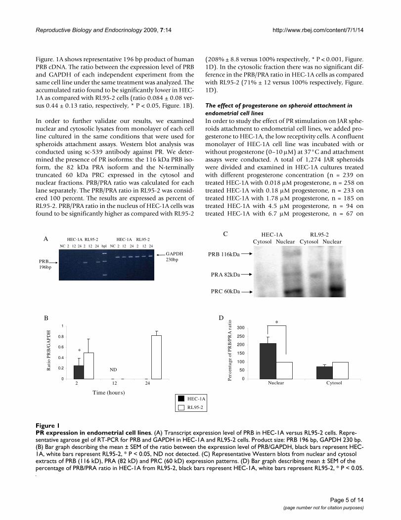
Figure. 1A shows representative 196 bp product of humanPRB cDNA. The ratio between the expression level of PRBand GAPDH of each independent experiment from thesame cell line under the same treatment was analyzed. Theaccumulated ratio found to be significantly lower in HEC-1A as compared with RL95-2 cells (ratio 0.084 ± 0.08 ver-sus 0.44 ± 0.13 ratio, respectively, * P < 0.05, Figure. 1B).
In order to further validate our results, we examinednuclear and cytosolic lysates from monolayer of each cellline cultured in the same conditions that were used forspheroids attachment assays. Western blot analysis wasconducted using sc-539 antibody against PR. We deter-mined the presence of PR isoforms: the 116 kDa PRB iso-form, the 82 kDa PRA isoform and the N-terminallytruncated 60 kDa PRC expressed in the cytosol andnuclear fractions. PRB/PRA ratio was calculated for eachlane separately. The PRB/PRA ratio in RL95-2 was consid-ered 100 percent. The results are expressed as percent ofRL95-2. PRB/PRA ratio in the nucleus of HEC-1A cells wasfound to be significantly higher as compared with RL95-2
(208% ± 8.8 versus 100% respectively, * P < 0.001, Figure.1D). In the cytosolic fraction there was no significant dif-ference in the PRB/PRA ratio in HEC-1A cells as comparedwith RL95-2 (71% ± 12 versus 100% respectively, Figure.1D).
The effect of progesterone on spheroid attachment in endometrial cell linesIn order to study the effect of PR stimulation on JAR sphe-roids attachment to endometrial cell lines, we added pro-gesterone to HEC-1A, the low receptivity cells. A confluentmonolayer of HEC-1A cell line was incubated with orwithout progesterone (0–10 μM) at 37°C and attachmentassays were conducted. A total of 1,274 JAR spheroidswere divided and examined in HEC-1A cultures treatedwith different progesterone concentration (n = 239 ontreated HEC-1A with 0.018 μM progesterone, n = 258 ontreated HEC-1A with 0.18 μM progesterone, n = 233 ontreated HEC-1A with 1.78 μM progesterone, n = 185 ontreated HEC-1A with 4.5 μM progesterone, n = 94 ontreated HEC-1A with 6.7 μM progesterone, n = 67 on
PR expression in endometrial cell linesFigure 1PR expression in endometrial cell lines. (A) Transcript expression level of PRB in HEC-1A versus RL95-2 cells. Repre-sentative agarose gel of RT-PCR for PRB and GAPDH in HEC-1A and RL95-2 cells. Product size: PRB 196 bp, GAPDH 230 bp. (B) Bar graph describing the mean ± SEM of the ratio between the expression level of PRB/GAPDH, black bars represent HEC-1A, white bars represent RL95-2, * P < 0.05, ND not detected. (C) Representative Western blots from nuclear and cytosol extracts of PRB (116 kD), PRA (82 kD) and PRC (60 kD) expression patterns. (D) Bar graph describing mean ± SEM of the percentage of PRB/PRA ratio in HEC-1A from RL95-2, black bars represent HEC-1A, white bars represent RL95-2, * P < 0.05.
B
*
NC 2 12 24 2 12 24 bpl NC 2 12 24 2 12 24
A
2 12 24
Time (hours)
GAPDH 230bpPRB
196bp
HEC-1A RL95-2 HEC-1A RL95-2
Rat
io P
RB
/GA
PD
H
HEC-1A
RL95-2
PRB 116kDa
PRA 82kDa
PRC 60kDa
HEC-1A RL95-2Cytosol Nuclear Cytosol Nuclear
0
50
100
150
200
250
300
Per
cen
tage
of
PR
B/P
RA
rat
io
Nuclear Cytosol
*
C
D
ND
0
0.2
0.4
0.6
0.8
1
Page 5 of 14(page number not for citation purposes)

Reproductive Biology and Endocrinology 2009, 7:14 http://www.rbej.com/content/7/1/14
treated HEC-1A with 10 μM progesterone, and n = 198 onun-treated HEC-1A cells). As shown in Figure. 2A, wefound a dual effect of progesterone, at low concentrationof 0.018 μM progesterone significantly inhibited spheroidattachment to treated HEC-1A cells as compared to non-treated HEC-1A cells (13.7% ± 0.47 versus 26.5% ± 3.78,respectively) while a higher progesterone concentration of6.7 μM and 10 μM increased the attachment rate as com-pared with non-treated HEC-1A cells (39.8% ± 3.87 and34.3% ± 0.43 versus 26.5% ± 3.78, respectively, * P <0.05). RL95-2 is high receptivity cells therefore we testedthe low concentration of progesterone, the inhibitory con-centrations, (0–0.18 μM). As shown in Figure. 2B JARspheroid attachment rate to RL95-2 was not affected byprogesterone.
The effect of PR antagonist RU-486 on spheroid attachment in endometrial cell linesIt is well known that progesterone effect is mediated by itscognate receptor – PR. In order to further investigate therole of the PR during the implantation process we testedthe effect of PR inhibition on spheroids attachment. Weconducted attachment assays to endometrial cell linestreated with RU-486 (10-6 M) PR antagonist. A confluentmonolayer of HEC-1A and RL95-2 cell lines was incu-bated with or without RU-486 for 24 hours before sphe-roids deliver. In three experiments a total of 270 JARspheroids were divided and examined in HEC-1A cell cul-tures (n = 134 on treated cells and n = 136 on non-treatedcells) The JAR spheroid attachment rate to HEC-1A treatedwith RU-486 increased as compared with non-treatedcells (89.5% ± 1.7 versus 75.5% ± 2.8 respectively, * P <
JAR spheroid attachment to HEC-1A and RL95-2 cells with or without ProgesteroneFigure 2JAR spheroid attachment to HEC-1A and RL95-2 cells with or without Progesterone. (A) Bar graph describes mean ± SEM of % attachment to HEC-1A cells treated with progesterone (0–10 μM), * P < 0.05. (B) Bar graph describes mean ± SEM of % attachment to RL95-2 cells treated with progesterone (0–0.18 μM). JAR spheroid attachment to HEC-1A and RL95-2 cells with or without PR antagonist RU-486. (C) Bar graph describes mean ± SEM of % attachment to HEC-1A cells treated with RU-486 (10-6 M). (D) Bar graph describes mean ± SEM of % attachment to RL95-2 cells treated with RU-486 (10-
6 M). * P < 0.05.
Page 6 of 14(page number not for citation purposes)

Reproductive Biology and Endocrinology 2009, 7:14 http://www.rbej.com/content/7/1/14
0.01, Figure. 2C). In four experiments a total of 302 JARspheroids were divided and examined in RL95-2 cell cul-tures (n = 148 on treated cells and n = 154 on un-treatedcells). In contrast, in RL95-2 cells, RU-486 decreased theattachment rates of JAR spheroids to treated cells as com-pared with non-treated cells (96.7% ± 2.1 versus 86.2% ±3.7 respectively, * P < 0.05, Figure. 2D).
c-Met expression in RL95-2 and HEC-1A cellsc-Met gene expression was studied by RT-PCR. For nor-malization we have used the levels of the housekeepinggene GAPDH. In order to exclude the possibility of fluctu-ation in gene expression during 24 h period, we have stud-ied the basal c-Met gene expression on 2, 12 and 24 h. 2 hafter medium replacement considered as starting period(time 0).
Figure. 3A shows representative 139 bp product of humanc-Met cDNA. The ratio between the expression level of c-Met and GAPDH of each independent experiment from
the same cell line under the same treatment was analyzed.The ratio in RL95-2 was considered as 100%, the resultsexpressed as % of RL95-2 (Figure. 3B). There was no dif-ference in transcript expression level of c-Met between thetwo cell lines.
In order to study whether c-Met protein is expressedequally in both cell lines, we examined cell lysates frommonolayer of each cell line cultured in the same condi-tions that were used for spheroids attachment assays.Western blot analysis was conducted using sc-161 anti-body against c-Met. We determined the presence of twobands in endometrial cell lines (140 and 50 kDa, Figure.3C). The ratio between the expression level of the shortform of c-Met (50 kDa) to the long form (140 kDa) wasanalyzed for each independent experiment, under thesame treatment. In both HEC-1A and HEC-1A-2 theexpression level of the 50 kDa form was lower as com-pared to the 140 kDa form of c-Met. These results are con-trary to c-Met's expression pattern in RL95-2 (Figure. 3C).
c-MET expression in endometrial cell linesFigure 3c-MET expression in endometrial cell lines. (A) Representative agarose gel of RT-PCR for c-MET and GAPDH in HEC-1A and RL95-2 cells. Product size: c-MET 139 bp, GAPDH 230 bp. (B) Bar graph describing mean ± SEM of the percentage of c-MET/GAPDH ratio in HEC-1A from RL95-2, black bars represent HEC-1A, white bars represent RL95-2. (C) Representative Western blots from whole cell extract of HEC-1A, RL95-2 and HEC-1A-2 cells. Full length of c-MET – 140 kD, short form – 50 kD. (B) Bar graph describing mean ± SEM of the 50 kDa/140 kDa ratio in HEC-1A, RL95-2 and HEC-1A-2, black bar represents HEC-1A; white bar represents RL95-2; gray bar represents HEC-1A-2, * P < 0.05.
Page 7 of 14(page number not for citation purposes)

Reproductive Biology and Endocrinology 2009, 7:14 http://www.rbej.com/content/7/1/14
The c-Met forms 50 kDa/140 kDa ratio for HEC-1A andHEC-1A-2 was 0.47 ± 0.07 and 0.43 ± 0.09 respectively,versus 2.56 ± 0.43 for RL95-2, * P < 0.05 (Figure. 3D).
The expression pattern of HGF, the specific ligand of c-Met, in JAR cells and endometrial cell lines was studied bywestern blot analysis. As shown in Figure. 4E all the testedcells expressed high level of HGF. This result directed us to
perform inhibition of c-Met in order to study the role ofthe receptor in endometrial receptivity.
The effect of c-Met inhibition on spheroid attachment to endometrial cell linesc-Met does not have any known specific inhibitor, so weused c-Met antisense to get the required inhibition. Todetermine if manipulation on c-Met expression using theantisense (AS) method might alter JAR spheroid attach-
JAR spheroid attachment to endometrial cell lines, with or without c-MET antisenseFigure 4JAR spheroid attachment to endometrial cell lines, with or without c-MET antisense. (A) Representative immuno-flouroscent labeling for c-MET expression in HEC-1A, RL95-2 and HEC-1A-2 cells with or without c-MET antisense. (B) Bar graph describes mean ± SEM of % attachment (black bars) to HEC-1A, RL95-2 and HEC-1A-2 cells treated with c-MET anti-sense (grey bars), * P < 0.05.
Page 8 of 14(page number not for citation purposes)

Reproductive Biology and Endocrinology 2009, 7:14 http://www.rbej.com/content/7/1/14
ment to endometrial cell lines we conducted attachmentassays to HEC-1A, RL95-2 and HEC-1A-2 cells, transfectedwith c-Met antisense. Figure. 4A shows immunofluro-cense staining for c-Met expression in endometrial celllines. The c-Met expression level in the transfected cells islower compared with the un-transfected cells.
JAR spheroids were added to a confluent monolayer oftransfected or un-transfected cells and the attachmentrates were measured. In five experiments a total of 482 JARspheroids were divided and examined in endometrial cellcultures (n = 83 on transfected HEC-1A cells and n = 78on un-transfected HEC-1A cells, n = 82 on transfectedRL95-2 cells and n = 82 on un-transfected RL95-2 cells, n= 79 on transfected HEC-1A-2 cells and n = 78 on un-transfected HEC-1A-2 cells). Inhibition of c-Met expres-sion by using the antisense method significantlydecreased the attachment rate of JAR spheroids to treatedHEC-1A-2 cells as compared to non-treated HEC-1A-2cells (71.3% ± 2.78 versus 91.7% ± 0.28 respectively, p <0.01, Figure. 4B). c-Met antisense treatment did not alterthe JAR spheroid attachment rate to treated HEC-1A andto treated RL95-2 cell lines (for HEC-1A: 78.2% ± 7.4 ver-sus 72.3% ± 4.5, for RL95-2: 92.2% ± 2.6 versus 96.5% ±2.6, treated and un-treated respectively, Figure. 4B). As itwas expected, according to our previous study (Harduf etal, 2007), the attachment rate of JAR spheroids to RL95-2
is significantly higher as compared to HEC-1A (Figure.4B).
Association of PB1 and c-MetWith regard to JAR spheroids attachment, HEC-1A-2,which expressed high level of PB1, was the only cell linethat affected following c-Met inhibition. In order to fur-ther investigate the possible function of c-Met in theimplantation process, we examined if c-Met associateswith the intracellular portion of PB1. In six independentexperiments, lysates of HEC-1A, RL95-2 and HEC-1A-2cells were immunoprecipitated for the PB1 receptor (IP:PB1) and immunoblotted for the presence of c-Met (WB:c-Met), using antibodies against c-Met.
As a negative control, PB1 immunoprecipitation was per-formed followed by Western blotting with GAPDH anti-body (WB: GAPDH). As shown in Figure. 5, the higherpanel represents the presence of the 140 kDa isoform of c-Met, while the mid panel represents the presence of the 50kDa isoform of c-Met. The input extract contains all theproteins of the tested cells while the IP extract containsjust the proteins associated with PB1. Complexes of PB1with the 140 kDa isoform of c-Met were observed in HEC-1A, RL95-2 and HEC-1A-2 cells. Complexes of PB1 withthe 50 kDa isoform of c-Met were observed in RL95-2 and
Immunoprecipitation of c-MET with PB1Figure 5Immunoprecipitation of c-MET with PB1. Representative Western blots from whole cell extract of HEC-1A, RL95-2 and HEC-1A-2 cells. Endometrial cells were lysed, and protein in equal amounts (500 μg) was subjected to immunoprecipitation with PB1 antibodies (IP: PB1), followed by Western blotting with c-MET antibody (WB: c-MET). c-MET was observed mainly in immunoprecipitates from RL95-2 and HEC-1A-2. Lower lane represents negative control of PB1 immunoprecipitation, and Western blotting with GAPDH antibody (WB: GAPDH).
Page 9 of 14(page number not for citation purposes)

Reproductive Biology and Endocrinology 2009, 7:14 http://www.rbej.com/content/7/1/14
HEC-1A-2 cells. Complexes of PB1 with GAPDH were notobserved in the tested cells (Figure. 5).
DiscussionProgesterone, estrogen and their cognate receptors are crit-ical and essential regulators of uterine receptivity [34,35].PR was identified in the nuclei of epithelial cells, stromalcells, and myometrial smooth muscle cells of the uterus.The PR content of endometrial epithelium and stromavaries with the menstrual cycle. The epithelium demon-strated very strong PR expression during the proliferativephase and in the early secretory phase while at the post-ovulation phase it decreased sharply [8]. In general, therewas homogeneous expression of the two isoforms innuclei within the same tissue compartment during moststages of the menstrual cycle, although there were excep-tions [36]. However according to another study expres-sion of only one isoform, either PRA or PRB, is common[37]. The mRNA expression of PRB was high in RL95-2and low in HEC-1A. However, at the protein level the PRBexpression was high in HEC-1A and low in RL95-2. Dis-crepancy between the abundance of RNA molecules andthe cognate protein has been recently reviewed [38]. Pro-tein and RNA represent different steps of a multi-steppedcellular genetic information process, in which under dif-ferential-regulatory steps they are dynamically producedand degraded. The current study results suggest posttran-scriptional modulation of PRB expression in these cells.During the proliferative phase of the menstrual cycle, highlevels of both PRA and PRB were noted in the epithelium.The intensity of PR staining increased during the prolifer-ative phase, reaching maximal levels in the mid- to lateproliferative phase, and a similar increase in levels of PRAand PRB was evident. By the mid-secretory phase,although overall PR protein concentrations were still fur-ther reduced, predominant expression of the PRB isoformwas demonstrated [36].
In our study, we found significantly lower PRB proteinexpression level in RL95-2 as compared with HEC-1Acells. Moreover, the PRB/PRA ratio in the nucleus of RL95-2 cells is significantly lower as compared with HEC-1A.RL95-2 represents receptive epithelial endometrium cellsduring window of implantation or early secretory phase,while HEC-1A represents epithelial endometrium cellsout of the window of implantation. Our results are seem-ingly contradictory with previous studies, which had dem-onstrated earlier down regulation of nuclear PRA duringearly secretory phase in epithelium endometrial cells,while PRB was the predominant isofrm at this stage[36,8]. This difference may be due to the difference inmeasurement techniques between the two studies.
The distribution of PR subtypes may have important clin-ical consequences. Both the A and B isoforms of PR are
capable of binding progesterone and dimerizing andinteracting with progesterone-responsive elements, aswell as the transcriptional machinery to regulate geneexpression. A growing body of evidence has accumulatedin recent years demonstrating that the PRA and PRB pro-teins are functionally different. PRB has a higher activityin response to progesterone stimulation [39]. In addition,they may regulate different physiological target genes inresponse to progesterone, and each protein may displaydifferent trans-activation capabilities in different target tis-sues [40]. It is the balance between these two forms thatmay make it possible for progesterone to affect suchdiverse physiological targets [41,39].
To facilitate uterine remodeling for embryo attachment,progesterone is known to attenuate estrogen-inducedgene expression in uterine epithelial cells. Intriguingly,this suppression is mediated by stromal PR, suggestingthat the coordinated action of estrogen and progesteronedepends on cross talk between the epithelial and stromalcompartments of the uterus. The mechanism by whichprogesterone suppresses estrogen's action remains poorlydefined [35]. PRA was implicated as the more importantmediator of stromal- epithelial interaction [41]. Morerecently, selective ablation of the PRA and PRB proteins inmice had facilitated examination of the contribution ofthe individual PR isoforms to the reproductive activities ofprogesterone. Ablation of PRA results in severe abnormal-ities in ovarian and uterine function leading to femaleinfertility, whereas ablation of PRB does not affect eitherovarian or uterine function. The anti-proliferative andanti-inflammatory roles of PR, both of which are anti-estrogenic, may be imparted primarily by the A protein[39]. Thus, PRA is both necessary and sufficient to elicitthe progesterone-dependent reproductive responses nec-essary for female fertility [42]. This report is consistentwith our finding that PRA is the dominant isoform inRL95-2.
In order to study the involvement of PR in spheroidattachment to endometrial cell lines, we stimulated PRwith progesterone. A dual progesterone effect was foundin HEC-1A cells (low concentration of progesterone sig-nificantly inhibited spheroids attachment, while higherconcentrations of progesterone increased attachmentrates to HEC-1A cells). It is well known that progesteronefunction in regard to trophoblast-endometrium interac-tion is dose-dependent [43] and that the progesteroneresponse is biphasic [44].
Attachment rates of JAR spheroids to RL95-2 were notaltered in response to low concentrations of progesterone.It might be explainable by the different dominant PR iso-form in those cells: PRA in RL95-2 and PRB in HEC-1Acells.
Page 10 of 14(page number not for citation purposes)

Reproductive Biology and Endocrinology 2009, 7:14 http://www.rbej.com/content/7/1/14
Progesterone is known to control cell adhesion proteinsincluding ECM proteins and their cellular receptors,integrins. It was shown that progesterone upregulates theexpression of these molecules [45,46]. It might be sug-gested that progesterone's action in increasing the attach-ment rate occurs via integrin stimulation.
In our study, PR antagonist RU-486 appeared to decreasethe JAR spheroid attachment rate to RL95-2 cells by 10%,while it increased the attachment rate of JAR spheroids toHEC-1A cells by 18.5%. In accordance with our study itwas shown that RU-486 inhibits the expression ofendometrial receptivity markers (LIF, Integrin αVβ3,MUC1) and also inhibits attachment of human embryosto an in-vitro endometrial construct [47,48]. It was shownthat the potent progesterone antagonist RU-486 has thepotential to acquire substantial agonist activity inresponse to stimulation of cAMP signaling pathways.Moreover, agonist activity appears to be the result ofauthentic RU-486 activity through the PR [49]. There issignificant implication for the PR subtype in regarding toRU-486 effect. It was documented that the ratio of PR iso-forms B/A strongly influences the direction of a tissue'sresponse to progesterone antagonists [50]. It might besuggested that RU-486 acts differently on each of theendometrial cell lines. In HEC-1A the dominant isoformis PRB. Inhibition of it by RU-486 changed the ratio PRB/PRA and elevated the relative amount of PRA, whichseemed to be involved in the attachment process. Never-theless it requires further investigation.
The expression pattern of c-Met and its biological rolehave been studied in various tumors, but, as yet, thesefindings have not been incorporated into the implanta-tion studies. We undertook this study to characterize theexpression pattern of c-Met and to determine whether c-Met is a potential target for trophoblast epithelial interac-tion. There was no difference in transcript expression levelof c-MET between the two cell lines. However at the pro-tein level we found a different expression profile in thetwo cell lines. The ratio of 50 kDa/140 kDa of c-Met iso-forms was found to be significantly lower in the HEC-1Acell line as compared to RL95-2. The c-Met expression pat-tern in HEC-1A-2 was similar to the pattern observed inHEC-1A. Nevertheless, the ratio 50 kDa/140 kDa of c-Metisoforms were lower in the first.
The Met proto-oncogene encodes a transmembrane tyro-sine kinase of 190 kDa (p190MET), which has recentlybeen identified as the receptor for hepatocyte growth fac-tor/scatter factor (HGF). p190Met is a heterodimer com-posed of two disulfide-linked chains of 50 kDa (p50alpha) and 145 kDa (p145 beta). It is believed that thetruncated 50 kDa form is a cytosolic form called cyto-Met[51]. This form is suggested to be degraded by rapidly
polyubiquitinated [52]. It was also hypothesized that theproduction of the C-terminal truncated Met forms mayhave a physiological role in modulating the Met receptorfunction [53]. They interfere with the Met receptor signaltransduction pathway by competing with the intact recep-tor for binding to the ligand. Such a negative regulatoryrole has already been shown. Moreover, the transmem-brane truncated receptors, devoid of tyrosine kinase activ-ity, may form inactive heterodimers with the intactreceptors [54,26].
It was suggested that in mouse the presence of the shortervariant transcript and its corresponding protein isoformin a variety of normal tissues has a unique physiologicalrole [55]. Thus it might explain our observation of a dif-ferent expression profile of the two forms of c-MET recep-tor between the cell lines, which differ in their receptivitypotential.
According to our results, c-Met AS was sufficient to inhibitspheroid attachment to HEC-1A-2 cells but not to HEC-1A or RL95-2. The only difference between RL95-2 andHEC-1A-2 is the higher expression level of the truncated c-Met form and between the HEC-1A and HEC-2A is thehigher PB1 expression level in the latter. It was publishedthat cells expressing the endogenous proteins, PB1 and c-Met, associate in a complex [56,57,15]. In addition, bind-ing of Sema 4D to PB1 stimulates the tyrosine kinaseactivity of c-Met, resulting in tyrosine phosphorylation ofboth receptors [15]. This suggests that integration of cell-restricted expression of receptor partners that modulatekinase outputs with the intrinsic signaling features ofreceptors is required for specification of biologicalresponses. In this study we suggest that c-Met interactswith PB1 in all endometrial cell lines. However, a highinteraction was observed in HEC-1A-2 and RL95-2, whichis not surprising since PB1 expression level is significantlyhigher in these cell lines. However this scenario is proba-bly more complex because plexins can interact with a vari-ety of growth factors and receptors [58].
ConclusionAttachment and invasion processes seem to be separatelyregulated. The findings of differential c-Met and PR profileexpressed during the receptivity period may have impor-tant implications. PRA is most probably involved inendometrial receptivity and in the attachment process. c-Met is creating a complex with PB1 contributing to theregulation of endometrial receptivity and the attachmentprocess.
We hypothesized that before cells attachment, an interac-tion between the endometrial blastocyst receptors occurs.Consequently cell-cell attachment followed by cell adhe-
Page 11 of 14(page number not for citation purposes)

Reproductive Biology and Endocrinology 2009, 7:14 http://www.rbej.com/content/7/1/14
sion open the door for the beginning of successfulimplantation (Figure. 6)
Competing interestsThe authors declare that they have no competing interests.
Authors' contributionsHH carried out the laboratory work, participated in designof the study, performed the statistical analysis and draftedthe manuscript. SG Participated in conceiving and design-ing of the study, directed the laboratory work, helped inthe statistical analysis and in drafting of the manuscript.ES Conceived and design the study and edited the manu-
script. All authors read and approved the final manu-script.
AcknowledgementsThe study is part of HH PhD research thesis submitted to the senate of the Technion, Israel Institute of Technology. The generous financial help of the Technion is gratefully acknowledged.
References1. Perrier d'Hauterive S: Implantation: the first maternal-embryo
crosstalk. J Gynecol Obstet Biol Reprod 2004, 33:5-8.2. Paria BC, Reese J, Das SK, Dey SK: Deciphering the cross-talk of
implantation: advances and challenges. Science 2002,21:2185-2188.
3. Simon C, Dominguez F, Remohi J, Pellicer A: Embryo effects inhuman implantation: embryonic regulation of endometrial
Proposed hypothesis of epithelial endometrial receptors role in endometrial receptivity and attachment stageFigure 6Proposed hypothesis of epithelial endometrial receptors role in endometrial receptivity and attachment stage. (A) Different expression pattern of endometrial receptors (PR, c-MET, PB1) in endometrial cells at the window of implantation and in endometrial cells before the window of implantation. (B) Attachment stage is mediated by epithelial endometrial recep-tors: PRA, hetrodimer of PB1 – c-MET (mainly 140 kDa form) and homodimer of PB1 (C) Signal transduction is activated in the embryo and endometrial cells. (D) Interaction of trophoblast embryo cells with epithelial endometrial cells leading to attach-ment and to implantation.
Page 12 of 14(page number not for citation purposes)

Reproductive Biology and Endocrinology 2009, 7:14 http://www.rbej.com/content/7/1/14
molecules in human implantation. Ann N Y Acad Sci 2001,943:1-16.
4. Glasser SR, Idrees Munir M, Soares MJ: Biological Markers DuringEarly Pregnancy: Trophoblastic Signals of the Pern-implan-tation Period. Environmental Health Perspectives 1987, 74:129-147.
5. Ghosh D, Sengupta J: Recent developments in endocrinologyand paracrinology of blastocyst implantation in the primate.Hum Reprod Update 1998, 4:153-168.
6. Nardo LG, Sabatini L, Rai R, Nardo F: Pinopode expression duringhuman implantation. European Journal of Obsterics and Gynecologyand Reproductive Biology 2002, 101:104-108.
7. Heneweer C, Adelmann G, Kruse L, Denker HW, Thie M: HumanUterine Epithelial RL95-2 Cells Reorganize Their Cytoplas-mic Architecture with Respect to Rho Protein and F-Actin inResponse to Trophoblast Binding. Cells Tissues Organs 2003,175:1-8.
8. Lessey BA, Yeh I, Castelbaum AJ, Fritz MA, Ilesanmi AO, Korzenio-wski P, Sun J, Chwalisz K: Endometrial progesterone receptorsand markers of uterine receptivity in the window of implan-tation. Fertil Steril 1996, 65:477-483.
9. Illera MJ, Lorenzo PL, Gui YT, beyler SA, Apparao KB, Lessey BA: Arole for alphavbeta3 integrin during implantation in the rab-bit model. Biol Repo 2003, 68:766-771.
10. Cavagna M, Mantese JC: Biomarkers of endometrial receptivity– a review. Placenta 2003, 24(Suppl B):39-47.
11. Kliman HJ, Feinberg RF: Human trophoblast-extracellularmatrix (ECM) interactions in vitro: ECM thickness modu-lates morphology and proteolytic activity. Proc Natl Acad SciUSA 1990, 87:3057-3061.
12. Carver J, Martin K, Spyropoulou I, Barlow D, Sargent I, Mardon H: Anin-vitro model for stromal invasion during implantation ofthe human blastocyst. Hum Reprod 2003, 18:283-290.
13. Popovici RM, Betzler NK, Krause MS, Luo M, Jauckus J, Germeyer A,Bloethner S, Schlotterer A, Kumar R, Strowitzki Th, Wolff M: GeneExpression Profiling of Human Endometrial-TrophoblastInteraction in a Coculture Model. Endocrinology 2006,147:5662-5675.
14. Harduf H, Goldman S, Shalev E: Human uterine epithelial RL95-2 and HEC-1A cell-line adhesiveness: the role of plexin B1.Fertil Steril 2007, 87:1419-1427.
15. Giordano S, Corso S, Conrotto P, Artigiani S, Gilestro G, Barberis D,Tamagnone L, Comoglio PM: The semaphorin 4D receptor con-trols invasive growth by coupling with Met. Nat Cell Biol 2002,4:720-724.
16. Tamagnone , Comoglio PM: To move or not to move? EMBO Rep2004, 5:356-361.
17. Way DL, Grosso DS, Davis JR, Surwit EA, Christian CD: Character-ization of a new human endometrial carcinoma (RL95-2)established in tissue culture. In Vitro 1983, 19:147-158.
18. Thie M, Herter P, Pommerenke H, Durr F, Sieckmann F, Nebe B:Adhesiveness of the free surface of a human endometrialmonolayer for trrophoblast as related to actin cytoskeleton.Mol Hum Repro 1997, 3:275-283.
19. Martin JC, Jasper MJ, Valbuena D, Meseguer M, Remohi J, Pellicer A:Increased adhesiveness in cultured endometrial-derivedcells is related to the absence of moesin expression. BiolReprod 2000, 63:1370-1376.
20. Thie M, Denker HW: In vitro studies on endometrial adhesive-ness for trophoblast: cellular dynamics in uterine epithelialcells. Cells Tissues Organs 2002, 172:237-252.
21. Kastner P, Krust A, Turcotte B, Stropp U, Tora L, Gronemeyer H:Two distinct estrogen-regulated promoters generate tran-scripts encoding the two functionally different human pro-gesterone receptor forms A and B. EMBO J 1990, 9:1603-1614.
22. Wen DX, Xu YF, Mais DE, Goldman ME, McDonnell DP: The A andB isoforms of the human progesterone receptor operatethrough distinct signaling pathways within target cells. MolCell Biol 1994, 14:8356-8364.
23. Makikallio K, Jouppila P, Tekay A: First trimester uterine, placen-tal and yolk sac haemodynamics in pre-eclampsia and pre-term labour. Hum Reprod 2004, 19:729-733.
24. Schindler AE: First trimester endocrinology: consequences fordiagnosis and treatment of pregnancy failure. Gynecol Endocri-nol 2004, 18:51-57.
25. Lessey BA: Two pathways of progesterone action in thehuman endometrium: implications for implantation andcontraception. Steroids 2003, 68:809-815.
26. Prat M, Crepaldi T, Gandino L, Giordano S, Longati P, Comoglio P: C-terminal truncated forms of Met, the hepatocyte growth fac-tor receptor. Mol Cell Biol 1991, 11:5954-5962.
27. Bardelli A, Comoglio PM: Scatter factor receptors are key play-ers in a unique multistep program leading to invasivegrowth. Ciba Found Symp 1997, 212:133-147.
28. Maggiora P, Gambarotta G, Olivero M, Giordano S, Di Renzo MF,Comoglio PM: Control of invasive growth by the HGF recep-tor family. J Cell Physiol 1997, 173:183-186.
29. Wolf HK, Zarnegar R, Oliver L, Michalopoulos GK: Hepatocytegrowth factor in human placenta and trophoblastic disease.Am J Pathol 1991, 138:1035-1043.
30. Yoshida S, Harada T, Mitsunari M, Iwabe T, Sakamoto Y, Tsukihara S,Iba Y, Horie S, Terakawa N: Hepatocyte growth factor/Met sys-tem promotes endometrial and endometriotic stromal cellinvasion via autocrine and paracrine pathways. J Clin EndocrinolMetab 2004, 89:823-832.
31. Thie M, Harrach-Ruprecht B, Sauer H, Fuchs P, Albers A, DenkerHW: Cell adhesion to the apical pole of epithelium: a functionof cell polarity. Eur J Cell Biol 1995, 66:180-191.
32. Kuramutu H, Tamura S, Notake Y: Establishment of a cell line ofhuman endometrial adenocarcinoma in vitro. Am J ObstetGynecol 1972, 114:1012-1019.
33. John NJ, Linke M, Denker HW: Retinoic acid decreases attach-ment of JAR choriocarcinoma spheroids to a humanendometrial cell monolayer in vitro. Placenta 1993, 14:13-24.
34. Petersen A, Bentin-Ley U, Ravn V, Qvortrup K, Sørensen S, Islin H,Sjögren A, Mosselmann S, Hamberger L: The antiprogesteroneOrg 31710 inhibits human blastocyst-endometrial interac-tions in vitro. Fertil Steril 2005, 83(Suppl 1):1255-1263.
35. Kurihara I, Lee DK, Petit F, Jeong J, Lee K, Lydon JP, DeMayo FJ, TsaiMJ, Tsai SY: COUP-TFII Mediates Progesterone Regulation ofUterine Implantation by Controlling ER Activity. PLoS Genet2007, 3:e102. 22
36. Mote PA, Balleine RL, McGowan EM, Clarke CL: Colocalization ofprogesterone receptors A and B by dual immunofluorescenthistochemistry in human endometrium during the men-strual cycle. J Clin Endocrinol Metab 1999, 84:2963-2971.
37. Arnett-Mansfield RL, Defazio A, Mote PA, Clarke CL: SubnuclearDistribution of Progesterone Receptors A and B in Normaland Malignant Endometrium. J Clin Endocrinol Metab 2004,89:1429-1442.
38. Wang D: Discrepancy between mRNA and protein abun-dance: insight from information retrieval process in comput-ers. Comput Biol Chem 2008, 32:462-468.
39. Graham JD, Clarke CL: Progesterone receptors – animal mod-els and cell signaling in breast cancer: Expression and tran-scriptional activity of progesterone receptor A andprogesterone receptor B in mammalian cells. Breast CancerRes 2002, 4:187-190.
40. Conneely OM, Lydon JP: Progesterone receptors in reproduc-tion: functional impact of the A and B isoforms. Steroids 2000,65:571-577.
41. Wang H, Critchley HD, Kelly RW, Shen D, Baird DT: Progesteronereceptor subtype B is differentially regulated in humanendometrial stroma. Molecular Human Reproduction 1998,4:407-412.
42. Conneely OM, Mulac-Jericevic B, Lydon JP: Progesterone-depend-ent regulation of female reproductive activity by two distinctprogesterone receptor isoforms. Steroids 2003, 68:771-778.
43. Yagel S, Hurwitz A, Rosenn B, Keizer N: Progesterone enhance-ment of prostaglandin E2 production by fetal placental mac-rophages. Am J Reprod Immunol Microbiol 1987, 14:45-48.
44. Gizard F, Robillard R, Gross B, Barbier O, Révillion F, Peyrat JP, Tor-pier G, Hum DW, Staels B: TReP-132 Is a Novel ProgesteroneReceptor Coactivator Required for the Inhibition of BreastCancer Cell Growth and Enhancement of Differentiation byProgesterone. Mol Cell Biol 2006, 26:7632-7644.
45. Haslam SZ, Woodward TL: Tumour-stromal interactions:Reciprocal regulation of extracellular matrix proteins andovarian steroid activity in the mammary gland. Breast CancerRes 2001, 3:365-372.
Page 13 of 14(page number not for citation purposes)

Reproductive Biology and Endocrinology 2009, 7:14 http://www.rbej.com/content/7/1/14
Publish with BioMed Central and every scientist can read your work free of charge
"BioMed Central will be the most significant development for disseminating the results of biomedical research in our lifetime."
Sir Paul Nurse, Cancer Research UK
Your research papers will be:
available free of charge to the entire biomedical community
peer reviewed and published immediately upon acceptance
cited in PubMed and archived on PubMed Central
yours — you keep the copyright
Submit your manuscript here:http://www.biomedcentral.com/info/publishing_adv.asp
BioMedcentral
46. Pang H, Rowan BG, Al-Dhaheri M, Faber LE: Epidermal growthfactor suppresses induction by progestin of the adhesion pro-tein desmoplakin in T47D breast cancer cells. Breast CancerRes 2004, 6:239-245.
47. Meng CX, Andersson KL, Bentin-Ley U, Gemzell-Danielsson K, Lalit-kumar PG: Effect of levonorgestrel and mifepristone onendometrial receptivity markers in a three-dimensionalhuman endometrial cell culture model. Fertil Steril 2009,91(1):256-64.
48. Lalitkumar PG, Lalitkumar S, Meng CX, Stavreus-Evers A, Hambiliki F,Bentin-Ley U, Gemzell-Danielsson K: Mifepristone, but not lev-onorgestrel, inhibits human blastocyst attachment to an invitro endometrial three-dimensional cell culture model.Hum Reprod 2007, 22:3031-3037.
49. Beck CA, Weigelt NL, Moyer ML, Nordeen SK, Edwards DP: Theprogesterone antagonist RU486 acquires agonist activityupon stimulation of cAMP signaling pathways. Proc Natl AcadSci USA 1993, 90:4441-4445.
50. Tung L, Mohamed MK, Hoeflier JP, Takimoto GS, Horwitz KB:Antagonist-occupied human progesterone B-receptors acti-vate transcription without binding to progesterone responseelements and are dominantly inhibited by A-receptors. MolEndocrin 1993, 7:1256-1265.
51. Wallenius V, Hisaoka M, Helou K, Levan G, Mandahl N, Meis-Kindb-lom JM, Kindblom LG, Jansson JO: Overexpression of the hepato-cyte growth factor (HGF) receptor (Met) and presence of atruncated and activated intracellular HGF receptor frag-ment in locally aggressive/malignant human musculoskeletaltumors. Am J Pathol 2000, 156:821-829.
52. Jeffers M, Taylor GA, Weidner KM, Omura S, Woude GF Vande:Degradation of the Met tyrosine kinase receptor by the ubiq-uitin-proteasome pathway. Mol Cell Biol 1997, 17:799-808.
53. Jeffers M, Koochekpour S, Fiscella M, Sathyanarayana BK, Woude GFVande: Signaling requirements for oncogenic forms of theMet tyrosine kinase receptor. Oncogene 1998, 17:2691-2700.
54. Rodrigues GA, Naujokas MA, Park M: Alternative splicing gener-ates isoforms of the met receptor tyrosine kinase whichundergo differential processing. Mol Cell Biol 1991,11:2962-2970.
55. Lee CC, Yamada KM: Identification of a Novel Type of Alterna-tive Splicing of a Tyrosine Kinase Receptor. Juxtamembranedeletion of the c-met protein kinase C serine phosphoryla-tion regulatory site. J Biol Chem 1994, 29:19457-19461.
56. Artigiani S, Comoglio PM, Tamagnone L: Plexins, semaphorins,and scatter factor receptors: a common root for cell guid-ance signals? IUBMB Life 1999, 48:477-482.
57. Danilkovitch A, Leonard EJ: Kinases involved in MSP/RON sign-aling. J Leukoc Biol 1999, 65:345-348.
58. Gómez Román JJ, Garay GO, Saenz P, Escuredo K, Sanz Ibayondo C,Gutkind S, Junquera C, Simón L, Martínez A, Fernández Luna JL, Val-Bernal JF: Plexin B1 is downregulated in renal cell carcinomasand modulates cell growth. Transl Res 2008, 151:134-140.
Page 14 of 14(page number not for citation purposes)


![Syntheses of [21-11C] and (21-13C) progesterone](https://img.pdfslide.net/doc/110x75/634d50fc7c06afa1b60d0a1c/syntheses-of-21-11c-and-21-13c-progesterone.jpg)


