Embed Size (px)
Citation preview

Biochimica et Biophysica Acta 1822 (2012) 1300–1307
Contents lists available at SciVerse ScienceDirect
Biochimica et Biophysica Acta
j ourna l homepage: www.e lsev ie r .com/ locate /bbad is
Prolongation of carrageenan-induced inflammation in human colonic epithelial cellsby activation of an NFκB‐BCL10 loop
Alip Borthakur 1, Sumit Bhattacharyya 1, Arivarasu N. Anbazhagan, Anoop Kumar,Pradeep K. Dudeja, Joanne K. Tobacman ⁎Department of Medicine, University of Illinois at Chicago, IL, USAJesse Brown VA Medical Center, Chicago, IL 60612, USA
Abbreviations: BCL10, B-cell leukemia/lymphomaester; CARMA, caspase recruitment domain membraneCBM, CARMA-BCL10-MALT complex; CGN, carrageenaprecipitation; DSS, dextran sulfate sodium; Hsp, heatsa-associated lymphoid tissue; IκB, inhibitor of κB;IL-8, Interleukin-8; IRAK, Interleukin-β receptor assocsensus oligonucleotide; NF-κB, nuclear factor kappabinding site in BCL10 promoter; NFEM, mutated expin BCL10 promoter; NIK, NF-κB inducing kinase; PROS, reactive oxygen species; TLR4, Toll-like receptor⁎ Corresponding author at: Department of Medicine,
go, 840 S. Wood St., CSN 440 M/C 718, Chicago, IL 67826; fax: +1 312 413 8283.
E-mail address: [email protected] (J.K. Tobacman).1 Co-first authors.
0925-4439/$ – see front matter. Published by Elsevier Bdoi:10.1016/j.bbadis.2012.05.001
a b s t r a c t
a r t i c l e i n f oArticle history:Received 13 December 2011Received in revised form 25 April 2012Accepted 1 May 2012Available online 8 May 2012
Keywords:NF-kappaBBCL10CarrageenanInflammationIL-8DSS
Carrageenan, a sulfated polysaccharide that is widely used as a food additive, induces inflammatory re-sponses in animal models and human cells. The carrageenan-induced inflammatory cascades involve toll-like receptor (TLR)4- and B-cell leukemia/lymphoma (BCL)10-dependent activation of NF-κB, leading to in-creased IL-8 production. Translocations involving BCL10 in the mucosa-associated lymphoid tissue (MALT)lymphomas are associated with constitutive activation of NF-κB. This report presents a mechanism bywhich carrageenan exposure leads to prolonged activation of both BCL10 and NF-κB in human colonic epithe-lial cells. Study findings demonstrate that nuclear RelA and RelB bind to an NF-κB binding motif in the BCL10promoter in human colonic epithelial NCM460 and HT-29 cells. In vitro oligonucleotide binding assay, non-radioactive gel shift assay, and chromatin immunoprecipitation (ChIP) indicate binding of RelA and RelB tothe BCL10 promoter. Prolonged inflammation follows activation of the BCL10-NFκB inflammatory loop in re-sponse to carrageenan, shown by increased BCL10, RelA, and IL-8 for 36 to 48 h and increased RelB for 24 hfollowing withdrawal of carrageenan after 12 h. In contrast, exposure to dextran sulfate sodium, whichdoes not cause inflammation through TLR4 and BCL10 in the colonic epithelial cells, did not provoke pro-longed activation of inflammation. The carrageenan-enhanced BCL10 promoter activity was blocked bycaffeic acid phenethyl ester (CAPE) and MB-132 which inhibit NF-κB activation. These results indicate thatNF-κB binding to the BCL10 promoter can lead to prolonged activation of the carrageenan-induced inflamma-tory cascade by a transcriptional mechanism involving an NF-κB‐BCL10 loop.
Published by Elsevier B.V.
1. Introduction
1.1. Background about carrageenan exposure
Carrageenans are highly sulfated polysaccharides that are ob-tained from red seaweeds (Rhodophycae). They have been widelyused for decades as a thickener, stabilizer, or emulsifying agent in
10; CAPE, caffeic phenethyl-associated guanylate kinase;n; ChIP, chromatic immuno--shock protein; MALT, muco-IKK, inhibitor of IκB kinase;iated kinase; NF, NF-κB con-B; NFE, experimental NF-κBerimental NF-κB binding siteAF, platelet-activating factor;4; Ub, ubiquitinUniversity of Illinois at Chica-0612, USA. Tel.: +1 312 569
.V.
many processed foods in the Western diet, including dairy products,processed meats, soymilk, and infant formula, and are also used in avariety of other products, such as cosmetics, toothpaste, room de-odorizers, and pharmaceuticals. Current data suggest an average con-sumption of 250 mg/day of carrageenan in the United States. Multiplestudies in mammals have demonstrated that carrageenan exposurepredictably causes inflammation, including development of ulcera-tions, polyps, colitis, and colorectal tumors, and carrageenan hasbeen used in thousands of cell-based and animal experiments tocause inflammation, primarily to study mediators of inflammationand anti-inflammatory therapeutics [1–3].
1.2. Carrageenan stimulates TLR4‐BCL10 mediated pathwayof inflammation
In human colonic epithelial cells and murine models, we havereported that carrageenan triggers innate immune pathways of in-flammation in which TLR4 and BCL10 are critical [4–8]. The in-flammatory response initiated by carrageenan exposure activatedboth canonical, involving RelA (p65) and p50, and non-canonical, in-volving RelB and p52, pathways of NF-κB activation. We have

1301A. Borthakur et al. / Biochimica et Biophysica Acta 1822 (2012) 1300–1307
demonstrated that carrageenan induces inflammatory responsesvia three cascades: 1) a TLR4, BCL10, IkappaB kinase (IKK)γ, andphospho-IκBα-mediated activation of RelA; 2) a TLR4, BCL10,phospho-NF-κB-inducing kinase (NIK), IKKα-mediated activationof the non-canonical pathway leading to nuclear translocation ofp52 and RelB; and 3) a reactive oxygen species (ROS)-mediatedpathway requiring Hsp27 and IKKβ (Fig. 1) [4–8]. Recent workalso demonstrated that carrageenan-induced inflammation causedglucose intolerance, insulin resistance, and impaired insulin signal-ing in mouse and cell-based studies [9]. These effects are consistentwith the role of TLR4-induced inflammation reported in diabetesand carrageenan stimulation of TLR4-mediated inflammatory cas-cades [10].
1.3. BCL10 is associated with constitutive activation of NF-κB in theMALT lymphomas
BCL10, which encodes a cytosolic protein composed of 233amino acids, has a pivotal role in the innate immune-mediatedpathways of inflammation that require TLR4. The BCL10 gene(locus 1p22) was originally identified from a recurrent breakpointt(1;14)(p22;q32) found in gastric mucosa-associated lymphoid tis-sue (MALT) lymphomas that was associated with constitutive acti-vation of NF-κB [11,12]. BCL10 was shown to be an adaptorprotein that mediated canonical NF-κB signaling in T and B lympho-cytes. Subsequently, a critical role for BCL10 in non-myeloid cellswas identified, including mediation of the inflammatory cascade inresponse to carrageenan, lipopolysaccharide, platelet-activating fac-tor (PAF), lysophosphatidic acid, and angiotensin II [4,13–17]. Ex-periments with BCL10 silencing and mutation in human colonicepithelial cells demonstrated a requirement for BCL10 in the pro-duction of canonical and non-canonical activation of NF-κB, involv-ing NF-κB components RelA and RelB, respectively [6,7,18]. In thisreport, we present a transcriptional mechanism by which carra-geenan initiates an inflammatory loop, involving the up-regulation
Fig. 1. Schematic illustration of carrageenan-stimulated inflammatory signaling path-ways in human colonocytes. Schematic illustration indicates the presence of a signalingloop following exposure to carrageenan. Carrageenan through TLR4 and BCL10 leads tonuclear translocation/activation of NF-κB, both RelA and RelB. In turn, these nuclearfactors bind to the putative NF-κB binding element in the BCL10 promoter, stimulatingincreased expression of BCL10, which again leads to increased nuclear translocation/activation of NF-κB through effects on the IKK signalosome.
of BCL10 expression and prolonged activation of canonical andnon-canonical NF-κB pathways of inflammation. This transcriptionaleffect is based on our previous identification of a putative NF-κBbinding sequence in the BCL10 promoter that was activated follow-ing exposure to PAF [17].
2. Materials and methods
2.1. Cell culture of colonic epithelial cells
NCM460 cells, a human colonic epithelial cell line derived fromnormal colonic mucosa, were grown in M3:10™ media (INCELL,San Antonio, TX) and maintained at 37 °C in a humidified 5%CO2 en-vironment with media changes at 2-day intervals [19]. The HT-29(ATCC #HTB-38) cell line, a human colonic adenocarcinoma cellline, was grown in DMEM media with 10% FBS.
2.2. BCL10 promoter activity by luciferase assay
A 1310 bp promoter region of BCL10 gene (NM_003921) that wascloned previously into pGL2 plasmid (Promega, Fitchburg, WI) wastransiently transfected into NCM460 cells using Lipofectamine 2000(Invitrogen, Carlsbad, CA) [17]. Twenty-four hours post-transfection,cells were treated with λ-carrageenan (1 μg/ml; Sigma ChemicalCompany, St. Louis, MO) for 24 h. Promoter activity was measuredutilizing firefly luciferase assay kit (Promega) as described previous-ly and expressed as relative luciferase units (RLU)/mg protein [17].Putative transcription factor binding sites in the promoter regionwere identified using TFSEARCH and Motif Search (http://motif.genome.jp/), as previously described [17].
2.3. BCL10 and phospho-BCL10 by ELISA
A standardized BCL10 ELISA was used to determine the BCL10content following stimulation by carrageenan, with or without caffeicacid phenethyl ester (CAPE), an inhibitor of NF-κB activation or MG-132, a proteasomal inhibitor [4,20,21]. Cells were pre-treated withCAPE (50 μM×1 h) or MG-132 (20 μM×2 h), and then in combina-tion with carrageenan (1 μg/ml×24 h). Phospho-BCL10 was detectedby cell-based ELISA using phospho(Ser138)-BCL10 antibody, as previ-ously described [6,7].
2.4. RelA, RelB and c-Rel binding to BCL10 promoter byoligonucleotide-based ELISA
Nuclear extracts were prepared from control or carrageenan-treated NCM460 cells by a nuclear extraction kit (Active Motif,Carlsbad, CA). Sense and antisense oligonucleotides encompassingthe putative NF-κB binding region in the BCL10 promoter andcorresponding mutated constructs were commercially synthesized.After being annealed, the double-stranded oligonucleotides [NF-κB(NF) consensus oligonucleotide: 5′‐GGGACTTTCC-3′; NF-κB Experi-mental binding site in BCL10 promoter (NFE): 5′‐GGAAACGCCC-3′;NFE Mutated (NFEM): 5′‐GTCCACGCCC-3′] were coated onto thewells of 96-well microtiter plates according to the reported proce-dure [22]. Treated and control nuclear extract samples were addedto the coated wells and incubated for 1 h. NF-κB components (RelA,RelB or c-Rel) bound to the coated oligonucleotides were capturedby anti-RelA, anti-RelB or anti-c-Rel antibodies and detected by ananti-rabbit-HRP-conjugated IgG (Active Motif). Color developmentwas performed with hydrogen peroxide/TMB chromogenic substrate,and intensity of the developed color proportionately represented thequantity of NF-κB component in each sample. The sample valueswere normalized with the total cell protein determined by proteinassay kit (Pierce, ThermoFisher Scientific, Rockford, IL).

1302 A. Borthakur et al. / Biochimica et Biophysica Acta 1822 (2012) 1300–1307
2.5. Detection of DNA–protein interactions by digoxigenin(DIG)-non-radioactive gel shift assay
The binding of RelA (p65) to the putative NF-κB binding elementwas detected utilizing the non‐radioactive DIG Gel Shift Kit (Roche,Indianapolis, IN). Nuclear extracts from control or carrageenan-treated cells were prepared using the nuclear extraction kit (ActiveMotif). Recombinant terminal transferase and digoxigenin (DIG)-11-ddUTP were used to label the 3′ end of the NF-κB oligonucleotidebinding site in the BCL10 promoter (NFE) which differed from theconsensus oligonucleotide for NFκB (NF) and the mutated NF-κBbinding sequence in BCL10 promoter (NFEM). The labeled probewas incubated with the nuclear extracts with or without competitionby unlabeled oligos, with or without specific RelA or p50 antibodies,for analysis of competition, as described previously [7]. Nuclear ex-tracts and probes were run on a 4% polyacrylamide gel. An alkalinephosphatase conjugated anti-digoxigenin antibody bound to theDIG-labeled oligonucleotide–protein complex, and the immobilizedalkaline phosphatase removed a phosphate group from the chemi-luminescent substrate CSPD, thereby emitting a signal that wasdetected and proportionate to the amount of bound NFE.
2.6. Detection of DNA–protein interactions by chromatinimmunoprecipitation (ChIP) assay
ChIP assay was performed utilizing a ChIP assay kit (Active Motif).Control and carrageenan-treated HT-29 cells were fixed with 1%formaldehyde for 10 min at room temperature, followed by shearingof chromatin by sonication. Sheared DNA was incubated with anti-RelA and anti-RelB antibodies (Active Motif) for 1 h. Protein–DNAcomplexes were precipitated by protein A-coupled agarose beads.After purification of the DNA from the immunoprecipitated complexesby reversal of cross-linking, followed by proteinase K treatment, real-time RT-PCRwas performed using Brilliant SYBR Green QRT-PCRmas-ter mix (Stratagene, La Jolla, CA) and Mx3000 (Stratagene) with theprimer pair (forward: 5′‐ACGGAAACGATGCCTTATGA-3′ and reverse:5′-TCCTTCCCTCTAGGCTCAGG-3′) that encompassed the putativeNF-κB binding element in the BCL10 promoter. Band intensity wascompared between the carrageenan-treated and the control sampleson a 1.5% agarose gel.
2.7. Carrageenan and dextran sulfate sodium (DSS) withdrawal studies
NCM460 and HT-29 cells grown to 60% confluency in 24-wellplates were treated with λ-carrageenan (1 μg/ml) or DSS (1 μg/ml;Sigma, St. Louis, MO) for 12 h. In selected wells, carrageenan orDSS treatment was continued for 60 h, with exchange of fresh, car-rageenan or DSS-containing media every 12 h. In other wells, carra-geenan or DSS was withdrawn after 12 h, and fresh media, withoutcarrageenan or DSS was exchanged every 12 h. Samples (cells orspent media) from all preparations were collected at 12 h intervals.Spent media were stored at −80 °C for subsequent IL-8 measure-ment; cells were washed with 1× PBS and stored at −80 °C forlater processing to measure RelA or RelB in the nuclear extracts.
2.8. Statistical analysis
Data are the mean±Standard Deviation (S.D.) of three inde-pendent biological samples with two technical replicates of each de-termination, unless stated otherwise. Statistical significance wasdetermined by one-way ANOVA followed by a post-hoc Tukey–Kramer test for multiple comparisons, unless stated otherwise inthe Results or figure legends, using Prism or InStat software (Gra-phPad, Carlsbad, CA). Asterisks represent significant differencescompared to control, with * for p≤0.05, ** for p≤0.01, and *** forp≤0.001.
3. Results
3.1. Carrageenan exposure increased BCL10 promoter activity
NCM460 cells, derived from normal colonic epithelial cells, wereexposed to λ-carrageenan (carrageenan 1 μg/ml×24 h) to examinethe effects on BCL10 promoter activity. A 1310 bp fragment of the5′-untranslated region of the BCL10 gene that has cis elements forvarious transcription factors, including NF-κB, was cloned previouslyin the pGL2 reporter plasmid [17]. When transiently transfectedinto NCM460 cells, the cloned fragment showed 10-fold greaterpromoter activity, as compared to pGL2 empty vector control.Carrageenan treatment for 24 h further increased the BCL10 pro-moter activity (~2-fold) (Fig. 2A), consistent with previously re-ported increases in BCL10 expression following carrageenan. Thecarrageenan-induced increase in BCL10 protein expression wascompletely inhibited when NCM460 cells were pre-incubated withcaffeic acid phenethyl ester (CAPE 50 μM×1 h, then in combinationwith carrageenan 1 μg/ml×24 h), an inhibitor of NF-κB nuclear trans-location (Fig. 2B). In contrast, the carrageenan-induced increase inphospho-BCL10 level was unaffected (Fig. 2C). In the colorectal ad-enocarcinoma cell‐line HT-29, a similar decline in BCL10 protein ex-pression and no change in phospho-BCL10 were demonstrated withCAPE and with the proteasomal inhibitor MG-132 (20 μM×2 h,then in combination with λ-carrageenan×24 h) (Fig. 2D, E),indicating that these effects are not cell-line specific. These findingsdemonstrate that the enhanced BCL10 expression following carra-geenan is a transcriptional effect that requires the nuclear translo-cation of NF-κB.
3.2. Carrageenan increased RelA and RelB binding to BCL10 promoter
3.2.1. Oligonucleotide-based ELISAThe BCL10 promoter construct contains a sequence with high
homology to the established NF-κB consensus sequence that was iden-tified as a putative NF-κB binding sequence [17]. By oligonucleotide-based ELISA, carrageenan exposure significantly increased binding ofRelA to the NFE (experimental NF-κB binding site in BCL10 promoter),as well as to the established NF-κB consensus sequence (NF)(pb0.001), but not to the mutated sequence (NFEM) in the NCM460cells (Fig. 3A). Carrageenan treatment also significantly increased bind-ing of RelB to the NFE and NF (pb0.001), but not the NFEM (Fig. 3B).Carrageenan treatment did not induce c-Rel binding to NF, NFE orNFEM (Fig. 3C).
3.2.2. Non-radioactive competitive oligonucleotide-protein gelshift assay
The carrageenan-induced enhancement of NF-κB binding to NFEwas also shown by competitive oligonucleotide-protein gel shiftassay in the NCM460 cells (Fig. 3D, E). Digoxigenin (DIG)-non-ra-dioactive gel shift assay was performed with NCM460 cell nuclearextract alone and following exposure of the cells to carrageenan.The nuclear extract-NFE (protein-oligonucleotide) band intensitywas increased in response to carrageenan treatment, compared tothe untreated control (Lane 3 vs. Lane 2, Fig. 3D). The addition ofRelA antibody or p50 antibody successfully competed for bindingbetween the endogenous RelA (Lane 4) or p50 (Lane 5) in thenuclear extract and the labeled oligonucleotide binding site in theBCL10 promoter (NFE), and eliminated the protein-oligonucleotideband. In contrast, the IgG negative control had no effect (Lane 6).
Nuclear extract binding to labeled NFE was competed out byunlabeled NFE oligonucleotide (lane 4, Fig. 3E), but not by the mu-tated unlabeled oligonucleotide (NFEM) (lane 5). In the antibodycompetition assay, the RelA antibody (lane 6) and the p50 antibody(lane 7) competed out the binding of the endogenous Rel A or p50in the nuclear extract to labeled NFE, as noted above.

0
750
1500
2250
3000A ***
BC
L10
Pro
mo
ter
Act
ivit
y(R
LU
/mg
pro
tein
)
***
PGL2-empty PGL2-BCL10 PGL2-BCL10+ carrageenan
B
C D
E
0
10
20
30
40
50
60
Ph
osp
ho
-BC
L10
(% c
on
tro
l)
0
1
2
3
4
5
control CGN CAPE MG-132 CGN+CAPE CGN+MG-132
control CGN CAPE MG-132 CGN+CAPE CGN+MG-132
BC
L10
(n
g/m
g P
rote
in)
00.5
11.5
22.5
33.5
44.5
control CGN CAPE CGN+CAPE
BC
L10
(n
g/m
g P
rote
in) ***
***
05
101520253035404550
control CGN CAPE CGN+CAPE
Ph
osp
ho
-BC
L10
(% B
CL
10 c
on
tro
l)
***
***
Fig. 2. Carrageenan enhances BCL10 promoter activity in NCM460 cells. A.When NCM460 cells were transiently transfectedwith the BCL10 promoter construct in the pGL2 vector or with theempty vector and exposed to carrageenan (1 μg/ml×24 h), promoter activity increased significantly. Results are mean±S.D. of five independent experiments (***pb0.001 vs. control).B. NCM460 cells were pre-treated with CAPE (50 μM×1 h, then in combination with carrageenan 1 μg/ml×24 h), and BCL10 protein was measured by ELISA in the cell lysate. Thecarrageenan-induced increase in BCL10 was inhibited in the presence of CAPE. C. Phospho(Ser138)-BCL10 protein expression in NCM460 cells, as detected by cell-based ELISA, increasedfollowing carrageenan and was unaffected by CAPE. D. In the HT-29 cells, both CAPE and MG-132 (20 μM for 2 h, then in combination with carrageenan 1 μg/ml×24 h) inhibited thecarrageenan-induced increase in BCL10, as detected by ELISA. E. In the HT-29 cells, neither CAPE nor MG-132 affected the carrageenan-induced increase in phospho(Ser138)-BCL10. Resultsshown are mean±S.D. of three independent experiments (***pb0.001 vs. control). [CGN= carrageenan; CAPE = caffeic phenethyl ester].
1303A. Borthakur et al. / Biochimica et Biophysica Acta 1822 (2012) 1300–1307
3.2.3. Chromatin immunoprecipitation assayChromatin immunoprecipitation (ChIP) assay examined the in
vivo interaction of RelA and RelB proteins with the BCL10 promoterfollowing carrageenan exposure in the HT-29 cells. The BCL10 pro-moter had increased interaction with RelA (Fig. 3F) and RelB(Fig. 3G) in carrageenan-treated cells, compared to control. The spec-ificity of the ChIP was verified using control IgG-precipitated chroma-tin, and no PCR-amplified product was visible.
3.3. Carrageenan exposure caused prolonged NF-κB activation inNCM460 and HT-29 cells
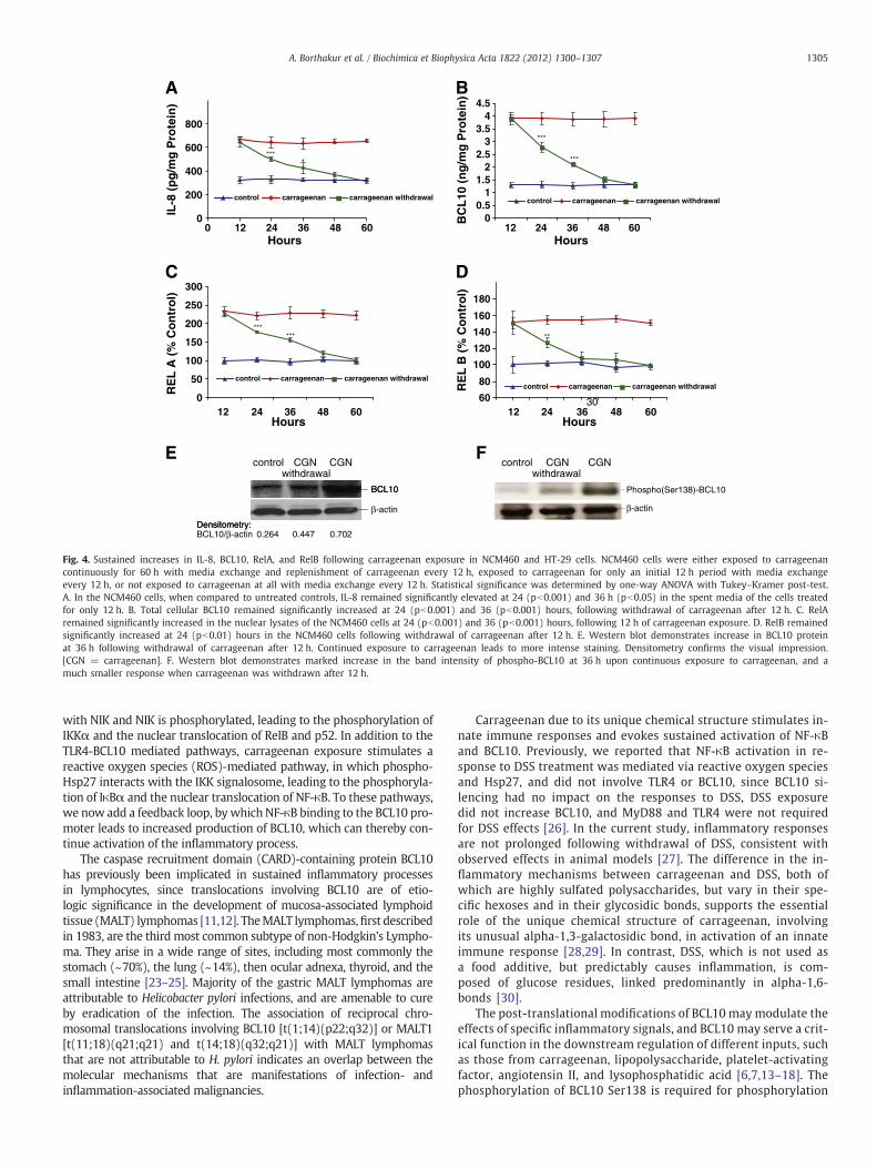
Since the function of the NF-κB-BCL10 loop was anticipated tolead to a prolonged inflammatory response following carrageenanexposure, the duration of the effects of carrageenan stimulationwas tested following carrageenan withdrawal. In the NCM460 cells,IL-8 secretion in the spent media (Fig. 4A), total cellular BCL10 pro-tein content (Fig. 4B), and nuclear RelA (Fig. 4C) remained signifi-cantly elevated compared to control for 24 h after carrageenanwithdrawal. Nuclear RelB (Fig. 4D) remained significantly elevatedfor 12 h after carrageenan withdrawal. A sustained increase in BCL10was also demonstrated by Western blot at 36 h which indicates signif-icantly increased BCL10 protein, compared to untreated control(Fig. 4E), although less than with continuous carrageenan treatment.Phospho(Ser138)-BCL10 was markedly increased at 36 h following
continuous exposure to carrageenan, as demonstrated by Westernblot (Fig. 4F). When carrageenan was withdrawn after 12 h, the inten-sity of the band for phospho-BCL10 at 36 hwasmuch reduced. In HT-29cells, even larger sustained effects were observed followingwithdrawalof carrageenan thanwere evident in the NCM460 cells (SupplementaryFig. 1A–D).
3.4. DSS treatment did not lead to sustained NF-κB activation
Effects of carrageenan differed from those obtained following ex-posure to dextran sodium sulfate (DSS), the sulfated polysaccharidethat is very commonly used to cause inflammation in cell-based andanimal experiments. DSS (1 μg/ml×24 h) increased RelA, but notRelB in NCM460 cells (Fig. 5A). Withdrawal experiments usingDSS in the NCM460 and HT-29 cells showed that DSS stimulationdid not produce sustained increases in RelA or IL-8. Treatmentwith DSS (1 μg/ml) for 12 h significantly increased IL-8 secretion(Fig. 5B) and nuclear RelA (Fig. 5C), compared to control. However,upon DSS withdrawal, IL-8 and RelA levels declined to the controllevels by 24 h, indicating that the inflammatory response was notsustained. RelB did not increase following exposure to DSS in theNCM460 cells (Fig. 5D). Similar lack of sustained increases in IL-8and RelA was demonstrated in the HT-29 cells following exposureto DSS (Supplementary Fig. 2A–B).

A
B
C
0
40
80
120
160
200
240
280
Rel
A B
ind
ing
(% u
ntr
eate
d t
o c
on
sen
sus)
no carrageenan
carrageenan
NF NFE NFEM
NF NFE NFEM
NF NFE NFEM
0
20
40
60
80
100
120
140
160
180
Rel
B B
ind
ing
(%
un
trea
ted
to
co
nse
nsu
s)
no carrageenan
carrageenan
0
20
40
60
80
100
120
c-R
el B
ind
ing
(%
un
trea
ted
co
nse
nsu
s)
no carrageenan
carrageenan
***
***
***
***
D
E
1 2 3 4 5 6 Lane + + + + + Nuclear extract
+ + + + carrageenan + + + + + + NFE labeled
+ RelA antibody + p50 antibody
+ IgG control
unbound oligonucleotide
protein-oligonucleotidecomplex
1 2 3 4 5 6 7 Lane + + + + + + + NFE labeled
+ NFE unlabeled
+ NFEM unlabeled
+ + + + + + Nuclear extract
+ RelA antibody + P50 antibody
+ carrageenan
protein-oligonucleotidecomplex
unbound oligonucleotide
RelA IgG Cn CGN
RelB IgG Cn CGN
F G
Fig. 3. Oligonucleotide-protein assays demonstrate binding to the NF-κB binding element in the BCL10 promoter. The double-stranded annealed nucleotides corresponding tothe established NF-κB consensus sequence (NF), the putative, experimental NF-κB binding element in the BCL10 promoter (NFE), or its mutated version (NFEM) were coatedonto the wells of microtiter plates. A. In the NCM460 cells, binding of RelA to the NF-κB consensus sequence (NF) and NF-κB binding element in the BCL10 promoter (NFE)was increased following exposure to carrageenan, whereas binding to the mutated sequence (NFEM) was unaffected. B. Similarly, binding of RelB to NF and NFE was increasedafter carrageenan, but binding to NFEM was unaffected. C. No increase in binding of c-Rel to either NF, NFE or NFEM was detected following carrageenan. D. Digoxigenin (DIG)-non-radioactive gel shift assay was performed with untreated NCM460 cell nuclear extract and following exposure of the cells to carrageenan. Lane 1: No nuclear extract; Lane2: nuclear extract and labeled NFE; Lane 3: carrageenan-treated nuclear extract and NFE; Lane 4: carrageenan-treated nuclear extract, NFE and RelA antibody; Lane 5;carrageenan-treated nuclear extract, NFE, and p50 antibody, and Lane 6: carrageenan-treated nuclear extract, NFE, and IgG control. Studies demonstrate increase in theoligonucleotide-protein band following carrageenan (Lane 3 vs. Lane 2). Exogenous RelA and p50 antibodies compete for binding between the endogenous RelA (Lane 4)or p50 (Lane 5) in the nuclear extract and the oligonucleotide binding site in the BCL10 promoter (NFE), eliminating the protein-oligonucleotide band. In contrast, IgGhas no effect on the binding of the nuclear extract and the labeled NFE (Lane 6). E. Digoxigenin (DIG)-non-radioactive gel shift assay showed carrageenan enhancementof RelA binding to the NF-κB binding element in the BCL10 promoter. Competition studies with cold (unlabeled) NFE or cold (unlabeled) and mutated NFEM or with RelAand p50 antibodies were performed to confirm the specificity of the protein-DNA interaction between RelA or p50 with the NF-κB binding site in the BCL10 promoter. Lane1: No nuclear extract; Lane 2: control nuclear extract and labeled NFE; Lane 3. carrageenan-treated nuclear extract and NFE; Lane 4: competition by cold NFE; Lane 5: compe-tition by cold NFEM; Lane 6: competition by RelA antibody; Lane 7: competition by p50 antibody. A representative blot of 4 independent experiments with similar results isshown. F, G. Increased binding of RelA and RelB to the BCL10 promoter by chromatin immunoprecipitation (ChIP) assay following carrageenan exposure. Control or carrageenan(1 μg/ml×24 h) treated NCM460 cells were fixed with formaldehyde, and chromatin prepared from these cells was sheared, and then immunoprecipitated with anti-RelA, anti-RelB antibody or with control IgG. Specific primers were used to amplify the promoter region containing the NF-κB binding element in the human BCL10 promoter. Amplifiedproducts were resolved on 1.5% agarose gel and indicate marked increase following carrageenan vs. control. [Cn = control; CGN = lambda-carrageenan; NF = NF-κB consensussequence; NFE = experimental NF-κB binding element in the BCL10 promoter; NFEM = mutated NF-κB binding element in the BCL10 promoter].
1304 A. Borthakur et al. / Biochimica et Biophysica Acta 1822 (2012) 1300–1307
4. Discussion
Previous reports have presented three pathways by which expo-sure to the common food additive carrageenan stimulates inflamma-tion in human colonic epithelial cells (Fig. 1) [4–8]. These includepathways initiated by interaction of carrageenan with the toll-like
receptor (TLR)-4, requiring MyD88, IRAK, and BCL10. The canonicalpathway of NF-κB activation involves BCL10 interaction with IKKγ,the regulatory subunit of the IKK signalosome, leading to IKKγubiquitination, IKKβ phosphorylation of IκBα, and nuclear transloca-tion of NF-κB RelA (p65) and p50. Alternatively, as in the non-canonical pathway of NF-κB activation, phospho-BCL10 interacts
0
200
400
600
800
0 12 24 36 48 60
12 24 36 48 60
IL-8
(p
g/m
g P
rote
in)
****
0
50
100
150
200
250
300
RE
L A
(%
Co
ntr
ol)
00.5
11.5
22.5
33.5
44.5
12 24 36 48 60
12 24 36 48 60
BC
L10
(n
g/m
g P
rote
in)
60
80
100
120
140
160
180
RE
L B
(%
Co
ntr
ol)
*** *** **
***
***
control carrageenan carrageenan withdrawalcontrol carrageenan carrageenan withdrawal
Hours
control carrageenan carrageenan withdrawal
Hours
control carrageenan carrageenan withdrawal
A
Hours
B
C D
30
Hours
control CGN CGNwithdrawal
BCL10
Densitometry:BCL10/β-actin 0.264 0.447 0.702
CGN CGNcontrol
BCL10
β-actin β-actin
Densitometry:
E
Phospho(Ser138)-BCL10
withdrawal
F
Fig. 4. Sustained increases in IL-8, BCL10, RelA, and RelB following carrageenan exposure in NCM460 and HT-29 cells. NCM460 cells were either exposed to carrageenancontinuously for 60 h with media exchange and replenishment of carrageenan every 12 h, exposed to carrageenan for only an initial 12 h period with media exchangeevery 12 h, or not exposed to carrageenan at all with media exchange every 12 h. Statistical significance was determined by one-way ANOVA with Tukey–Kramer post-test.A. In the NCM460 cells, when compared to untreated controls, IL-8 remained significantly elevated at 24 (pb0.001) and 36 h (pb0.05) in the spent media of the cells treatedfor only 12 h. B. Total cellular BCL10 remained significantly increased at 24 (pb0.001) and 36 (pb0.001) hours, following withdrawal of carrageenan after 12 h. C. RelAremained significantly increased in the nuclear lysates of the NCM460 cells at 24 (pb0.001) and 36 (pb0.001) hours, following 12 h of carrageenan exposure. D. RelB remainedsignificantly increased at 24 (pb0.01) hours in the NCM460 cells following withdrawal of carrageenan after 12 h. E. Western blot demonstrates increase in BCL10 proteinat 36 h following withdrawal of carrageenan after 12 h. Continued exposure to carrageenan leads to more intense staining. Densitometry confirms the visual impression.[CGN = carrageenan]. F. Western blot demonstrates marked increase in the band intensity of phospho-BCL10 at 36 h upon continuous exposure to carrageenan, and amuch smaller response when carrageenan was withdrawn after 12 h.
1305A. Borthakur et al. / Biochimica et Biophysica Acta 1822 (2012) 1300–1307
with NIK and NIK is phosphorylated, leading to the phosphorylation ofIKKα and the nuclear translocation of RelB and p52. In addition to theTLR4-BCL10 mediated pathways, carrageenan exposure stimulates areactive oxygen species (ROS)-mediated pathway, in which phospho-Hsp27 interacts with the IKK signalosome, leading to the phosphoryla-tion of IκBα and the nuclear translocation of NF-κB. To these pathways,we now add a feedback loop, by which NF-κB binding to the BCL10 pro-moter leads to increased production of BCL10, which can thereby con-tinue activation of the inflammatory process.
The caspase recruitment domain (CARD)-containing protein BCL10has previously been implicated in sustained inflammatory processesin lymphocytes, since translocations involving BCL10 are of etio-logic significance in the development of mucosa-associated lymphoidtissue (MALT) lymphomas [11,12]. TheMALT lymphomas,first describedin 1983, are the third most common subtype of non-Hodgkin's Lympho-ma. They arise in a wide range of sites, including most commonly thestomach (~70%), the lung (~14%), then ocular adnexa, thyroid, and thesmall intestine [23–25]. Majority of the gastric MALT lymphomas areattributable to Helicobacter pylori infections, and are amenable to cureby eradication of the infection. The association of reciprocal chro-mosomal translocations involving BCL10 [t(1;14)(p22;q32)] or MALT1[t(11;18)(q21;q21) and t(14;18)(q32;q21)] with MALT lymphomasthat are not attributable to H. pylori indicates an overlap between themolecular mechanisms that are manifestations of infection- andinflammation-associated malignancies.
Carrageenan due to its unique chemical structure stimulates in-nate immune responses and evokes sustained activation of NF-κBand BCL10. Previously, we reported that NF-κB activation in re-sponse to DSS treatment was mediated via reactive oxygen speciesand Hsp27, and did not involve TLR4 or BCL10, since BCL10 si-lencing had no impact on the responses to DSS, DSS exposuredid not increase BCL10, and MyD88 and TLR4 were not requiredfor DSS effects [26]. In the current study, inflammatory responsesare not prolonged following withdrawal of DSS, consistent withobserved effects in animal models [27]. The difference in the in-flammatory mechanisms between carrageenan and DSS, both ofwhich are highly sulfated polysaccharides, but vary in their spe-cific hexoses and in their glycosidic bonds, supports the essentialrole of the unique chemical structure of carrageenan, involvingits unusual alpha-1,3-galactosidic bond, in activation of an innateimmune response [28,29]. In contrast, DSS, which is not used asa food additive, but predictably causes inflammation, is com-posed of glucose residues, linked predominantly in alpha-1,6-bonds [30].
The post-translational modifications of BCL10 may modulate theeffects of specific inflammatory signals, and BCL10 may serve a crit-ical function in the downstream regulation of different inputs, suchas those from carrageenan, lipopolysaccharide, platelet-activatingfactor, angiotensin II, and lysophosphatidic acid [6,7,13–18]. Thephosphorylation of BCL10 Ser138 is required for phosphorylation

control DSS DSS withdrawal
0
50
100
150
200
250
300
control DSS
Act
ivat
ed N
F-κ
B (
% c
on
tro
l)
RelA
RelB
Rel
A (
%co
ntr
ol)
0
200
400
600
800
1000
IL-8
(p
g/m
g p
rote
in)
A B control
12 24 36 48 60Hours
12 24 36 48 60Hours
12 24 36 48 60Hours
0
50
100
150
200
250
C
DSS DSS withdrawal
0
20
40
60
80
100
120
140
Rel
B (
% c
on
tro
l)
control DSS DSS withdrawalD
Fig. 5. DSS-induced increase in IL-8 and RelA (p65) are not sustained upon DSS withdrawal in NCM460 or HT-29 cells. A. DSS exposure produced no increase in RelB in the NCM460cells. NCM460 cells were initially exposed to DSS (1 μg/ml) for 12 h. This was followed by either replenishment with fresh DSS‐containing media every 12 h, or with media withoutDSS. Samples (spent media or cells) were harvested every 12 h. B. IL-8 declined to untreated level by 24 h in the NCM460 cells. C. RelA declined to untreated level by 24 h in theNCM460 cells. [DSS = dextran sulfate sodium]. D. RelB did not increase following exposure to DSS in the NCM460 cells.
1306 A. Borthakur et al. / Biochimica et Biophysica Acta 1822 (2012) 1300–1307
of NIK in the non-canonical pathway of NF-κB activation, andwas not affected by proteasomal inhibition by MG-132 or byCAPE, indicating that BCL10 phosphorylation occurs indepen-dently of NF-κB nuclear translocation, in contrast to BCL10 ex-pression [6,7,18,21]. The study finding that the RelB activationis sustained for a shorter interval than the RelA activation sug-gests that another process, likely involving the phosphorylationof NIK, BCL10, or IKKα, is also required to prolong the activationof the non-canonical pathway.
Exposure of cells to inflammatory stimuli results in rapid phos-phorylation and degradation of IκBα and subsequent translocationof NF-κB to the nucleus. These events enable prompt cellular re-sponse in the absence of de novo protein synthesis [31,32]. Giventhe central role of NF-κB in the pathogenesis and maintenance ofchronic inflammatory states, examination of mechanisms that con-tribute to the prolonged activation of NF-κB is of considerable rel-evance to human disease. In the current report, carrageenanexposure for 12 h caused sustained RelA activation, IL-8 produc-tion, and BCL10 protein level for 48 h. Since up‐regulation ofBCL10 expression results from NF-κB activation, and NF-κB activa-tion following carrageenan exposure involves BCL10, a positivefeedback loop involving BCL10 and NF-κB that causes prolongedactivation of NF-κB in response to carrageenan may have clinicalsignificance, leading to extended inflammation in intestinal epi-thelial cells. The relationship between infectious or inflammatoryprocesses and malignancy, such as H. pylori and gastric cancer,human papillomavirus and cervical cancer, or ulcerative colitisand colorectal carcinoma, is also of interest, since therapeutic in-terventions with anti-microbial and anti-inflammatory agentsthat retard inflammation may impact upon neoplastic transforma-tion. Identification of specific transcriptional regulatory mecha-nisms, such as this NF-κB-BCL10 loop that is set in motion by
carrageenan exposure, may contribute to improved understandingof the origins of malignancy, as well as of chronic inflammation.
Supplementary data to this article can be found online at http://dx.doi.org/10.1016/j.bbadis.2012.05.001.
Acknowledgements
Supported by the Department of Veterans Affairs and the NIDDK(R01-DK54016, R01-DK81858 and PO1-DK67887).
References
[1] J.K. Tobacman, Environ. Health Perspect. 109 (2001) 983–994.[2] Encyclopædia Britannica, “algae” Encyclopædia Britannica Online, Encyclopædia
Britannica, 2010. Web. 22 Jan 2011, http://search.eb.com/eb.article-31714.[3] J. West, K.N. Miller, California's living marine resources: a status report.
Agarophytes and carrageenophytes, California Department of Fish and Game,2001, pp. 286–287.
[4] A. Borthakur, S. Bhattacharyya, P.K. Dudeja, J.K. Tobacman, Am. J. Physiol. Gas-trointest. Liver Physiol. 292 (2007) G829–G838.
[5] S. Bhattacharyya, R. Gill, M.L. Chen, F. Zhang, R.J. Linhardt, P.K. Dudeja, J.K.Tobacman, J. Biol. Chem. 283 (2008) 10550–10558.
[6] S. Bhattacharyya, A. Borthakur, A.N. Anbazhagan, S. Katyal, P.K. Dudeja, J.K.Tobacman, Am. J. Physiol. Gastrointest. Liver Physiol. 301 (2011) G475–G486.
[7] S. Bhattacharyya, A. Borthakur, S. Tyagi, R. Gill, M.L. Chen, P.K. Dudeja, J.K.Tobacman, J. Biol. Chem. 285 (2010) 522–530.
[8] S. Bhattacharyya, P.K. Dudeja, J.K. Tobacman, Biochim. Biophys. Acta 1780 (2008)973–982.
[9] S. Bhattacharyya, I. O-Sullivan, S. Katyal, T. Unterman, J.K. Tobacman, Diabetologia55 (2012) 194–203.
[10] H. Shi, M.V. Kokoeva, K. Inouye, I. Tzameli, H. Yin, J.S. Flier, J. Clin. Invest. 116(2006) 3015–3025.
[11] T.G. Willis, D.M. Jadayel, M.Q. Du, H. Peng, A.R. Perry, M. Abdul-Rauf, H. Price, L.Karran, O. Majekodunmi, I. Wlodarska, L. Pan, T. Crook, R. Hamoudi, P.G.Isaacson, J.F. Dyer, Cell 96 (1999) 35–45.
[12] Q. Zhang, R. Siebert, M. Yan, B. Hinzmann, X. Cui, L. Xue, K.M. Rakestraw, C.W.Naeve, G. Beckmann, D.D. Weisenburger, W.G. Sanger, H. Nowotny, M. Vesely,

1307A. Borthakur et al. / Biochimica et Biophysica Acta 1822 (2012) 1300–1307
E. Callet-Bauchu, G. Salles, V.M. Dixit, A. Rosenthal, B. Schlegelberger, S.W. Morris,Nat. Genet. 22 (1999) 63–68.
[13] S. Bhattacharyya, A. Borthakur, N. Pant, P.K. Dudeja, J.K. Tobacman, Am. J. Physiol.Gastrointest. Liver Physiol. 293 (2007) G429–G437.
[14] D. Wang, Y. You, P.C. Lin, L. Xue, S.W. Morris, H. Zeng, R. Wen, X. Lin, Proc. Natl.Acad. Sci. U. S. A. 104 (2007) 145–150.
[15] L.M. McAllister-Lucas, J. Ruland, K. Siu, X. Jin, S. Gu, D.S. Kim, P. Kuffa, D. Kohrt,T.W. Mak, G. Nuñez, P.C. Lucas, Proc. Natl. Acad. Sci. U. S. A. 104 (2007) 139–144.
[16] S. Klemm, S. Zimmermann, C. Peschel, T.W. Mak, J. Ruland, Proc. Natl. Acad. Sci.U. S. A. 104 (2007) 134–138.
[17] A. Borthakur, S. Bhattacharyya, W. Alrefai, J.K. Tobacman, K. Ramaswamy, P.K.Dudeja, Inflamm. Bowel Dis. 16 (2010) 594–603.
[18] S. Bhattacharyya, A. Borthakur, P.K. Dudeja, J.K. Tobacman, Exp. Cell Res. 316(2010) 3317–3327.
[19] M.P. Moyer, L.A. Manzano, R.L. Merriman, J.S. Stauffer, L.R. Tanzer, In Vitro Cell.Dev. Biol. Anim. 32 (1996) 315–317.
[20] S. Bhattacharyya, N. Pant, P.K. Dudeja, J.K. Tobacman, J. Immunoassay Immuno-chem. 28 (2007) 173–188.
[21] S. Bhattacharyya, P.K. Dudeja, J.K. Tobacman, Am. J. Physiol. Gastrointest. LiverPhysiol. 293 (2) (2007) G429–G437.
[22] A. Zafiropoulos, G. Hatzidakis, L. Mavrogiannis, A. Klinakis, M. Kandilogiannaki, E.Krambovitis, Biotechniques 23 (1997) 1104–1109.
[23] P. Isaacson, D.H. Wright, Cancer 52 (1983) 1410–1416.[24] X. Sagaert, E. Van Cutsem, G. De Hertogh, K. Geboes, T. Tousseyn, Nat. Rev.
Gastroenterol. Hepatol. 7 (2010) 336–346.[25] http://www.cancer.gov/cancertopics/factsheet/Risk/h-pylori-cancer.[26] S. Bhattacharyya, P.K. Dudeja, J.K. Tobacman, Inflamm. Bowel Dis. 15 (2009)
673–683.[27] Y. Yan, V. Kolachala, G. Dalmasso, H. Nguyen, H. Laroui, S.V. Sitaraman, D. Merlin,
PLoS One 4 (2009) e6073.[28] S. Bhattacharyya, H. Liu, Z. Zhang, M. Jam, P.K. Dudeja, G. Michel, R.J. Linhardt, J.K.
Tobacman, J. Nutr. Biochem. 21 (2009) 906–913.[29] B.A. Macher, U. Galili, Biochim. Biophys. Acta 1780 (2008) 75–88.[30] http://www.gelifesciences.com/aptrix/upp00919.nsf/Content/A7BB06EF3C4EEE8EC12
57628001CEC60/$file/18115175AA.pdf.[31] A. Bierhaus, S. Schiekofer, M. Schwaninger, M. Andrassy, P.M. Humpert, J. Chen,
M. Hong, T. Luther, T. Henle, I. Klöting, M. Morcos, M. Hofmann, H. Tritschler, B.Weigle, M. Kasper, M. Smith, G. Perry, A.M. Schmidt, D.M. Stern, H.U. Häring, E.Schleicher, P.P. Nawroth, Diabetes 50 (2001) 2792–2808.
[32] P.J. Barnes, M. Karin, N. Engl. J. Med. 336 (1997) 1066–1071.


