Embed Size (px)
Citation preview

RESEARCH Open Access
Prolonged control of replication-competentdual- tropic human immunodeficiency virus-1following cessation of highly activeantiretroviral therapyMaria Salgado1, S Alireza Rabi1, Karen A O’Connell1, Robert W Buckheit III1, Justin R Bailey1, Amina A Chaudhry1,Autumn R Breaud2, Mark A Marzinke2, William Clarke2, Joseph B Margolick3, Robert F Siliciano1,4 andJoel N Blankson1*
Abstract
Background: While initiation of highly active antiretroviral therapy (HAART) during primary HIV-1 infectionoccasionally results in transient control of viral replication after treatment interruption, the vast majority of patientseventually experience a rebound in plasma viremia.
Results: Here we report a case of a patient who was started on HAART during symptomatic primary infection andwho has subsequently maintained viral loads of < 50 copies/mL for more than nine years after the cessation oftreatment. This patient had a high baseline viral load and has maintained a relatively high frequency of latentlyinfected CD4+ T cells. In addition, he does not have any known protective HLA alleles. Thus it is unlikely that hewas destined to become a natural elite controller or suppressor. The mechanism of control of viral replication isunclear; he is infected with a CCR5/CXCR4 dual-tropic virus that is fully replication-competent in vitro. In addition,his spouse, who transmitted the virus to him, developed AIDS. The patient’s CD4+ T cells are fully susceptible toHIV-1 infection, and he has low titers of neutralizing antibodies to heterologous and autologous HIV-1 isolates.Furthermore, his CD8+ T cells do not have potent HIV suppressive activity.
Conclusion: This report suggests that some patients may be capable of controlling pathogenic HIV-1 isolates forextended periods of time after the cessation of HAART through a mechanism that is distinct from the potentcytotoxic T lymphocyte (CTL) mediated suppression that has been reported in many elite suppressors.
Keywords: HIV-1, elite suppressor, elite controller, viral replication
BackgroundHIV-1 infection results in extensive viral replication andprogressive CD4+ T cell depletion in the vast majorityof patients. However, rare subjects, known as elite con-trollers or suppressors (ES), spontaneously control viralreplication without antiretroviral treatment [1]. Themechanisms involved in elite control are not fullyunderstood, but some ES appear to be infected withfully replication-competent virus [2-5] that continues to
evolve during chronic infection [6-8]. Thus infectionwith attenuated virus does not appear to be a commoncause of elite control. In contrast, many studies lookingat host factors have shown that the HLA-B*27 and 57alleles are overrepresented in ES [9-14]. This hasstrongly suggested a role for CD8+ T cell responses inelite control, and indeed, potent HIV-specific CD8+ Tcell responses [15-17] that are capable of inhibiting viralreplication [18,19] have been documented in many ES.It is not clear whether it will be possible to elicit simi-
lar levels of immune control in patients with progressiveHIV-1 disease. However, some studies have suggestedthat rare individuals who are treated early in primary
* Correspondence: [email protected] of Medicine, Johns Hopkins University School of Medicine,Baltimore, Maryland 21287, USAFull list of author information is available at the end of the article
Salgado et al. Retrovirology 2011, 8:97http://www.retrovirology.com/content/8/1/97
© 2011 Salgado et al; licensee BioMed Central Ltd. This is an Open Access article distributed under the terms of the Creative CommonsAttribution License (http://creativecommons.org/licenses/by/2.0), which permits unrestricted use, distribution, and reproduction inany medium, provided the original work is properly cited.

infection with highly active antiretroviral therapy(HAART) are able to control viral replication whentherapy is discontinued. Rosenberg and colleaguesdemonstrated that five of eight patients who were trea-ted before or shortly after seroconversion were able tosuppress HIV RNA levels to below 500 copies/mL for amedian of 6.5 months after therapy was interrupted[20]. However, a follow up study showed that this con-trol was of limited duration as only three of 14 patientswho started HAART during primary infection main-tained viral loads of < 5000 copies/mL two years aftertreatment interruption [21]. In another study, a patientwho was started on HAART a month after seroconver-sion was treated for four years prior to a treatmentinterruption which resulted in a rapid rebound in vire-mia. HAART was reinitiated and ultra-low doses ofinterleukin-2 (1.2 mIU/m2/day) were added to the regi-men. Interestingly, he maintained viral loads of < 50copies/mL for 14 months after both HAART and IL-2were discontinued [22]. In a recent study, five of thirty-two patients treated during primary HIV-1 infectionmaintained control of viral replication for more than sixmonths after treatment was interrupted [23]. While thisphenomenon is not routinely seen with early treatment[24-26], these cases strongly suggest that the immunesystem can be manipulated to control HIV-1 replicationin some patients. Thus, this could be the basis for thedesign of a successful therapeutic vaccine.We present a case of a patient infected with a replica-
tion-competent, dual-tropic HIV-1 isolate who wasstarted on treatment during primary infection. He hasmaintained stable CD4+ T cell counts and viral loads of< 50 copies/ml for more than nine years since HAARTwas discontinued. To our knowledge, this represents thelongest period of control of HIV-1 replication in apatient after the cessation of treatment. We performeddetailed analyses of the patient’s viral isolates and lookedat multiple aspects of his HIV-specific immuneresponse. While no clear mechanism of immune controlwas identified, this case suggests that long term controlof pathogenic HIV-1 isolates is possible in some patientswho were destined to become chronic progressors (CP).
ResultsPatientsPatient 169 is a 57 year old male who was diagnosedwith primary HIV-1 infection when he admitted to theintensive care unit at Johns Hopkins Hospital in 1999with severe HIV-1 meningoencephalitis that resulted inintubation for airway protection [27]. He was found tohave an indeterminate Western blot (only bands to p24were present) and an HIV-1 viral load of > 750,000copies/mL. He reported having tested negative for HIV-1 two years prior to admission. He was enrolled into the
Acute Infection and Early Disease Research Program(AIEDRP) study and started on a regimen of zidovudine,lamivudine, and indinavir within 48 hours of admission.This regimen was changed to abacavir, lamivudine andefavirenz at week four and by week 16, his viral loadwas < 50 copies/ml. He stopped taking his antiretrovir-als at week 36 for a 2 week period, and his viral loadrebounded to 22,000 copies/ml. The same regimen wasre-initiated, and he was adherent until week 92 at whichtime he stopped taking all of the antiretroviral drugs.His CD4+ T cell count, which was 412 cell/μL at thetime of diagnosis, has been stable at greater than 1000cells/μL over the last five years, and his viral load, whichhas been consistently less than 50 copies/ml since thediscontinuation of HAART, was measured at 1 copy/mLin 2011 using a highly sensitive single copy assay [28,29](Figure 1). The patient was incarcerated between 2004and 2005, and medical records confirmed that he wasnot on antiretroviral therapy at this point. Furthermore,qualitative testing for antiretroviral drugs on plasmasamples from 2009, 2010 and 2011 was performed torule out surreptitious use of antiretroviral therapy. Allsamples were negative whereas nevirapine and lamivu-dine were detected in a plasma sample from his spouse.The patient’s spouse was diagnosed with HIV-1 infec-
tion 3 years before subject 169 was admitted with acuteretroviral syndrome. Her CD4+ T cell count nadir was84 cells/μL, and her baseline viral load prior to theinitiation of HAART was 122,000 copies/ml.
Patient 169 has a high frequency of HIV-1infected CD4+ T cellsIn order to determine whether the patient was infectedwith a defective virus and whether his spouse trans-mitted the virus to him, we amplified virus from aplasma sample from the time of diagnosis. In addition,virus was cultured from CD4+ T cells isolated fromPBMCs obtained from the patient and his spouse in2010. The frequency of latently infected resting CD4+ Tcells in patient 169 was 1.61 infectious units per million,which is more than a log higher than the frequencyfound in our cohort of ES [3] and similar to the fre-quencies found in chronic progressors on suppressiveHAART regimens [30,31].
Patient 169 is infected with fully replication-competent,dual-tropic virusWe next analyzed the fitness of isolates obtained frompatient 169 and his spouse. For patient 169, full genomesequencing of replication-competent virus cultured from1999 plasma and three independent replication-compe-tent isolates obtained from CD4+ T cells in 2010 wasperformed. One of the isolates from 2010 (2B) was iden-tical to the 1999 isolate with the exception of a single
Salgado et al. Retrovirology 2011, 8:97http://www.retrovirology.com/content/8/1/97
Page 2 of 14

nucleotide difference in the HIV-1 LTR. The two otherisolates from 2010 (1A, 1B) were identical although theywere isolated from independent culture wells. The dif-ferences between the identical 2010 isolates and the1999 isolate are summarized in Table 1. For thepatient’s spouse, full genome sequencing of two inde-pendent isolates cultured from her resting CD4+ T cellsin 2010 was performed. No large deletions were foundin any gene and no drug resistance mutations werefound in any of the isolates obtained from either patient.Phylogenetic analysis of the env gene showed that theisolates from 169 and his spouse were more closelyrelated to each other than to any other isolate in theLos Alamos database, confirming that the two patientswere a transmission pair [Figure 2].
Sequence analysis of the env gene suggested that allisolates cultured from both patients were CXCR4- tropic(data not shown). To confirm this, we amplified andcloned the env gene from the 1999 and 2010 isolatesfrom patient 169, and made GFP-expressing NL4-3pseudotyped virus as previously described [32]. Infectionstudies were then performed with GHOST cells expres-sing CCR5 and/or CXCR4. As shown in Figure 3A,pseudotyped virus containing env from 1999 and 2010was able to infect GHOST cells expressing either co-receptor, demonstrating that each viral clone was dual-tropic (Figure 3A).We next compared the replication capacity of virus cul-
tured from patient 169 to that of CCR5-tropic (Ba-L) andCXCR4-tropic (IIIB) laboratory isolates. As shown in Fig-ure 3B, the isolates from 1999 and 2010 replicated as wellas IIIB in MT-2 cells whereas Ba-L did not replicate inthese cells, which do not express the CCR5 co-receptor. Inprimary CD4+ T cells, the two isolates from Patient 169replicated as well as Ba-L. Thus control of viral replicationin this patient was not due to infection with an attenuatedvirus, and viral fitness was stable over time.
Patient 169 does not have known genetic factors thatcontribute to the control of viral replicationHaving ruled out viral attenuation, we focused on hostfactors as potential causes of the observed virologic con-trol. Heterozygosity for the 32 base pair deletion inCCR5 has been associated with slow HIV-1 progression[33,34]. This gene was thus analyzed by PCR, andpatient 169 was determined to have two wild type CCR5alleles. The most consistent finding in different cohortsof ES has been the over-representation of protective
Year
0
1
2
3
4
5
6
7
0
200
400
600
800
1000
1200
1400
1600
1999 2000 2001 2002 2003 2004 2005 2006 2007 2008 2009 2010 2011 2012
Log VL (copies/m
L)CD
4+ T
cel
l cou
nts
(cel
ls/u
l)
Time on HAART
Isolated virus
Figure 1 Clinical characteristics of Patient 169. The patient’s CD4 counts and viral load are shown. Viral load measurements below the limitof detection are denoted by open symbols. The time on HAART is denoted by the shaded region.
Table 1 Differences in sequence of replication-competent1999 and 2010 isolates.
Differences between Pt-169 1999 and 2010-1A/1B isolates
Nucleotides Amino Acids
LTR 2
Gag Δ18* Δ6*
Pol 2 2
Vif 0 0
Vpr 1 1
Vpu 0 0
Env 2, Δ21 (V4)* 1, Δ7 (V4)*
Nef 1 1
Total 47 18
Isolate 2B from 2010 is identical to the 1999 isolate with the exception of asingle nucleotide in the LTR. Isolates 1A and 1B are identical to each other.
* The triangle denotes deletions in the 2010-1A/1B isolates.
Salgado et al. Retrovirology 2011, 8:97http://www.retrovirology.com/content/8/1/97
Page 3 of 14

HLA alleles such as HLA-B*27 and B*57 [9-14,35,36]. Inaddition, genome wide association studies have docu-mented a protective single nucleotide polymorphism
(SNP) in the HLA-C promoter [35,36]. Furthermore,HLA-Bw4-80Ile alleles have been shown to be associatedwith slowly progressive disease when inherited in
V1 V2
V3
V4
V5
* 150 * 170 * 190 * 210 * 230 CONSENSUS B Env: CTDLMNATNTNTTIIYRW------------RGEIKNCSFNITTSIRDKVQKEYALFYKLDVVPIDNDNTSYRLISCNTSVITQACPKVSFEPIPIHYCAPAGFA : 92Pt169-1999 : ....R.V...TNNTNGNATSATNSSRGMMEGV.M..........M-...R..H...NS...I.MEG..............V......I................ : 103Pt169-2010-2B : ....R.V...TNNTNGNATSATNSSRGMMEGV.M..........M-...R..H...NS...I.MEG..............V......I................ : 103Pt169-2010-1A/B: ....R.V...TNNTNGNATSATNSSRGMMEGV.M..........M-...R..H...NS...I.MEG..............V......I................ : 103Spouse-2010-4A : ....R.....TN---GNATSATNSSRGMMEGV.M..........M-...R..H...NS...I.MEG..............V......I................ : 100 * 250 * 270 * 290 * 310 * 330 CONSENSUS B Env: ILKCNDKKFNGTGPCTNVSTVQCTHGIRPVVSTQLLLNGSLAEEEVVIRSENFTDNAKTIIVQLNESVEINCTRPNNNTRKSIHIGPGRAFYTTGEIIGDIRQA : 196Pt169-1999 : ................S..........K................G.........N...I......Q....H.L...KY..QR...........ARD......K. : 207Pt169-2010-2B : ................S..........K................G.........N...I......Q....H.L...KY..QR...........ARD......K. : 207Pt169-2010-1A/B: ................S..........K................G.........N...I......Q....H.L...KY..QR...........ARD......K. : 207Spouse-2010-4A : ......N.........S..........K...............K..........N...I......Q....H.I...KY..QR.......S...AKNT.....K. : 204 * 350 * 370 * 390 * 410 * 430 * CONSENSUS B Env: HCNISRAKWNNTLKQIVKKLREQFGNKTIVFNQSSGGDPEIVMHSFNCGGEFFYCNTTQLFNSTWNGTW------NNTEGNITLPCRIKQIINMWQEVGKAMYA : 294Pt169-1999 : Y....KQN.T...Y.VS.......S....I...........................AG.......S..SLNDTKR.GNE................G....... : 311Pt169-2010-2B : Y....KQN.T...Y.VS.......S....I...........................AG.......S..SLNDTKR.GNE................G....... : 311Pt169-2010-1A/B: Y....KQN.T...Y.VS.......S....I...........................AG.......S..S-------GNE................G....... : 304Spouse-2010-4A : Y...NKQN.T...Y.VSN......-....I...........................AG.......S.RSLNDTKR.GNE................G....... : 307 450 * 470 CONSENSUS B Env : PPIRGQIRCSSNITGLLLTRDGGN-NET---EIFRP : 326 Pt169-1999 : ...S....................G.I.NTT.T... : 347 Pt169-2010-2B : ...S....................G.I.NTT.T... : 347 Pt169-2010-1A/B: ...S....................G.I.NTT.T... : 340 Spouse-2010-4A : ...E....................G.T.NTN.T... : 343
A)
B) Consensus Clade C
B.gi|3256384
B.gi|38477|g
B.JP.1989.
B.JP.1994.
B.JP.1995.
B.IT.14
B.JP.2004.
Pt169-1999
Pt169-2010-1A/1B
Spouse-2010-4A
B.gi|6253257
B.gi|4102285
B.FR.HXB2
B.FR.1985.
B.gi|4002173
B.gi|4002177
B.gi|4002186
B.gi|1465777
Consensus Clade B�����
Figure 2 Phylogenetic Analysis: An alignment of the variable regions of env is shown for replication-competent isolates obtainedfrom Patient 169 and his spouse. Numbering is from the first amino acid in gp120. (A). The sequences are also compared to other Clade Bsequences(B). Phylogenies were estimated by using a classical approach, functioning under a maximum-likelihood (ML) optimality criterion.
Salgado et al. Retrovirology 2011, 8:97http://www.retrovirology.com/content/8/1/97
Page 4 of 14

conjunction with the KIR3DS1 and/or KIR3DL1 naturalkiller cell receptor alleles [37,38]. Patient 169 does nothave an HLA-Bw4-80Ile allele or any other HLA allelethat has been previously associated with attenuatedHIV-1 disease. He also does not have the protective C/C HLA-C SNP (Table 2).
CD4+ T cells from Patient 169 are fully susceptible toinfectionSome studies [39,40] have suggested that ES CD4+ T cellsthat have been activated ex vivo are resistant to viral infec-tion while others have shown that unstimulated CD4+ Tcells from these patients are fully susceptible to viral entry
Pt169-1999Pt169-2010Ba-L IIIB
-1A
0.1
1
10
100
1000
10000
0 1 2 3 4 5 6 7
P24
[ng/
ml]
Days post infection
B)CD4+ T Cells
A)
0
5
10
15
20
25
30
35
GHOST CCR5 GHOST CXCR4 GHOST CCR5/CXCR4
% In
fect
ion
Uninfected
Ba-L
IIIB
Pt169-1999
Pt169-2010-1A
Pt169-1999Pt169-2010Ba-L IIIB
-1A
0.1
1
10
100
1000
10000
0 1 2 3 4 5 6 7
P24
[ng/
ml]
MT- 2 cells
Days post infectionFigure 3 Phenotypic analyses of viral isolates from Patient 169. (A) Viral tropism was determined using GHOST cells expressing CCR5 and/orCXCR4. Open bars were below the limit of the detection (B) Replication kinetics was determined in primary CD4+ T cells (left) and the MT-2 cellline (right).
Salgado et al. Retrovirology 2011, 8:97http://www.retrovirology.com/content/8/1/97
Page 5 of 14

and productive infection [41,42]. In order to determinewhether CD4+ T cells from patient 169 were resistant toinfection, we purified primary CD4+ T cells from 169 andfive HIV-1 seronegative donors and infected them directlyex vivo by spinoculation with CCR5 (Ba-L) and CXCR4(NL4-3) tropic isolates as previously described. As shownin Figure 4, CD4+ T cells from 169 were as susceptible toinfection with both types of isolates as were the CD4+ Tcells from the seronegative donors. In order to determineif spinoculation was masking subtle differences in the sus-ceptibility to infection, we infected CD4+ T cells withCXCR4-tropic virus without spinoculation [41] and againfound that CD4+ T cells from patient 169 were fully sus-ceptible to infection. We also looked at susceptibility toinfection with serial dilutions of both lab strains and apseudotyped virus containing dual tropic envelope thatwas amplified from the patient in 1999. In all cases, patient169’s cells were found to be as susceptible to infection ascells from HIV-negative donors.
Patient 169 has low titers of HIV-specific neutralizingantibodiesTo determine whether a robust humoral response wasplaying a role in the control of viral replication, we com-pared reciprocal IC50 titers of neutralizing antibodies(Nab) in patient 169 to titers in viremic patients and ESas previously described [43]. Patient 169 had the lowesttiters of Nab to laboratory strain SF162 Env as shown inFigure 5. To determine how well the patient neutralizedautologous virus, we measured titers of Nab to pseudo-typed virus expressing Env cloned from the 1999 and2010 replication-competent isolates. His reciprocal IC 50titers of Nab to autologous Env from 1999 was 1:195which is comparable to the titers seen in ES, but lowerthan the titers seen in viremic patients [43]. In contrast,his Nab titer to contemporaneous Env was >1: 4 (Figure5B). Thus it appears that neutralizing antibodies were notthe cause of control of viral replication in this patient.
Characteristics of the HIV-specific CD8+ T cell response inPatient 169Many ES have been found to possess potent HIV-speci-fic CD8+ T cell activity [16-19]. We thus looked at CD8
+ T cell responses in Patient 169. An ELISPOT assaywas performed following stimulation with Gag and Nefpeptides. As shown in Figure 6, two independent non-overlapping epitopes were targeted in Nef, and and sixsuch epitopes were targeted in Gag. Escape mutations incertain Gag epitopes have been associated with viralattenuation [44-46], and we therefore examinedsequences in targeted epitopes to look for signs of viro-logic escape. A comparison of the sequences from the1999 and 2010 isolates showed an R18G substitution ina Nef epitope and a G17W substitution in a Gag epitopein isolates 1A and 1B. Both substitutions were absent inisolate 2B, and thus even if these mutations caused areduction in viral fitness in some isolates, escape muta-tions would not explain virologic control in this patient.To determine whether CD8+ T cells were involved in
the direct control of viral replication, we attempted toculture autologous virus from Patient 169 with andwithout the depletion of CD8+ T cells. As shown in Fig-ure 7A, virus culture was successful only when CD8+ Tcells were depleted, but the same phenomenon was seenin chronic progressors who had substantial levels of vir-emia and in patients on suppressive HAART regimens.There was no viral outgrowth from ES CD4+ T cellsconsistent with the low frequency of infected CD4+ Tcells in these patients [3]. We next compared therespective abilities of CD8+ T cells from Patient 169 andES in HIV-1 inhibition assays in which pseudotypedvirus was used to superinfect autologous CD4+ T cells.As shown in Figure 7B, while primary CD8+ T cellsfrom most ES caused a significant reduction in virusreplication, CD8+ T cells from Patient 169 had very littleeffect in this assay.
DiscussionWe present here a patient who has controlled HIV-1replication for more than nine years after the cessationof HAART. While some studies have reported thatinitiation of HAART during primary infection can leadto the control of viral replication once therapy is discon-tinued, most of these patients eventually experienced arebound in viremia [21,22]. To our knowledge, the nineyears of control seen in patient 169 is the longest periodof control reported in a patient who was treated in pri-mary infection and who subsequently underwent treat-ment interruption. We extend prior studies byperforming full genome sequence analysis and phenoty-pic studies of viral isolates obtained at the time of infec-tion and eight years after the cessation of HAART. Weshow that Patient 169 was infected by virus from apatient with AIDS, and we demonstrate that the viralisolates from patient 169 are dual-tropic and replica-tion-competent, which makes it unlikely that an attenu-ated virus was transmitted. It should be noted that
Table 2 Analysis of genetic factors associated withprotection in HIV-1 infection
Genetic factors
Factor Genotype
CCR5 wild type
HLA-AHLA-BHLA-C
*3001, *6801*4201*0602, *1701
HLA C SNP (rs9264942) T/C
Salgado et al. Retrovirology 2011, 8:97http://www.retrovirology.com/content/8/1/97
Page 6 of 14

0
1
2
3
4
5
6
7
X4 Virus (NL43) R5 Virus (Ba-L)
% in
fect
ed C
D4
00.10.20.30.40.50.60.70.80.9
1
% in
fect
ed C
D4
HIV- DonorsPt-169
HIV- DonorsPt-169
0
1
2
3
4
5
110100100010000
% in
fect
ed C
D4
X4 Virus (NL43) (ng/mL)
HIV- Donors
Pt-169
0
0.1
0.2
0.3
0.4
1101001000
% in
fect
ed C
D4
R5 Virus (Bal) (ng/mL)
HIV- Donors
Pt-169
0
1
2
3
4
5
110100100010000
% in
fect
ed C
D4
R5/X4 Virus (Pt-169-1999) (ng/mL)
HIV- Donors
Pt-169
a)
b)
c) d)
e)
X4 Virus (NL43)
Figure 4 CD4+ T cell susceptibility assay. CD4+ T cells from five healthy donors (grey columns) and Patient 169 (shaded column) wereinfected with CCR5 tropic (Bal) and CXCR4 tropic (NL43) pseudotypevirus by spinoculation (A) or with NL43pseudotype virus withoutspinoculation (B). Infection with serial dilutions of CXCR4-tropic (C), CCR5-tropic (D) and dual-tropic virus (E) was also performed by spinoculation.The percent of infected cells (GFP positive) are shown.
Salgado et al. Retrovirology 2011, 8:97http://www.retrovirology.com/content/8/1/97
Page 7 of 14

B)
0
50
100
-0.60-1.20-1.80-2.40-3.00-3.60
Plasma dilution (Log)
Pt-169 1999
Pt-169 2010-1A/1B
Rel
ativ
e in
fect
ion
(%)
1
10
100
1000
10000
100000
ES Viremic
Reci
proc
al IC
50 ti
ter
Pt-169
A)
Figure 5 Titers of HIV-specific neutralizing antibodies. (A) Neutralizing activity of plasma from viremic patients, ES, and patient 169 againstrecombinant virus with Env from the laboratory strain SF162. The open symbols denote titers that were greater than 1:4. (B) Neutralizing activityof plasma from Patient 169 against recombinant virus with Env from 1999 and 2010-1A isolates. Relative infection of TZMb1 cells by therecombinant isolates is shown in the presence of four fold dilutions of plasma.
Salgado et al. Retrovirology 2011, 8:97http://www.retrovirology.com/content/8/1/97
Page 8 of 14

infection with dual-tropic virus is associated with morerapid progression than infection with CCR5-tropic virus[47,48]. Thus the long term control seen in this patientis even more remarkable.We hypothesized that the patient’s virus may have
developed drug resistance mutations or escape muta-tions that led to viral attenuation later in his diseasecourse. However sequence analysis did not reveal anydrug resistance mutations, and potential escape muta-tions in Gag and Nef were seen in only some isolates.While it is possible that escape mutations in other viralgenes caused a reduction in viral fitness, the fact thatisolates obtained from 2010 replicated as well in vitro asviral isolates present during primary infection makesthis unlikely.We considered the possibility that this patient was
destined to become a natural ES. However severalobservations suggest that this is not the case. He had aviral load of > 750,000 copies/mL and was very sympto-matic during seroconversion. Studies have shown thatpatients with severe acute retroviral syndrome have amore rapid rate of disease progression [49]. In contrast,natural ES tend to have limited symptoms and low viral
loads during primary infection [50-52]. ES also invari-ably have extremely low frequencies of latently infectedCD4+ T cells [3] whereas Patient 169 had a very largenumber of HIV-1 infected CD4+ T cells during primaryinfection [27], and his current frequency of latentlyinfected cells is currently similar to that seen in patientswith progressive disease on HAART. Finally, he did nothave any of the HLA alleles that are overrepresentedin ES.We show here that CD4+ T cells from Patient 169 are
fully susceptible to infection and that he had very lowtiters of neutralizing antibodies to heterologous andautologous virus. Interestingly, depletion of CD8+ Tcells resulted in efficient outgrowth of virus from CD4+
T cells. While this suggests that CD8+ T cells are play-ing a role in the control of viral replication, it is unlikelyto be the only mechanism involved as CD8+ T cellsfrom patients with progressive disease were also effectiveat preventing outgrowth of autologous virus. In contrast,CD8+ T cells from Patient 169 were not as effective asthose from ES at inhibiting replication of recombinantvirus carrying GFP. Thus it appears that this patient iscontrolling replication of pathogenic dual-tropic virus by
B*42 epitope
A)
120 * 140 * 160 * 180 * 200 *
* 20 * 40 * 60 * 80 * 100 *CONSENSUS B Nef : MGGKWSKRSVVGWPTVRERM---------RRAEPAADGVGAVSRDLEKHGAITSSNTAANNADCAWLEAQEEEEVGFPVRPQVPLRPMTYKGALDLSHFLKEKGGLEGLIYSQKRQD : 108 Pt-169 1999 : .......S.I....AI....RRAAPAAER.Q................R........---...A........D.....................................V....... : 114 Pt-169 2010-2B : .......S.I....AI....RRAAPAAER.Q................R........---...A........D.....................................V....... : 114 Pt-169 2010-1A/B: .......S.I....AIG...RRAAPAAER.Q................R........---...A........D.....................................V....... : 114
CONSENSUS B Nef : ILDLWVYHTQGYFPDWQNYTPGPGIRYPLTFGWCFKLVPVEPEKVEEANEGENNSLLHPMSLHGMDDPEREVLVWKFDSRLAFHHMARELHPEYYKDC : 206 Pt-169 1999 : .......................................M..............C....V.........K.........L...R.............. : 212 Pt-169 2010-2B : .......................................M..............C....V.........K.........L...R.............. : 212 Pt-169 2010-1A/B: .......................................M..............C....V.........K.........L...R.............. : 212
YTPGPGIRYP..............................
B)CONSENSUS B Gag : MGARASVLSGGELDRWEKIRLRPGGKKKYKLKHIVWASRELERFAVNPGLLETSEGCRQILGQLQPSLQTGSEELRSLYNTVATLYCVHQRIEVKDTKEALEK : 103Pt-169 1999 : ...............G...........Q..........................K......E.............K.....I........N.D........D. : 100Pt-169 2010-2B : ...........................Q..........................K......E.............K.....I........N.D........D. : 98 Pt-169 2010-1A/B: ... ......................Q..........................K......E.............K.....I........N.D........D. : 98.. * 120 * 140 * 160 * 180 * 200CONSENSUS B Gag : IEEEQNKSKKKAQQAAADTGNSSQVSQNYPIVQNLQGQMVHQAISPRTLNAWVKVVEEKAFSPEVIPMFSALSEGATPQDLNTMLNTVGGHQAAMQMLKETIN : 206Pt-169 1999 : .....................N............M....................I......................G........................ : 203Pt-169 2010-2B : .....................N............M....................I......................G........................ : 201 Pt-169 2010-1A/B: ..........------.....N............M....................I......................G........................ : 195 * 220 * 240 * 260 * 280 * 300CONSENSUS B Gag : EEAAEWDRLHPVHAGPIAPGQMREPRGSDIAGTTSTLQEQIGWMTNNPPIPVGEIYKRWIILGLNKIVRMYSPTSILDIRQGPKEPFRDYVDRFYKTLRAEQA : 309Pt-169 1999 : ............Q...V............................S.................................K....................... : 306Pt-169 2010-2B : ............Q...V............................S.................................K....................... : 304 Pt-169 2010-1A/B: ............Q...V............................S.................................K....................... : 298 * 320 * 340 * 360 * 380 * 400 *CONSENSUS B Gag : SQEVKNWMTETLLVQNANPDCKTILKALGPAATLEEMMTACQGVGGPGHKARVLAEAMSQVTNSATIMMQRGNFRNQRKTVKCFNCGKEGHIAKNCRAPRKKG : 412Pt-169 1999 : ................S.............G................A......................K...K..KRNI..........L.R.......R. : 409Pt-169 2010-2B : ................S.............G................A......................K...K..KRNI..........L.R.......R. : 407 Pt-169 2010-1A/B: ................S.............G................A......................K...K..KRNI..........L.R.......R. : 401 420 * 440 * 460 * 480 * 500 CONSENSUS B Gag : CWKCGKEGHQMKDCTERQANFLGKIWPSHKGRPGNFLQSRPEPTAPPEESFRFGEETTTPSQKQEPIDKELYPLASLRSLFGNDPSSQ : 500 Pt-169 1999 : ............................Y...............................P...D.....M...T............. : 489 Pt-169 2010-2B : ............................Y...............................P...D.....M...T............. : 489 Pt-169 2010-1A/B: ............................Y...............................P...D.....M...T............. : 489
TPQDLNTMLNT..G..........G..........G........
B*42 epitope
Figure 6 CD8+ T cell epitope analysis. Epitopes in Nef (A) and Gag (B) targeted by CD8+ T cells as determined by an IFN-g ELISPOT assayusing overlapping 15 mers. Open boxes represent actual peptides targeted in the assay whereas the shaded boxes represent predicted optimalHLA-B*42 restricted epiotpes which were not targeted.
Salgado et al. Retrovirology 2011, 8:97http://www.retrovirology.com/content/8/1/97
Page 9 of 14
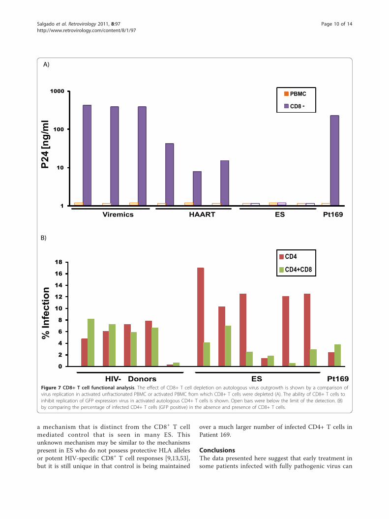
a mechanism that is distinct from the CD8+ T cellmediated control that is seen in many ES. Thisunknown mechanism may be similar to the mechanismspresent in ES who do not possess protective HLA allelesor potent HIV-specific CD8+ T cell responses [9,13,53],but it is still unique in that control is being maintained
over a much larger number of infected CD4+ T cells inPatient 169.
ConclusionsThe data presented here suggest that early treatment insome patients infected with fully pathogenic virus can
0
2
4
6
8
10
12
14
16
18
% In
fect
ion
CD4
CD4+CD8
HIV- Donors ES Pt169
A)
B)
PBMC
CD8 -
1
10
100
1000
P24
[ng/
ml
Viremics ES Pt169HAART
Figure 7 CD8+ T cell functional analysis. The effect of CD8+ T cell depletion on autologous virus outgrowth is shown by a comparison ofvirus replication in activated unfractionated PBMC or activated PBMC from which CD8+ T cells were depleted (A). The ability of CD8+ T cells toinhibit replication of GFP expression virus in activated autologous CD4+ T cells is shown. Open bars were below the limit of the detection. (B)by comparing the percentage of infected CD4+ T cells (GFP positive) in the absence and presence of CD8+ T cells.
Salgado et al. Retrovirology 2011, 8:97http://www.retrovirology.com/content/8/1/97
Page 10 of 14

lead to control of viral replication for extended periodsof time. Understanding the mechanisms involved in thiscontrol may lead to vaccine development and effectiveimmunotherapy in patients with progressive disease.
Availability of supporting dataThe data sets supporting the results of this article areavailable in the Gen Bank repository (accession numbersJN599164 and JN599165)
MethodsVirus Isolation and Sequence AnalysisCulture of replication-competent virus from CD4+ T cellswas performed as previously described [3]. Replication-competent virus from 1999 was obtained by spinoculatingCD4+ T cells from an uninfected donor with the patient’splasma. Full genome sequence analysis of viral isolateswas performed as previously described [3]. Classical, max-imum likelihood and Bayesian phylogenetic analysis wereperformed as described previously [7].
Antiretroviral drug testing100 μl of patient serum were treated with 300 μl of coldacetonitrile, stored at -20°C for 20 minutes and subse-quently centrifuged at 12,000 × rpm for 5 minutes. Spe-cimen supernatants were evaporated to dryness andreconstituted with 100 μl water. 10 uL of each treatedsample were injected onto the liquid chromatographysystem equipped with Transcend pumps (Thermo FisherScientific) for analytical separation. The chromato-graphic run began with 60 seconds of 5% methanol con-taining 10 mM ammonium acetate (mobile phase B),followed by a 10 minute ramp to 95% B. Analytes wereeluted from a Hypersil Gold 50 × 2.1 mm; 3 μm particlesize HPLC column (ThermoFisher Scientific) during thisgradient and the column was washed for 60 secondswith 2:2:1 acetonitrile:isopropanol:acetone and re-equili-brated with 5% mobile phase B for 180 seconds. Ana-lytes were detected over a 14.9 minute run using theExactive Orbitrap mass analyzer (Thermo Fisher Scienti-fic) with a heated electrospray ionization (HESI) source.The source parameters were as follows: sheath gas: 40,auxillary gas: 10, sweep gas: 0, spray voltage: 3.5 kV,capillary temperature: 270 °C, capillary voltage: 60 V,tube lens voltage: 120 V, skimmer voltage: 15 V, heatertemperature: 350 °C. The mass spectrometer methodincluded two positive-mode scan events: one full scanevent with ultra-high resolution (100000 @ 1 Hz) andone in-source collision-induced dissociation (CID) eventwith enhanced resolution (25000 @ 4 Hz) and collisionenergy of 45 eV. Both scan events were programmed for100 ms maximum inject time and balanced ACG tar-gets. The analytical method was found to have a limit ofdetection of <20 ng/ml for amprenavir, atazanavir,
darunavir, efavirenz, emtricitabine, indinavir, lamivudine,lopinavir, nelfinavir, nevirapine, ritonavir, saquinavir,tenofovir and tipranavir. Positive identification wasdetermined by exact mass detection at 5 ppm discrimi-nation, analyte retention time and identification of masstransitions when possible.
Viral Tropism assayRFP expressing recombinant pseudotype virus was madewith env genes amplified from 1999 and 2010 isolates.These viruses were used to infect GHOST cell linestransfected with CCR5 and/or CXCR4 (obtained fromthe NIH AIDS Research and Reference Program) andthe percentage of RFP positive cells was determined intriplicates on day three. GHOST cells express low levelsof endogenous CXCR4 and therefore infection of cellstransfected with CCR5 alone was performed in the pre-sence of the CXCR4 antagonist AMD 3100 at a dose of1 uM (obtained from the NIH AIDS Research andReference Program).
Viral Fitness AssayViral fitness was analyzed as described previously [3].PBMCs from healthy donors were activated for two dayswith IL-2 and PHA. CD4+ T cells were isolated (MACS,CD4+ T cell isolation Kit) and infected by spinoculation[54] (1200 × g for 2 hours) with equal quantities (200ng/mL) of p24 from primary patient isolates, or withBa-L or IIIB laboratory HIV-1 strains as controls. Super-natant samples were taken over the course of 7 days.Viral replication was quantified using p24 ELISA (PerkinElmer).
Genetic PolymorphismsHLA-A, B, and C allele identification was performed atthe Johns Hopkins University Immunonogeneticslaboratory. CCR5 was amplified from genomic DNAusing gene specific primers. The presence or absence ofthe CCR5 Δ32 mutation was determined by relative sizeof the resulting PCR fragment. HLA-C single nucleotidepolymorphism genotyping (rs9264942) was performedutilizing the Applied Biosystems 7300 real-time PCRSystem allelic discrimination assay, following the manu-facturer’s guidelines. Primers and probes were developedby Custom TaqMan SNP Genotyping assays (ABI).Determination of the HLA-B Bw4-80Ile allele was per-formed using the Olerup SSP 104.101 KIR Genotyping12 Lot71E and 104.201 KIR ligand genotyping Lot85Ekits, following the manufacturer’s guidelines.
CD4+ T cell susceptibility assayCD4+ T cells from the patient and five healthy donorswere purified by negative selection using Miltenyi beadsand were infected directly ex vivo. Spinoculation [55]
Salgado et al. Retrovirology 2011, 8:97http://www.retrovirology.com/content/8/1/97
Page 11 of 14

was performed with pseudotyped CCR5 and CXCR4 tro-pic viruses and GFP expression was measured in tripli-cates as previously described [41,42]. Infection withoutspinoculation was also performed with CXCR4 tropicvirus.
Neutralization assayNeutralization assays were performed as described pre-viously [43]. Briefly, recombinant pseudoviruses contain-ing SF162, or Patient 169 env were titrated on TZM b1cells to determine a linear range of infection for eachpseudovirus. Infections were then performed in dupli-cate with a concentration of virus within this linearrange, along with serial dilutions of patient plasma thathad been heat inactivated at 56°C for 60 min. All assayswere performed in the presence of 10% total humanplasma. Each virus was pre-incubated with 5% testplasma and with four-fold serial dilutions of test plasmain normal human plasma. To determine neutralization,each test plasma well was compared to wells containingan equal concentration of normal human plasma.
CD8+ T cell assaysReactive CTL epitopes were defined by IFN-g Elispot. Aspreviously described [55], whole blood was taken fromeach patient and PBMCs were isolated by Ficoll gradientcentrifugation. PBMCs were aliquoted into each well of96 well MultiScreen (Millipore) plates with conjugatedIFN-g antibody. Cells were activated with overlappingpeptides spanning the entire amino acid sequence of Bclade consensus gag and nef at a concentration of 5 μg/ml (obtained from the NIH AIDS Research and Refer-ence Program). PBMCs were cultured overnight, andsubsequently analyzed. Quantification of spot formingunits (SFU) was performed in a blinded fashion by Zell-net Consulting (Fort Lee, NJ). Positive responses weredefined as greater than 50 SFU per million PBMCs.Negative controls (wells with medium alone) routinelyhad less than 15 SFU per million PBMC.The effect of CD8+ T cells on autologous virus out-
growth was determined by measuring p24 values onunfractionated PBMC and PBMC depleted of CD8+ Tcells. The cells were stimulated for 48 hours with PHA at1 μg/ml and culture supernatant was obtained on day 10.The cytolytic T cell effect was determined by a CD8
suppression assay. PBMCs were isolated from patients,and CD8+ T cells were positively selected using Miltenyimagnetic beads (MACS, CD8+ T cell Isolation kit). CD8+ T cells were depleted of CD16+ cells (Invitrogen,Dynal Beads) to remove contaminating NK cells. CD4+
T cells were isolated by negative selection using Miltenyimagnetic beads. Purity of depletion was analyzed byflow cytometry. CD4+ T cells were infected by spinocu-lation at 1200 × g for two hours with replication
competent NL4-3 virus, in which GFP is engineeredinto nef. Flow cytometry was performed five days afterinfection to assess the percentage of GFP positive cells.
AcknowledgementsSupported by HHMI (RFS) and NIH grants R01AI056990-01A1 (JBM) and R01AI080328 (JNB)
Author details1Department of Medicine, Johns Hopkins University School of Medicine,Baltimore, Maryland 21287, USA. 2Department of Pathology, Johns HopkinsUniversity School of Medicine, Baltimore, Maryland 21287, USA. 3Departmentof Molecular Microbiology and Immunology, Johns Hopkins BloombergSchool of Public Health, Baltimore, Maryland 21287, USA. 4Howard HughesMedical Institute, Johns Hopkins University School of Medicine, Baltimore,Maryland 21287, USA.
Authors’ contributionsMS, SAR, KOC, and RWB performed all the experiments and helped draft themanuscript. JRB helped to design the neutralization antibody assay. ARB,MAM, and WC tested plasma samples for antiretroviral drugs. AAC and JBMprovided clinical samples and helped draft the manuscript. RFS participatedin the study design and helped to draft the manuscript. JNB conceived ofthe study, participated in its design and coordination and helped to draftthe manuscript. All authors read and approved the final manuscript.
Competing interestsThe authors declare that they have no competing interests.
Received: 12 September 2011 Accepted: 5 December 2011Published: 5 December 2011
References1. O’Connell KA, Bailey JR, Blankson JN: Elucidating the elite: mechanisms of
control in HIV-1 infection. Trends Pharmacol Sci 2009, 30:631-637.2. Bailey JR, O’Connell K, Yang HC, Han Y, Xu J, Jilek B, Williams TM, Ray SC,
Siliciano RF, Blankson JN: Transmission of human immunodeficiency virustype 1 from a patient who developed AIDS to an elite suppressor. J Virol2008, 82:7395-7410.
3. Blankson JN, Bailey JR, Thayil S, Yang HC, Lassen K, Lai J, Gandhi SK,Siliciano JD, Williams TM, Siliciano RF: Isolation and characterization ofreplication-competent human immunodeficiency virus type 1 from asubset of elite suppressors. J Virol 2007, 81:2508-2518.
4. Julg B, Pereyra F, Buzon MJ, Piechocka-Trocha A, Clark MJ, Baker BM, Lian J,Miura T, Martinez-Picado J, Addo MM, Walker BD: Infrequent recovery ofHIV from but robust exogenous infection of activated CD4(+) T cells inHIV elite controllers. Clin Infect Dis 2010, 51:233-238.
5. Lamine A, Caumont-Sarcos A, Chaix ML, Saez-Cirion A, Rouzioux C,Delfraissy JF, Pancino G, Lambotte O: Replication-competent HIV strainsinfect HIV controllers despite undetectable viremia (ANRS EP36 study).AIDS 2007, 21:1043-1045.
6. Mens H, Kearney M, Wiegand A, Shao W, Schonning K, Gerstoft J, Obel N,Maldarelli F, Mellors JW, Benfield T, Coffin JM: HIV-1 continues to replicateand evolve in patients with natural control of HIV infection. J Virol 2010,84:12971-12981.
7. O’Connell KA, Brennan TP, Bailey JR, Ray SC, Siliciano RF, Blankson JN:Control of HIV-1 in elite suppressors despite ongoing replication andevolution in plasma virus. J Virol 2010, 84:7018-7028.
8. Salgado M, Brennan TP, O’Connell KA, Bailey JR, Ray SC, Siliciano RF,Blankson JN: Evolution of the HIV-1 nef gene in HLA-B*57 positive elitesuppressors. Retrovirology 2010, 7:94.
9. Emu B, Sinclair E, Hatano H, Ferre A, Shacklett B, Martin JN, McCune JM,Deeks SG: HLA Class I-Restricted T Cell Responses May Contribute to theControl of HIV Infection, but Such Responses are Not Always Necessaryfor Long-term Virus Control. J Virol 2008.
10. Han Y, Lai J, Barditch-Crovo P, Gallant JE, Williams TM, Siliciano RF,Blankson JN: The role of protective HCP5 and HLA-C associatedpolymorphisms in the control of HIV-1 replication in a subset of elitesuppressors. AIDS 2008, 22:541-544.
Salgado et al. Retrovirology 2011, 8:97http://www.retrovirology.com/content/8/1/97
Page 12 of 14

11. Lambotte O, Boufassa F, Madec Y, Nguyen A, Goujard C, Rouzioux L,Meyer C, Venet A, Delfraissy JF, SEROCO-HEMOCO Study Group: HIVcontrollers: a homogeneous group of HIV-1-infected patients withspontaneous control of viral replication. Clin Infect Dis 2005, 41:1053-1056.
12. Migueles SA, Sabbaghian MS, Shupert WL, Bettinotti MP, Marincola FM,Martino L, Hallahan CW, Selig SM, Schwartz D, Sullivan J, Connors M: HLAB*5701 is highly associated with restriction of virus replication in asubgroup of HIV-infected long term nonprogressors. Proc Natl Acad SciUSA 2000, 97:2709-2714.
13. Pereyra F, Addo MM, Kaufmann DE, Liu Y, Miura T, Rathod A, Baker B,Trocha A, Rosenberg R, Mackey E, Ueda P, Lu Z, Cohen D, Wrin T,Petropoulos CJ, Rosenberg ES, Walker BD: Genetic and immunologicheterogeneity among persons who control HIV infection in the absenceof therapy. J Infect Dis 2008, 197:563-571.
14. Sajadi MM, Constantine NT, Mann DL, Charurat M, Dadzan E, Kadlecik P,Redfield RR: Epidemiologic characteristics and natural history of HIV-1natural viral suppressors. J Acquir Immune Defic Syndr 2009, 50:403-408.
15. Betts MR, Nason MC, West SM, De Rosa SC, Migueles SA, Abraham J,Lederman MM, Benito JM, Goepfert PA, Connors M, Roederer M, Koup RA:HIV nonprogressors preferentially maintain highly functional HIV-specificCD8+ T cells. Blood 2006, 107:4781-4789.
16. Hersperger AR, Pereyra F, Nason M, Demers K, Sheth P, Shin LY, Kovacs CM,Rodriguez B, Sieg SF, Teixeira-Johnson L, Gudonis D, Goepfert PA,Lederman MM, Frank I, Makedonas G, Kaul R, Walker BD, Betts MR: Perforinexpression directly ex vivo by HIV-specific CD8 T-cells is a correlate ofHIV elite control. PLoS Pathog 2010, 6:e1000917.
17. Migueles SA, Laborico AC, Shupert WL, Sabbaghian MS, Rabin R,Hallahan CW, Van Baarle D, Kostense S, Miedema F, McLaughlin M, Ehler L,Metcalf J, Liu S, Connors M: HIV-specific CD8+ T cell proliferation iscoupled to perforin expression and is maintained in nonprogressors. NatImmunol 2002, 3:1061-1068.
18. Migueles SA, Osborne CM, Royce C, Compton AA, Joshi RP, Weeks KA,Rood JE, Berkley AM, Sacha JB, Cogliano-Shutta NA, Lloyd M, Roby G,Kwan R, McLaughlin M, Stallings S, Rehm C, O’Shea MA, Mican J,Packard BZ, Komoriya A, Palmer S, Wiegand AP, Maldarelli F, Coffin JM,Mellors JW, Hallahan CW, Follman DA, Connors M: Lytic granule loading ofCD8+ T cells is required for HIV-infected cell elimination associated withimmune control. Immunity 2008, 29:1009-1021.
19. Saez-Cirion A, Lacabaratz C, Lambotte O, Versmisse P, Urrutia A, Boufassa F,Barre-Sinoussi F, Delfraissy JF, Sinet M, Pancino G, Venet A, AgenceNationale de Recherches sur le Sida EP36 HIV Controllers Study Group: HIVcontrollers exhibit potent CD8 T cell capacity to suppress HIV infectionex vivo and peculiar cytotoxic T lymphocyte activation phenotype. ProcNatl Acad Sci USA 104:6776-6781.
20. Rosenberg ES, Altfeld M, Poon SH, Phillips MN, Wilkes BM, Eldridge RL,Robbins GK, D’Aquila RT, Goulder PJ, Walker BD: Immune control of HIV-1after early treatment of acute infection. Nature 2000, 407:523-526.
21. Kaufmann DE, Lichterfeld M, Altfeld M, Addo MM, Johnston MN, Lee PK,Wagner BS, Kalife ET, Strick D, Rosenberg ES, Walker BD: Limited durabilityof viral control following treated acute HIV infection. PLoS Med 2004, 1:e36.
22. Margolick JB, Imteyaz H, Gallant JE, Langan SJ, Dinoso JB, Siliciano J,Blankson J, Nilles TL, Smith KA, Apuzzo LG: Prolonged viral suppressionwithout therapy in an HIV-1 seroconverter following early antiretroviraltherapy and daily interleukin-2. AIDS 2010, 24:932-935.
23. Hocqueloux L, Prazuck T, Avettand-Fenoel V, Lafeuillade A, Cardon B,Viard JP, Rouzioux C: Long-term immunovirologic control followingantiretroviral therapy interruption in patients treated at the time ofprimary HIV-1 infection. AIDS 2010, 24:1598-1601.
24. Blankson JN: Primary HIV-1 infection: to treat or not to treat? AIDS Read2005, 15:245-6, 249-51.
25. Smith DE, Walker BD, Cooper DA, Rosenberg ES, Kaldor JM: Is antiretroviraltreatment of primary HIV infection clinically justified on the basis ofcurrent evidence? AIDS 2004, 18:709-718.
26. Bell SK, Little SJ, Rosenberg ES: Clinical management of acute HIV infection:best practice remains unknown. J Infect Dis 2010, 202(Suppl 2):S278-88.
27. Blankson JN, Finzi D, Pierson TC, Sabundayo BP, Chadwick K, Margolick JB,Quinn TC, Siliciano RF: Biphasic decay of latently infected CD4+ T cells inacute human immunodeficiency virus type 1 infection. J Infect Dis 2000,182:1636-1642.
28. Dinoso JB, Kim SY, Siliciano RF, Blankson JN: A comparison of viral loadsbetween HIV-1-infected elite suppressors and individuals who receivesuppressive highly active antiretroviral therapy. Clin Infect Dis 2008,47:102-104.
29. Palmer S, Wiegand AP, Maldarelli F, Bazmi H, Mican JM, Polis M, Dewar RL,Planta A, Liu S, Metcalf JA, Mellors JW, Coffin JM: New real-time reversetranscriptase-initiated PCR assay with single-copy sensitivity for humanimmunodeficiency virus type 1 RNA in plasma. J Clin Microbiol 2003,41:4531-4536.
30. Finzi D, Blankson J, Siliciano JD, Margolick JB, Chadwick K, Pierson T,Smith K, Lisziewicz J, Lori F, Flexner C, Quinn TC, Chaisson RE, Rosenberg E,Walker B, Gange S, Gallant J, Siliciano RF: Latent infection of CD4+ T cellsprovides a mechanism for lifelong persistence of HIV-1, even in patientson effective combination therapy. Nat Med 1999, 5:512-517.
31. Siliciano JD, Kajdas J, Finzi D, Quinn TC, Chadwick K, Margolick JB, Kovacs C,Gange SJ, Siliciano RF: Long-term follow-up studies confirm the stabilityof the latent reservoir for HIV-1 in resting CD4+ T cells. Nat Med 2003,9:727-728.
32. Zhang H, Zhou Y, Alcock C, Kiefer T, Monie D, Siliciano J, Li Q, Pham P,Cofrancesco J, Persaud D, Siliciano RF: Novel single-cell-level phenotypicassay for residual drug susceptibility and reduced replication capacity ofdrug-resistant human immunodeficiency virus type 1. J Virol 2004,78:1718-1729.
33. Huang Y, Paxton WA, Wolinsky SM, Neumann AU, Zhang L, He T, Kang S,Ceradini D, Jin Z, Yazdanbakhsh K, Kunstman K, Erickson D, Dragon E,Landau NR, Phair J, Ho DD, Koup RA: The role of a mutant CCR5 allele inHIV-1 transmission and disease progression. Nat Med 1996, 2:1240-1243.
34. Ashton LJ, Biti RA, Ffrench RA, Bennetts BH, Newcombe NR, Benson EM,Carr A, Cooper DA, Kaldor JM: Increased frequency of CCR-5 delta 32heterozygotes among long-term non-progressors with HIV-1 infection.The Australian Long-Term Non-Progressor Study Group. AIDS 1997,11:1833-1838.
35. International HIV Controllers Study, Pereyra F, Jia X, McLaren PJ, Telenti A,de Bakker PI, Walker BD, Ripke S, Brumme CJ, Pulit SL, Carrington M,Kadie CM, Carlson JM, Heckerman D, Graham RR, Plenge RM, Deeks SG,Gianniny L, Crawford G, Sullivan J, Gonzalez E, Davies L, Camargo A,Moore JM, Beattie N, Gupta S, Crenshaw A, Burtt NP, Guiducci C, Gupta N,Gao X, Qi Y, Yuki Y, Piechocka-Trocha A, Cutrell E, Rosenberg R, Moss KL,Lemay P, O’Leary J, Schaefer T, Verma P, Toth I, Block B, Baker B,Rothchild A, Lian J, Proudfoot J, Alvino DM, Vine S, Addo MM, Allen TM,Altfeld M, Henn MR, Le Gall S, Streeck H, Haas DW, Kuritzkes DR,Robbins GK, Shafer RW, Gulick RM, Shikuma CM, Haubrich R, Riddler S,Sax PE, Daar ES, Ribaudo HJ, Agan B, Agarwal S, Ahern RL, Allen BL,Altidor S, Altschuler EL, Ambardar S, Anastos K, Anderson B, Anderson V,Andrady U, Antoniskis D, Bangsberg D, Barbaro D, Barrie W, Bartczak J,Barton S, Basden P, Basgoz N, Bazner S, Bellos NC, Benson AM, Berger J,Bernard NF, Bernard AM, Birch C, Bodner SJ, Bolan RK, Boudreaux ET,Bradley M, Braun JF, Brndjar JE, Brown SJ, Brown K, Brown ST, Burack J,Bush LM, Cafaro V, Campbell O, Campbell J, Carlson RH, Carmichael JK,Casey KK, Cavacuiti C, Celestin G, Chambers ST, Chez N, Chirch LM,Cimoch PJ, Cohen D, Cohn LE, Conway B, Cooper DA, Cornelson B, Cox DT,Cristofano MV, Cuchural G Jr, Czartoski JL, Dahman JM, Daly JS, Davis BT,Davis K, Davod SM, DeJesus E, Dietz CA, Dunham E, Dunn ME, Ellerin TB,Eron JJ, Fangman JJ, Farel CE, Ferlazzo H, Fidler S, Fleenor-Ford A, Frankel R,Freedberg KA, French NK, Fuchs JD, Fuller JD, Gaberman J, Gallant JE,Gandhi RT, Garcia E, Garmon D, Gathe JC Jr, Gaultier CR, Gebre W,Gilman FD, Gilson I, Goepfert PA, Gottlieb MS, Goulston C, Groger RK,Gurley TD, Haber S, Hardwicke R, Hardy WD, Harrigan PR, Hawkins TN,Heath S, Hecht FM, Henry WK, Hladek M, Hoffman RP, Horton JM, Hsu RK,Huhn GD, Hunt P, Hupert MJ, Illeman ML, Jaeger H, Jellinger RM, John M,Johnson JA, Johnson KL, Johnson H, Johnson K, Joly J, Jordan WC,Kauffman CA, Khanlou H, Killian RK, Kim AY, Kim DD, Kinder CA, Kirchner JT,Kogelman L, Kojic EM, Korthuis PT, Kurisu W, Kwon DS, LaMar M,Lampiris H, Lanzafame M, Lederman MM, Lee DM, Lee JM, Lee MJ, Lee ET,Lemoine J, Levy JA, Llibre JM, Liguori MA, Little SJ, Liu AY, Lopez AJ,Loutfy MR, Loy D, Mohammed DY, Man A, Mansour MK, Marconi VC,Markowitz M, Marques R, Martin JN, Martin HL Jr, Mayer KH, McElrath MJ,McGhee TA, McGovern BH, McGowan K, McIntyre D, Mcleod GX,Menezes P, Mesa G, Metroka CE, Meyer-Olson D, Miller AO, Montgomery K,Mounzer KC, Nagami EH, Nagin I, Nahass RG, Nelson MO, Nielsen C,Norene DL, O’Connor DH, Ojikutu BO, Okulicz J, Oladehin OO, Oldfield EC,
Salgado et al. Retrovirology 2011, 8:97http://www.retrovirology.com/content/8/1/97
Page 13 of 14

Olender SA, Ostrowski M, Owen WF Jr, Pae E, Parsonnet J, Pavlatos AM,Perlmutter AM, Pierce MN, Pincus JM, Pisani L, Price LJ, Proia L, Prokesch RC,Pujet HC, Ramgopal M, Rathod A, Rausch M, Ravishankar J, Rhame FS,Richards CS, Richman DD, Rodes B, Rodriguez M, Rose RC, Rosenberg ES,Rosenthal D, Ross PE, Rubin DS, Rumbaugh E, Saenz L, Salvaggio MR,Sanchez WC, Sanjana VM, Santiago S, Schmidt W, Schuitemaker H,Sestak PM, Shalit P, Shay W, Shirvani VN, Silebi VI, Sizemore JM Jr,Skolnik PR, Sokol-Anderson M, Sosman JM, Stabile P, Stapleton JT, Starrett S,Stein F, Stellbrink HJ, Sterman FL, Stone VE, Stone DR, Tambussi G,Taplitz RA, Tedaldi EM, Telenti A, Theisen W, Torres R, Tosiello L, Tremblay C,Tribble MA, Trinh PD, Tsao A, Ueda P, Vaccaro A, Valadas E, Vanig TJ,Vecino I, Vega VM, Veikley W, Wade BH, Walworth C, Wanidworanun C,Ward DJ, Warner DA, Weber RD, Webster D, Weis S, Wheeler DA, White DJ,Wilkins E, Winston A, Wlodaver CG, van’t Wout A, Wright DP, Yang OO,Yurdin DL, Zabukovic BW, Zachary KC, Zeeman B, Zhao M: The majorgenetic determinants of HIV-1 control affect HLA class I peptidepresentation. Science 2010, 330:1551-1557.
36. Shianna KV, Ge D, Colombo S, Ledergerber B, Weale M, Zhang K, Gumbs C,Castagna A, Cossarizza A, Cozzi-Lepri A, De Luca A, Easterbrook P,Francioli P, Mallal S, Martinez-Picado J, Miro JM, Obel N, Smith JP,Wyniger J, Descombes P, Antonarakis SE, Letvin NL, McMichael AJ,Haynes BF, Telenti A, Goldstein DB: A whole-genome association study ofmajor determinants for host control of HIV-1. Science 2007, 317:944-947.
37. Martin MP, Gao X, Lee JH, Nelson GW, Detels R, Goedert JJ, Buchbinder S,Hoots K, Vlahov D, Trowsdale J, Wilson M, O’Brien SJ, Carrington M:Epistatic interaction between KIR3DS1 and HLA-B delays the progressionto AIDS Nat. Genet 2002, 31:429-434.
38. Martin MP, Qi Y, Gao X, Yamada E, Martin JN, Pereyra F, Colombo S,Brown EE, Shupert WL, Phair J, Goedert JJ, Buchbinder S, Kirk GD, Telenti A,Connors M, O’Brien SJ, Walker BD, Parham P, Deeks SG, McVicar DW,Carrington M: Innate partnership of HLA-B and KIR3DL1 subtypes againstHIV-1. Nat Genet 2007, 39:733-740.
39. Chen H, Li C, Huang J, Cung T, Seiss K, Beamon J, Carrington MF, Porter LC,Burke PS, Yang Y, Ryan BJ, Liu R, Weiss RH, Pereyra F, Cress WD, Brass AL,Rosenberg ES, Walker BD, Yu XG, Lichterfeld M: CD4+ T cells from elitecontrollers resist HIV-1 infection by selective upregulation of p21. J ClinInvest 2011, 121:1549-1560.
40. Saez-Cirion A, Hamimi C, Bergamaschi A, David A, Versmisse P, Melard A,Boufassa F, Barre-Sinoussi F, Lambotte O, Rouzioux C, Pancino G, for theANRS CO18 Cohort: Restriction of HIV-1 replication in macrophages andCD4+ T cells from HIV controllers. Blood 2011, 118:955-964.
41. O’Connell KA, Rabi SA, Siliciano RF, Blankson JN: CD4+ T cells from elitesuppressors are more susceptible to HIV-1 but produce fewer virionsthan cells from chronic progressors. Proc Natl Acad Sci USA 2011.
42. Rabi SA, O’Connell KA, Nikolaeva D, Bailey JR, Jilek BL, Shen L, Page KR,Siliciano RF, Blankson JN: Unstimulated primary CD4+ T cells from HIV-1-positive elite suppressors are fully susceptible to HIV-1 entry andproductive infection. J Virol 2011, 85:979-986.
43. Bailey JR, Lassen KG, Yang HC, Quinn TC, Ray SC, Blankson JN, Siliciano RF:Neutralizing antibodies do not mediate suppression of humanimmunodeficiency virus type 1 in elite suppressors or selection ofplasma virus variants in patients on highly active antiretroviral therapy. JVirol 2006, 80:4758-4770.
44. Brockman MA, Schneidewind A, Lahaie M, Schmidt A, Miura T, Desouza I,Ryvkin F, Derdeyn CA, Allen S, Hunter E, Mulenga J, Goepfert PA, Walker BD,Allen TM: Escape and compensation from early HLA-B57-mediatedcytotoxic T-lymphocyte pressure on human immunodeficiency virustype 1 Gag alter capsid interactions with cyclophilin. A J Virol 2007,81:12608-12618.
45. Leslie AJ, Pfafferott KJ, Chetty P, Draenert R, Addo MM, Feeney M, Tang Y,Holmes EC, Allen T, Prado JG, Altfeld M, Brander C, Dixon C, Ramduth D,Jeena P, Thomas SA, St John A, Roach TA, Kupfer B, Luzzi G, Edwards A,Taylor G, Lyall H, Tudor-Williams G, Novelli V, Martinez-Picado J, Kiepiela P,Walker BD, Goulder PJ: HIV evolution: CTL escape mutation and reversionafter transmission. Nat Med 2004, 10:282-289.
46. Martinez-Picado J, Prado JG, Fry EE, Pfafferott K, Leslie A, Chetty S,Thobakgale C, Honeyborne I, Crawford H, Matthews P, Pillay T, Rousseau C,Mullins JI, Brander C, Walker BD, Stuart DI, Kiepiela P, Goulder P: Fitnesscost of escape mutations in p24 Gag in association with control ofhuman immunodeficiency virus type 1. J Virol 2006, 80:3617-3623.
47. Raymond S, Delobel P, Mavigner M, Cazabat M, Encinas S, Souyris C,Bruel P, Sandres-Saune K, Marchou B, Massip P, Izopet J: CXCR4-usingviruses in plasma and peripheral blood mononuclear cells duringprimary HIV-1 infection and impact on disease progression. AIDS 2010,24:2305-2312.
48. Yu XF, Wang Z, Vlahov D, Markham RB, Farzadegan H, Margolick JB:Infection with dual-tropic human immunodeficiency virus type 1variants associated with rapid total T cell decline and diseaseprogression in injection drug users. J Infect Dis 1998, 178:388-396.
49. Vanhems P, Lambert J, Cooper DA, Perrin L, Carr A, Hirschel B, Vizzard J,Kinloch-de Loes S, Allard R: Severity and prognosis of acute humanimmunodeficiency virus type 1 illness: a dose-response relationship. ClinInfect Dis 1998, 26:323-329.
50. Altfeld M, Addo MM, Rosenberg ES, Hecht FM, Lee PK, Vogel M, Yu XG,Draenert R, Johnston MN, Strick D, Allen TM, Feeney ME, Kahn JO,Sekaly RP, Levy JA, Rockstroh JK, Goulder PJ, Walker BD: Influence of HLA-B57 on clinical presentation and viral control during acute HIV-1infection. AIDS 2003, 17:2581-2591.
51. Goujard C, Chaix ML, Lambotte O, Deveau C, Sinet M, Guergnon J,Courgnaud V, Rouzioux C, Delfraissy JF, Venet A, Meyer L, Agence Nationalede Recherche sur le Sida PRIMO Study Group: Spontaneous control of viralreplication during primary HIV infection: when is “HIV controller” statusestablished? Clin Infect Dis 2009, 49:982-986.
52. Okulicz JF, Marconi VC, Landrum ML, Wegner S, Weintrob A, Ganesan A,Hale B, Crum-Cianflone N, Delmar J, Barthel V, Quinnan G, Agan BK,Dolan MJ, Infectious Disease Clinical Research Program (IDCRP) HIV WorkingGroup: Clinical outcomes of elite controllers, viremic controllers, andlong-term nonprogressors in the US Department of Defense HIV naturalhistory study. J Infect Dis 2009, 200:1714-1723.
53. Saez-Cirion A, Sinet M, Shin SY, Urrutia A, Versmisse P, Lacabaratz C,Boufassa F, Avettand-Fenoel V, Rouzioux C, Delfraissy JF, Barre-Sinoussi F,Lambotte O, Venet A, Pancino G, ANRS EP36 HIV Controllers Study Group:Heterogeneity in HIV suppression by CD8 T cells from HIV controllers:association with Gag-specific CD8 T cell responses. J Immunol 2009,182:7828-7837.
54. O’Doherty U, Swiggard WJ, Malim MH: Human immunodeficiency virustype 1 spinoculation enhances infection through virus binding. J Virol2000, 74:10074-10080.
55. O’Connell KA, Xu J, Durbin AP, Apuzzo LG, Imteyaz H, Williams TM, Ray SC,Margolick JB, Siliciano RF, Blankson JN: HIV-1 evolution followingtransmission to an HLA-B*5801-positive patient. J Infect Dis 2009,200:1820-1824.
doi:10.1186/1742-4690-8-97Cite this article as: Salgado et al.: Prolonged control of replication-competent dual- tropic human immunodeficiency virus-1 followingcessation of highly active antiretroviral therapy. Retrovirology 2011 8:97.
Submit your next manuscript to BioMed Centraland take full advantage of:
• Convenient online submission
• Thorough peer review
• No space constraints or color figure charges
• Immediate publication on acceptance
• Inclusion in PubMed, CAS, Scopus and Google Scholar
• Research which is freely available for redistribution
Submit your manuscript at www.biomedcentral.com/submit
Salgado et al. Retrovirology 2011, 8:97http://www.retrovirology.com/content/8/1/97
Page 14 of 14