Embed Size (px)
Citation preview
Molecular Biology of the CellVol. 18, 3993–4002, October 2007
Prophase Microtubule Arrays Undergo Flux-like Behaviorin Mammalian Cells□D □V
Nick P. Ferenz and Patricia Wadsworth
Department of Biology and Program in Molecular and Cellular Biology, University of Massachusetts,Amherst, MA 01003
Submitted May 8, 2007; Revised July 12, 2007; Accepted July 20, 2007Monitoring Editor: Kerry Bloom
In higher eukaryotic cells, microtubules within metaphase and anaphase spindles undergo poleward flux, the slow,poleward movement of tubulin subunits through the spindle microtubule lattice. Although a number of studies havedocumented this phenomenon across a wide range of model systems, the possibility of poleward flux before nuclearenvelope breakdown (NEB) has not been examined. Using a mammalian cell line expressing photoactivatable greenfluorescent protein (GFP)-tubulin, we observe microtubule motion, both toward and away from centrosomes, at a widerange of rates (0.5–4.5 �m/min) in prophase cells. Rapid microtubule motion in both directions is dynein dependent. Incontrast, slow microtubule motion, which occurs at rates consistent with metaphase flux, is insensitive to inhibition ofdynein but sensitive to perturbation of Eg5 and Kif2a, two proteins with previously documented roles in flux. Our resultsdemonstrate that microtubules in prophase cells are unexpectedly dynamic and that a subpopulation of these microtu-bules shows motion that is consistent with flux. We propose that the marked reduction in rate and directionality ofmicrotubule motion from prophase to metaphase results from changes in microtubule organization during spindleformation.
INTRODUCTION
After spindle formation, microtubule marking experimentshave revealed a unique form of motion called flux, whichresults from the coordinated addition and loss of tubulinsubunits at opposite ends of spindle microtubules duringmetaphase (Mitchison, 1989). During anaphase, flux contrib-utes to chromosome-to-pole motion, although the extent offlux-dependent chromosome motion varies by model sys-tem (Rogers et al., 2005). To date, flux has been observed inall eukaryotic cells examined except yeast (Mitchison, 1989;Sawin and Mitchison, 1991; Mitchison and Salmon, 1992;Maddox et al., 2000, 2002; LaFountain et al., 2004); and re-cently, a number of contributing molecular componentshave been identified, namely, Eg5 and Kif2a.
Eg5 is a homotetrameric, plus end-directed member of thekinesin-5 family that is capable of sliding antiparallel micro-tubules relative to one another in vitro (Kapitein et al., 2005).Kif2a is a microtubule destabilizing member of the kine-sin-13 family that lacks inherent motility (Desai et al., 1999).Both proteins are enriched at spindle poles during meta-phase, although Eg5 additionally stains spindle microtu-bules (Kapoor et al., 2000; Ganem and Compton, 2004). Im-portantly, each of these proteins has been implicated in theflux mechanism. Inhibition of Eg5, for example, has been
shown to reduce the rate of flux in mammalian cells (Cameronet al., 2006) and to eliminate flux in Xenopus egg extracts(Miyamoto et al., 2004; Shirasu-Hiza et al., 2004). Likewise, theperturbation of Kif2a has been reported to abolish flux inmammalian cells (Ganem et al., 2005), Drosophila embryos (Rogerset al., 2004) and Xenopus egg extracts (Gaetz and Kapoor, 2004).Mechanistically, flux has been modeled as a combination ofKif2a-mediated depolymerization of microtubule minus endsand Eg5-mediated sliding of overlapping, antiparallel micro-tubules. Recently, this model has been challenged by new datadocumenting the continuation of flux, albeit at reduced rates,in mammalian monopolar spindles lacking antiparallel micro-tubules (Cameron et al., 2006). Considering these data, as wellas the centrosomal enrichment of Eg5, it is possible that inmammalian cells, Eg5 may contribute to flux by reeling in andfeeding microtubules to the Kif2a depolymerase as suggestedpreviously (Cassimeris, 2004; Gadde and Heald, 2004).
Despite the extensive work concerning the occurrence andmechanics of flux, all studies have been conducted afternuclear envelope breakdown (NEB), leaving the possibilityof prophase flux unaddressed. Here, we examine microtu-bule behavior after photoactivation of green fluorescent pro-tein (GFP)-tubulin in prophase cells, and we compare theresults with experiments performed in early prometaphaseand metaphase cells. We show that prophase microtubulesare remarkably dynamic, because photoactivated marksmove toward and away from spindle poles at a wide rangeof rates, and that the variability of this motion decreases asmitosis progresses, with only slow, poleward-directed mo-tion remaining at metaphase. Additionally, a subset ofprophase and early prometaphase microtubule motion oc-curs at rates consistent with flux, and it is sensitive to per-turbation of Eg5 and Kif2a, but not dynein. These datademonstrate that a population of microtubules in prophaseand early prometaphase cells exhibits behavior consistentwith flux.
This article was published online ahead of print in MBC in Press(http://www.molbiolcell.org/cgi/doi/10.1091/mbc.E07–05–0420)on August 1, 2007.□D □V The online version of this article contains supplemental mate-rial at MBC Online (http://www.molbiolcell.org).
Address correspondence to: Patricia Wadsworth ([email protected]).
Abbreviations used: AP, away from a spindle pole; NEB, nuclearenvelope breakdown; P, toward a spindle pole.
© 2007 by The American Society for Cell Biology 3993 http://www.molbiolcell.org/content/suppl/2007/08/01/E07-05-0420.DC1.htmlSupplemental Material can be found at:
MATERIALS AND METHODS
MaterialsAll materials for cell culture were obtained from Sigma-Aldrich (St. Louis,MO) with the exception of Opti-MEM, which was obtained from Invitrogen(Carlsbad, CA) and fetal bovine serum, which was obtained from AtlantaBiologicals (Norcross, GA). Unless otherwise noted, all other chemicals wereobtained from Sigma-Aldrich (St. Louis, MO).
Cell CultureLLC-Pk1 cells expressing photoactivatable (PA)-GFP-tubulin were cultured asdescribed previously (Tulu et al., 2003). Cells were plated on glass coverslips(Corning Life Sciences, Acton, MA) 2 d before imaging. For live imaging, cellswere mounted in chambers containing non-CO2 minimal essential mediumsupplemented with 0.3 U/ml Oxyrase (EC Oxyrase, Oxyrase, Mansfield, OH),and they were maintained at �37°C.
Inhibitorsp150-CC1 plasmid, a gift from Dr. T. Kapoor (The Rockefeller University,New York, NY), was prepared according to protocol (King et al., 2003). Afterdilution with injection buffer (50 nM K-Glu and 1 mM MgCl2, pH 7.0), it wasinjected at 15 �M. Monastrol was used at 200 �M. Kif2a and MCAK antibod-ies, kind gifts from Drs. D. Compton (Dartmouth Medical School, Hanover,NH) and C. Walczak (Indiana University, Bloomington, IN), respectively,were combined 1:1 at full strength and injected.
ImmunofluorescenceCells were rinsed twice in calcium and magnesium-free phosphate-bufferedsaline (PBS), fixed in either paraglutaraldehyde (4.0% paraformaldehyde,0.10% glutaraldehyde, and 0.5% Triton X-100 in calcium- and magnesium-freePBS) for 5 min or glutaraldehyde (0.25% glutaraldehyde in calcium- andmagnesium-free PBS) for 1 min, treated with 1 mg/ml sodium borohydridefor 10 min, and rehydrated in calcium- and magnesium-free PBS containing0.1% Tween and 0.02% sodium azide. For the glutaraldehyde fixation, cellswere additionally lysed in Karsenti’s [0.5% Triton X-100, 80 mM piperazine-N,N�-bis(2-ethanesulfonic acid), 1 mM MgSO4, and 5 mM EGTA] for 1 minafter the initial glutaraldehyde incubation, and then they were fixed again for5–10 min before sodium borohydride treatment. The following primary an-tibodies were used in these experiments: YL1⁄2 (Accurate Chemical, Westbury,NY) used at 1:2; anti-Eg5, gift from Dr. D. Compton (Dartmouth MedicalSchool, Hanover, NH) used at 1:200; and anti-Kif2a (Novus Biologicals, Little-ton, CO) used at 1:10,000. Incubations with primary antibodies were per-formed overnight at room temperature or for 1 h at 37°C. Cy3- (JacksonImmunoResearch Laboratories, West Grove, PA) or fluorescein isothiocya-nate-labeled (Sigma-Aldrich, St. Louis, MO) secondary antibodies were usedat the recommended dilution for 30 or 90 min at room temperature, respec-tively. Coverslips were mounted in Vectashield (Vector Laboratories, Burlin-game, CA) and sealed with nail polish.
Image AcquisitionImages were acquired using a Nikon Eclipse TE 300 microscope equippedwith a 100� phase, numerical aperture 1.4 objective lens, a spinning diskconfocal scan head (PerkinElmer-Cetus, Wellesley, MA), and a HamamatsuOrca ER cooled charge-coupled device camera (Hamamatsu, Bridgewater,NJ). All images were taken with a single wavelength (488) filter cube. Imageacquisition was controlled by MetaMorph software (Molecular Devices,Sunnyvale, CA). Time-lapse sequences were acquired at 5-s intervals using anexposure time of 800 ms. Z-stacks were acquired at 0.2-�m steps using anexposure time of 800 ms.
For photoactivation experiments, cells were photoactivated (under the nu-cleus during prophase and along spindle fibers post-NEB) by a 5-s exposureto 413-nm light using an X-Cite 120 light source (EXFO America, Plano, TX)and a D405/20 filter cube (Chroma Technology, Rockingham, VT). The area ofphotoactivation was restricted using a slit (Lennox Laser, Glen Arm, MD)mounted in a Ludl filter wheel placed in a conjugate image plane in theepi-illumination light path. To activate the entire field of view, an openposition in the filter wheel was selected. After photoactivation, confocalimage acquisition proceeded as described above. Images were acquired�1–10 min after p150-CC1 injection, �5–15 min after Kif2a/MCAK injection,and �1–3.5 h after monastrol treatment.
Images of fixed cells were acquired by capturing optical sections every 0.2�m using exposure times of 200–400 ms (at 488 nm) and 800 ms (at 568 nm),and they were displayed as maximum intensity projections. Deconvolvedimages were acquired using AutoDeblur & AutoVisualize software, version9.3.6 (AutoQuant Imaging, Watervliet, NY).
Data AnalysisImmediately after each time-lapse sequence, the entire cell was photoacti-vated, and a Z-stack was acquired to determine the location of each centro-some. Frequently, centrosomes (or spindle poles, depending on the mitotic
stage) in early mitotic cells were located in different focal planes. However,because the relative location of the initial photoactivated region could becompared in X, Y, and Z to the fully photoactivated Z-stack, photoactivatedmarks could be determined to be associated with microtubules of a particularcentrosome. With a single centrosome as a reference point, motion could becategorized as toward that particular centrosome, not away from the other(and vice versa). To calculate rates of motion, a rectangular box, typically 11pixels in height (defined as the dimension perpendicular to the long axis ofthe photoactivated mark), was placed around a fluorescent mark of interest.The dimensions of the box were selected so that, during the time lapse, themark of interest remained within the defined region. Montages were createdfor each boxed region, and rates were extrapolated from the montage’s slope.All data were plotted using Excel (Microsoft, Redmond, WA). All statisticswere analyzed using a Student’s t test.
RESULTS
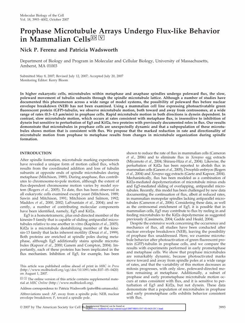
Metaphase Flux in LLC-Pk1-PA CellsTo determine whether microtubules undergo flux duringprophase, flux was first characterized in metaphase LLC-Pk1cells. To do this, a permanent cell line expressing photoactivat-able GFP-tubulin (hereafter LLC-Pk1-PA) was used (Pattersonand Lippincott-Schwartz, 2002; Tulu et al., 2003). Metaphasecells were photoactivated, and time-lapse sequences of theresulting fluorescent marks were acquired (Figure 1A and Sup-plemental Video 1). The location of each centrosome was iden-tified from a Z-stack obtained after the entire cell was photo-activated, and each clear mark was assigned a rate anddirectionality, scored as either toward (P) or away from (AP)the spindle poles (see Materials and Methods).
In metaphase cells, photoactivated marks on spindle mi-crotubules moved poleward between 0.50 and 2.24 �m/min,with an average rate of 1.37 �m/min (Figure 1B and Table1). AP motion was not detected. Although published rates offlux in LLC-Pk1 cells are significantly lower than the ratereported here (Mitchison, 1989; Zhai et al., 1995), those mea-surements were based on data collected at 30°C. Indeed,when photoactivations were performed at this reduced tem-perature, the average rate of metaphase flux decreased to0.75 �m/min, a value more in line with the literature. Fur-thermore, despite the substantial difference between the ratesof chromosomal oscillations in the spindle center (1.96 �m/min) and periphery (1.45 �m/min), flux rates were identicalregardless of location (unpublished data), consistent with re-cent observations in PtK1 cells (Cameron et al., 2006).
Prophase Microtubule Motion Is Extremely VariableNext, photoactivations were performed before NEB to exam-ine microtubule behavior during early mitosis (Figure 1A andSupplemental Video 2). In most prophase cells, the data re-vealed a surprisingly wide distribution of rates (0.50–4.49 �m/min) and the co-occupancy of nearly each populated rangewith both directionalities of motion (Figure 1B). Approxi-mately 61% of this motion was P, and �39% was AP (Table 2).This distribution did not seem to depend on the location of thephotoactivation, because activations gave similar results re-gardless of the distance from the centrosomes. However, invery early prophase cells with little chromatin condensation,photoactivated marks were static, indicating that the onset ofmicrotubule motion is abrupt and takes place between mid-and late prophase (unpublished data).
To establish a link between our prophase data and theearliest mitotic stage at which flux is known to occur (lateprometaphase cells with nearly all chromosomes aligned atthe metaphase plate), photoactivations also were performedduring early prometaphase (i.e., post-NEB cells with numer-ous unaligned chromosomes) (Figure 1A and SupplementalVideo 3). In such cells, the distribution of rates was similar tothat seen during prophase (0.50–3.49 �m/min), although
N. P. Ferenz and P. Wadsworth
Molecular Biology of the Cell3994
AP motion was practically undetectable (Figure 1B). Ap-proximately 99% of all early prometaphase motion was P,and �1% was AP (Table 2).
In each of the above-mentioned cases, a subpopulation ofphotoactivated marks could be seen moving poleward atrates consistent with metaphase flux (0.50–2.24 �m/min).To directly analyze this subset, motion was classified not
only as P or AP but also as slow (rates within the metaphaseflux range) or fast (rates beyond the metaphase flux range).Using these criteria, the average rates of slow P (flux-like)motion in prophase and early prometaphase cells were notsignificantly different from the metaphase flux value (Table1). We therefore hypothesized that early mitotic slow Pmotion represented flux.
Figure 1. Microtubule motion fromprophase to metaphase. (A) Microtubules werephotoactivated during prophase, early promet-aphase and metaphase (Supplemental Videos1–3). In metaphase cells, photoactivated micro-tubules flux toward the left pole. During earlyprometaphase and prophase, an increasing va-riety of motion is present, directed either to-ward or away from spindle poles. The initial,postactivation image is 0:00. Phase contrast im-ages before photoactivation are displayed asinsets. Yellow bars serve as fiduciary marksagainst which movement can be visualized;large asterisks mark spindle poles that are inthe same optical plane as the fluorescentmarks, whereas small asterisks mark spindlepoles that are not. Time points are in minutes:seconds. (B) The percentage of total motion (thenumber of photoactivated marks analyzed fora given rate, directionality and mitotic stagedivided by the total number of photoactivatedmarks analyzed for that same mitotic stage) isplotted against rates. Solid bars denote P mo-tion; hatched bars denote AP motion. Thedashed vertical line divides slow and fast mo-tion. Bar, 10 �m.
Prophase Microtubule Motion
Vol. 18, October 2007 3995
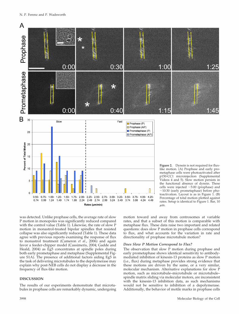
Flux-like Motion Is Dynein IndependentSmall molecule inhibition and RNA interference knockdownexperiments have established flux as a dynein-independentevent (Sawin and Mitchison, 1991; Maiato et al., 2005). Incontrast, rapid inward motion (i.e., sliding) of peripheralmicrotubules has been shown to depend on dynein (Rusanet al., 2002). We predicted that if the slow P component ofearly mitotic motion corresponded to flux, it too would bedynein-independent. To test this, LLC-Pk1-PA cells weremicroinjected before photoactivation with p150-CC1, a pro-tein fragment that binds dynein IC and disrupts dynein/dynactin interactions (Quintyne et al., 1999; Gaetz andKapoor, 2004) (Figure 2A and Supplemental Videos 4 and 5).Importantly, we found that, as in Ptk1 cells, p150-CC1 doesnot mislocalize Kif2a in LLC-Pk1 cells (unpublished data) asit does in Xenopus egg extracts (Gaetz and Kapoor, 2004;Cameron et al., 2006). Moreover, p150-CC1 does not disruptcentrosome integrity during the experimental time course.
As predicted, slow P motion in prophase, early promet-aphase, and metaphase cells was unaffected by dynein inhi-bition. Average rates were not significantly different fromthe control value (Table 1), and there was no noticeablechange in the frequency of this motion (Table 2). In contrast,microinjection of p150-CC1 essentially abolished all fast mo-tion, both toward and away from the poles, during prophaseand early prometaphase (Figure 2B). In control prophasecells, �20% of all motion was fast, and this was decreased to�5% after injection (Table 2). In control early prometaphasecells, �16% of all motion was fast, and this was decreased to�3% after injection (Table 2). The residual fast motion mayresult from incomplete inhibition of dynein, or it may be theconsequence of other motors that generate rapid movement(DeLuca et al., 2001). Consistent with previous reports(Salina et al., 2002), NEB was delayed in injected cells (un-published data). These data demonstrate that fast P and APmotion is dynein dependent, and they support a model inwhich microtubules can be the cargo of cytoplasmic dynein(Rusan et al., 2002; Wadsworth and Khodjakov, 2004). Ad-ditionally, the insensitivity of slow P motion to dynein in-
hibition is consistent with flux; however, it does not excludedynein-independent sliding as the underlying basis for suchmotion.
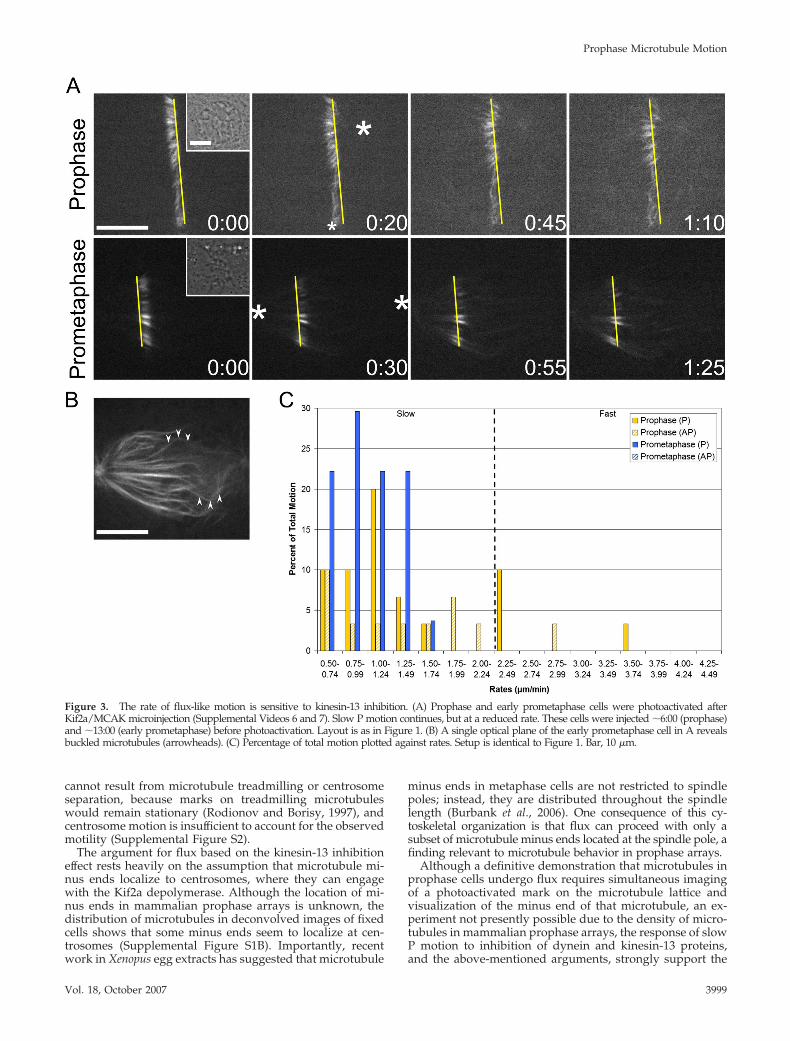
Kinesin-13 Inhibition Decreases the Rate of Flux-likeMotionExperiments in mammalian tissue culture cells, Drosophilaembryos, and Xenopus egg extracts have demonstrated thatmembers of the kinesin-13 family contribute to polewardflux (Gaetz and Kapoor, 2004; Rogers et al., 2004; Ganem etal., 2005). Accordingly, we predicted that the rate of slow Pmotion would be sensitive to inhibition of Kif2a, a mamma-lian kinesin-13. To test this, the strategy of Ganem et al.(2005) was followed by microinjecting a mixture of Kif2aand MCAK antibodies before photoactivation (Figure 3Aand Supplemental Videos 6 and 7). Such injections resultedin extensive astral microtubule formation and kinetochorefiber buckling, demonstrating that the antibodies alter mi-crotubule dynamics in LLC-Pk1-PA cells (Figure 3B).
As anticipated, the average rates of prophase, early pro-metaphase, and metaphase slow P motion were significantlyreduced from the control value (Table 1). However, theoverall distribution of rates remained unaltered in prophasecells, and it was only slightly modified in early promet-aphase cells (Figure 3C and Table 2). These data demon-strate that prophase and early prometaphase slow P motionis affected in an identical manner as metaphase flux. Doublelabel immunofluorescence further shows that Kif2a localizesto centrosomes during prophase and spindle poles afterNEB in LLC-Pk1-PA cells and that numerous microtubulesterminate at the centrosome (Supplemental Figure S1). Theseobservations, in addition to the well-established minus enddepolymerizing activity of Kif2a, suggest that slow pole-ward motion in prophase cells corresponds to flux.
Eg5 Inhibition Decreases the Frequency of Flux-likeMotion during ProphaseEvidence from Xenopus and mammalian systems has indi-cated the involvement of Eg5 in flux (Miyamoto et al., 2004;
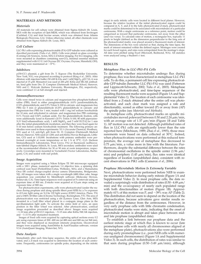
Table 1. Average rates (�m/min) of photoactivated marks in mitotic LLC-Pk1-PA cells
Slow P Fast P Slow AP Fast AP
ControlProphase 1.34 � 0.45 2.69 � 0.24 1.56 � 0.46 3.12 � 0.65Prometaphase 1.52 � 0.45 2.63 � 0.36 �2 ratesMetaphase 1.37 � 0.47
p150-CC1Prophase 1.24 � 0.46 �2 rates 1.35 � 0.51 �2 ratesPrometaphase 1.32 � 0.46 �2 rates �2 ratesMetaphase 1.22 � 0.36
Kif2a/MCAKProphase 1.02 � 0.28a 2.68 � 0.58 1.23 � 0.59 �2 ratesPrometaphase 1.01 � 0.30a
Metaphase 1.17 � 0.29b
MonastrolProphase 1.41 � 0.33 �2 rates 1.55 � 0.43 3.43 � 0.55Monopole 1.06 � 0.42c
Metaphase 0.95 � 0.18a
Rates � SDs. Unless otherwise indicated, values in any one column are not statistically different from that column’s control value (denotedin bold).a Statistically significant at p � 0.001.b Statistically significant at p � 0.05.c Statistically significant at p � 0.01.
N. P. Ferenz and P. Wadsworth
Molecular Biology of the Cell3996
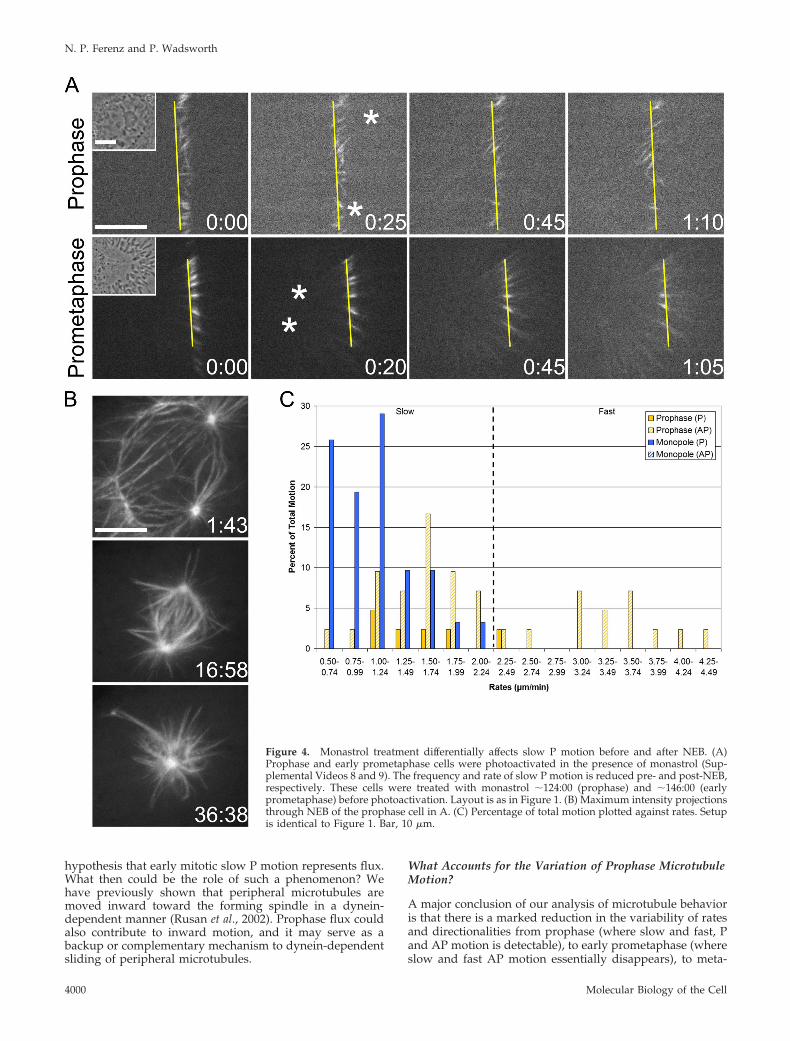
Shirasu-Hiza et al., 2004; Cameron et al., 2006). For thisreason, we predicted that the rate of slow P motion would bereduced after inhibition of Eg5. To test this, Eg5 activity wasinhibited using the small molecule monastrol (Mayer et al.,1999), and cells were photoactivated in the continued pres-ence of the inhibitor (Figure 4A and Supplemental Video 8).Prophase cells were imaged through NEB to ensure theformation of monopolar spindles, the hallmark of Eg5 inhi-bition (Figure 4B).
Unexpectedly, the rate of slow P motion during prophasewas not different from the control value (Table 1); however,the frequency of this particular motion was strongly de-creased from �54% in controls to �12% (Table 2). In fact,across the full range of rates (which was indistinguishablefrom the untreated prophase range), the proportion of P andAP motion was significantly shifted (Figure 4C). Approxi-mately 14% of all motion was P (compared with �61% incontrols), and �86% was AP (compared with �39% in con-trols) (Table 2). Importantly, both the decrease in P motionand the increase in AP motion represent true shifts; neither
are artificial consequences of changes in the opposing direc-tionality (Table 2). These data reveal that, during prophase,Eg5 activity is required to generate slow P motion and main-tain a balance between P and AP motion. Because Eg5 is highlyconcentrated at centrosomes during prophase (SupplementalFigure S1A), these results favor a model where Eg5 functions atleast in part to reel in microtubules at spindle poles(Cassimeris, 2004; Gadde and Heald, 2004), thus accounting forthe decreased frequency of flux-like motion.
Eg5 Inhibition Decreases the Rate of Flux-like Motionafter NEBLast, we examined microtubule behavior in monastrol-treated LLC-Pk1-PA cells that had undergone NEB andformed monopolar spindles (Figure 4A and SupplementalVideo 9). In 11 of 14 monopoles examined, we found thatphotoactivated marks moved exclusively in the P direction,confirming that flux occurs in monopolar spindles (Cameronet al., 2006), across a range of rates slightly reduced fromcontrols (Figure 4C). In the remaining three cells, no motion
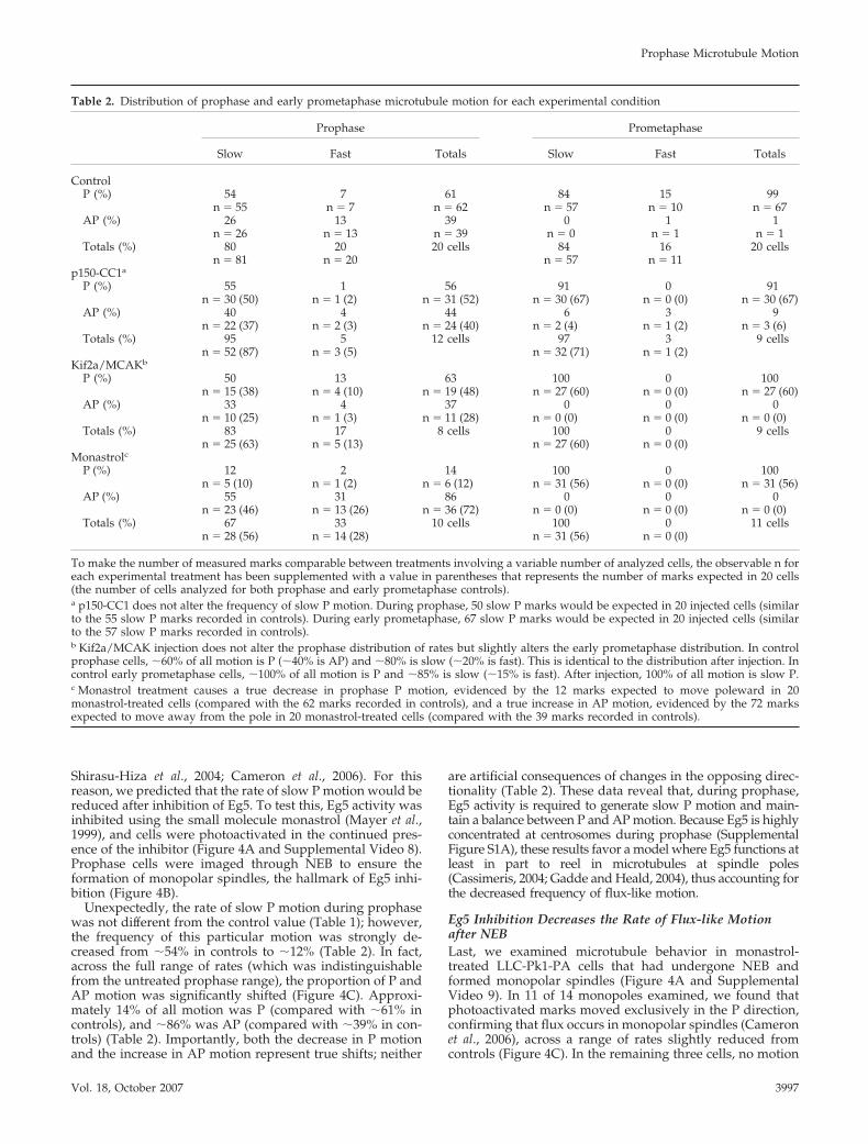
Table 2. Distribution of prophase and early prometaphase microtubule motion for each experimental condition
Prophase Prometaphase
Slow Fast Totals Slow Fast Totals
ControlP (%) 54 7 61 84 15 99
n � 55 n � 7 n � 62 n � 57 n � 10 n � 67AP (%) 26 13 39 0 1 1
n � 26 n � 13 n � 39 n � 0 n � 1 n � 1Totals (%) 80 20 20 cells 84 16 20 cells
n � 81 n � 20 n � 57 n � 11p150-CC1a
P (%) 55 1 56 91 0 91n � 30 (50) n � 1 (2) n � 31 (52) n � 30 (67) n � 0 (0) n � 30 (67)
AP (%) 40 4 44 6 3 9n � 22 (37) n � 2 (3) n � 24 (40) n � 2 (4) n � 1 (2) n � 3 (6)
Totals (%) 95 5 12 cells 97 3 9 cellsn � 52 (87) n � 3 (5) n � 32 (71) n � 1 (2)
Kif2a/MCAKb
P (%) 50 13 63 100 0 100n � 15 (38) n � 4 (10) n � 19 (48) n � 27 (60) n � 0 (0) n � 27 (60)
AP (%) 33 4 37 0 0 0n � 10 (25) n � 1 (3) n � 11 (28) n � 0 (0) n � 0 (0) n � 0 (0)
Totals (%) 83 17 8 cells 100 0 9 cellsn � 25 (63) n � 5 (13) n � 27 (60) n � 0 (0)
MonastrolcP (%) 12 2 14 100 0 100
n � 5 (10) n � 1 (2) n � 6 (12) n � 31 (56) n � 0 (0) n � 31 (56)AP (%) 55 31 86 0 0 0
n � 23 (46) n � 13 (26) n � 36 (72) n � 0 (0) n � 0 (0) n � 0 (0)Totals (%) 67 33 10 cells 100 0 11 cells
n � 28 (56) n � 14 (28) n � 31 (56) n � 0 (0)
To make the number of measured marks comparable between treatments involving a variable number of analyzed cells, the observable n foreach experimental treatment has been supplemented with a value in parentheses that represents the number of marks expected in 20 cells(the number of cells analyzed for both prophase and early prometaphase controls).a p150-CC1 does not alter the frequency of slow P motion. During prophase, 50 slow P marks would be expected in 20 injected cells (similarto the 55 slow P marks recorded in controls). During early prometaphase, 67 slow P marks would be expected in 20 injected cells (similarto the 57 slow P marks recorded in controls).b Kif2a/MCAK injection does not alter the prophase distribution of rates but slightly alters the early prometaphase distribution. In controlprophase cells, �60% of all motion is P (�40% is AP) and �80% is slow (�20% is fast). This is identical to the distribution after injection. Incontrol early prometaphase cells, �100% of all motion is P and �85% is slow (�15% is fast). After injection, 100% of all motion is slow P.c Monastrol treatment causes a true decrease in prophase P motion, evidenced by the 12 marks expected to move poleward in 20monastrol-treated cells (compared with the 62 marks recorded in controls), and a true increase in AP motion, evidenced by the 72 marksexpected to move away from the pole in 20 monastrol-treated cells (compared with the 39 marks recorded in controls).
Prophase Microtubule Motion
Vol. 18, October 2007 3997
was detected. Unlike prophase cells, the average rate of slowP motion in monopoles was significantly reduced comparedwith the control value (Table 1). Likewise, the rate of slow Pmotion in monastrol-treated bipolar spindles that resistedcollapse was also significantly reduced (Table 1). These dataagree with previous reports examining the response of fluxto monastrol treatment (Cameron et al., 2006) and againfavor a feeder-chipper model (Cassimeris, 2004; Gadde andHeald, 2004) as Eg5 concentrates at spindle poles duringboth early prometaphase and metaphase (Supplemental Fig-ure S1A). The presence of additional factors aiding Eg5 inthe task of delivering microtubules to the depolymerase mayexplain why post-NEB cells do not display a decrease in thefrequency of flux-like motion.
DISCUSSION
The results of our experiments demonstrate that microtu-bules in prophase cells are remarkably dynamic, undergoing
motion toward and away from centrosomes at variablerates, and that a subset of this motion is comparable withmetaphase flux. These data raise two important and relatedquestions: does slow P motion in prophase cells correspondto flux, and what accounts for the variation in rate anddirectionality of prophase microtubule motion?
Does Slow P Motion Correspond to Flux?The observation that slow P motion during prophase andearly prometaphase shows identical sensitivity to antibody-mediated inhibition of kinesin-13 proteins as slow P motion(i.e., flux) during metaphase provides strong evidence thatthese motions are driven by the same, or a very similar,molecular mechanism. Alternative explanations for slow Pmotion, such as microtubule–microtubule or microtubule–spindle matrix sliding via molecular motors, are inconsistentwith the kinesin-13 inhibition data, as such mechanismswould not be sensitive to inhibition of a depolymerase.Additionally, the behavior of motile marks in prophase cells
Figure 2. Dynein is not required for flux-like motion. (A) Prophase and early pro-metaphase cells were photoactivated afterp150-CC1 microinjection (SupplementalVideos 4 and 5). Slow motion persists inthe functional absence of dynein. Thesecells were injected �5:00 (prophase) and�10:30 (early prometaphase) before pho-toactivation. Layout is as in Figure 1. (B)Percentage of total motion plotted againstrates. Setup is identical to Figure 1. Bar, 10�m.
N. P. Ferenz and P. Wadsworth
Molecular Biology of the Cell3998
cannot result from microtubule treadmilling or centrosomeseparation, because marks on treadmilling microtubuleswould remain stationary (Rodionov and Borisy, 1997), andcentrosome motion is insufficient to account for the observedmotility (Supplemental Figure S2).
The argument for flux based on the kinesin-13 inhibitioneffect rests heavily on the assumption that microtubule mi-nus ends localize to centrosomes, where they can engagewith the Kif2a depolymerase. Although the location of mi-nus ends in mammalian prophase arrays is unknown, thedistribution of microtubules in deconvolved images of fixedcells shows that some minus ends seem to localize at cen-trosomes (Supplemental Figure S1B). Importantly, recentwork in Xenopus egg extracts has suggested that microtubule
minus ends in metaphase cells are not restricted to spindlepoles; instead, they are distributed throughout the spindlelength (Burbank et al., 2006). One consequence of this cy-toskeletal organization is that flux can proceed with only asubset of microtubule minus ends located at the spindle pole, afinding relevant to microtubule behavior in prophase arrays.
Although a definitive demonstration that microtubules inprophase cells undergo flux requires simultaneous imagingof a photoactivated mark on the microtubule lattice andvisualization of the minus end of that microtubule, an ex-periment not presently possible due to the density of micro-tubules in mammalian prophase arrays, the response of slowP motion to inhibition of dynein and kinesin-13 proteins,and the above-mentioned arguments, strongly support the
Figure 3. The rate of flux-like motion is sensitive to kinesin-13 inhibition. (A) Prophase and early prometaphase cells were photoactivated afterKif2a/MCAK microinjection (Supplemental Videos 6 and 7). Slow P motion continues, but at a reduced rate. These cells were injected �6:00 (prophase)and �13:00 (early prometaphase) before photoactivation. Layout is as in Figure 1. (B) A single optical plane of the early prometaphase cell in A revealsbuckled microtubules (arrowheads). (C) Percentage of total motion plotted against rates. Setup is identical to Figure 1. Bar, 10 �m.
Prophase Microtubule Motion
Vol. 18, October 2007 3999
hypothesis that early mitotic slow P motion represents flux.What then could be the role of such a phenomenon? Wehave previously shown that peripheral microtubules aremoved inward toward the forming spindle in a dynein-dependent manner (Rusan et al., 2002). Prophase flux couldalso contribute to inward motion, and it may serve as abackup or complementary mechanism to dynein-dependentsliding of peripheral microtubules.
What Accounts for the Variation of Prophase MicrotubuleMotion?
A major conclusion of our analysis of microtubule behavioris that there is a marked reduction in the variability of ratesand directionalities from prophase (where slow and fast, Pand AP motion is detectable), to early prometaphase (whereslow and fast AP motion essentially disappears), to meta-
Figure 4. Monastrol treatment differentially affects slow P motion before and after NEB. (A)Prophase and early prometaphase cells were photoactivated in the presence of monastrol (Sup-plemental Videos 8 and 9). The frequency and rate of slow P motion is reduced pre- and post-NEB,respectively. These cells were treated with monastrol �124:00 (prophase) and �146:00 (earlyprometaphase) before photoactivation. Layout is as in Figure 1. (B) Maximum intensity projectionsthrough NEB of the prophase cell in A. (C) Percentage of total motion plotted against rates. Setupis identical to Figure 1. Bar, 10 �m.
N. P. Ferenz and P. Wadsworth
Molecular Biology of the Cell4000
phase (where only slow P motion remains). We propose thatthe reduction in microtubule motion results not fromchanges in the active/inactive state of mitotic motors, butfrom progressive changes in microtubule organization dur-ing spindle formation. This possibility is supported by thefact that mitotic motors are activated as cells enter mitosisand Cdk1 activity rises, and they are thought to remainactive until exit from mitosis (Verde et al., 1990, 1991; Blangyet al., 1997). Microtubule dynamics are similarly activated atentry into mitosis (Verde et al., 1990; Verde et al., 1992). Thus,the suppression of microtubule motion is likely to resultfrom the progressive establishment of interactions betweenmicrotubules and spindle components (i.e., centrosomes andkinetochores) as well as from microtubule–microtubule in-teractions.
The clearest support for this possibility is the loss of fastmicrotubule motion as cells progress through mitosis. Ourresults demonstrate that fast motion is dynein dependent,yet substantial evidence supports the view that dynein re-mains active throughout mitosis. It is possible that only free,untethered microtubules (defined here as microtubules thatare not linked, or weakly linked, to other microtubules, spindlepoles, or kinetochores) undergo fast motion. In support of this,exogenous microtubule pieces added to asters in Xenopus ex-tracts move rapidly poleward in a dynein-dependent manner,presumably because they are not tethered to spindle compo-nents (Heald et al., 1997). As mitosis progresses, free microtu-bules that are moved poleward by dynein could become teth-ered to the spindle pole by dynein/NuMA/dynactincomplexes (Merdes et al., 1996). Conversely, microtubules thatare moved away from the poles may undergo catastrophe andrapid disassembly in the peripheral cytoplasm (Rusan et al.,2001).
Progressive changes in microtubule organization may alsoaccount for the differential response of pre- and post-NEBcells to Eg5 inhibition. Our data support the possibility thatEg5 functions as both a feeder, delivering microtubules tothe kinesin-13 depolymerase (Cassimeris, 2004; Gadde andHeald, 2004), and a tether, cross-linking neighboring micro-tubules (Kapitein et al., 2005). During prophase, other mo-lecular components that would normally contribute to teth-ering may be unable to. NuMA, for example, plays a majorrole in organizing spindle poles (Merdes et al., 1996), butis nuclear, and therefore unavailable, in prophase cells(Compton et al., 1992). If Eg5 acts as the dominant tetherduring prophase, then monastrol treatment could result inmicrotubules becoming untethered, leading to the observedincrease in the frequency of AP motion, because antagonisticmotors can slide free microtubules. Importantly, although itis recognized that monastrol treatment does not disruptEg5–microtubule interactions (Kapoor et al., 2000), Eg5-bound microtubules are capable of sliding in the presence ofmonastrol (Crevel et al., 2004). After NEB, proteins otherthan Eg5 (i.e., NuMA) may tether spindle microtubules andEg5 inhibition would not result in AP motion because thesemicrotubules would remain tethered. Furthermore, Eg5 in-hibition may limit the rate of flux-like motion in post-NEB,but not prophase cells, because its activity could be requiredto overcome antagonistic forces that oppose slow P motion(Sharp et al., 2000); such forces could come about, for exam-ple, through antiparallel microtubule cross-links, of whichthere are few in prophase.
Considering the increasing degree of coordination of mi-crotubule motion from prophase to metaphase, it is an in-teresting possibility that the flux machinery is operationalthroughout all of mitosis and only becomes obviously ap-
parent when other motion has been suppressed or elimi-nated, as the spindle matures.
ACKNOWLEDGMENTS
p150-CC1 plasmid was a kind gift of Dr. Tarun Kapoor. Antibodies weregenerously provided by Drs. Duane Compton (Kif2a and Eg5) and ClaireWalczak (MCAK). We thank Dr. Wei-Lih Lee for helpful suggestions on thismanuscript. This work was supported by a grant from the National Institutesof Health (to P.W.).
REFERENCES
Blangy, A., Arnaud, L., and Nigg, E. A. (1997). Phosphorylation by p34cdc2
protein kinase regulates binding of the kinesin related motor HsEg5 to thedynactin subunit p150glued. J. Biol. Chem. 272, 19418–19424.
Burbank, K. S., Groen, A. C., Perlman, Z. E., Fisher, D. S., and Mitchison, T. J.(2006). A new method reveals microtubule minus ends throughout the mei-otic spindle. J. Cell Biol. 175, 369–375.
Cameron, L. A., Yang, G., Cimini, D., Canman, J. C., Kisurina-Evgenieva, O.,Khodjakov, O., Danuser, G., and Salmon, E. D. (2006). Kinesin 5-independentpoleward flux of kinetochore microtubules in PtK1 cells. J. Cell Biol. 173,173–179.
Cassimeris, L. (2004). Cell division: eg’ing on microtubule flux. Curr. Biol. 14,R1000–R1002.
Compton, D. A., Szilak, I., and Cleveland, D. W. (1992). Primary structure ofNuMA, an intranuclear protein that defines a novel pathway for segregationof proteins at mitosis. J. Cell Biol. 116, 1395–1408.
Crevel, I. M., Alonso, M. C., and Cross, R. A. (2004). Monastrol stabilises anattached low-friction mode of Eg5. Curr. Biol. 1, R411–R412.
DeLuca, J. G., Newton, C. N., Himes, R. H., Jordan, M. A., and Wilson, L.(2001). Purification and characterization of native conventional kinesin,HSET, and CENP-E from mitotic HeLa cells. J. Biol. Chem. 276, 28014–28021.
Desai, A., Verma, S., Mitchison, T. J., and Walczak, C. E. (1999). Kin I kinesinsare microtubule-destabilizing enzymes. Cell 96, 69–78.
Gadde, S., and Heald, R. (2004). Mechanisms and molecules of the mitoticspindle. Curr. Biol. 14, R797–R805.
Gaetz, J., and Kapoor, T. M. (2004). Dynein/dynactin regulate metaphasespindle length by targeting depolymerizing activities to spindle poles. J. CellBiol. 166, 465–471.
Ganem, N. J., and Compton, D. A. (2004). The KinI kinesin Kif2a is requiredfor bipolar spindle assembly through a functional relationship with MCAK. J.Cell Biol. 166, 473–478.
Ganem, N. J., Upton, K., and Compton, D. A. (2005). Efficient mitosis inhuman cells lacking poleward microtubule flux. Curr. Biol. 15, 1827–1832.
Heald, R., Tournebize, R., Habermann, A., Karsenti, E., and Hyman, A. (1997).Spindle assembly in Xenopus egg extracts: respective roles of centrosomes andmicrotubule self-organization. J. Cell Biol. 138, 615–628.
Kapitein, L. C., Peterman, E.J.G., Kwok, B. H., Kim, J. H., Kapoor, T. M., andSchmidt, C. F. (2005). The bipolar mitotic kinesin Eg5 moves on both micro-tubules that it crosslinks. Nature 435, 114–118.
Kapoor, T. M., Mayer, T. U., Coughlin, M. L., and Mitchison, T. J. (2000).Probing spindle assembly mechanics with monastrol, a small molecule inhib-itor of the mitotic kinesin, Eg5. J. Cell Biol. 150, 975–988.
King, S. J., Brown, C. L., Maier, K. C., Quintyne, N. J., and Schroer, T. A.(2003). Analysis of the dynein-dynactin interaction in vitro and in vivo. Mol.Biol. Cell 14, 5089–5097.
LaFountain, J. R., Jr., Cohan, C. S., Siegel, A. J., and LaFountain, D. J. (2004).Direct visualization of microtubule flux during metaphase and anaphase incrane-fly spermatocytes. Mol. Biol. Cell 15, 5724–5732.
Maddox, P. S., Bloom, K. S., and Salmon, E. D. (2000). The polarity anddynamics of microtubule assembly in the budding yeast Saccharomyces cerevi-siae. Nat. Cell Biol. 2, 36–41.
Maddox, P., Desai, A., Oegema, K., Mitchison, T. J., and Salmon, E. D. (2002).Poleward microtubule flux is a major component of spindle dynamics andanaphase A in mitotic Drosophila embryos. Curr. Biol. 12, 1670–1674.
Maiato, H., Khodjakov, A., and Rieder, C. L. (2005). Drosophila CLASP isrequired for the incorporation of microtubule subunits into fluxing kineto-chore fibres. Nat. Cell Biol. 7, 42–47.
Prophase Microtubule Motion
Vol. 18, October 2007 4001
Mayer, T. U., Kapoor, T. M., Haggarty, S. J., King, R. W., Schreiber, S. L., andMitchison, T. J. (1999). Small molecule inhibitor of mitotic spindle bipolarityidentified in a phenotype-based screen. Science 286, 971–974.
Merdes, A., Ramyar, K., Vechio, J. D., and Cleveland, D. W. (1996). A complexof NuMA and cytoplasmic dynein is essential for mitotic spindle assembly.Cell 87, 447–458.
Mitchison, T. J. (1989). Polewards microtubule flux in the mitotic spindle:evidence from photoactivation of fluorescence. J. Cell Biol. 109, 637–652.
Mitchison, T. J., and Salmon, E. D. (1992). Poleward kinetochore fiber move-ment occurs during both metaphase and anaphase-A in newt lung cell mito-sis. J. Cell Biol. 119, 569–582.
Miyamoto, D. T., Perlman, Z. E., Burbank, K. S., Groen, A. C., and Mitchison,T. J. (2004). The kinesin Eg5 drives poleward microtubule flux in Xenopuslaevis egg extract spindles. J. Cell Biol. 167, 813–818.
Patterson, G. H., and Lippincott-Schwartz, J. (2002). A photoactivatable GFPfor selective photolabeling of proteins and cells. Science 297, 1873–1877.
Quintyne, N. J., Gill, S. R., Eckley, D. M., Crego, C. L., Compton, D. A., andSchroer, T. A. (1999). Dynactin is required for microtubule anchoring atcentrosomes. J. Cell Biol. 147, 321–334.
Rodionov, V. I., and Borisy, G. G. (1997). Microtubule treadmilling in vivo.Science 275, 215–218.
Rogers, G. C., Rogers, S. L., and Sharp, D. J. (2005). Spindle microtubules influx. J. Cell Sci. 118, 1105–1116.
Rogers, G. C., Rogers, S. L., Schwimmer, T. A., Ems-McClung, S. C., Walczak,C. E., Vale, R. D., Scholey, J. M., and Sharp, D. J. (2004). Two mitotic kinesinscooperate to drive sister chromatid separation during anaphase. Nature 427,364–370.
Rusan, N. M., Fagerstom, C. J., Yvon, A. M., and Wadsworth, P. (2001). Cellcycle-dependent changes in microtubule dynamics in living cells expressinggreen fluorescent protein-alpha tubulin. Mol. Biol. Cell 12, 971–980.
Rusan, N. M., Tulu, U. S., Fagerstrom, C., and Wadsworth, P. (2002). Reor-ganization of the microtubule array in prophase/prometaphase requires cy-toplasmic dynein-dependent microtubule transport. J. Cell Biol. 158, 997–1003.
Salina, D., Bodoor, K., Eckley, D. M., Schroer, T. A., Rattner, J. B., and Burke,B. (2002). Cytoplasmic dynein as a facilitator of nuclear envelope breakdown.Cell 108, 97–107.
Sawin, K. E., and Mitchison, T. J. (1991). Poleward microtubule flux in mitoticspindles assembled in vitro. J. Cell Biol. 112, 941–954.
Sharp, D. J., Brown, H. M., Kwon, M., Rogers, G. C., Holland, G., and Scholey,J. M. (2000). Functional coordination of three mitotic motors in Drosophilaembryos. Mol. Biol. Cell 11, 241–253.
Shirasu-Hiza, M., Perlman, Z. E., Wittmann, T., Karsenti, E., and Mitchison,T. J. (2004). Eg5 causes elongation of meiotic spindles when flux-associatedmicrotubule depolymerization is blocked. Curr. Biol. 14, 1941–1945.
Tulu, U. S., Rusan, N. M., and Wadsworth, P. (2003). Peripheral, non-centro-some-associated microtubules contribute to spindle formation in centrosome-containing cells. Curr. Biol. 13, 1894–1899.
Verde, F., Berrez, J.-M., Antony, C., and Karsenti, E. (1991). Taxol-inducedmicrotubule asters in mitotic extracts of Xenopus eggs: requirements forphosphorylated factors and cytoplasmic dynein. J. Cell Biol. 112, 1177–1187.
Verde, F., Dogterom, M., Stelzer, E., Karsenti, E., and Leibler, S. (1992).Control of microtubule dynamics and length by cyclin A- and cyclin B-dependent kinases in Xenopus egg extracts. J. Cell Biol. 118, 1097–1108.
Verde, F., Labbe, J.-C., Doree, M., and Karsenti, E. (1990). Regulation ofmicrotubule dynamics by cdc2 protein kinase in cell-free extracts of Xenopuseggs. Nature 343, 233–237.
Wadsworth, P., and Khodjakov, A. (2004). E pluribus unum: towards auniversal mechanism for spindle assembly. Trends Cell Biol. 14, 413–419.
Zhai, Y., Kronebusch, P. J., and Borisy, G. G. (1995). Kinetochore microtubuledynamics and the metaphase-anaphase transition. J. Cell Biol. 131, 721–734.
N. P. Ferenz and P. Wadsworth
Molecular Biology of the Cell4002


