Embed Size (px)
Citation preview

Protease Inhibitors from Marine Actinobacteria as aPotential Source for Antimalarial CompoundL. Karthik1, Gaurav Kumar1, Tarun Keswani2, Arindam Bhattacharyya2, S. Sarath Chandar3, K. V. Bhaskara
Rao1*
1 Environmental Biotechnology Division, School of Bio Sciences and Technology, VIT University, Vellore, Tamil nadu, India, 2 Immunology Lab, Department of Zoology,
University of Calcutta, Kolkata, West Bengal, India, 3 Genetics Division, School of Bio Sciences and Technology, VIT University, Vellore, Tamil nadu, India
Abstract
The study was planned to screen the marine actinobacterial extract for the protease inhibitor activity and its anti- Pf activityunder in vitro and in vivo conditions. Out of 100 isolates, only 3 isolates exhibited moderate to high protease inhibitoractivities on trypsin, chymotrypsin and proteinase K. Based on protease inhibitor activity 3 isolates were chosen for furtherstudies. The potential isolate was characterized by polyphasic approach and identified as Streptomyces sp LK3 (JF710608).The lead compound was identified as peptide from Streptomyces sp LK3. The double-reciprocal plot displayed inhibitionmode is non-competitive and it confirms the irreversible nature of protease inhibitor. The peptide from Streptomyces sp LK3extract showed significant anti plasmodial activity (IC50: 25.78 mg/ml). In in vivo model, the highest level of parasitemiasuppression (<45%) was observed in 600 mg/kg of the peptide. These analyses revealed no significant changes wereobserved in the spleen and liver tissue during 8 dpi. The results confirmed up-regulation of TGF-b and down regulation ofTNF-a in tissue and serum level in PbA infected peptide treated mice compared to PbA infection. The results obtained inferthat the peptide possesses anti- Pf activity activity. It suggests that the extracts have novel metabolites and could beconsidered as a potential source for drug development.
Citation: Karthik L, Kumar G, Keswani T, Bhattacharyya A, Chandar SS, et al. (2014) Protease Inhibitors from Marine Actinobacteria as a Potential Source forAntimalarial Compound. PLoS ONE 9(3): e90972. doi:10.1371/journal.pone.0090972
Editor: Olivier Neyrolles, Institut de Pharmacologie et de Biologie Structurale, France
Received September 20, 2013; Accepted February 6, 2014; Published March 11, 2014
Copyright: � 2014 Karthik et al. This is an open-access article distributed under the terms of the Creative Commons Attribution License, which permitsunrestricted use, distribution, and reproduction in any medium, provided the original author and source are credited.
Funding: These authors have no support or funding to report.
Competing Interests: The authors have declared that no competing interests exist.
* E-mail: [email protected]
Introduction
Malaria is a highly infectious disease caused by a protozoan
parasite of the genus Plasmodium. These parasites are transmitted
by the bite of infectious female Anopheles sp mosquitoes. There are
totally five species of Plasmodium associated with malarial fever
viz., P. falciparum, P. vivax, P. ovale, P. malariae and P. knowlesi.
Among them, P. falciparum is highly virulent and it is the
predominant agent in Africa. While, P. vivax is comparatively less
virulent and is more prevalent throughout the world and
remaining three species are associated with the minor outbreaks
in several parts of the world. Malaria is a major cause of morbidity
and mortality and it is projected that around 3.3 billion people
were at risk of malaria in 2010. Likewise, among 91% of deaths
are estimated in the WHO African Region, with children under
five years of age and pregnant women being severely affected [1].
World Malaria Report (2012) summarizes that 106 countries are
malaria-endemic in 2011 [2].
Three different approaches were considered for the control of
more virulent malarial parasite, Plasmodium falciparum. They are
widely exploited development of effective vaccines, vector control
and development of new drugs. It is very difficult to develop a
vaccine due to their exhibition of their multiple antigenicity. Based
on several factors, the vector control shows limited success. On the
other hand, there is an increasing resistance of malarial parasites
to the existing drug hence, there is an urgent need demand for new
antimalarial agents [3,4].
Due to the evolution of drug resistance in Plasmodium sp, which
necessitates the need for new drugs, ideally directed against new
targets such as heme and malarial proteases. The life cycle of
malarial parasite exhibits two stages: exoerythrocytic cycle and
erythrocytes life cycle. The erythrocytes life cycle was responsible
for all clinical manifestations and it begins when free merozoites
invade erythrocytes. The free merozoites will enter into the RBC
cells and develop from small ring-stage organisms to larger, more
metabolically active trophozoites followed by multinucleated
schizonts [5]. The schizonts will ruptures the erythrocytes and
releases 30,000 invasive merozoites in P. falciparum, 10,000 for P.
vivax and P. ovale and 2,000 for P. malariae. This step is called as
egress. At this stage, proteases are required for the rupture and
subsequent invasion of erythrocytes by merozoite stage parasites
and for the degradation of hemoglobin by intraerythrocytic
trophozoites.
The merozoites form of P. falciparum express a number of
merozoite surface proteins (MSPs). These may be considered as
target antigens for vaccine preparation [6]. The merozoites
synthesize a B195-kDa glycosyl phosphatidy- linositol-anchored
precursor that assembles as a complex with two peripheral
membrane proteins such as MSP6 and MSP7 [7–10]. This
complex (MSP1/6/7) is uniformly present in the merozoite surface
and it initiates the erythrocyte invasion [11]. This complex was
involving ‘primary’ proteolytic cleavage events earlier to egress
stage [12] and the cleavage products remain associated with the
surface of the released merozoite, to the complex is finally shed at
PLOS ONE | www.plosone.org 1 March 2014 | Volume 9 | Issue 3 | e90972
the point of erythrocyte invasion in an essential secondary
processing step by the action of a membrane-bound parasite
protease called PfSUB2 [13]. The primary proteolysis and the
positional conservation of the cleavage sites in MSP1 orthologues
across the Plasmodium genus [14] proposed that prime processing is
essential for the function of the MSP1/6/7 complex and for
merozoite viability.
The exonemes, specialized merozoite organelles releases the
subtilisin-like serine protease called PfSUB1 [15] and it mediates
the proteolytic maturation of members of a family of abundant,
papain-like putative proteases called SERA, previously implicated
in egress [16]. The inhibition of PfSUB1 prevents SERA
maturation and block egress. This indicates a role for PfSUB1 in
triggering egress, probably through activation of the SERA
enzymes.
Enzyme inhibitors are the third important product of marine
actinobacteria. So far, it is used for the study of enzyme structures
and reaction mechanisms, but recently it has been used in
pharmacology [17]. These selective inhibitors can be used as a
powerful tool for inactivating target proteases in the pathogenic
processes of human diseases such as malaria, emphysema, arthritis,
pancreatitis, thrombosis, high blood pressure, muscular dystrophy,
cancer, and AIDS [18]. Enzyme inhibitors from marine micro-
organisms were sparsely studied. However, terrestrial isolated
Streptomyces is a one of the potential producers of enzyme inhibitors
[19]. The isolation of novel enzyme inhibitor from terrestrial
sources is rare hence marine actinobacteria will provide new
potential inhibitors.
Proteases are essential constituents found in prokaryotes, fungi,
plants and animals. Serine, cysteine, metalloproteases is widely
spread in many pathogenic parasites, where they play critical
functions related to evasion of host immune defenses, acquisition
of nutrient for growth and proliferation, facilitation of dissemina-
tion or tissue damage during infection [20]. Thus, proteases play a
foremost role in pathogenesis. Moreover, protease enzymes are
used for a long time in various forms of clinical therapies. Their
application in medicine is gaining more and more attention as
several clinical studies are indicating their benefits in oncology,
inflammatory conditions, blood rheology control and immune
regulation. Hence, this study was focused on the screening of the
protease inhibitor from marine actinobacteria for anti- Pf activity
under in vitro and in vivo conditions.
Materials and Methods
Ethics statementAnimal experiments were carried out as per the guidelines of the
Committee for the Purpose of Control and Supervision of
Experimental Animals (CPCSEA), Government of India (Regis-
tration No: 885/ac/05/ CPCSEA) and as approved by the
Institutional Animal Ethics Committee (IAEC) University of
Calcutta, and confirms with the Guide for the Care and Use of
Laboratory Animals published by the US National Institutes of
Health (NIH Publication No. 85–23, revised 1985).
Media, chemicals and reagentsAll the media were purchased from Himedia, India. Giemsa’s
azur eosin methylene blue solution and Hematoxylin and eosin
stains for microscopy were obtained from Merck KGaA Frank-
furter Str., Germany. A phosphate buffer solution (PBS), sodium
bicarbonate, sodium azide, RNase A, NBT (nitro-blue tetrazolium
chloride) and BCIP (5-bromo-4-chloro-39-indolyphosphate p-
toluidine salt) (Cat# RM 578, RM 2577) were procured from
Himedia chemicals, India. Antibodies against TGF-b, TNF-a, b-
actin, AP-linked anti-rabbit, AP-linked anti-mouse secondary
antibodies were purchased from the Cell signaling Technology
Cell Signaling Technology, Inc (Danvers, MA, USA). Pre stain
molecular weight protein markers were purchased from Bangalore
Genei (India) and the remaining chemicals were purchased in an
analytical grade of the highest purity (India). All solutions were
prepared with commercial reagents of at least pro-analysis quality
and with sterilized 18 MV milliQ water. When necessary, the
specific origins of reagents are listed in the text.
In silico molecular docking analysisTo predict the inhibiting characteristics of respective inhibitors/
ligands (S. albogriseolus, S. longisporus, S. fradiae, S. lividans and S.
griseus) on PLASMEPSIN- II and FALCIPAIN-2 receptors, in silico
molecular docking approach was opted. Initially before docking
calculations, both receptors and ligands were subjected to energy
minimization by GROMOS97 available in a Swiss PDB viewer.
For the above in silico approach, Hex Server (http://hexserver.
loria.fr), an interactive molecular graphics program was used for
calculating and visualizing feasible docking modes of pairs of
macromolecules. It adopts Spherical Polar Fourier (SPF) correla-
tions to increase speed of the docking calculations. In the first step
as an input, respective PDB files of ligands and receptors were
specified in the Hex server [21]. Further, the docking correlation
type calculation parameters were set to ‘‘Shape and electrostatics’’
mode. Later, the origin and interface residues for the docking
calculations were set to their respective default values. Further, the
range angles for receptor and ligands were set to 180u. The step
sizes for the docking calculations were set to 10. Finally, the
docking evaluation solutions were set to evaluate 10 best docking
solutions. All the docking calculations were performed using GPU
mode. The best docking orientations were selected based on the
binding energy (kJ/mol) for the further intermolecular interaction
studies. The results were analyzed with Hex 6.0, a stand-alone
graphical application to visualize the intermolecular interactions of
receptors and ligands.
Actinobacteria strainsHundred actinobacteria strains were isolated from the island of
Nicobar (9u 099 N, 92u 499 E). These isolation procedures were
already published [22]. Further, these isolated strains were used
for screening to detect the protease inhibitor production. These
strains were designated as LK1-LK100. All the strains were
inoculated into 50 ml SS medium in 250 ml Erlenmeyer flasks
containing the sea water 50%, distilled water 50%, pH 7.5 and
incubated for 2 days in a rotary shaker incubator (200 rpm) at
28uC. The seed inoculums (10%) were transferred into 200 ml
production medium in 1 L Erlenmeyer flasks. The inoculated
cultures in the production medium were incubated for 72 h on a
rotary shaker (2000 rpm) at 28uC. After fermentation the broth
was centrifuged at 4000 rpm for 10 min and the filtrate was
separated.
Assay for protease inhibitor activityTo a mixture of mercuric chloride, phosphate buffer and trypsin
solution, marine actinobacterial extracts were added. After
adjusting the pH to 7.5 BAPNA (N-a-benzoyl-DL-arginine-p-
nitroanilide) was added and incubated at 37uC for 20 mins. After
incubation, 5% of the TCA was added and incubated at room
temperature for 20 mins. Later, the solution was filtered with
Whatmann no. 1 filter paper and the absorbance was taken at
280 nm. The inhibiting activity of protease inhibitor was
expressed using following formula.
Inhibiting activity (%) = C-T/C X 100
Protease Inhibitor for Antimalarial Treatment
PLOS ONE | www.plosone.org 2 March 2014 | Volume 9 | Issue 3 | e90972
Kinetics of protease inhibition by protease inhibitorThe mode of inhibition of protease inhibitor against trypsin
activity was measured with increasing concentrations of BAPNA
(0.5,1,2 and 4 mM) as a substrate in the absence and presence of
protease inhibitors at 0.5 mg/ml and 1 mg/ml. Optimal amounts
of protease inhibitor were determined based on the enzyme
inhibitory activity assay. Mode of inhibition of protease inhibitor
was determined by Lineweaver–Burk plot analysis of the data
calculated following Michaelis-Menten kinetics.
Dialysis for reversibility of protease inhibitor actionTrypsin (100 U/ml) was incubated with protease inhibitor
(23.5 mg/ml) in 0.5 ml of tris HCL buffer (10 mM, pH 7.5) for
2 h at 37uC and dialyzed against tris HCL buffer (1 mM, pH 7.5)
at 4uC for 24 h, changing the buffer every 12 h. Another
premixed-enzyme solution (0.5 ml) was kept at 4uC for 24 h
without dialysis for the control experiment. Reversibility of
protease inhibitor has been determined by comparing the residual
enzyme activity after dialysis with that of non-dialyzed one.
Identification of the actinobacterial IsolateThe strain was screened based on the protease inhibitor
production and the efficient isolate was subjected to molecular
characterization based on 16s rRNA sequencing chromous
biotech, Bangalore, India. The 16s rRNA fragment was amplified
using PCR polymerase. The PCR product was sequenced bi-
directionally using the forward (59-AGAGTRTGATCMTYGCT-
WAC-39) and reverse (59-CGYTAMCTTWTTACGRCT-39)
primers. The sequence was analysed by ABI3730XL capillary
DNA sequencer (ABI Prism 310 Genetic Analyzer, Tokyo, Japan).
Further, the genus and species were successfully identified.
Extraction, purification and characterization of proteaseinhibitor
The Streptomyces sp LK3 was subjected to fermentation for 7 days
in production medium. After fermentation, the supernatant was
collected and purified by four sub-sequential steps viz., ammonium
sulphate precipitation, dialysis, Ion exchange chromatography and
preparative HPLC chromatography. The functional group of the
compound was measured using Fourier transform infrared (FT-
IR) spectroscopy (Thermo Nicolet -330, USA) using a KBr pellet.
The structure of the compound was established by using the
spectral data obtained from gas chromatography (Perkin Elmer,
Claeus 680) -mass spectrometry (Claeus 600) at a flow rate of
1 ml/min with a carrier gas of helium and Liquid Chromatog-
raphy-Mass Spectrometer (LC-MS) (Thermo LCQ Deca XP
MAX). The proton NMR (1H NMR), carbon NMR, H,H-COSY
and HSQC (13C NMR, V Bruker Avance III 500 MHz (AV 500))
spectra of the compounds were obtained by using a dimethyl
sulfoxide d6 (DMSO-d6) as solvent.
In vitro anti- Pf activityP. falciparum isolates NZR-2 was obtained from National
Institute of Malaria Research, New Delhi, India. The experiment
was carried out in a 96-well microtitre plate by adding 150 ml of
infected human red blood cell suspension to each well. The
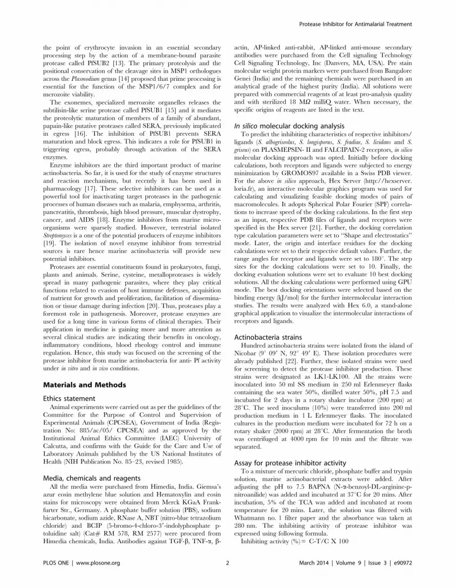
Figure 1. Intermolecular interactions between PLASMEPSIN- II and Protease inhibitor of marine actinobacteria A. S. albogriseolus PI;B. S. longisporus PI; C. S. fradiae PI; D. S. lividans; E. S. griseus. Enzyme PLASMEPSIN-II (indicated in red color) and their corresponding inhibitors(indicated in green color) are represented in ribbon configuration.doi:10.1371/journal.pone.0090972.g001
Table 1. In silico studies of protease inhibitor from marine actinobacteria.
Receptors Ligands-Binding Affinity(kJ/mol)
A.S. albogriseolus B.S. longisporus C.S. fradiae D.S. lividans E.S. griseus
PLASMEPSIN- II 2692.4540 2458.0594 2440.7606 2574.2310 2673.7805
FALCIPAIN-2 2192.4911 2590.0973 2269.6229 2420.6248 2491.3755
The docking revealed the S. albogriseolus has a tendency to inhibit the PLASMEPSIN- II receptor effectively. In addition, S. longisporous tends to inhibit FALCIPAIN-2receptors.doi:10.1371/journal.pone.0090972.t001
Protease Inhibitor for Antimalarial Treatment
PLOS ONE | www.plosone.org 3 March 2014 | Volume 9 | Issue 3 | e90972

peptide was dissolved separately in RPMI-1640 to obtain a
concentration range of 20, 40, 60, 80 and 100 mg/ml and 50 ml of
each dilution was poured into individual wells separately.
Artemisinin was used as a positive control and water as a negative
control. The microtitre plate was incubated for 48 hours. The
percentage of parasitemia was determined microscopically after
Giemsa staining.
In vivo anti- Pf activityMale Swiss albino mice (,25 g each; aged 6 to 8 weeks) were
maintained in sterilized cages and absorbent media; food and
water were provided ad libitum. Animal experiments were carried
out as per the guidelines of the Committee for the Purpose of
Control and Supervision of Experimental Animals (CPCSEA),
Government of India (Registration No: 885/ac/05/ CPCSEA)
and as approved by the Institutional Animal Ethics Committee
(IAEC) University of Calcutta, and confirms with the Guide for
the Care and Use of Laboratory Animals published by the US
National Institutes of Health (NIH Publication No. 85–23, revised
1985).
Parasite strain Plasmodim berghei ANKA obtained from National
Institute Malaria Research Center, New Delhi. Parasitized mouse
red blood cells (pRBC) from a liquid N2 preserved stabilize were
injected (16106 pRBC, in 100 ml phosphate buffer solution) into
mice of the same background. Mice were infected with PbA (16106
pRBC), in 100 ml PBS by intraperitoneal injection, after ampli-
fication an equal number of uninfected erythrocytes were injected
into control mice. Parasitemia was monitored daily in all
experimental groups by Giemsa-stained thin blood smears made
from tail snips. Survivability and parasitemia of mice (n = 60) were
also observed daily. The percentage of parasitemia was calculated
as follows: Parasitemia (%) = [(number of infected erythrocytes)/
(total number of erythrocytes counted)] x 100.
Experimental groups parasite clearance and curativeefficacy
For optimization of drug concentration, a stock solution of LK1,
LK2 and LK3 (5000 mg/ml) were dissolved in (PBS), and 50 mice
received 100 ml i.p. injections each of LK1, LK2 and LK3 (600,
800, 1000 and 1200 mg/kg/ BW respectively) from 2 dpi till the
last day of survival. The control group received injections of
vehicle on the same treatment schedule. Parasitological evaluation,
Figure 2. Streptomyces sp LK3 strain showed high protease inhibitor activity against trypsin and chymotrypsin with IC50 values ofLK3 strain A. trypsin (96.77 mg/ml) and B. chymotrypsin (161.8 mg/ml) B. Mode of protease inhibition by peptide. The double-reciprocal plotdisplayed non-competitive inhibition mode C. Reversibility of peptide action was confirmed by the continuous steep decline in Vmax with anincrease in inhibitor concentration with Km value remaining confined.doi:10.1371/journal.pone.0090972.g002
Table 2. Protease inhibitor activity of marine actinobacteria.
LK1 90 89 30
LK2 91 91 42
LK3 98 95 40
doi:10.1371/journal.pone.0090972.t002
Protease Inhibitor for Antimalarial Treatment
PLOS ONE | www.plosone.org 4 March 2014 | Volume 9 | Issue 3 | e90972

survivability indicated that LK1 (800 mg/kg/BW), LK2
(1000 mg/kg/BW) and LK3 (600 mg/kg/BW) was the best drug
concentration. Parasitamia and survivability in mice were mon-
itored daily (n = 60). The experimental animals were divided into
groups such as (1) uninfected control, (2) PbA (16106 pRBC)
infected, PbA infected and treated with LK1 (800 mg/kg/BW) (3)
PbA infected and treated with LK2 (1000 mg/kg/BW) and (4) PbA
infected and treated with LK3 (600 mg/kg/BW). For an analysis
on respective dpi, 5 mice from each experimental group were
used.
The ‘8-day suppressive test’ was adopted for the determination
of the parasite clearance and curative efficacy, respectively. The
doses for LK1 (800 mg/kg/BW) (3) PbA infected and treated with
LK2 (1000 mg/kg/BW) and (4) PbA infected and treated with
LK3 (600 mg/kg/BW). Groups of 5 mice each were administered
vehicle only. Tests were performed in a 8 day Suppressive
standard test. The Plasmodium berghei is most widely employed in
the rodent malaria parasite.
Thin smears of blood films were obtained from the peripheral
blood on the tail from each mouse on day four after infection
[23,24]. The smears were placed on microscope slides, fixed with
methanol and stained with Gemsa at pH 7.2, for parasitemia. The
microscope had an Ehrlich’s eyepiece and a nose diaphragm
showing about 100 red blood cells per field. The number of
parasitized erythrocytes in each of the 10–50 such fields were
counted three times and the average was calculated to give the
Parasitemia of each individual animal. Percentage of suppression
was calculated by using the following formula [23,24,25].
% Suppression~ Parasitemia in negative control�
Parasitemia in study group=Parasitemia in negative
control
Histopathology of spleenThe spleen was aseptically removed from each host and
immediately washed in phosphate buffered saline (PBS, pH 7.4).
The tissues were then fixed for 24 h in buffered formaldehyde
solution (10% in PBS) at room temperature, dehydrated by graded
ethanol, and embedded in paraffin (MERCK, solidification point
60–62uC). Tissue sections (5-mm thickness) were then deparaffi-
nized with xylene, re-hydrated with graded alcohols (100–50%
ethanol), stained with eosin and hematoxylin, and then mounted
in DPX resin (Merck, Mumbai, India). Digital images were
captured with Olympus CAMEDIA digital camera, Model C-
7070 wide zoom.
Western blot analysis and cytokine assaysThe mechanisms were elucidated by Western blot analysis and
cytokine assays as described earlier [26].
Statistical analysisValues between groups on same or different dpi were analyzed
using one or two way ANOVA. All values are shown as mean 6
SD, except where otherwise indicated. Data were analyzed and
when appropriate, the significance of the differences between
mean values was determined using Student’s test. *P values ,0.05
were considered significant for all statistical analysis, otherwise
stated.
Results and Discussion
Estimation of protease inhibitors efficacies using in silicomolecular docking
To validate the efficacy of protease inhibitors from marine
actinobacteria as a suitable targets for the malarial protease, five
inhibitors (S. albogriseolus, S. longisporus, S. fradiae, S. lividans and S.
griseus) were assayed against PLASMEPSIN- II and FALCIPAIN-2
using Hex server (Table.1). Docking calculations revealed that S.
albogriseolus have a probable tendency to inhibit the PLASMEP-
SIN- II receptor effectively and may outperform the other
inhibitors. On the other hand, S. longisporous tends to inhibit
FALCIPAIN-2 receptors, when compared to the other inhibitors.
The preliminary study revealed that the protease inhibitors from
five actinobacteria inhibit the respective receptors more effectively
(Fig. 1). Hence, we had hypothesized the inhibiting characteristics
of protease inhibitors obtained from marine actinobacteria
comparatively and their roles in malaria chemotherapeutic
development were elucidated using in silico studies.Figure 3. The 16s RNA sequencing confirmed the strain LK3 is anovel species with G+C content of the isolated DNA at 57.29mol%.doi:10.1371/journal.pone.0090972.g003
Table 3. Kinetic analysis of protease inhibition by peptide.
Peptide (mg/ml) Vmax (mM/min) Km (mM)
0 0.24 1.64
0.5 0.19 1.54
1 0.14 1.65
2 0.10 1.7
doi:10.1371/journal.pone.0090972.t003
Protease Inhibitor for Antimalarial Treatment
PLOS ONE | www.plosone.org 5 March 2014 | Volume 9 | Issue 3 | e90972
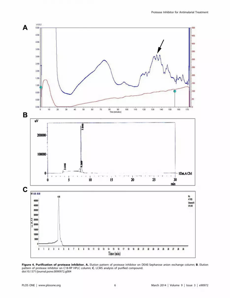
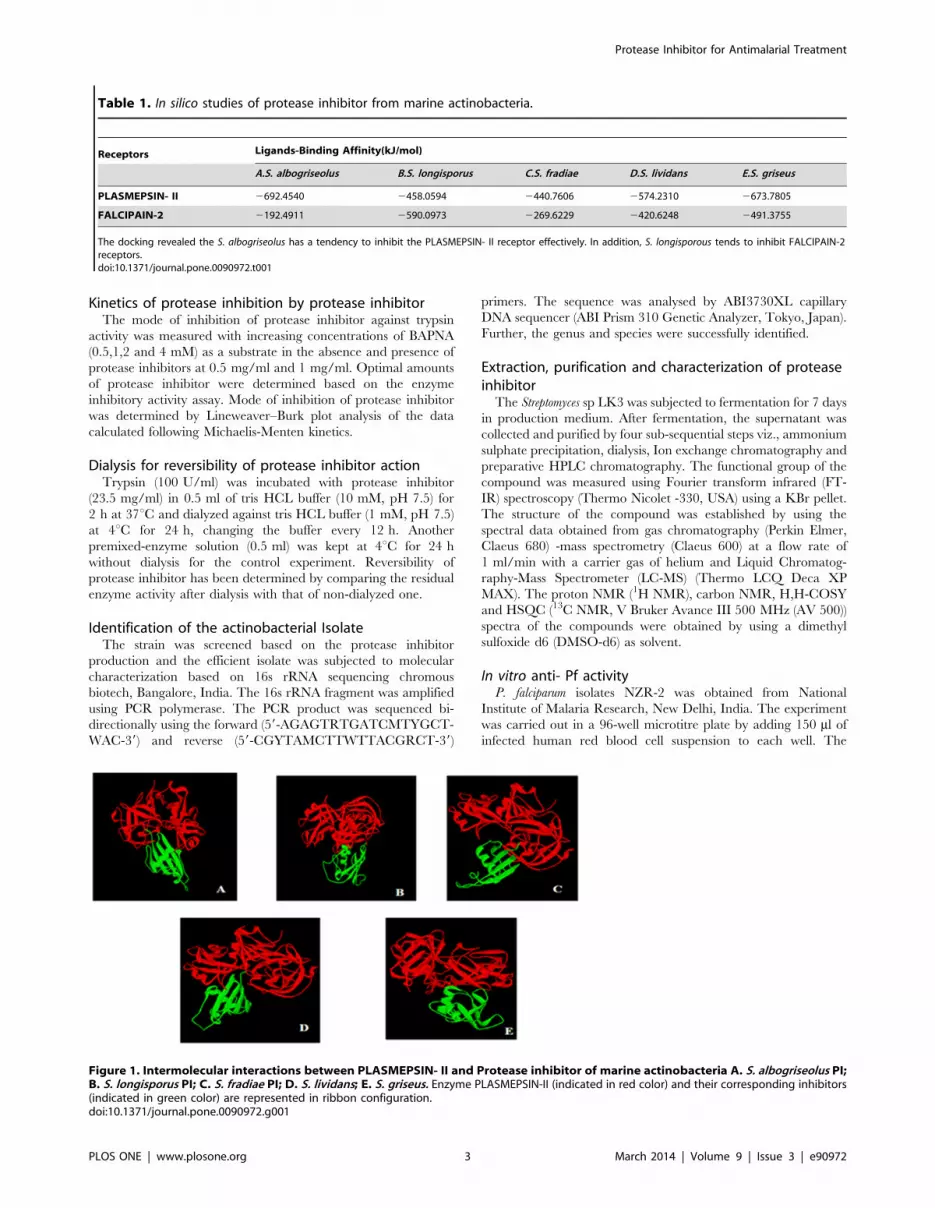
Figure 4. Purification of protease inhibitor. A. Elution pattern of protease inhibitor on DEAE-Sepharose anion exchange column; B. Elutionpattern of protease inhibitor on C18-RP HPLC column; C. LCMS analysis of purified compound.doi:10.1371/journal.pone.0090972.g004
Protease Inhibitor for Antimalarial Treatment
PLOS ONE | www.plosone.org 6 March 2014 | Volume 9 | Issue 3 | e90972

Figure 5. Characterization of protease inhibitor. A. Mass spectrum of peptide compound obtained from the GCMS clearly revealed that it maybe a small peptide with 5 or 6 aminoacids (Molecular Weight-568da) The amino acids may be present in the peptide are: AILKRVMGNC; B. FTIRspectrum of peptide compound. Absorbance at 1632 corresponded to the carbonyl functions whereas the absorbance from 3300–3600cm-1 may bedue to the NH function from the aminoacid functional groups.doi:10.1371/journal.pone.0090972.g005
Protease Inhibitor for Antimalarial Treatment
PLOS ONE | www.plosone.org 7 March 2014 | Volume 9 | Issue 3 | e90972
Figure 6. Characteristics of PbA infection in Swiss Albino Mice and effect of LK1 (800 mg/kg/BW), LK2 (1000 mg/kg/BW) and LK3(600 mg/kg/BW) respectively. (A) Resulting parasitemias were expressed as a percent parasitemia (mean 6 SD), measured daily on Giemsastained blood smears. Parasitemia was evaluated statistically by Two-way ANOVA with Bonferroni post-test showed that the parasitemias weresignificantly different (*p,0.05) between PbA infected groups and LK3 treated groups, whereas parasitemias were not significantly different in theLK1, LK-2 group w.r.t. PbA infected controls and the matched controls (P.0.05). (B) Effect of LK1 (800 mg/kg/BW), LK2 (1000 mg/kg/BW) and LK3(600 mg/kg/BW) respectively on survival of mice infected with P. berghei ANKA. Each value in Y axis represents the percentage (%) of survival of thetreated groups compared to PbA treated control mice. The treatment groups represent mice injected intraperitoneally from 2 dpi till the last day ofsurvival. Results are presented as arithmetic mean (6SE) of five mice per group. (C) Percentage parasitemia suppression after administration of LK1
Protease Inhibitor for Antimalarial Treatment
PLOS ONE | www.plosone.org 8 March 2014 | Volume 9 | Issue 3 | e90972

Screening of protease inhibitor producing marineactinobacteria
Among the 100 isolates tested for protease inhibitor activity,
LK-1, LK-2 and LK-3 strain showed substantial protease inhibitor
activity. The optimized 3 strains were subjected to fermentation
and the protease inhibitor extracts were lyophilized. These 3
strains were initially assayed against trypsin, chymotrypsin and
proteinase K (table 2). In these, Streptomyces sp LK3 strain showed
high protease inhibitor activity against trypsin and chymotrypsin.
The potential strain with 50% inhibiton were further subjected to
determine an IC50 value. The LK3 extract inhibited trypsin and
chymotrypsin with IC50 values of 96.77 mg/ml and 161.8 mg/ml
respectively (Fig. 2 A).
Out of 100, 3 isolates exhibited moderate to high protease
inhibitor activity of trypsin and chymotrypsin. Many reports are
available on the production of inhibitors from Streptomyces sp viz.,
S.mauvecolor, S.michiganensis, S.violaceus and S. yokosukaensis were
produced antipain against papain enzyme. It is also capable to
inhibit trypsin and cathepsin B. S.hygroscopicus and S.lavendulae
produced chymostatin against chymotrypsin. S.griseoruber produced
elastatinal against elastase [27,28]. So far, only few reports only
available for anti- Pf activity of marine actinobacteria. Maskey et
al. (2004) reported that the trioxacarcins A and D isolated from the
marine Streptomyces sp. isolate B8652 BCC 5149 possessed
‘‘extremely high antiplasmodial activity’’ against the parasite P.
falciparum K1 and NF54 strains which was much higher than the
clinically used compound chloroquine [29]. Peraud (2006)
reported that the manzamines isolated from the marine Micro-
monospora sp. strain M42 possessed antimalarial activity [30].
Prudhomme et al (2008) reported that the salinosporamide A,
isolated from the marine Salinispora tropica possessed antimalarial
activity [31]. It is also acts as a potent proteasome inhibitor in
development for treating cancer [32].
Mode of protease inhibitionThe mode of inhibition of peptide on trypsin activity was
analyzed using LB plot. The double-reciprocal plot displayed non-
competitive inhibition mode (Fig. 2B) as there is a continous steep
decline in Vmax with an increase in inhibitor concentration with
Km value remaining confined (Table 3). Enzyme activity of trypsin
was completely recovered after the dialysis, as showed by the
enzyme mixed inhibitor curve (EID) which was similar to the
curves of enzyme control without dialysis (EC) and dialysis (ED)
(Fig. 2C). A dialyzed mixture of enzyme and extract confirmed
that processes are not affected the enzyme activity dialysis.
However, the non-dialyzed mixture of enzyme and extract (EIC)
showed less inhibited activity and it confirms the irreversible
nature of protease inhibitor.
The main role of irreversible inhibitor is once target enzyme
inhibited, it cannot reactivate and the organism must reproduce
the enzyme. Several reversible protease inhibitors reached the
market as a drug but only few drugs of irreversible protease
inhibitors are available such as aspirin and b-lactam antibiotics.
Sajid and McKerrow (2002) reported irreversible inhibitor could
be used in bacterial, viral and parasitic diseases and in the future,
irreversible inhibitors are promising source for disease treatment
[33].
Identification of potential strainThe 16s rRNA sequencing is a method of choice for tracing
bacterial phylogeny and definite the taxonomy [34]. Hence, the
potential strain was identified as Streptomyces sp LK-3 through 16S
rRNA Sequencing. The BLAST search showed only 93%
similarity with Streptomyces mutabilis. The strain was deposited in
the Bankit (GenBank, NCBI, USA) under the accession number
JF710608. The 16s rRNA sequencing of strain LK3 was
confirmed that it occupies a distinctive phylogenetic position
within the radiation including representatives of the family
Streptomycetes using neighbor-joining tree (Fig. 3) and it forms
a separate subclade from other member of Streptomyces. It confirms
the strain LK3 is a novel species. The G+C content of the isolated
DNA is 57.29 mol% (http://tubic.tju.edu.cn/GC-Profile/). Based
on the chemotaxonomy, morphological and 16S rRNA gene
sequence data suggest that strain LK3 represents a novel species
when compared with type strains of species in nonomura key. The
polyphasic taxonomic study of strain LK3 merits classification as
the type strain of a novel species within the genus Streptomyces, and
hence the strain were named as Streptomyces sp LK3.
Purification and characterization of protease inhibitorThe RSM optimized Streptomyces sp LK3 [35] was subjected to
fermentation (7 days) and extracted protease inhibitor was purified
by a four sub-sequential steps viz., Ammonium sulphate precip-
itation, dialysis, Ion exchange chromatography and preparative
HPLC chromatography. The protease inhibitor was achieved 80%
saturation. Following this, dialysis and anion exchange chroma-
tography was performed.
The eighth peak contains the highest protein concentration
(0.15 mg/ml). Similarly, Angelova et al (2005) was purified and
(800 mg/Kg), LK2 (1000 mg/Kg) and LK3 (600 mg/Kg) respectively. Data from five replicates are presented as arithmetic mean 6 standard deviationand indicate p,0.05 with respect to the controls. Data shown are one representative experiment of five (n).doi:10.1371/journal.pone.0090972.g006
Figure 7. Histopathological changes in hepatic and splenictissues in response to LK3 treatment during PbA infection.Hematoxylin and eosin stains were used to prepare sections of the liverand spleen from mice treated with vehicle, PbA infected + 600 mg/KgLK3 and PbA infected respectively. Sections of control (A) and PbA+LK3infected (B) dpi liver showing, surrounding hepatocytes with nuclei andblood sinusoids lined with Kupffer cells. Liver from infected mice (C)shows, moriform vacuolization of hepatocytes and hemozoin pigment-ed Kupffer cells on 8 dpi. In control (D) and PbA+ LK3 infected spleentissue section (E), distinct and defined separate cluster of white pulpmarked by yellow arrows. In treatment tissue sections white pulp andred pulp are not distinct (F). Appearances of hollow spaces without cellsobserved in spleen tissue sections compared to that of control (redarrows). Black arrows represent hemozoin accumulation in liver andspleen tissue sections. Magnification indicated 406.doi:10.1371/journal.pone.0090972.g007
Protease Inhibitor for Antimalarial Treatment
PLOS ONE | www.plosone.org 9 March 2014 | Volume 9 | Issue 3 | e90972

characterized a novel protease inhibitor (PISC-2002) isolated from
culture supernatants of Streptomyces chromofuscus using DEAE-
Sepharose [36]. Figure 4A shows the elution pattern of the
protease inhibitor and a broad active peak was obtained, when the
dialyzed sample was passed through a DEAE Sepharose column
with 0.4 M NaCl Tris-HCl buffer with NaCl pH 7.5. Further
purification was carried out by preparative HPLC on a C18
column (Fig. 4B) and a single peak was confirmed by LCMS
(Fig. 4C). About 10 mg of pure protease inhibitor was obtained
from 3 g of lyophilised supernatant.
Mass spectrum obtained from the GCMS clearly revealed that it
may be a small peptide with 5 or 6 Aminoacids (Molecular
Weight-568da) (Fig. 5A). The 1H, 13C, H,H-COSY, HSQC also
showed the chemical shift values in the aliphatic region (2.5–4.0
and 5.0). This chemical shift may be due to the alpha hydrogens
and NH hydrogens. The compound doesn’t have any chemical
shift values in the aromatic region. The careful examination of IR
spectra also revealed the presence of peptides. The IR spectra
showed absorbance in the region of 1632 cm21, 3300–3600 cm21
(Fig. 5B). The absorbance around the 1632 corresponded to the
carbonyl functions where as the absorbance between 3300–
3600 cm21 may be due to the NH function. Apart from this two
characteristic peak, no other characteristic peaks have been
observed. The amino acids may be present in the peptide are:
AILKRVMGNC.
Many reports are available on the production of inhibitors from
Streptomyces sp viz., S.mauvecolor, S.michiganensis, S.violaceus and
S.yokosukaensis were produced antipain against papain enzyme
and it can also capable to inhibit trypsin and cathepsin B [37,38].
Inhibitors from Streptomyces sp was classified as SSI family inhibitors
and designated as SSI –like [39]. Hence, the protease inhibitor
from Streptomyces sp LK3 was belongs to the SSI family inhibitors.
The microbial origin inhibitors are low molecular weight
peptides of unusual structures [40]. Similarly, protease inhibitor
from Streptomyces sp LK3 is a low molecular weight peptide of
unusual structure. In 1969, Umezawa discovered first low
molecular weight enzyme inhibitor from Streptomyces sp [41]. At
present, more than 100 inhibitors were reported but this is the first
report on marine Streptomyces sp.
In vitro anti- Pf activityA total of 3 different actinobacterial isolates were chosen for
anti- Pf activity based on the protease inhibitor activity. Extracts
were tested at five initial concentrations: 20 mg/ml, 40 mg/ml,
60 mg/ml, 80 mg/ml and 100 mg/ml. Three extracts showed a 50
to 85% inhibition. The peptide from Streptomyces sp LK3 extract
showed significant anti plasmodial activity (IC50: 25.78 mg/ml).
The rest of the other actinobacterial extracts showed weak activity
(IC50 .200 mg/ml).
In vivo anti- Pf activityEffect of LK1, LK2 and LK3 on survival and parasite
clearance efficacy during Plasmodium berghei ANKA
infection. The clinical course of infection in the experimental
mice is summarized in Figure 5. Mice from both experimental
groups were age matched and had, PbA infected + LK2 treated set
and in PbA infected + LK3 treated set (Fig. 6A) and continued to
rise till mice dies. Most of the mice succumbed to CM during 4–9
Figure 8. Cell lysate from respective control, treatment spleen and liver were subjected to western blot analysis and ELISA (A) liverand (B) spleen during PbA infection and treated with peptide (C) ELISA results (p,0.05, ANOVA followed by post hoc LSD test).doi:10.1371/journal.pone.0090972.g008
Protease Inhibitor for Antimalarial Treatment
PLOS ONE | www.plosone.org 10 March 2014 | Volume 9 | Issue 3 | e90972

dpi, except in LK3 treated group (*p,0.05) compared to other
experimental groups and matched controls. Statistically significant
differences in the percentage survival of mice were observed
(Fig. 6B).
The ‘8 -day suppressive test’ were adopted for the determination
of the percentage of parasite clearance. The doses were LK1
(800 mg/kg/BW), LK2 (1000 mg/kg/BW) and LK3 (600 mg/
kg/BW) respectively. Groups of 5 mice each were administered
vehicle only. The results of the study indicated that in vivo, LK3
displayed a considerable activity against the P. berghei ANKA
malaria parasite (Fig. 6C).
PbA infection induces histological changes in the
spleen. To confirm the unpleasant effect of PbA infection and
effect of LK3 on the spleen and liver, mice were sacrificed, spleen
and liver tissues were isolated on 8 dpi, along with controls and
HE staining was done. Liver sections of uninfected mice (Fig. 7 A)
showed typical architecture and color, clean sinusoids with few
cells and resting Kupffer cells. On day 8 of infection, PbA infected
liver sections showed extensive histopathological changes, present-
ed an enlarged liver laden with malaria pigment (Fig. 7C). In
contrast, in liver sections of LK3 treated mice shows significant
reduction of malarial pigment and the mice were able to survive
the infection (Fig. 7B). Spleen of control mice presented distinct T
and B cell zones in the white pulp, resting B-cell follicles, with
small lymphocytes, surrounded by well defined marginal zones
(MZ), trabeculae (T) and red pulp (RP)(Fig. 7D). In the spleens of
PbA infected hosts, cells in the white pulp had proliferated
considerably and enlarged to the limits wherein the margin
between white and red pulp began to disappear and hollow spaces
without cells appeared. Follicle germinal centers (Gc) lost the
typical architecture acquiring a disorganized aspect, and several
phagocytic-centers with hemozoin (Hz) accumulation (Fig.7 F). In
PbA infected + LK3 treated set, we observed less damaged white
pulp and red pulp, small clusters of hemozoin pigments compared
to PbA infected and control mice (Fig. 7E).
TGF-b a known anti-inflammatory agent, acts to down regulate
the production of proinflamatory cytokines. On the other hand
much of the pathology of malaria is mediated by proinflamatory
cytokine such as TNF-a. From western blot results, significant
upregulation of TNF-a and down regulation of TGF-b were
observed in the spleen and liver during PbA infection with respect
to control. The ELISA results were strongly supported by
statistically significant western blot results. These observations
further support the proposed hypothesis, suggesting an antagonist
relationship between TNF-a and TGF-b and their involvement
during PbA infection (Fig. 8).
More recently, artemisinins resistance P. falciparum was con-
firmed at the Cambodia–Thailand border [42,43]. Hence, there is
a need of new drugs. The in silico study revealed that protease
inhibitors from five actinobacteria liable to show very high affinity
towards the respective receptors. Several researchers demonstrated
peptidyl fluoromethyl ketone [44], vinyl sulfone [45] and aldehyde
inhibitors [46] capable to protect Plasmodium-infected mice
against lethal malaria, which act as cysteine protease inhibitors.
Likewise, Gelhaus et al. (2005) reported Biotinylated dibenzyl
aziridine-2,3-dicarboxylate, it can block host erythrocyte rupture
and subsequent merozoite release [47]. In 2006, Ersmark et al
found aspartic proteases also very good antimalarial drug target
[48]. More recently, Yeoh et al (2007) reported serine protease
primes the malaria parasite for red blood cell invasion [49]. Senior
(2005) reported HIV aspartic protease inhibitors showed activity
against Plasmodium falciparum [50]. Protease inhibitors have also
drug of choice shown utility against other infectious diseases
caused by protozoans. Similarly, protease inhibitor from Strepto-
myces sp LK3 may be the drug of choice against Plasmodium
falciparum.
McCoubrie et al (2007) and Putrianti et al (2010) reported
SERA family members play a major role in malaria life cycle
[51,52]. PfSUB1 is a serine protease involved in both schizont
rupture and erythrocyte reinvasion in the P.falciparum life cycle. It
can be blocked by serine protease inhibitors. The inhibition of
PfSUB confirms the egress blocking and reduces the invasion of
merozoite. Hence, PfSUB1 is a drug target enzyme [49]. While,
compared to other malarial drug target, PfSUB1 is the best choice
because no human enzyme homolog is available. Recently,
Withers-Martinez et al (2012) also suggested that SUB1 inhibitors
could be used in the current dwindling armory of antimalarial
agents [53]. Based on our results, we confirmed the protease
inhibitor from Streptomyces sp LK3 capable of degrading serine
protease (Fig. 9).
As anticipated in the PbA infected mice in our study along with
haemozoin accumulation, spleen histology showed an increase in
the cellularity of both red and white pulp and the progressive loss
of a defined marginal zone. These expected histological changes
were associated with increasing parasite burden. The increase in
red pulp cellularity is reported to occur as the phagocytic and
erythropoietic activity of the spleen is enhanced to replace infected
erythrocytes with a healthy population of cells. Although primate
models provide a better prediction of efficacy in human than the
rodent models, the latter has also been validated through the
Figure 9. Possible mechanism of peptide from Streptomyces spLK3 against malaria.doi:10.1371/journal.pone.0090972.g009
Protease Inhibitor for Antimalarial Treatment
PLOS ONE | www.plosone.org 11 March 2014 | Volume 9 | Issue 3 | e90972

identification of several conventional antimalarials, such as
chloroquine, halofantrine, mefloquine and more recently artemi-
sinin derivatives. P. berghei are used in the prediction of treatment
outcomes. Hence, it was an appropriate parasite for the study.
The 8-day suppressive test is a standard test commonly used for
antimalarial screening, and the determination of percent inhibition
of parasitemia is the most reliable parameter. Therefore, it is clear
from the result that in P. berghei infected mice treated with the
LK1, LK2 and LK3, the percentage of parasitemia changed
significantly from those in the control animals in LK3 treated
group. This significant suppression of parasitemia by the LK3
group on day 8 was also in agreement with P. falciparum isolates
NZR-2 in vitro. The observed anti- Pf activity is consistent with the
traditional use of artemisinin. From the present study, it can be
concluded that the LK3 have shown parasite suppressive effects on
P.berghei infected Swiss albino mice in a dose related fashion,
whereas the underlying mechanisms of protease inhibitor LK3
need to study in detail for future therapeutic approach.
However, the studies offer additional justification for the use of
protease inhibitors as antimalarials and suggest that differences
between P. falciparum and P berghei targets may contribute to the
limited in vivo efficacies of some protease inhibitors. The results
further suggest a need for rethinking traditional approaches. Anti-
malarial drug discovery has typically relied on validation with
rodent models before advancement to full development. However,
in cases where human and rodent parasite targets differ, it may be
appropriate to bring promising compounds forward without
validation in rodents, perhaps after extensive pharmacokinetic
studies, and then to evaluate their efficacy in P. falciparum models.
Conclusion
The actinobacteria isolate, Streptomyces sp LK3 was isolated from
Nicobar marine sediment samples, which yielded peptide com-
pound. Peptide exhibits anti- Pf activity in both in vitro and in vivo
experiments. Our results confirmed up-regulation of TGF-b and
down-regulation of TNF-a in tissue and serum level in PbA
infected peptide treated mice compared to PbA infection. In
conclusion, the peptide is effective in delaying parasitemia rise,
survivility caused by P. berghei ANKA. The results obtained suggest
that the peptide possess anti-Pf activity and could be considered as
a potential source for anti-Pf drug development.
Acknowledgments
The authors wish to thank the Management of VIT University and
University of Calcutta for providing necessary facilities to carry out this
study.
Author Contributions
Conceived and designed the experiments: LK GK TK AB SC KVB.
Performed the experiments: LK TK SC. Analyzed the data: LK GK TK
SC. Contributed reagents/materials/analysis tools: LK GK TK AB SC
KVB. Wrote the paper: LK TK.
References
1. WMR, 2011. World Malaria Report 2011. In: WHO (Ed.) Geneva.
2. WMR, 2011. World Malaria Report. Geneva.
3. Oaks SC, Mitchell VS, Pearson GW, Carpenter CCJ, editors. Malaria: obstaclesand opportunities. Washington: National Academy Press; 1991.
4. Olliaro P, Cattani J, Wirth D (1996) Malaria, the submerged disease. JAMA 275:230–3.
5. McKerrow JH, Sun E, Rosenthal PJ, Bouvier J (1993) The proteases and
pathogenicity of parasitic protozoa. Annu Rev Microbiol 47: 821–853.
6. Vekemans J, Ballou WR (2008) Plasmodium falciparum malaria vaccines in
development, Expert Rev Vaccines 7(2): 223–240
7. Holder AA, Lockyer MJ, Odink KG, Sandhu JS, Riveros-Moreno V, et al.
(1985) Primary structure of the precursor to the three major surface antigens of
Plasmodium falciparum merozoites. Nature 317: 270–273.
8. Kauth CW, Woehlbier U, Kern M, Mekonnen Z, Lutz R, et al. (2006)
Interactions between merozoite surface proteins 1, 6, and 7 of the malariaparasite Plasmodium falciparum. J Biol Chem 281: 31517–31527.
9. Pachebat JA, Ling IT, Grainger M, Trucco C, Howell S, et al. (2001) The 22
kDa component of the protein complex on the surface of Plasmodiumfalciparum merozoites is derived from a larger precursor, merozoite surface
protein 7. Mol Biochem Parasitol 117: 83–89.
10. Stafford WH, Gunder B, Harris A, Heidrich HG, Holder AA, et al. (1996) A 22
kDa protein associated with the Plasmodium falciparum merozoite surface
protein-1 complex. Mol Biochem Parasitol 80: 159–169.
11. Li X, Chen H, Oo TH, Daly TM, Bergman LW, et al. (2004) A Co-ligand
complex anchors Plasmodium falciparum merozoites to the erythrocyte invasionreceptor band 3. J Biol Chem 279: 5765–5771.
12. Pachebat JA, Kadekoppala M, Grainger M, Dluzewski AR, Gunaratne RS,
et al. (2007) Extensive proteolytic processing of the malaria parasite merozoitesurface protein 7 during biosynthesis and parasite release from erythrocytes. Mol
Biochem Parasitol 151: 59–69.
13. Harris PK, Yeoh S, Dluzewski AR, O’Donnell RA, Withers-Martinez C, et al.
(2005) Molecular identification of a malaria merozoite surface sheddase. PLoSPathog 1: 241–251.
14. Blackman MJ (2000) Proteases involved in erythrocyte invasion by the malaria
parasite: function and potential as chemotherapeutic targets. Curr Drug Targets1: 59–83.
15. Yeoh S, O’Donnell RA, Koussis K, Dluzewski AR, Ansell KH, et al. (2007)Subcellular discharge of a serine protease mediates release of invasive malaria
parasites from host erythrocytes. Cell 131:1072–1083.
16. Aly AS, Matuschewski K (2005) A malarial cysteine protease is necessary forPlasmodium sporozoite egress from oocysts. J Exp Med 202: 225–230.
17. Bode W, Huber R (1992) Natural protein proteinase inhibitors and theirinteraction with proteinases. Eur J Biochem 204:433–451.
18. Demuth HU (1990) Recent developments in inhibiting cysteine and serine
proteases. J Enzyme Inhib 3:249–278.
19. Umezawa H (1972) Enzyme Inhibitor of Microbial Origin. Tokyo, Japan:University of Tokyo Press.
20. Drag M, Salvesen GS (2010) Emerging principles in protease-based drug
discovery. Nat Rev Drug Discov 9: 690–701.
21. Ritchie DW, Kemp GJL (2000) Protein Docking Using Spherical Polar FourierCorrelations. PROTEINS: Struct Funct Genet 39: 178–194.
22. Karthik L, Gaurav Kumar, Bhaskara Rao KV (2010) Diversity of Marine
Actinomycetes from Nicobar Marine sediments and its Antifungal Activity.Int J of Pharmacy and Pharmaceutical Sciences 2(1):199–203.
23. Peter IT, Anatoli VK (1998) The current global malaria situation. Malaria
parasite biology, pathogenesis, and protection. ASM press. W.D.C. PP. 11–22.
24. David AF, Philip JR, Simon LC, Reto B, Solomon N (2004) Antimalarial drugdiscovery: Efficacy models for compound screening. Nat Rev Drug Discov
3:509–520.
25. WHO. (1980) The biology of malaria parasites: Report of a WHO ScientificGroup. WHO Technical Report Series, 1980; 2.
26. Karthik L, Gaurav K, Tarun K, Arindam B, Palakshi R, et al. (2013) Marine
Actinobacterial mediated Gold nanoparticles synthesis and their antimalarialactivity. Nanomedicine 9(7):951–60.
27. Ohno H, Saheki T, Awaya J, Nakagawa A, Omura S (1978) Isolation and
characterization of elasnin, a new human granulocyte elastase inhibitorproduced by a strain of Streptomyces. J Antibiot (Tokyo) 31(11): 1116–23.
28. Nakagawa A, Ohno H, Miyano K, Omura S (1980) Structure of elasnin, a novel
elastase inhibitor containing an. alpha.-pyrone ring. J Org Chem 45(16): 3268–
3274.
29. Maskey RP, Halmke E, Kayser O, Fiebig HH, Maier A, et al. (2004) Anti-cancer
and antibacterial trioxacarcins with anti-malaria activity from a marine
Streptomycete and their absolute stereochemistry. J Nat Prod 57 (12): 771–779.
30. Peraud O (2006) Isolation and Characterization of a Sponge-AssociatedActinomycete that Produces Manzamines. University of Maryland, College
Park.
31. Prudhomme J, McDaniel E, Ponts N, Bertani S, Fenical W, et al. (2008) Marineactinomycetes: a new source of compounds against the human malaria parasite.
PLoS One 3: 2335.
32. Macherla VR, Mitchell SS, Manam RR, Reed KA, Chao TH, et al. (2005)Structure-activity relationship studies of salinosporamide A (NPI-0052), a novel
marine derived proteasome inhibitor. J Med Chem 48 (11): 3684–3687.
33. Sajid M, McKerrow JH (2002) Cysteine proteases of parasitic organisms. MolBiochem Parasitol 120(1):1–21.
34. Yin H, Cao L, Xie M, Chen Q, Qiu G, et al. (2008) Bacterial diversity based on
16S rRNA and gyr B genes at Yinshan mine, China. Syst Appl Microbiol 31(4):302–311.
35. Karthik L, Gaurav Kumar, Bhaskara Rao KV (2013) Antioxidant activity of
newly discovered lineage of Marine actinobacteria. Asian Pac J Trop Med 6(4):
325–332.
Protease Inhibitor for Antimalarial Treatment
PLOS ONE | www.plosone.org 12 March 2014 | Volume 9 | Issue 3 | e90972

36. Angelova L, Danova S, Iliev I, Ivanova I, Serkedjieva J (2005) Characterization
of production of an extracellular proteinase inhibitor from Streptomyces
chromofuscus 34–1 with alkaline phosphatase activity and antiviral effect.
Biotechnol Biotechnol Equip 19 (2): 126–131.
37. Suda H, Aoyagi T, Hamada M, Takeuchi T, Umezawa H (1972) Antipain, anew protease inhibitor isolated from actinomycetes. J Antibiot (Tokyo) 25 (4):
263–266.38. Umezawa S, Tatsuta K, Fujimoto K, Tsuchiya T, Umezawa H (1972) Structure
of antipain, a new Sakaguchi-positive product of Streptomyces. J Antibiot
(Tokyo) 25 (4): 267–270.39. Taguchi S, Kikuchi H, Suzuki M, Kojima S, Terabe M, et al. (1993)
Streptomyces subtilisin inhibitor-like proteins are distributed widely inStreptomycetes. Appl Environ Microbiol 59 (12): 4338–4341.
40. Umezawa H (1982) Low-molecular-weight enzyme inhibitors of microbialorigin. Annu Rev Microbiol 36:75–99.
41. Umezawa H, Aoyagi T, Morishima H, Matsuzaki M, Hamada M (1970)
Pepstatin, a new pepsin inhibitor produced by Actinomycetes. J Antibiot (Tokyo)23 (5): 259–62.
42. Dondorp AM, Nosten F, Yi P, Das D, Phyo AP, et al. (2009) Artemisininresistance in Plasmodium falciparum malaria. N Engl J Med 361 (5): 455–467.
43. WHO (2010) World malaria report. WHO Press, Geneva, ISBN 978 92 4
156410 6:58–62.44. Rosenthal PJ (2004) Cysteine proteases of malaria parasites. Int J Parasitol 34:
1489–1499.45. Olson JE, Lee GK, Semenov A, Rosenthal PJ (1999) Antimalarial Effects in
Mice of Orally Administered Peptidyl Cysteine Protease Inhibitors. Bioorg MedChem 7 (4): 633–8.
46. Lee M, Fridman R, Mobashery S (2004) Extracellular proteases as targets for
treatment of cancer metastases. Chem Soc Rev 33 (7): 401–409.
47. Gelhaus C, Vicik R, Schirmeister T, Leippe M (2005) Blocking effect of a
biotinylated protease inhibitor on the egress of Plasmodium falciparum merozoites
from infected red blood cells. Biol Chem 386 (5): 499–502.
48. Ersmark K, Samuelsson B, Hallberg A (2006) Plasmepsins as potential targets for
new antimalarial therapy. Med Res Rev 26 (5): 626–666.
49. Yeoh S, O’Donnell RA, Koussis K, Dluzewski AR, Ansell KH, et al. (2007)
Subcellular discharge of a serine protease mediates release of invasive malaria
parasites from host erythrocytes. Cell 131 (6): 1072–1083.
50. Senior K (2005) Battle against malaria could involve anti-HIV drugs. Drug
Discov Today 10 (18): 1210.
51. McCoubrie JE, Miller SK, Sargeant T, Good RT, Hodder AN, et al. (2007)
Evidence for a common role for the serine-type Plasmodium falciparum serine
repeat antigen proteases: implications for vaccine and drug design. Infect
Immun 75 (12): 5565–5574.
52. Putrianti ED, Schmidt-Christensen A, Arnold I, Heussler VT, Matuschewski K,
et al. (2010) The Plasmodium serine-type SERA proteases display distinct
expression patterns and non-essential in vivo roles during life cycle progression of
the malaria parasite. Cell Microbiol 12 (6): 725–739.
53. Withers-Martinez C, Suarez C, Fulle S, Kher S, Penzo M, et al. (2012).
Plasmodium subtilisin-like protease 1 (SUB1): insights into the active-site
structure, specificity and function of a pan-malaria drug target. Int J Parasitol
42(6):597–612.
Protease Inhibitor for Antimalarial Treatment
PLOS ONE | www.plosone.org 13 March 2014 | Volume 9 | Issue 3 | e90972


