Embed Size (px)
Citation preview
Protection by imidazol(ine) drugs and agmatine ofglutamate-induced neurotoxicity in cultured cerebellar granule cellsthrough blockade of NMDA receptor
1Gabriel Olmos, 2Nuria DeGregorio-Rocasolano, 3M. Paz Regalado, 2Teresa Gasull,1M.Assumpcio Boronat, 2Ramo nTrullas, 3AlvaroVillarroel, 3JuanLerma&*,1Jesu s A.Garcõ a-Sevilla
1Laboratory of Neuropharmacology, Associate Unit of the Institute Cajal/Consejo Superior de Investigaciones CientõÂ ®cas,Department of Biology, University of the Balearic Islands, Cra. Valldemossa Km 7.5, E-07071 Palma de Mallorca, Spain;2Neurobiology Unit, I.I.B.B., Consejo Superior de Investigaciones CientõÂ ®cas/IDIBAPS, E-08034 Barcelona, Spain and 3InstitutoCajal/Consejo Superior de Investigaciones CientõÂ ®cas, E-28002 Madrid, Spain
1 This study was designed to assess the potential neuroprotective e�ect of several imidazol(ine)drugs and agmatine on glutamate-induced necrosis and on apoptosis induced by low extracellularK+ in cultured cerebellar granule cells.
2 Exposure (30 min) of energy deprived cells to L-glutamate (1 ± 100 mM) caused a concentration-dependent neurotoxicity, as determined 24 h later by a decrease in the ability of the cells tometabolize 3-(4,5-dimethylthiazol-2-yl)-2,5-diphenyltetrazoliumbromide (MTT) into a reducedformazan product. L-glutamate-induced neurotoxicity (EC50=5 mM) was blocked by the speci®cNMDA receptor antagonist MK-801 (dizocilpine).
3 Imidazol(ine) drugs and agmatine fully prevented neurotoxicity induced by 20 mM (EC100) L-glutamate with the rank order (EC50 in mM): antazoline (13)4cirazoline (44)4LSL 61122 [2-styryl-2-imidazoline] (54)4LSL 60101 [2-(2-benzofuranyl) imidazole] (75)4idazoxan (90)4LSL 60129 [2-(1,4-benzodioxan-6-yl)-4,5-dihydroimidazole] (101)4RX821002 (2-methoxy idazoxan) (106)4agma-tine (196). No neuroprotective e�ect of these drugs was observed in a model of apoptotic neuronalcell death (reduction of extracellular K+) which does not involve stimulation of NMDA receptors.
4 Imidazol(ine) drugs and agmatine fully inhibited [3H]-(+)-MK-801 binding to the phencyclidinesite of NMDA receptors in rat brain. The pro®le of drug potency protecting against L-glutamateneurotoxicity correlated well (r=0.90) with the potency of the same compounds competing against[3H]-(+)-MK-801 binding.
5 In HEK-293 cells transfected to express the NR1-1a and NR2C subunits of the NMDA receptor,antazoline and agmatine produced a voltage- and concentration-dependent block of glutamate-induced currents. Analysis of the voltage dependence of the block was consistent with the presenceof a binding site for antazoline located within the NMDA channel pore with an IC50 of 10 ± 12 mMat 0 mV.
6 It is concluded that imidazol(ine) drugs and agmatine are neuroprotective against glutamate-induced necrotic neuronal cell death in vitro and that this e�ect is mediated through NMDAreceptor blockade by interacting with a site located within the NMDA channel pore.
Keywords: Imidazoline; agmatine; neurotoxicity; neuroprotection; glutamate; N-methyl-D-aspartate receptors; cerebellargranule cells
Abbreviations: G-LH bu�er, modi®ed Locke-HEPES (M-LH) bu�er containing 10 mM glucose; K+ATP, ATP-sensitive K+
channels; LSL 60101, 2-(2-benzofuranyl)imidazole; LSL 60129, 2-(1,4-benzodioxan-6-yl)-4,5-dihydroimidazole;LSL 61122, 2-styryl-2-imidazoline; MTT, 3-(4,5-dimethylthiazol-2-yl)-2,5-diphenyltetrazoliumbromide; MTTR,reduced MTT; NMDA, N-methyl-D-aspartate; PCP, phencyclidine; RX821002, 2-methoxy idazoxan
Introduction
Several studies have demonstrated that imidazol(ine)/guani-dine compounds display nanomolar a�nities for various non-
adrenoceptor sites, the so-called imidazoline receptors, andthat the interaction with these recognition sites elicits variouscentral and peripheral e�ects (for a review see Bousquet, 1995;
French, 1995; Regunathan & Reis, 1996; Molderings, 1997).The amine agmatine (decarboxylated arginine) has beenproposed as an endogenous agonist at imidazoline receptors
(Li et al., 1994). Independently of their a�nities on thesereceptors, imidazol(ine)/guanidine compounds also interact (inthe micromolar range) with the monoamine oxidase enzymes
(Ozaita et al., 1997) and various cation channels. Thesecompounds block ATP-sensitive K+ channels (K+
ATP chan-
nels) in pancreatic b-cells and rat insulinoma (RIN) cells andthis leads to the stimulation of insulin release (Jonas et al.,1992; Olmos et al., 1994; Berdeu et al., 1997; Proks & Ashcroft,
1997). Imidazol(ine) drugs inhibit the acetylcholine-inducedsecretion of catecholamines in adrenal chroma�n cells (Ohara-Imaizumi & Kumakura, 1992) by blocking nicotinic acetylcho-
line receptors (Musgrave et al., 1995). These compounds alsointeract with 5-HT3 receptors in N1E-115 cells inhibiting theveratridine-induced in¯ux of guanidinium to these cells
(Molderings et al., 1996). Finally, an interaction with red cellGardos channels (Coupry et al., 1996) and with rat brain N-methyl-D-aspartate (NMDA) receptors (Olmos et al., 1996)has also been demonstrated.*Author for correspondence.
British Journal of Pharmacology (1999) 127, 1317 ± 1326 ã 1999 Stockton Press All rights reserved 0007 ± 1188/99 $12.00
http://www.stockton-press.co.uk/bjp
Glutamate, the major excitatory neurotransmitter in thebrain, is also a potent excitotoxin, inasmuch as prolongedexposure of most cultured neurones to micromolar concentra-
tions of this aminoacid leads to neurotoxicity (Choi, 1988). Inthis sense, glutamate exposure of primary cultures of cerebellargranule cells is a well characterized model of neurotoxicitywhich results from the overstimulation of the NMDA receptor
(Lysko et al., 1989; Schramm et al., 1990; Berman & Murray,1996), leading to alterations in Ca2+ homeostasis (Milani et al.,1991). In this context, imidazol(ine) drugs and agmatine have
been shown to be neuroprotective in brain injuries of necrotic(Gustafson et al., 1990; Maiese et al., 1992; Gilad et al., 1996)and apoptotic neuronal cell death (Olmos et al., 1999) in which
glutamate-mediated neurotoxicity is clearly involved as themechanism determining cell death. Thus, the present study wasdesigned (1) to assess the potential neuroprotective e�ect of
several imidazol(ine) drugs and agmatine on glutamate-induced necrosis and on apoptosis induced by low extracellularK+ in cultured cerebellar granule cells and (2) to seek whetherimidazol(ine) drugs could exert neuroprotective e�ects through
NMDA receptor blockade. A preliminary report of a portionof this study has been previously presented in abstract form(DeGregorio-Rocasolano et al., 1999).
Methods
Cerebellar granule cell cultures
Primary cultures of granule cells were prepared from cerebellaof 7-day-old Wistar rat pups as previously described (Lysko etal., 1989; Boje et al., 1993; Fossom et al., 1995). Proceduresinvolving animals and their care were conducted in conformity
with institutional guidelines that are in compliance withnational and international laws and policies. Dissociated cellswere plated in 24-well plastic plates (2 cm2) previously coated
with poly-L-lisine hydrobromide (10 mg ml71, MW4300,000)and the density was adjusted to give approximately 56105 cellsper cm2. Cells were cultured in Eagle's Basal Medium with the
following additions: 10% heat-inactivated foetal calf serum,2 mM L-glutamine, 0.1 mg ml71 gentamicin, and 25 mM KCl.
The replication of non-neuronal cells was prevented byadding cytosine arabinoside (10 mM) 18 ± 24 h after plating.
The cultures were incubated at 378C in 5% CO2 in airsaturated with water vapour. Conditioned media from naivecultures were reserved for later use. The cells were used for
experiments after 8 ± 11 days in culture.
Glutamate induction of necrotic neuronal death
In cerebellar granule cells depleted of energy resources,glutamate induces a rapid necrosis, whereas development of
the apoptotic programme of neuronal death requires theintegrity of mitochondrial function (Dessi et al., 1993;Ankarcrona et al., 1995). Thus, cultured neurones were washedonce with 0.75 ml aliquots of modi®ed Locke-HEPES (M-LH)
bu�er without magnesium and glucose (in mM: NaCl 154, KCl5.6, NaHCO3 3.6, CaCl2 1.3, and HEPES 10, (pH 7.35) andthen preincubated in the same bu�er for 25 min; thereafter the
imidazol(ine) drugs or agmatine (see Table 1 for their chemicalstructures) were added for 15 min where appropriate. Thispreincubation time (40 min) in M-LH bu�er depleted
cerebellar granule cells of energy resources and rendered themsusceptible to L-glutamate toxicity (Lysko et al., 1989).Neurotoxicity was then induced by a 30 min incubation withL-glutamate in M-LH bu�er in the presence or absence of
experimental drugs. Each experimental condition was repli-cated in at least three independent preparations. The cultureswere washed twice with 0.75 ml aliquots of M-LH bu�er
containing 10 mM glucose (G-LH bu�er) at the end of thisincubation period. Conditioned media (0.75 ml) was added tothe cultures and the cultures were returned to the incubator.
Low K+ induction of apoptotic neuronal death
Cerebellar granule cells maintained in serum-free medium with
25 mM K+ undergo an apoptotic death when switched to5 mM K+ (D'Mello et al., 1993; Ishitani et al., 1997). Toinvestigate the e�ects of imidazol(ine) drugs on apoptosis
induced by low extracellular K+, cerebellar granule cells werewashed twice and maintained in serum-free Eagle's BasalMedium containing 5 mM K+ (low [K+]out) for 24 h in the
presence or absence of drugs where indicated. Other cellcultures were washed identically and maintained in serum-freeEagle's Basal Medium with 25 mM K+ (high [K+]out) in thepresence or absence of drugs until neurotoxicity assays were
performed.
Assessment of neurotoxicity and neuroprotection
Neurotoxicity was quantitatively determined 24 h aftertreatment using a tetrazolium salt colorimetric assay with
3 - (4,5 - dimethylthiazol -2-yl)-2,5-diphenyltetrazoliumbromide(MTT) (Mosmann, 1983). Mitochondrial enzymes have thecapacity to transform MTT into a reduced insoluble
formazan product (MTTR), thus, the MTT assay providesan indication of mitochondrial metabolic function. De-creased MTT metabolism 24 h after neurotoxic treatmentindicates loss of functional mitochondria in viable neurons
because results obtained with this technique correlate wellwith cell viability estimated by staining with ¯uoresceindiacetate (Lysko et al., 1989) or propidium iodide
(Ankarcrona et al., 1995). Cells were incubated for 30 minat 378C with 1 ml of G-LH bu�er containing 0.4 mM MTT.The bu�er was aspirated and replaced with 300 ml of
dimethyl sulphoxide to extract the blue formazan complex.The entire extract was transferred to a 96-well plate and theoptical density of samples was measured. The MTTR
formation was calculated as the di�erence between the
optical densities measured at 490 and 630 nm. To normalizefor di�erences in the absorbance values among di�erentculture plates and assays, values were expressed relative to
unexposed control cells (see below).Per cent neurotoxicity was de®ned as the per cent decrease
in the formation of MTTR in cells exposed to L-glutamate with
respect to unexposed control cells and was calculated asfollows:
% Neurotoxicity � 100ÿG100 �1�where G=MTTR in L-glutamate exposed cells/MTTR incontrol cells.
Per cent neuroprotection was de®ned as the per cent
increase in the formation of MTTR in cells exposed to L-glutamate plus experimental drugs with respect to cellsexposed to L-glutamate and was calculated as follows:
% Neuroprotection � 100�T100ÿG100�100ÿ �G100� �2�
where T=MTTR in L-glutamate plus experimental drugsexposed cells/MTTR in control cells.
Data from neurotoxicity and neuroprotection curves were
®tted to a four-parameter logistic equation by computer-
Imidazol(ine) drugs protect against glutamate neurotoxicity1318 G. Olmos et al
assisted non-linear regression using the Prism programme(GraphPAD, San Diego, CA, U.S.A.).
[3H]-(+)-MK-801 binding assays and analyses ofbinding data
Radioligand binding assays with [3H]-(+)-MK-801 and
preparation of P2 membrane fractions from the ratparieto-occipital cortex were done as previously described(Olmos et al., 1996). Drug competition studies were
performed in a total volume of 500 ml, containing 400 mlof membrane suspension and [3H]-(+)-MK-801 (461079
M),in the absence or presence of various concentrations of the
competing drugs (1076 ± 1072M; 11 concentrations). Non-
speci®c binding was determined in the presence of 1074M
ketamine. Glutamate and glycine were not included in the
assays because the content of endogenous aminoacidscontaminating the crude (non washed) membrane prepara-tions is su�cient to occupy the transmitter recognition sitesof the NMDA receptors and cause partial enhancement of
[3H]-(+)-MK-801 binding (see Foster & Wong, 1987 fordetails). Furthermore, the addition of 10 mM glutamate torat brain synaptic membrane preparations did not sig-
ni®cantly change the a�nity of agmatine for [3H]-(+)-MK-801 binding sites (Anis et al., 1990). The mixture wasincubated for 45 min at 238C and then subjected to rapid
®ltration through Whatman GF/C ®lters using a Brandel48 R cell harvester (Biomedical Research and DevelopmentLaboratories, U.S.A.). The ®lters were then rinsed twice
with 5 ml of ice-cold incubation bu�er and counted forradioactivity by liquid scintillation spectrometry at 50%e�ciency. Analysis of competition experiments as well as the®tting of data to the appropriate binding models were
performed by computer-assisted non-linear regression usingthe EBDA-LIGAND programmes (Munson & Rodbard,1980; McPherson, 1985).
Electrophysiology of NMDA receptor
Experiments were carried out on the cell line HEK-293. Cellswere grown in Dulbecco's modi®ed Eagle Medium supple-mented with 10% foetal calf serum, 1% glutamine and 1%penicillin/streptomycin. Cell-transfection was done as pre-
viously described (Villarroel et al., 1998) by electroporating thecDNAs encoding for the NR1-1a and NR2C subunits of theNMDA receptor together with the cDNA encoding for Green
Fluorescent Protein (GFP) (1 : 2 : 1 ratio) for visualization ofpositively transfected cells and then seeded in plastic Petridishes containing 2 ml of medium. Non-transfected cells do
not express any kind of glutamate receptor. After 3 ± 5 h themedium was replaced with a glutamine-free medium contain-ing the NMDA receptor glycine site antagonist 7-chloroky-
nurenic acid (100 mM) to prevent NMDA-receptor-mediatedcell death. The cDNAs employed were a generous gift of Dr S.Nakanishi (University of Kyoto).
HEK-293 cells were recorded 1 day after transfection.
Currents activated by 200 mM L-glutamate in the presence of20 mM glycine were measured at 770 mV, unless otherwiseindicated, in the whole-cell con®guration of the patch-clamp
technique using an EPC7 ampli®er. The borosilicate glasselectrodes had resistances of 3 ± 6 MO and the series resistancewas compensated by 30 ± 60% when the current responses
exceeded 400 pA. Solutions were delivered using a fastperfusion system (Lerma et al., 1998). The composition of theextracellular solution was (in mM): NaCl 160, KCl 2.5, CaCl20.5, glucose 10, HEPES 10 and glycine 0.02 (pH 8.4 with
NaOH; 325 mOsM). An alkaline pH was used to evoke largercurrents. The composition of the intracellular solution was (inmM): CsCH4SO3 126, CsCl 10, MgCl2 5, CaCl2 0.5, EGTA 10
and HEPES 10 (pH 7.3 with CsOH; 310 mOsM).To evaluate channel blockade at di�erent potentials, the
current in response of a voltage ramp from770 to +70 mV of450 ms of duration was ®tted to the Boltzman equation:
I � �Vÿ Vrev�G1� e�VÿV0:5�=S �3�
where V is the membrane potential, Vrev is the reversalpotential, G is the conductance, V0.5 is the voltage at which50% of the current is blocked at a given blocker concentration,
and S is the slope factor. The values for G, V0.5 and S wereallowed to vary during the ®tting procedure, while Vrev was®xed to a value estimated by a linear ®t of the I ±V relation on
the control ramp. The parameters were used to generateidealized I ±V relations, that in turn were used to generateidealized relations of the blocker e�ect at di�erent potentials.This procedure was repeated at di�erent concentrations of the
blocker, and the concentration-response relation at a givenpotential was ®tted to the equation:
I � 1
1��
IC50
�B�
�n �4�
where IC50 is the concentration of the compound that produces50% blockade, [B] is the drug concentration, and n is the Hillcoe�cient.
The relation of the IC50 with voltage was ®tted to theWoodhull model (Woodhull, 1973) with the linear equation:
pIC50 � pIC50�0� � z�:FV
RT�5�
where pIC50 is the negative logarithm of the IC50, IC50(0) is theIC50 value at 0 mV, RT/F=25.4 mV at 228C and zd is theproduct of the charge of the blocker and the fraction of the
electrical distance within the plane of the membrane.
Statistics
Results are expressed as mean+s.e.mean. Student's unpairedt-test was used for the statistical evaluations. Correlationcoe�cients were calculated by the method of least squares. The
level of signi®cance was P=0.05.
Drugs
[3H]-(+)-MK-801 (23.9 Ci mmol71) was supplied by NewEngland Nuclear Du Pont (U.S.A.). Other drugs (and their
sources) included: agmatine sulphate (Aldrich Chemical Co.,U.S.A.), antazoline HCl (Sigma Chemical Co., U.S.A.); 7-chlorokynurenic acid (Tocris Cookson Ltd, U.K.); cirazolineHCl (Synthe labo Recherche, France); L-glutamic monosodium
salt (Sigma); glycine (Tocris Cookson Ltd, U.K.); idazoxanHCl, LSL 60101 [2-(2-benzofuranyl)imidazole HCl], LSL60129 [2-(1,4-benzodioxan-6-yl)-4,5-dihydroimidazole HCl]
and LSL 61122 [2-styryl-2-imidazoline HCl, valldemossine](synthesized by Dr F. Geijo at S.A. Lasa Laboratorios,Barcelona, Spain); (+)-MK-801 (dizocilpine) maleate (RBI,
Natick, U.S.A.); 3-(4,5-dimethylthiazol-2-yl)-2,5-diphenylte-trazoliumbromide (MTT) (Sigma) and RX821002 (2-methoxyidazoxan) HCl (synthesized by Dr F. Geijo at S.A. LasaLaboratorios). Cell culture media were obtained from Gibco-
BRL (Life technologies Ltd, U.K.). Other reagents wereobtained from Sigma Chemical Co. (U.S.A.).
Imidazol(ine) drugs protect against glutamate neurotoxicity 1319G. Olmos et al
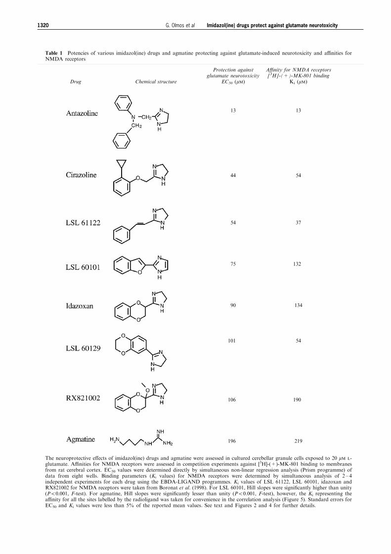
Table 1 Potencies of various imidazol(ine) drugs and agmatine protecting against glutamate-induced neurotoxicity and a�nities forNMDA receptors
Drug Chemical structure
Protection againstglutamate neurotoxicity
EC50 (mM)
A�nity for NMDA receptors[3H]-(+)-MK-801 binding
Ki (mM)
13
44
54
75
90
101
106
196
13
54
37
132
134
54
190
219
The neuroprotective e�ects of imidazol(ine) drugs and agmatine were assessed in cultured cerebellar granule cells exposed to 20 mM L-glutamate. A�nities for NMDA receptors were assessed in competition experiments against [3H]-(+)-MK-801 binding to membranesfrom rat cerebral cortex. EC50 values were determined directly by simultaneous non-linear regression analysis (Prism programme) ofdata from eight wells. Binding parameters (Ki values) for NMDA receptors were determined by simultaneous analysis of 2 ± 4independent experiments for each drug using the EBDA-LIGAND programmes. Ki values of LSL 61122, LSL 60101, idazoxan andRX821002 for NMDA receptors were taken from Boronat et al. (1998). For LSL 60101, Hill slopes were signi®cantly higher than unity(P50.001, F-test). For agmatine, Hill slopes were signi®cantly lesser than unity (P50.001, F-test), however, the Ki representing thea�nity for all the sites labelled by the radioligand was taken for convenience in the correlation analysis (Figure 5). Standard errors forEC50 and Ki values were less than 5% of the reported mean values. See text and Figures 2 and 4 for further details.
Imidazol(ine) drugs protect against glutamate neurotoxicity1320 G. Olmos et al
Results
Neuroprotective e�ects of imidazol(ine) drugs andagmatine against L-glutamate-induced neurotoxicity incultured cerebellar granule cells
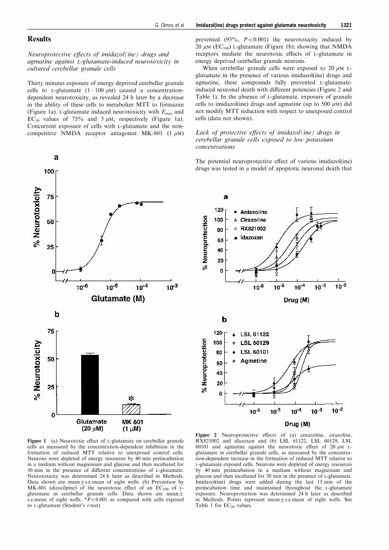
Thirty minutes exposure of energy deprived cerebellar granulecells to L-glutamate (1 ± 100 mM) caused a concentration-
dependent neurotoxicity, as revealed 24 h later by a decreasein the ability of these cells to metabolize MTT to formazan(Figure 1a). L-glutamate induced neurotoxicity with Emax and
EC50 values of 75% and 5 mM, respectively (Figure 1a).Concurrent exposure of cells with L-glutamate and the non-competitive NMDA receptor antagonist MK-801 (1 mM)
prevented (93%, P50.001) the neurotoxicity induced by20 mM (EC100) L-glutamate (Figure 1b); showing that NMDAreceptors mediate the neurotoxic e�ects of L-glutamate in
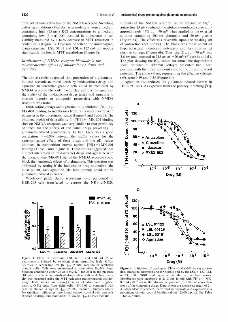
energy deprived cerebellar granule neurons.When cerebellar granule cells were exposed to 20 mM L-
glutamate in the presence of various imidazol(ine) drugs andagmatine, these compounds fully prevented L-glutamate-
induced neuronal death with di�erent potencies (Figure 2 andTable 1). In the absence of L-glutamate, exposure of granulecells to imidazol(ine) drugs and agmatine (up to 500 mM) did
not modify MTT reduction with respect to unexposed controlcells (data not shown).
Lack of protective e�ects of imidazol(ine) drugs incerebellar granule cells exposed to low potassiumconcentrations
The potential neuroprotective e�ect of various imidazol(ine)drugs was tested in a model of apoptotic neuronal death that
Figure 1 (a) Neurotoxic e�ect of L-glutamate on cerebellar granulecells as measured by the concentration-dependent inhibition in theformation of reduced MTT relative to unexposed control cells.Neurons were depleted of energy resources by 40 min preincubationin a medium without magnesium and glucose and then incubated for30 min in the presence of di�erent concentrations of L-glutamate.Neurotoxicity was determined 24 h later as described in Methods.Data shown are mean+s.e.mean of eight wells. (b) Prevention byMK-801 (dizocilpine) of the neurotoxic e�ect of an EC100 of L-glutamate in cerebellar granule cells. Data shown are mean+s.e.mean of eight wells. *P50.001 as compared with cells exposedto L-glutamate (Student's t-test).
Figure 2 Neuroprotective e�ects of (a) antazoline, cirazoline,RX821002 and idazoxan and (b) LSL 61122, LSL 60129, LSL60101 and agmatine against the neurotoxic e�ect of 20 mM L-glutamate in cerebellar granule cells, as measured by the concentra-tion-dependent increase in the formation of reduced MTT relative toL-glutamate exposed cells. Neurons were depleted of energy resourcesby 40 min preincubation in a medium without magnesium andglucose and then incubated for 30 min in the presence of L-glutamate.Imidazol(ine) drugs were added during the last 15 min of thepreincubation time and maintained throughout the L-glutamateexposure. Neuroprotection was determined 24 h later as describedin Methods. Points represent mean+s.e.mean of eight wells. SeeTable 1 for EC50 values.
Imidazol(ine) drugs protect against glutamate neurotoxicity 1321G. Olmos et al
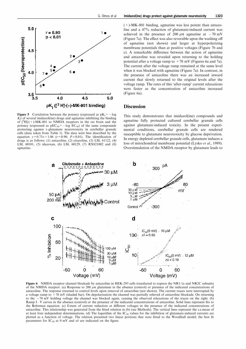
does not involve activation of the NMDA receptor. Switchingculturing conditions of cerebellar granule cells from a mediumcontaining high (25 mM) KCl concentrations to a medium
containing low (5 mM) KCl resulted in a decrease in cellviability measured by a 64% decrease in MTT reduction incontrol cells (Figure 3). Exposure of cells to the imidazol(ine)drugs cirazoline, LSL 60101 and LSL 61122 did not modify
signi®cantly the loss in MTT metabolism (Figure 3).
Involvement of NMDA receptor blockade in theneuroprotective e�ects of imidazol(ine) drugs andagmatine
The above results suggested that prevention of L-glutamate-induced necrotic neuronal death by imidazol(ine) drugs andagmatine in cerebellar granule cells could be mediated by
NMDA receptor blockade. To further address this question,the ability of the imidazol(ine) drugs tested and agmatine tointeract (agonist or antagonist properties) with NMDAreceptors was tested.
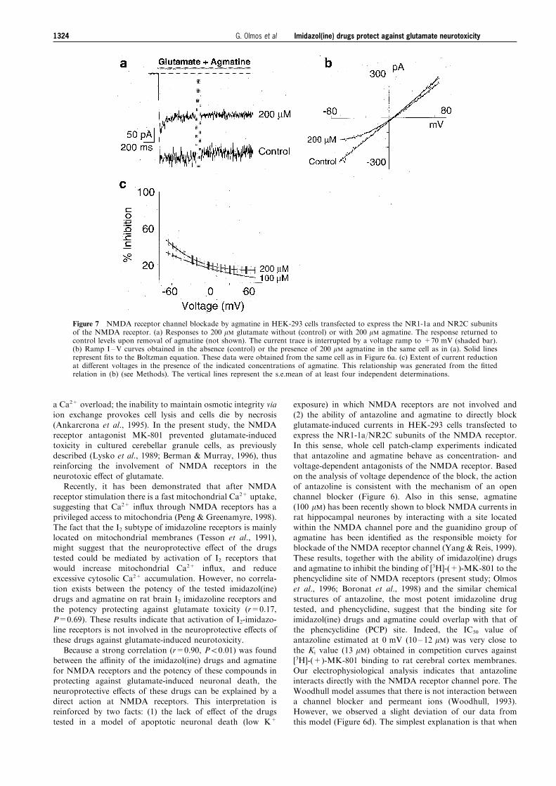
Imidazol(ine) drugs and agmatine fully inhibited [3H]-(+)-MK-801 binding to membranes from rat cerebral cortex withpotencies in the micromolar range (Figure 4 and Table 1). The
obtained pro®le of drug a�nity for [3H]-(+)-MK-801 bindingsites on NMDA receptors was very similar to that previouslyobtained for the e�ects of the same drugs preventing L-
glutamate-induced neurotoxicity. In fact, there was a goodcorrelation (r=0.90) between the pEC50 values for theneuroprotective e�ects of these drugs and the pKi values
obtained in competition curves against [3H]-(+)-MK-801binding (Table 1 and Figure 5). These results suggested thata direct interaction of imidazol(ine) drugs and agmatine withthe phencyclidine/MK-801 site of the NMDA receptor could
block the neurotoxic e�ects of L-glutamate. This question wasaddressed by testing if the imidazoline drug antazoline (themost potent) and agmatine (the least potent) could inhibit
glutamate-induced currents.Whole-cell patch clamp recordings were performed in
HEK-293 cells transfected to express the NR1-1a/NR2C
subunits of the NMDA receptor. In the absence of Mg2+,antazoline (3 mM) reduced the glutamate-induced current byapproximately 45% at 770 mV when applied in the external
solution containing 200 mM glutamate and 20 mM glycine(Figure 6a). The e�ect was reversible upon the washing o�of antazoline (not shown). The block was most potent athyperpolarizing membrane potentials and less e�ective at
positive voltages (Figure 6b). Thus, the IC50 at 770 mV was3.6 mM and increased to 215 mM at +70 mV (Figure 6c and d ).The plot showing the IC50 values for antazoline (logarithmic
scale) obtained at di�erent voltages presented two linearportions, with the in¯ection point close to the current reversalpotential. The slope values, representing the e�ective valences
(zd), were 0.19 and 0.55 (Figure 6d).Agmatine also reduced the glutamate-induced current in
HEK-293 cells. As expected from the potency inhibiting [3H]-
Figure 3 E�ect of cirazoline, LSL 60101 and LSL 61122 onneurotoxicity induced by switching from serum-free high [K+]out(25 mM) to serum-free low [K+]out (5 mM) medium in cerebellargranule cells. Cells were maintained in serum-free Eagle's BasalMedium containing either 25 or 5 mM K+ for 24 h in the presence(100 mM) or absence (control) of drugs where indicated. Neurotoxi-city was measured using the MTT reduction (mitochondrial activity)assay. Data shown are mean+s.e.mean of absorbance (opticaldensity, O.D.) units from eight wells. *P50.05 as compared withcells maintained in high [K+]out (25 mM) medium (Student's t-test).No signi®cant di�erences were found between control cells and cellsexposed to drugs and maintained in low [K+]out (5 mM) medium.
Figure 4 Inhibition of binding of [3H]-(+)-MK-801 by (a) antazo-line, cirazoline, idazoxan and RX821002 and by (b) LSL 61122, LSL60129, LSL 60101 and agmatine in the rat cerebral cortex.Membranes were incubated at 238C for 45 min with [3H]-(+)-MK-801 (461079
M) in the absence or presence of di�erent concentra-tions of the competing drugs. Data shown are mean+s.e.mean of 2 ±4 independent experiments performed in triplicate and expressed as apercentage of total control binding (about 12,000 d.p.m.). See Table1 for Ki values.
Imidazol(ine) drugs protect against glutamate neurotoxicity1322 G. Olmos et al
(+)-MK-801 binding, agmatine was less potent than antazo-line and a 47% reduction of glutamate-induced current wasachieved in the presence of 200 mM agmatine at 770 mV
(Figure 7a). The e�ect was also reversible upon the washing o�of agmatine (not shown) and larger at hyperpolarizingmembrane potentials than at positive voltages (Figure 7b andc). A remarkable di�erence between the action of agmatine
and antazoline was revealed upon returning to the holdingpotential after a voltage ramp to +70 mV (Figures 6a and 7a).The current after the voltage ramp remained at the same level
when it was blocked with agmatine (Figure 7a). In contrast, inthe presence of antazoline there was an increased inwardcurrent that slowly returned to the original levels after the
voltage ramp. The rates of this `after-ramp' current relaxationswere faster as the concentration of antazoline increased(Figure 6a).
Discussion
This study demonstrates that imidazol(ine) compounds andagmatine fully protected cultured cerebellar granule cellsagainst glutamate-induced toxicity. In the present experi-
mental conditions, cerebellar granule cells are renderedsusceptible to glutamate neurotoxicity by glucose deprivation.In energy depleted cerebellar granule cells, glutamate induces a
loss of mitochondrial membrane potential (Lysko et al., 1989).Overstimulation of the NMDA receptor by glutamate leads to
Figure 5 Correlation between the potency (expressed as pKi=7logKi) of several imidazol(ine) drugs and agmatine inhibiting the bindingof [3H]-(+)-MK-801 to NMDA receptors in the rat brain and thepotency (expressed as pEC50=7log EC50) of the same compoundsprotecting against L-glutamate neurotoxicity in cerebellar granulecells (data taken from Table 1). The data were best described by theequation y=0.75x+1.06 (r=0.90; P50.01). The identi®cation ofdrugs is as follows: (1) antazoline, (2) cirazoline, (3) LSL 61122, (4)LSL 60101, (5) idazoxan, (6) LSL 60129, (7) RX821002 and (8)agmatine.
Figure 6 NMDA receptor channel blockade by antazoline in HEK-293 cells transfected to express the NR1-1a and NR2C subunitsof the NMDA receptor. (a) Response to 200 mM glutamate in the absence (control) or presence of the indicated concentrations ofantazoline. The response returned to control levels upon removal of antazoline (not shown). The current traces were interrupted bya voltage ramp to +70 mV (shaded bar). On depolarization the channel was partially relieved of antazoline blockade. On returningto the 770 mV holding voltage the channel was blocked again, causing the observed relaxations of the traces on the right. (b)Ramp I ±V curves in the absence (control) or the presence of the indicated concentrations of antazoline. Solid lines represent ®ts tothe Boltzman equation. (c) Extent of current reduction at di�erent voltages in the presence of the indicated concentrations ofantazoline. This relationship was generated from the ®tted relation in (b) (see Methods). The vertical lines represent the s.e.mean ofat least four independent determinations. (d) The logarithm of the IC50 values for the inhibition of glutamate-induced currents areplotted as a function of voltage. The relation presented two linear portions that were ®tted to the Woodhull model; the best ®tparameters for IC50 at 0 mV and zd are indicated on the ®gure.
Imidazol(ine) drugs protect against glutamate neurotoxicity 1323G. Olmos et al
a Ca2+ overload; the inability to maintain osmotic integrity via
ion exchange provokes cell lysis and cells die by necrosis(Ankarcrona et al., 1995). In the present study, the NMDAreceptor antagonist MK-801 prevented glutamate-inducedtoxicity in cultured cerebellar granule cells, as previously
described (Lysko et al., 1989; Berman & Murray, 1996), thusreinforcing the involvement of NMDA receptors in theneurotoxic e�ect of glutamate.
Recently, it has been demonstrated that after NMDAreceptor stimulation there is a fast mitochondrial Ca2+ uptake,suggesting that Ca2+ in¯ux through NMDA receptors has a
privileged access to mitochondria (Peng & Greenamyre, 1998).The fact that the I2 subtype of imidazoline receptors is mainlylocated on mitochondrial membranes (Tesson et al., 1991),might suggest that the neuroprotective e�ect of the drugs
tested could be mediated by activation of I2 receptors thatwould increase mitochondrial Ca2+ in¯ux, and reduceexcessive cytosolic Ca2+ accumulation. However, no correla-
tion exists between the potency of the tested imidazol(ine)drugs and agmatine on rat brain I2 imidazoline receptors andthe potency protecting against glutamate toxicity (r=0.17,
P=0.69). These results indicate that activation of I2-imidazo-line receptors is not involved in the neuroprotective e�ects ofthese drugs against glutamate-induced neurotoxicity.
Because a strong correlation (r=0.90, P50.01) was foundbetween the a�nity of the imidazol(ine) drugs and agmatinefor NMDA receptors and the potency of these compounds inprotecting against glutamate-induced neuronal death, the
neuroprotective e�ects of these drugs can be explained by adirect action at NMDA receptors. This interpretation isreinforced by two facts: (1) the lack of e�ect of the drugs
tested in a model of apoptotic neuronal death (low K+
exposure) in which NMDA receptors are not involved and
(2) the ability of antazoline and agmatine to directly blockglutamate-induced currents in HEK-293 cells transfected toexpress the NR1-1a/NR2C subunits of the NMDA receptor.In this sense, whole cell patch-clamp experiments indicated
that antazoline and agmatine behave as concentration- andvoltage-dependent antagonists of the NMDA receptor. Basedon the analysis of voltage dependence of the block, the action
of antazoline is consistent with the mechanism of an openchannel blocker (Figure 6). Also in this sense, agmatine(100 mM) has been recently shown to block NMDA currents in
rat hippocampal neurones by interacting with a site locatedwithin the NMDA channel pore and the guanidino group ofagmatine has been identi®ed as the responsible moiety forblockade of the NMDA receptor channel (Yang & Reis, 1999).
These results, together with the ability of imidazol(ine) drugsand agmatine to inhibit the binding of [3H]-(+)-MK-801 to thephencyclidine site of NMDA receptors (present study; Olmos
et al., 1996; Boronat et al., 1998) and the similar chemicalstructures of antazoline, the most potent imidazoline drugtested, and phencyclidine, suggest that the binding site for
imidazol(ine) drugs and agmatine could overlap with that ofthe phencyclidine (PCP) site. Indeed, the IC50 value ofantazoline estimated at 0 mV (10 ± 12 mM) was very close to
the Ki value (13 mM) obtained in competition curves against[3H]-(+)-MK-801 binding to rat cerebral cortex membranes.Our electrophysiological analysis indicates that antazolineinteracts directly with the NMDA receptor channel pore. The
Woodhull model assumes that there is not interaction betweena channel blocker and permeant ions (Woodhull, 1993).However, we observed a slight deviation of our data from
this model (Figure 6d). The simplest explanation is that when
Figure 7 NMDA receptor channel blockade by agmatine in HEK-293 cells transfected to express the NR1-1a and NR2C subunitsof the NMDA receptor. (a) Responses to 200 mM glutamate without (control) or with 200 mM agmatine. The response returned tocontrol levels upon removal of agmatine (not shown). The current trace is interrupted by a voltage ramp to +70 mV (shaded bar).(b) Ramp I ±V curves obtained in the absence (control) or the presence of 200 mM agmatine in the same cell as in (a). Solid linesrepresent ®ts to the Boltzman equation. These data were obtained from the same cell as in Figure 6a. (c) Extent of current reductionat di�erent voltages in the presence of the indicated concentrations of agmatine. This relationship was generated from the ®ttedrelation in (b) (see Methods). The vertical lines represent the s.e.mean of at least four independent determinations.
Imidazol(ine) drugs protect against glutamate neurotoxicity1324 G. Olmos et al
the net current is outward (many ions ¯ow in the outwarddirection) the permeating ions expel the blocker from itsbinding site on the pore (Antonov et al., 1998) or alternatively,
that divalent cations interact with the blocker binding sitewithin the pore (Lerma et al., 1991). The neuroprotective e�ectof imidazoline drugs and agmatine against glutamate-inducedneurotoxicity is speci®c and related to NMDA receptor
blockade, since neurotoxicity in cerebellar granule cells byother mechanisms not involving glutamate receptor activationcannot be prevented by glutamate receptor antagonists
(Dargent et al., 1996) nor imidazol(ine) drugs.The potencies (micromolar range) of imidazol(ine) drugs
and agmatine at blocking NMDA receptors ([3H]-(+)-MK-
801 binding) are in good agreement with the potencies of thesame compounds at blocking other cation channels, such asK+
ATP channels (Jonas et al., 1992; Shepherd et al., 1996),
nicotinic acetylcholine (Musgrave et al., 1995) and 5-HT3
receptors (Molderings et al., 1996). Furthermore, as alsodemonstrated for these cation channels, imidazol(ine) drugspossessing two benzene rings (e.g. antazoline) are more potent
on the NMDA receptor than others with only one aromaticring (e.g. idazoxan) and these, in turn, are more potent thanother molecules lacking an aromatic ring (e.g. agmatine)
(Table 1). Hydrocarbon substituents on the benzene ring (e.g.cirazoline) increase the a�nity of imidazol(ine) drugs for theNMDA receptor, as demonstrated for the interaction of these
drugs with other cation channels (see Sakuta & Okamoto, 1994for comparison). These ®ndings, together with the observationthat there are good correlations between the pro®les of drug
a�nity of imidazol(ine) compounds at blocking di�erentcation channels (K+
ATP channels, NMDA and 5-HT3
receptors) (Olmos et al., 1996; Molderings, 1997), reinforcethe hypothesis of the existence of a common imidazol(ine)/
agmatine binding site on all these cation channels (Olmos etal., 1996).
The present results indicate that NMDA receptor blockade
by imidazol(ine) drugs and agmatine is anti-excitotoxic in an in
vitro model of necrotic neuronal death. The imidazoline drugidazoxan and agmatine can also exert neuroprotection afterfocal or global ischaemia (Gustafson et al., 1989; 1990; Maiese
et al., 1992; Gilad et al., 1996), two in vivo models of necroticneuronal death in which clear protective e�ects of NMDAreceptor antagonists have been demonstrated (Meldrum, 1990;Holt, 1997). However, the relatively low a�nity of idazoxan
and agmatine for NMDA receptors and the fact that the extentof agmatine transport into the brain is unknown, make itdi�cult that the weak interaction of these drugs with NMDA
receptors would also explain the survival-promoting e�ectsfound in vivo. In fact, it has been suggested that theneuroprotective e�ect of idazoxan after global ischaemia in
the rat is related to the hypothermic e�ects of this drug(Craven & Conway, 1997). Also in this context, competitiveand non-competitive NMDA receptor antagonists have been
proved to attenuate the development of tolerance to morphine-induced antinociception (Trujillo & Akil, 1991; Tiseo et al.,1994). Although the same e�ect has been observed afterconcurrent chronic treatment of morphine with various
imidazol(ine) drugs, no relationship between the potency ofthe drugs tested on NMDA receptors and their ability toattenuate morphine tolerance was found (Boronat et al., 1998).
In conclusion, the present study indicates that imidazol(ine)drugs and agmatine are neuroprotective in an in vitro model ofglutamate-induced necrotic neuronal death and that this e�ect
is mediated through NMDA receptor blockade. Furtherstudies are needed to establish the possible in vivo functionalimplications of these ®ndings.
This study was supported by DGICYT Grants PB94-0002-Mod C(J.A. Garcõ a-Sevilla), PB94-0017 (R. Trullas) and PM-0008/96 (J.Lerma) from the Ministerio de Educacio n y Cultura. M.A. Boronatwas supported by a fellowship from the Universitat de les IllesBalears. We are grateful to Consell Insular de Mallorca for supportand to the pharmaceuticals ®rms for gifts of drugs. J.A. Garcõ a-Sevilla is a member of the Institut d'Estudis Catalans.
References
ANIS, N., SHERBY, S., GOODNOW, JR R., NIWA, M., KONNO, K.,
KALLIMOPOULOS, T., BUKOWNIK, R., NAKANISHI, K., USHER-
WOOD, P., ELDEFRAWI, A. & ELDEFRAWI, M. (1990). Structure-activity relationships of philanthotoxin analogs and polyamineson N-methyl-D-aspartate and nicotinic acetylcholine receptors.J. Pharmacol. Exp. Ther., 254, 764 ± 773.
ANKARCRONA, M., DYPBUKT, J.M., BONFOCO, E., ZHIVOTOVSKY,
B., ORRENIUS, S., LIPTON, S.A. & NICOTERA, P. (1995).Glutamate-induced neuronal death: a succession of necrosis orapoptosis depending on mitochondrial function. Neuron, 15,961 ± 973.
ANTONOV, S.M., GMIRO, V.E. & JOHNSON, J.W. (1998). Binding sitesfor permeant ions in the channel of NMDA receptors and theire�ect on channel block. Nature Neurosci., 1, 451 ± 461.
BERDEU, D., PUECH, R., RIBES, G., LOUBATIEÁ RES-MARIANI, M.-M.
& BERTRAND, G. (1997). Antazoline increases insulin secretionand improves glucose tolerance in rats and dogs. Eur. J.Pharmacol., 324, 233 ± 239.
BERMAN, F.W. & MURRAY, T.F. (1996). Characterization of[3H]MK-801 binding to N-methyl-D-aspartate receptors incultured rat cerebellar granule neurons and involvement inglutamate-mediated toxicity. J. Biochem. Toxicol., 11, 217 ± 226.
BOJE, K.M., WONG, G. & SKOLNICK, P. (1993). Desensitization ofthe NMDA receptor complex by glycinergic ligands in cerebellargranule cell cultures. Brain Res., 603, 207 ± 214.
BORONAT, M.A., OLMOS, G. & GARCIÂ A-SEVILLA, J.A. (1998).Attenuation of tolerance to opioid-induced antinociception andprotection against morphine-induced decrease of neuro®lamentproteins by idazoxan and other I2-imidazoline ligands. Br. J.Pharmacol., 125, 175 ± 185.
BOUSQUET, P. (1995). Imidazoline receptors: from basic concepts torecent developments. J. Cardiovasc. Pharmacol., 26, S1 ± S6.
CHOI, D.W. (1988). Calcium-mediated neurotoxicity: relationship tospeci®c channel types and role in ischemic damage. TrendsNeurosci., 11, 465 ± 469.
COUPRY, I., ARMSBY, C.C., ALPER, S.L., BRUGNARA, C. & PARINI,
A. (1996). Clotrimazole and efaroxan inhibit red cell Gardoschannel independently of imidazoline I1 and I2 binding sites. Eur.J. Pharmacol., 295, 109 ± 112.
CRAVEN, J.A. & CONWAY, E.L. (1997). E�ects of a2-adrenoceptorantagonists and imidazoline2-receptor ligands on neuronaldamage in global ischaemia in the rat. Clin. Exp. Pharmacol.Physiol., 24, 204 ± 207.
DARGENT, B., ARSAC, C., TRICAUD, N. & COURAUD, F. (1996).Activation of voltage-dependent sodium channels in culturedcerebellar granule cells induces neurotoxicity that is not mediatedby glutamate release. Neuroscience, 73, 209 ± 216.
DEGREGORIO-ROCASOLANO, N., OLMOS, G., GASULL, T., BOR-
ONAT, M., TRULLAS, R. & GARCIÂ A-SEVILLA, J.A. (1999).Protection by imidazol(ine) compounds of L-glutamate neuro-toxicity through NMDA receptor blockade. Ann. N.Y. Acad.Sci., in press.
DESSI, F., CHARRIAUT-MARLANGUE, C., KHRESTCHATISKY, M.
& BEN-ARI, Y. (1993). Glutamate-induced neuronal death is not aprogrammed cell death in cerebellar culture. J. Neurochem., 60,1953 ± 1955.
D'MELLO, S.R., GALLI, C., CIOTTI, T. & CALISSANO, P. (1993).Induction of apoptosis in cerebellar granule neurons by lowpotassium: inhibition of death by insulin-like growth factor I andcAMP. Proc. Natl. Acad. Sci. U.S.A., 90, 10989 ± 10993.
Imidazol(ine) drugs protect against glutamate neurotoxicity 1325G. Olmos et al
FOSSOM, L.H., BASILE, A.S. & SKOLNICK, P. (1995). Sustainedexposure to 1-aminocyclopropanecarboxylic acid, a glycinepartial agonist, alters N-methyl-D-aspartate receptor functionand subunit composition. Mol. Pharmacol., 48, 981 ± 987.
FOSTER, A.C. & WONG, E.H.F. (1987). The novel anticonvulsantMK-801 binds to the activated state of the N-methyl-D-aspartatereceptor in rat brain. Br. J. Pharmacol., 91, 403 ± 409.
FRENCH, N. (1995) a2-adrenoceptors and I2 sites in the mammaliancentral nervous system. Pharmacol. Ther., 68, 175 ± 208.
GILAD, G.M., SALAME, K., RABEY, J.M. & GILAD, V.H. (1996).Agmatine treatment is neuroprotective in rodent brain injurymodels. Life Sci., 58, PL41 ± 46.
GUSTAFSON, I., WESTERBORG, E. & WIELOCH, T.W. (1990).Protection against ischemia-induced neuronal damage by a2-adrenoceptor antagonist idazoxan: In¯uence of time of admin-istration and possible mechanisms of action. J. Cereb. FlowMetab., 10, 885 ± 894.
GUSTAFSON, I., YOSHITOYO, M. & WIELOCH, T.W. (1989).Postischemic administration of idazoxan, an a2-adrenergicreceptor antagonist, decreases neuronal damage in the rat brain.J. Cereb. Blood Flow Metab., 9, 171 ± 174.
HOLT, W.F. (1997). Glutamate in health and disease: the role ofinhibitors. In Neuroprotection in CNS diseases, eds. BaÈ r, P.R. &Beal, M.F. pp. 87 ± 119. New-York: Marcel Dekker, Inc.
ISHITANI, R., SUNAGA, K., TANAKA, M., AISHITA, H. & CHUANG,
D.-W. (1997). Overexpression of glyceraldehyde-3-phosphatedehydrogenase is involved in low K+-induced apoptosis butnot necrosis of cultured cerebellar granule cells.Mol. Pharmacol.,51, 542 ± 550.
JONAS, J.C., PLANT, T.D. & HENQUIN, J.C. (1992). Imidazolineantagonists of a2-adrenoceptors increase insulin release in vitroby inhibiting ATP-sensitive K+ channels in pancreatic b-cells.Br. J. Pharmacol., 107, 8 ± 14.
LERMA, J.L., PATERNAIN, A.V., SALVADOR, N., SOMOHANO, F.,
MORALES, M. & CASADO, M. (1998). Excitatory amino acid-activated channels. In Ion Channel Pharmacology. eds. Soria, B.& CenÄ a, V. pp. 399 ± 421. Oxford: Oxford University Press.
LERMA, J., ZUKIN, R.S. & BENNETT, M.V.L. (1991). Interaction ofMg2+ and phencyclidine in use-dependent block of NMDAchannels. Neurosci. Lett., 123, 187 ± 191.
LI, G., REGUNATHAN, S., BARROW, C.J., ESHRAGHI, J., COOPER, R.
& REIS, D.J. (1994). Agmatine ± an endogenous clonidine-displacing substance in the brain. Science, 263, 966 ± 969.
LYSKO, P.G., COX, J.A., VIGANO, A. & HENNEBERRY, R.C. (1989).Excitatory amino acid neurotoxicity at the N-methyl-D-aspar-tate receptor in cultured neurons: pharmacological characteriza-tion. Brain Res., 499, 258 ± 266.
MAIESE, K., PEK, L.. BERGER, S.B. & REIS, D.J. (1992). Reduction infocal cerebral ischemia by agents acting at imidazole receptors. J.Cereb. Blood Flow Metab., 12, 53 ± 63.
MCPHERSON, G.A. (1985). Analysis of radioligand binding experi-ments: a collection of computer programs for IBM PC. J.Pharmacol. Meth., 14, 213 ± 228.
MELDRUM, B. (1990). Protection against ischemic neuronal damageby drugs acting on excitatory neurotransmission. Cerebrovasc.Brain Metab. Rev., 2, 27 ± 57.
MILANI, D., GUIDOLIN, D., FACCI, L., POZZAN, T., BUSO, M., LEON,
A. & SKAPER, S.D. (1991). Excitatory amino acid-inducedalterations of cytoplasmic free Ca2+ in individual cerebellargranule neurons: role in neurotoxicity. J. Neurosci. Res., 28,434 ± 441.
MOLDERINGS, G.J. (1997). Imidazoline receptors: basic knowledge,recent advances and future prospects for therapy and diagnosis.Drugs Fut., 22, 757 ± 772.
MOLDERINGS, G.J., SCHMIDT, K., BOÈ NISCH, H. & GOÈ THERT, M.
(1996). Inhibition of 5-HT3 receptor function by imidazolines inmouse neuroblastoma cells: potential involvement of s2 bindingsites. Nauynn-Schmied. Arch. Pharmacol., 354, 245 ± 252.
MOSMANN, T. (1983). Rapid colorimetric assay for cellular growthand survival: application to proliferation and cytotoxicity assays.J. Immunol. Meth., 65, 55 ± 63.
MUNSON, P.J. & RODBARD, D. (1980). LIGAND: a versatilecomputerized approach for the characterization of ligandbinding systems. Anal. Biochem., 107, 220 ± 239.
MUSGRAVE, I.F., KRAUTWURST, D., HESCHELER, J. & SCHULTZ,
G. (1995). Clonidine and cirazoline inhibit activation of nicotinicchannels in PC-12 cells. Ann. N.Y. Acad. Sci., 763, 272 ± 283.
OHARA-IMAIZUMI, M. & KUMAKURA, K. (1992). E�ects ofimidazole compounds on catecholamine release in adrenalchroma�n cells. Cell. Mol. Neurobiol., 12, 273 ± 283.
OLMOS, G., CASANOVAS, A., RIBERA, J., BORONAT, M.A., ESQUER-
DA, J.E. & GARCIÂ A-SEVILLA, J.A. (1999). Prevention byLSL 60101 [2-(2-benzofuranyl) imidazole) of motoneuron celldeath after neonatal axotomy. Ann. N.Y. Acad. Sci., in press.
OLMOS, G., KULKARNI, R.N., HAQUE, M. & MACDERMOT, J.
(1994). Imidazolines stimulate release of insulin from RIN-5AH cells independently from imidazoline I1 and I2 receptors.Eur. J. Pharmacol., 262, 41 ± 48.
OLMOS, G., RIBERA, J. & GARCIÂ A-SEVILLA, J.A. (1996). Imidazo-li(di)ne compounds interact with the phencyclidine site ofNMDA receptors in the rat brain. Eur. J. Pharmacol., 310,273 ± 276.
OZAITA, A., OLMOS, G., BORONAT, M.A., LIZCANO, J.M., UNZETA,
M. & GARCIÂ A-SEVILLA, J.A. (1997). Inhibition of monoamineoxidase A and B activities by imidazol(ine)/guanidine drugs,nature of the interaction and distinction from I2-imidazolinereceptors in rat liver. Br. J. Pharmacol., 121, 901 ± 912.
PENG, T.-I. & GREENAMYRE, J.T. (1998). Privileged access tomitochondria of calcium in¯ux through N-methyl-D-aspartate-receptors. Mol. Pharmacol., 53, 974 ± 980.
PROKS, P. & ASHCROFT, F.M. (1997). Phentolamine block KATP
channels is mediated by Kir6.2. Proc. Natl. Acad. Sci. U.S.A., 94,11716 ± 11720.
REGUNATHAN, S. & REIS, D.J. (1996). Imidazoline receptors andtheir endogenous ligands. Annu. Rev. Pharmacol. Toxicol., 36,511 ± 544.
SAKUTA, H. & OKAMOTO, K. (1994). Inhibition by imidazoline andimidazolidine derivatives of glibenclamide-sensitive K+ currentsin Xenopus oocytes. Eur. J. Pharmacol., 259, 223 ± 231.
SCHRAMM, M., EIMERL, S. & COSTA, E. (1990). Serum anddepolarizing agents cause acute neurotoxicity in culturedcerebellar granule cells: role of glutamate receptor responsiveto N-methyl-D-aspartate. Proc. Natl. Acad. Sci. U.S.A., 87,1193 ± 1197.
SHEPHERD, R.M., HASHMI, M.N., KANE, C., SQUIRES, P.E. &
DUNNE, M.J. (1996). Elevation of cytosolic calcium byimidazolines in mouse islets of Langerhans: implications forstimulus response coupling of insulin release. Br. J. Pharmacol.,119, 911 ± 916.
TESSON, F., PRIP-BUUS, C., LEMOINE, A., PEGORIER, J.-P. &
PARINI, A. (1991). Subcellular distribution of imidazoline-guanidinium-receptive sites in human and rabbit liver. J. Biol.Chem., 266, 155 ± 160.
TISEO, P.J., CHENG, J., PASTERNAK, G.W. & INTURRISI, C.E. (1994).Modulation of morphine tolerance by the competitive N-methyl-D-aspartate receptor antagonist LY274614: assessment of opiodreceptor changes. J. Pharmacol. Exp. Ther., 268, 195 ± 201.
TRUJILLO, K.A. & AKIL, H. (1991). Inhibition of morphine toleranceand dependence by the NMDA receptor antagonist MK-801.Science, 251, 85 ± 87.
VILLARROEL, A., REGALADO, M.P. & LERMA, J. (1998). Glycine-dependent desensitization: localization of molecular determi-nants. Neuron, 20, 329 ± 339.
WOODHULL, A.M. (1973). Ionic blockade of sodium channel innerve. J. Gen. Physiol., 61, 687 ± 708.
YANG, X.C. & REIS, D.J. (1999). Agmatine selectively blocks the N-methyl-D-aspartate subclass of glutamate receptor channels inrat hippocampal neurons. J. Pharmacol. Exp. Ther., 288, 544 ±549.
(Received March 22, 1999Revised April 21, 1999
Accepted April 23, 1999)
Imidazol(ine) drugs protect against glutamate neurotoxicity1326 G. Olmos et al


![Substituted Imidazole of 5-Fluoro-2-[4-[(2-phenyl-1H-imidazol-5-yl)methyl]-1-piperazinyl]pyrimidine Inactivates Cytochrome P450 2D6 by Protein Adduction](https://img.pdfslide.net/doc/110x75/635224a47a164e65570ac17f/substituted-imidazole-of-5-fluoro-2-4-2-phenyl-1h-imidazol-5-ylmethyl-1-piperazinylpyrimidine.jpg)


