Embed Size (px)
Citation preview

arX
iv:1
109.
4232
v1 [
q-bi
o.B
M]
20
Sep
2011
Protein multi-scale organization through graph partitioning and robustness analysis:
Application to the myosin-myosin light chain interaction
A. Delmotte,1, 3, ∗ E.W. Tate,2, 3 S.N. Yaliraki,2, 3 and M. Barahona1, 3, †
1Department of Mathematics, Imperial College London, London SW7 2AZ, UK2Department of Chemistry, Imperial College London, London SW7 2AZ, UK
3British Heart Foundation Centre of Research Excellence,
Imperial College London, London SW7 2AZ, UK
Despite the recognized importance of the multi-scale spatio-temporal organization of proteins,most computational tools can only access a limited spectrum of time and spatial scales, thereby ig-noring the effects on protein behavior of the intricate coupling between the different scales. Startingfrom a physico-chemical atomistic network of interactions that encodes the structure of the protein,we introduce a methodology based on multi-scale graph partitioning that can uncover partitions andlevels of organization of proteins that span the whole range of scales, revealing biological featuresoccurring at different levels of organization and tracking their effect across scales. Additionally, weintroduce a measure of robustness to quantify the relevance of the partitions through the generationof biochemically-motivated surrogate random graph models. We apply the method to four distinctconformations of myosin tail interacting protein, a protein from the molecular motor of the malariaparasite, and study properties that have been experimentally addressed such as the closing mecha-nism, the presence of conserved clusters, and the identification through computational mutationalanalysis of key residues for binding.
Keywords: multi-scale graph partitioning, robustness analysis, variation of information, myosin tail interact-ing protein, myosin-myosin light chains interactions, random graph surrogates
I. INTRODUCTION
A. The methodology: Multi-scale analysis of
protein structures
Proteins are complex structures characterized by mul-tiple scales in time and space [1–4]. Atoms, functionalchemical groups, amino acids, the ensuing secondarystructures, the large conformational domains: each definedifferent, yet coupled, levels of structural and dynami-cal organization linked to behaviors occurring at differ-ent time and spatial scales. Molecular dynamics simula-tions [5] can deal successfully with the very short timescales but such methods cannot be applied to long times(or large systems) due to their exorbitant computationalcost. On the other hand, because many of the key bi-ological functions take place at the micro- to millisec-ond time scales, strongly simplified coarse-grained sys-tems have been proposed as a means to reaching the bi-ologically relevant regimes [6–9]. However, these simpli-fied coarse-grainings often ignore the detailed physico-chemical atomic interactions and, consequently, cannotprovide a picture that emerges seamlessly from the small-est scales.Yet the different levels of organization in proteins do
not behave independently: the dynamics at long timeand length scales, which is in many cases crucial for bi-ological function, is the result of the integrative interac-tion of the finer organizational levels. Analyzing proteinsfrom this multi-scale perspective can reveal the intricate
∗ [email protected]† [email protected]
linkage between the levels and give insight into the be-havior of the protein starting from the bottom-up. Thispicture can also aid in the understanding of the effectsthat small-scale changes like mutations have on large-scale organization. To achieve this, we apply a recentlyproposed general methodology for multi-scale graph par-titioning [10] that uses dynamical processes on graphs touncover the multi-level organization of graph communi-ties relevant at different time scales. This method allowsus to bridge the gap across scales, thus relating the be-havior at a certain level to the consequences it has atcoarser scales. In the case of proteins, our analysis startsfrom a fully atomistic description of the protein whichis transformed into a graph theoretical formalism. Themethod is then able to find increasingly large clustersof atoms that behave coherently over increasingly longtime scales and quantifies the time scales over which thosegroupings are relevant. This leads to a multi-level hier-archical organization of the protein structure at differ-ent scales: from chemical groups through amino acids, tothe appearance of secondary structures and intermediatestructural elements, such as clusters of several helices, tothe eventual emergence of large conformational units [11].Hence the picture at larger scales emerges directly fromthe detailed physico-chemical information at the smallestatomic scales.
In this paper, we extend this multi-scale methodologyand then apply it to understand the multi-scale dynam-ical features of a class of biologically relevant proteinsand to infer possible mechanisms of functional motions.The present work extends the methodology in two ways:firstly, we introduce a tool to quantify the relevance of alevel of organization through a novel measure of its ro-bustness as compared to that of relevant biochemically-motivated surrogate null models; secondly, we introduce

2
a measure which estimates the effect of mutations of aparticular residue on the structure and dynamics of theglobal properties of the protein and therefore suggestskey residues or “hotspots” that could be targeted throughmutagenesis. This extended method is first exemplifiedon adenylate kinase (AdK), a model protein for whichthere is extensive experimental data. We then applyour analysis to the detailed study of a particular myosin-myosin light chain interaction, an example of biologicalimportance in which protein interactions lead to signif-icant changes in their functionality. We now give somebiological background for this biological system.
B. The biological system: Myosin-myosin light
chain interaction
Myosin light chains (MLCs) are small proteins knownto play a major role in the regulation of motor complexes.In mammalian muscle cells, the essential and regulatorylight chains regulate the actin-myosin motor complex bybinding to the myosin heavy chains. Each of the myosinheavy chains consists of a long tail terminated by a glob-ular head, where the actin binds upon activation by ATP.Preceding the head is the neck region, formed by a sin-gle α-helix, which serves as the binding domain for thetwo light chains (Fig. 1) [12, 13]. Crystal structures ofthe myosin head and neck regions [14] have highlightedthe importance of the light chains in stabilizing the leverarm formed by these two regions, which allows a morepowerful stroke in the cross-bridge cycle [15]. The lightchains have also been suggested to be responsible for thefine tuning of the motor apparatus and even to interactdirectly with the actin filaments [16]. However, the struc-ture and dynamics of the myosin light chains and theireffects on regulation are still poorly understood.Here we focus on myosin tail interacting protein
(MTIP), a myosin light chain involved in the invasionmachinery of Plasmodium species, which include thecausative agents of malaria. Therefore, MTIP is of par-ticular interest as a potential target for the design of anti-malarial drugs [18]. MTIP binds myosin A (MyoA), anunconventional class XIV myosin, which was first foundto be part of the motor complex responsible for the glid-ing mobility of Toxoplasma gondii [19]. Later on, MyoAwas also identified in Plasmodium species and discoveredto be responsible for their ability to invaginate red bloodcells [20]. MyoA was then found to be anchored to an in-ner membrane complex, located just behind the plasmamembrane, via the MTIP protein [21–26].A first crystal structure containing three conformations
of Plasmodium knowlesi MTIP (PkMTIP) suggested thatits binding to MyoA should essentially be realized by thetwo lobes of the C-terminal domain wrapping around theMyoA tail [17]. However, subsequent crystal structures ofPlasmodium falciparium MTIP (PfMTIP), and bindingassays demonstrated the importance of the N-terminaldomain, suggesting that it should also change its confor-mation to bind with the MyoA tail [18, 27]. Althoughinitial assays suggested the last fifteen residues to be re-
FIG. 1. Crystal structure of the scallop muscle myosin es-sential (ELC) and regulatory (RLC) light chains in complexwith the myosin heavy chain (PDB ID: 1QVI). This ELC isthe closest structural MTIP homolog [17] and, when boundto the myosin heavy chain, adopts a conformation similar tothe way MTIP wraps around the MyoA tail.
sponsible for most of the interactions [21], subsequentstudies showed that the last nineteen residues give a muchstronger binding [18].
To help resolve these discrepancies, we study here howdifferent parts of MTIP interact dynamically at differ-ent time scales, and how these interactions are influencedupon binding with the MyoA tail. We investigate how themechanism by which MTIP wraps around the MyoA tailis related to the multi-scale structure of the protein. Ourgoal is to understand the changes induced in the struc-ture upon binding with the MyoA and to identify aminoacids of the MyoA tail that play a key role in the binding.
The paper is organized as follows. Firstly, we describeour multi-scale graph partitioning algorithm and intro-duce two novel biochemically-motivated random graphmodels, which allow our analysis to target a specific levelof organization by including in the null model biochemi-cal properties dominant at the different scales considered.Secondly, the method is tested on Escherichia coli adeny-late kinase, since its structure and closing mechanism arewell documented [2]. Thirdly, the dynamic behavior ofMTIP is investigated by identifying parts of the proteinsharing the same dynamics at a particular scale, leadingto a hypothetical closing mechanism. The same tool isthen used to explain the differences observed between dif-ferent conformations of PkMTIP and between the struc-tures of PkMTIP and PfMTP. Finally, the role of eachamino acid of the MyoA tail is probed through computa-tional mutagenesis.
3
II. MATERIALS AND METHODS
A. Structural data
We first apply our extended methodology to a crys-tal structure of Escherichia coli AdK (PDB ID code4AKE). We then study in detail four crystal structuresof Plasmodium knowlesi (Pk) and Plasmodium falcipar-ium (Pf) MTIPs, either unliganded or in complex with aMyoA tail peptide. The two unliganded PkMTIP and thecomplexed PkMTIP/MyoA structures were obtained byBosch et al [17] through 2.6 A resolution X-ray crystallog-raphy and comprise residues K79 to L204 of P. knowlesiMTIP and residues S803 to A817 of P. yoelii MyoA tailsolved at pH 5.3 (PDB ID code 2AUC). The three confor-mations were found within the same asymmetric unit inthe crystal. The structure of PfMTIP in complex with a15-amino acid MyoA tail peptide, which has been deter-mined by the same group [27], is a 1.7 A resolution crys-tal structure comprising residues E60 to Q204 in complexwith the same P. yoelii MyoA tail peptide and solved atpH 7.5 (PDB ID code 2QAC).
B. Multi-scale graph partitioning through Stability
When dealing with complex graphs, it is sometimesdesirable to obtain simplified reduced representationsin terms of subgraphs or communities, i.e., meaningfulgroupings of nodes that are significantly related. For in-stance, the nodes of a network are likely to belong to-gether if they are part of a tightly-knit group with manyconnections within the group and fewer to external nodes.Such communities can then be used as coarse-grainedrepresentations of the network [28]. Community detec-tion and graph clustering has a long history, and recentresearch has both rediscovered classic results and intro-duced novel methods [29, 30].In this work, we use Stability, a recently introduced
method for multi-scale graph partitioning [10, 31] that isparticularly suited to the analysis of structures, such asproteins, with an intrinsic multi-scale organization. Sta-bility uses a dynamical (Markov) process taking placeon the graph to establish its community structure. Acommunity is relevant over a particular time scale if thedynamical process tends to be more contained inside thatgroup over that time scale than would otherwise be ex-pected at stationarity. Hence, our method has an in-trinsic Markov time associated with the dynamics thatreveals the community structure at different scales. Theanalysis can be viewed as following the time evolution ofa linear probabilistic process on the graph and identifyingthe subgraphs where the probabilistic flow gets trapped.This is measured in terms of the Stability R(t), whichcan be seen as a clustered autocovariance:
R(t) =∑
C
∑
i,j∈C
[
(
e−t L/〈k〉)
i,j
1
N−
1
N2
]
, (1)
where C extends over the set of communities and i, j
extend over the N nodes of the graph. Each node of thegraph has a degree ki. Here 〈k〉 is the average degree,and L is the Laplacian matrix: L = diag(ki)− A, whereA is the adjacency matrix of the graph. Hence, as theMarkov time increases, Stability follows the expandingtransient of this dynamics towards stationarity and, indoing so, it allows us to reveal naturally a sequence ofcoarser partitions that uncovers the multi-scale structureof the graph, if it exists.
1. Application of multi-scale Stability partitioning to
proteins.
In our original work, we already indicated how thisgeneric methodology for graph analysis provides a routefor the analysis of the multi-scale organization of proteinstructures [10]. Subsequently, the method has been re-fined and tested extensively on a variety of protein struc-tures encoded in terms of a weighted graph formalismthat is built bottom-up from the atomistic description ofthe protein [11]. In this abstraction, each atom is rep-resented by a node, and each covalent bond or weak in-teraction (hydrogen bonds, hydrophobic tethers and saltbridges) by an edge associated with a weight related tothe potential energy of that interaction.The graph is generated as follows. We start from a
PDB file [32] containing the spatial coordinates of eachatom of the molecule and relax it through energy min-imization using the molecular dynamics package GRO-MACS [33]. When necessary, GROMACS is first usedto add missing hydrogens. The protein is then placed ina cubic box of spc216 water separated from the walls ofthe box by a distance of 0.6 nm, and an energy mini-mization of the structure is carried out using the steep-est descent and conjugate gradient algorithms includedin GROMACS. Once the structure is relaxed, we use thesoftware FIRST [34] to identify the bonds and interac-tions present in the network. These constitute the edgesof the graph. The interaction potentials are here approx-imated by mass-spring systems, with a specific springconstant that defines the weight of the correspondingedge [11, 35]. Ours is a distinct variation from otherapproaches to generate network representations of pro-teins [34, 36–38], in that most of those other approachesgenerate unweighted graphs or graphs based on proximityedges and often coarse-grain to the level of amino acids(see also [39] and references therein). Our weighted net-work of bonds and weak interactions includes full detailsof the chemistry as well as information about the spatialconformation since the location of edges partly encodesthe relative position of atoms and has proved to be anefficient description for the study of biomolecules.The structural and dynamical organization of the pro-
tein is then extracted by identifying the communities (i.e.,groups of atoms) that are relevant at different time scalesaccording to the Stability (1) which, importantly, appliesdirectly to weighted graphs. Previous work [10, 11] hasshown that this approach is able to identify meaningfulpartitions at different scales, from bonds and chemical

4
groups to large functional domains, by sweeping the in-trinsic Markov time of the algorithm. The Markov timecan be related monotonically to the biophysical time ofmotion of the corresponding groups of atoms as comparedto experiment or atomistic simulation [11, 35]. Hence themulti-scale groupings found through Stability establisha link between substructures at particular spatial scalesand dynamics at time scales specified by the Markov time.In particular, we then establish computationally the ef-fect of mutations on this multiscale organization.Chennubhotla and Bahar [37] studied allostery in the
complex GroEL-GroES using a coarse-graining algorithmthat preserves properties of the stationary distribution ofa Markov process on the network of residues. Sehti etal. [40] constructed a network based on correlations offluctuations in 20 ns molecular dynamics simulations andused graph partitioning based on another quality func-tion, the modularity, to identify residues critical for al-lostery. However, our work differs from these methodsin two important aspects: Firstly, the network in thosemethods is coarse-grained at the level of residues, whereasin our method amino acids emerge as a natural partitionfrom the network of atomistic interactions. Secondly, thedistinctive feature of our method (i.e., the uncovering ofthe multi-scale spatial structure relevant over differenttime scales) is not present in any of those methods.
2. Algorithm for the optimization of Stability.
Stability, as defined in (1), provides a measure to assessthe quality of a defined partition. However, the global op-timization of Stability is computationally hard—a com-mon occurrence in the study of complex landscapes. Wecan use a variety of heuristic algorithms to obtain goodpartitions which can then be ranked by Stability to pro-vide us with near-optimal partitions at different timescales. Different such algorithms exist, either greedy ordivisive, depending on whether nodes are progressivelygrouped to form communities, or whether the wholegraph is gradually divided into smaller groups of nodes.Here, we use a greedy agglomerative method, the Lou-
vain algorithm [41, 42], which has been shown to providean extremely efficient optimization of Stability. Briefly,Louvain works as follows. Initially, each node is assignedto its own community. The nodes are successively trans-ferred into the neighboring community where the increaseof Stability is the biggest, as long as it improves the Sta-bility of the overall partition. This step is repeated untilno transfer can increase the stability. At that point, anew meta-graph of communities is generated, and the al-gorithm repeats these two steps until a graph is obtainedwhere no further grouping can improve the stability. Thisheuristic has been observed to require little computa-tional effort and to find partitions close to the optimalsolution. Note that the method is deterministic but thefinal solution found depends on the order in which thedifferent nodes are scanned for the grouping step. Thiscan be chosen at random every time the algorithm is run,and we will refer to it in what follows as the Louvain ini-
tial condition. Indeed, we will use the variability of theobserved solution induced by our random choice of theLouvain initial condition to estimate the robustness of apartition, a measure of its relevance.
C. Robustness tools for Stability analysis
At each Markov time, a different partition with op-timal Stability can potentially be obtained. However,not all optimal partitions are meaningful. Therefore, thequestion that now needs to be addressed is: which par-titions, among all those generated across Markov times,are relevant? In this paper, we introduce robustness toolsto address this issue, which is of general importance inmulti-scale analysis methods, and we provide specific ro-bustness tools for the analysis of proteins.
1. Identification of relevant partitions and robustness
analysis.
As suggested by Karrer et al [43], the defining prop-erty of a significant community structure should be itsrobustness with regard to small perturbations. A parti-tion is robust if, when introducing an alteration, eitherof the graph itself or of the partitioning method, the newpartition found by any method is very similar to the oneobtained originally. In this sense, the “Markov lifetime”of a partition, i.e., how long the partition is optimal interms of Stability, is a straightforward way to obtain aninitial assessment of its robustness and relevance [11, 44].An alternative way of measuring the robustness, and
thus the significance, of a partition consists in quantify-ing to what extent the result is changed by a perturba-tion [43, 45, 46]. This can be done by measuring thedistance between the solutions found before and afterthe perturbation. The distance between two partitionscan be measured by the variation of information, a truemetric of the amount of information not shared by twopartitions [47]. Consider a community Ck containing nk
nodes among the N of the whole network. Let fk = nk
Nbe the fraction of the nodes belonging to community Ck.The amount of information contained in a partition Pcan then be defined by its Shannon entropy
H(P) = −∑
k
fk log fk. (2)
The variation of information (VI) between two partitionsP and P ′, relating to how much information is not sharedby P and P ′, can be expressed as a function of theirmarginal (H(P), H(P ′)) and joint (H(P ,P ′)) entropies
VI(P ,P ′) = 2H(P ,P ′)−H(P)−H(P ′), (3)
where
H(P ,P ′) = −∑
k
∑
k′
fk,k′ log fk,k′ . (4)

5
As defined, this measure depends on the size of the net-work: larger networks contain more information. There-fore, in this work, we use a normalized version of the VIby dividing it by its maximum, logN .As stated above, the perturbation used here consists
in changing the Louvain initial condition. By computingthe partitions with 100 different initial conditions, thedistances between all pairs of solutions are calculated,and the average is used as a measure of how much thepartitions are affected by the perturbation, which we thenuse as an estimate of their robustness.
2. Surrogate random graph models.
The normalized variation of information does not, initself, give an absolute value of the robustness of thepartitions since the number of possible partitions varieswith the number of communities found, which changeswith the Markov time. This problem can be overcome bycomparing the VI at each Markov time against a surro-gate control group, obtained from a random graph model.The use of random graph surrogate models is a classicalbootstrapping tool in graph theory [48] to test the emer-gence of particular statistical properties in a certain typeof graph, to classify graphs into different categories, or tohighlight differences in the properties of different typesof graphs. Here we use the z-score statistic to comparethe robustness of the partitions of a particular graph withan ensemble of graphs from the random graph model, asfollows. For each Markov time t, generate K surrogategraphs from the random model. For each of those Kgraphs, obtain the average VI(t) computed between allpairs of partitions obtained by starting from 100 Louvaininitial conditions. Then compute the mean µ(t) and stan-dard deviation σ(t) over the K average VI values of thesurrogates. The z-score of the variation of informationthen reads:
Z(t) =V I(t)− µ(t)
σ(t), (5)
which we can then use as an estimate of the robustnessof the partition independent from the number of commu-nities detected.
III. RESULTS AND DISCUSSION
A. Biochemical null models to estimate the
robustness of protein partitions at different scales
We introduce our extended method through the anal-ysis of an example that has been well studied both ex-perimentally and computationally [2], namely the adeny-late kinase (AdK) from Escherichia coli. The analysisproceeds as described in section II. We start from thecorresponding PDB file, relax the structure through en-ergy minimization, and then obtain a weighted graph
representation with edges based on identifying physico-chemical interactions. We then find partitions that op-timize Stability at different Markov times. In Fig. 2 weshow that as the Markov time increases, the optimal par-tition gets coarser: at very small values, each atom isidentified as a distinct community; at very large times,the graph is partitioned into two large communities. Bothat low times and large times, it is apparent that certainpartitions have long persistence, i.e., they remain optimalover long intervals of the Markov time. This persistenceis an indication of their relevance at the correspondingtime scales.
However, it is difficult to establish the persistence ofpartitions in the intermediate regime of the Markov time.This is partly due to the fact that the number of possiblepartitions of intermediate size grows combinatorially. Inorder to refine the evaluation of the robustness of the par-titions, we calculate, at each Markov time, the variationof information (VI) between 100 optimal solutions foundstarting from 100 random Louvain initial conditions andcompare it with the VI of surrogate random graph mod-els using a z-score statistic. The ensemble of surrogatemodels can be designed to test the null hypothesis.
In this particular case, we use our intrinsic structuralknowledge of the physico-chemical structure of proteinsto formulate surrogates that can probe the emergence ofbiochemically relevant substructures at different scales.Indeed, the multi-scale organization observed in the caseof proteins is particularly interesting because communi-ties at different levels are linked to the presence of edgesof different biophysical origin. For instance, the organi-zation of the protein in the form of a chain of amino acidsis only defined by the network of covalent bonds, whilehigher levels of organization, such as conformational andtertiary structures, only depend on the position of theweak interactions and are essentially independent fromthe organization of the covalent bonds. The biophysicalorigin of the different forms of structural organizations,which can either be chemical or spatial, leads to the defi-nition of two types of surrogate random graph models forthe robustness analysis.
a. Robustness analysis at short scales: the chemical
configuration model. Our first surrogate set is basedon a random graph that preserves the local chemistryof the protein while randomizing all other interactions.This can be used as a chemical null model that should beidentical to our original graph at short time and lengthscales but will highlight the differences that emerge withthe longer scale organization. The random graph modelis designed to preserve the chemical attributes of the pro-tein including the chemical composition of the moleculepreserving the valence of the atoms, encoded in the degreeof the nodes, and the energies of the bonds and interac-tions, encoded in the weights of the edges. All the basicchemical properties of the graph can be kept using a sim-ple randomization scheme similar to the one proposed byMaslov and Sneppen [49], in which pairs of bonds cho-sen at random exchange one of the two nodes they link.By doing this repeatedly, a new random graph keepingthe number but also the weights of the connections of

6
each node is generated. The same method is used here,with the additional constraints that the pairs of bondsare of the same kind (covalent bonds of the same energy,or weak interactions of the same nature), and that theexchange keeps the whole network of covalent bonds con-nected. This randomization thus also keeps the chemicalnature of the neighbors of each atom. Consequently, froma chemical point of view, the small chemical groups arekept, and, from a graph theoretical point of view, the de-gree of each node is also maintained. In that respect, thismodel is similar to the configuration model [50] and canbe thought of as the “chemical configuration model”.
b. Robustness analysis at long scales: randomized
weak interactions. The large-scale spatial organizationof the protein is mainly determined by the weak inter-actions such as hydrogen bonds, hydrophobic tethers orsalt bridges. The second type of surrogate random graphtherefore conserves the whole network of covalent bondsdefining the primary structure of the protein, but ran-domizes the positions of the weak interactions which de-termine the secondary and tertiary structures. The ran-domization of these interactions is carried out preserv-ing the necessary chemical constraints: hydrogen bondsshould only bind oxygen or nitrogen with hydrogen atomsand hydrophobic tethers, carbon and sulphur atoms. Theweak interactions are then re-positioned between nodesof the required nature selected at random.
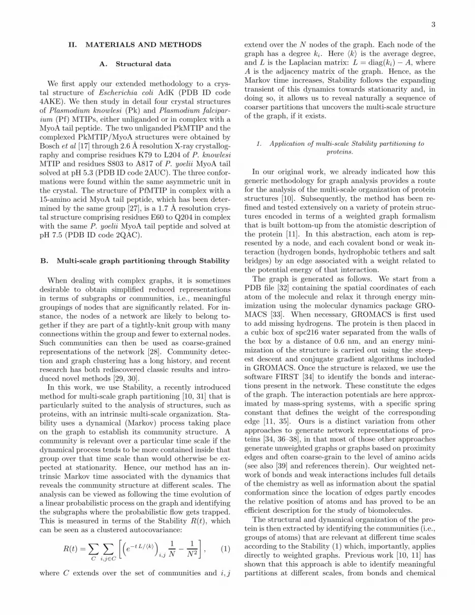
c. Application to AdK. Results of the partition-ing and robustness analysis for AdK are summarized inFig. 2. At each Markov time, the Stability was optimizeda hundred times with different Louvain initial conditions.For each Markov time, the optimal partition is shown inthe top panel of Fig. 2(a) by its number of communities,and the variation of information between all the parti-tions found at this Markov time is shown in the bottompanel. Partitions at very small and very high Markovtimes remain optimal for extended Markov times and cor-respond to biochemically meaningful components: smallchemical groups (small times) or the three functionaldomains (LID, NMP and CORE domains) [10, 11, 35].This is confirmed by our robustness analysis, which showssmall values of VI for the long-lived partitions.
Fig. 2 also shows the comparison of the robustness ofthe partitions of the protein against that of ensembles ofrandom graphs from our surrogate models. As expected,the random graphs obtained from the chemical config-uration model are indistinguishable from the protein atshort Markov times, since their local chemical structureis identical. However, at longer times the comparisonreveals two additional partitions of strong biochemicalsignificance corresponding to the peptide bonds betweenthe amino acids (at Markov times around 10−2) and tothe emergence of amino acids at Markov times around10−1. At the local minimum of VI, 63% of the aminoacids were grouped as a community, while most of theothers, essentially small amino acids, were grouped withanother residue due to the slight tendency of Stability tofind communities of about the same size.
The robustness of the protein is indistinguishable fromthe ensemble of graphs obtained by randomization of
weak interactions until Markov times of around 2. Thisestablishes the spatial and time scale at which the weakinteractions start having an influence on the communities,and, by extension, on the conformation of the protein inspace. Interestingly, the communities found at this scaleusually contain four amino acids, which is the number ofresidues usually found in one turn of an α-helix.At long Markov times, Stability finds partitions into a
few subunits that are much more robust for the proteinthan for the random surrogates. The study of their ro-bustness indicates the relevance of partitions into 2, 3,4, and 8 communities, which can be linked to the well-known folding of AdK and to an existent hinge analy-sis [2, 11]. As expected, the two random models convergeat long Markov times since in both cases the composi-tion of the molecule is conserved, and the weak interac-tions have been randomized. At Markov times above 30we observe an increase in the variability and a decay inthe value of the VI for the surrogates from the weak in-teraction randomization. This indicates the point whereweak interactions placed at random begin to induce ro-bust compact subgroups in the structure in an effect akinto undirected packing. In contrast, the specific locationof the weak interactions in the structure of the protein in-duces robust and reproducible partitions that reveal thespecific organization of the protein conformation.
B. The succession of partitions of PfMTIP suggests
a rigid cluster and the dynamics of the closing
mechanism
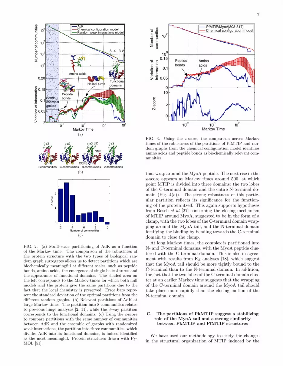
We have used the methodology introduced above tostudy the structure and dynamics of PfMTIP/MyoA[803-817], i.e., PfMTIP in complex with a peptide of the last15 amino acids of the MyoA tail (PDB ID code 2QAC).Again, at small Markov times, we find partitions of highrobustness corresponding to peptide bonds and aminoacids (Fig. 3). The relevant partitions at long Markovtimes are summarized in Fig. 4(a). Starting from the de-tection of the secondary structure, the different α-helicesand β sheets are progressively incorporated in a quasi-hierarchical manner into bigger clusters as the Markovtime increases. Some of the groupings lead to a markedincrease in the robustness of the partition. This is thecase for the first community to appear that incorpo-rates two secondary structures: helices α6 and α7. Thiscommunity is conserved across a broad range of Markovtimes, more than any other community of multiple el-ements of secondary structure. This suggests a strongdynamical linkage between these two α-helices over anextended time scale of motion. This is in agreementwith the PkMTIP crystal structure from Bosch et al [17],where these helices keep the same relative position inall three conformations observed, thereby suggesting thepresence of a rigid cluster.Another important community is the one formed by
helices α5 and α8, which also leads to an increase in therobustness of the partition. Together with the α6 − α7cluster, they divide the C-terminal domain into two lobes
7
(a)
(b)
(c)
FIG. 2. (a) Multi-scale partitioning of AdK as a functionof the Markov time. The comparison of the robustness ofthe protein structure with the two types of biological ran-dom graph surrogates allows us to detect partitions which arebiochemically meaningful at different scales, such as peptidebonds, amino acids, the emergence of single helical turns andthe appearance of functional domains. The shaded area onthe left corresponds to the Markov times for which both nullmodels and the protein give the same partitions due to thefact that the local chemistry is preserved. Error bars repre-sent the standard deviation of the optimal partitions from thedifferent random graphs. (b) Relevant partitions of AdK atlarge Markov times. The partition into 8 communities relatesto previous hinge analyses [2, 11], while the 3-way partitioncorresponds to the functional domains. (c) Using the z-scoreto compare partitions with the same number of communitiesbetween AdK and the ensemble of graphs with randomizedweak interactions, the partition into three communities, whichdivides AdK into its functional domains, is indeed identifiedas the most meaningful. Protein structures drawn with Py-MOL [51].
FIG. 3. Using the z-score, the comparison across Markovtimes of the robustness of the partitions of PfMTIP and ran-dom graphs from the chemical configuration model identifiesamino acids and peptide bonds as biochemically relevant com-munities.
that wrap around the MyoA peptide. The next rise in thez-score appears at Markov times around 500, at whichpoint MTIP is divided into three domains: the two lobesof the C-terminal domain and the entire N-terminal do-main (Fig. 4(c)). The strong robustness of this partic-ular partition reflects its significance for the function-ing of the protein itself. This again supports hypothesesfrom Bosch et al [27] concerning the closing mechanismof MTIP around MyoA, suggested to be in the form of aclamp, with the two lobes of the C-terminal domain wrap-ping around the MyoA tail, and the N-terminal domainfortifying the binding by bending towards the C-terminaldomain to close the clamp.
At long Markov times, the complex is partitioned intoN- and C-terminal domains, with the MyoA peptide clus-tered with the C-terminal domain. This is also in agree-ment with results from Kd analyses [18], which suggestthat the MyoA tail should be more tightly bound to theC-terminal than to the N-terminal domain. In addition,the fact that the two lobes of the C-terminal domain clus-ter at an earlier Markov time suggests that the wrappingof the C-terminal domain around the MyoA tail shouldtake place more rapidly than the closing motion of theN-terminal domain.
C. The partitions of PkMTIP suggest a stabilizing
role of the MyoA tail and a strong similarity
between PkMTIP and PfMTIP structures
We have used our methodology to study the changesin the structural organization of MTIP induced by the

8
(a)
(b) (c)
FIG. 4. (a) Multi-scale partitioning of PfMTIP/MyoA as afunction of Markov time. The elements of the secondary struc-ture are progressively grouped into larger communities as theMarkov times evolves. Although in general our methodologydoes not pre-impose a hierarchical community structure, inthis case the succession of community groupings is close toa strict hierarchy. Clusters kept for a long range of Markovtimes, such as the group of helices α6 and α7 are well-definedpartitions. The identification of the rigid cluster, and of thefunctional domains leads to an increase in the robustness (z-score) of the partitions. (b) The comparison of the z-scoreper number of communities suggests that the partitions into9 communities, where the rigid cluster is found, and the par-tition into two communities, where the N and C-terminal do-mains are identified, are significant. (c) Detection of the func-tional domains in the four-way partition of PfMTIP/MyoA.Protein structures drawn with PyMOL [51].
presence of the MyoA peptide by comparing unliganded(free) conformations of MTIP (PkMTIP1 and PkMTIP2)with ‘liganded’ conformations of MTIP (PkMTIP3 andPfMTIP), which are obtained from MTIP-MyoA com-plexes by deleting the MyoA peptide together with all itsinteractions when generating the graph. Liganded con-formations thus reflect the change of shape induced bythe MyoA peptide with none of the direct constraints.Fig. 5(a) and 5(b) show that the partitions for the lig-anded conformations obtained from the complexed formsare in general much more robust than the partitions ofthe two unliganded structures, especially at the level ofthe secondary structure (8, 9, and 10 communities) andof functional domains (3 communities). Such increase ofthe robustness of the partitions in the liganded confor-mations emerges naturally from the change in the spatialstructure induced by the MyoA peptide.
Importantly, although the partitions differ significantlyin their robustness and the Markov time of their predom-inance, they are very similar among the different confor-mations, especially between the two liganded forms. Inparticular, the important communities identified in sec-tion III B, such as the α6−α7 cluster and the functionaldomains (Fig. 4(c)), are also detected in all three con-formations with high robustness (the only exception be-ing the functional domains of PkMTIP2). The fact thatthe similar partitions are found in all structures suggeststhat the overall organization of the protein is not changedmuch between the conformations and corroborates theidea that the space of conformations that can be takenby a proteins is inscribed within its own structure [1].However, changes in the robustness and Markov lifetimeof the partitions suggests that the secondary and tertiarystructures get better structured upon binding with theMyoA tail since the corresponding partitions are betterdefined in this case. Note also that in comparing theliganded conformations, PfMTIP has more robust parti-tions than PkMTIP3, possibly a result of the stabilizingrole of the N-terminal domain, which in PfMTIP alsobinds the peptide and closes the clamp. On the otherhand, the unliganded form PkMTIP2 possesses the leastrobust partitions, in accordance with the hypothesis [17]that it should be an intermediate conformation betweenthe fully opened and fully closed forms.
The similarity between the partitions of the complexedforms of PfMTIP and PkMTIP supports the expectationthat their structural organization should not be very dif-ferent. However, there is a current open debate in theliterature regarding the difference in the hinge region be-tween the N- and C-terminal domains, with PkMTIPpresenting a long central α-helix where PfMTIP onlyhas a loop (Fig. 5(a)). The classification of this cen-tral domain in PkMTIP as a helix is however contro-versial since the structure of PkMTIP was measured ata non-physiological pH and does not correspond to thestructure observed in other MLCs [17]. Interestingly, ourpartitioning consistently divides the central α-helix ofPkMTIP into two different communities at all Markovtimes. Furthermore, in the partitions, the separation be-tween the two halves of this central α-helix is constrained

9
(a)
(b)
FIG. 5. (a) Robustness of the partitions of MTIP in differentconformations as a function of the Markov time. The ligandedconformations (PfMTIP and PkMTIP3) show better proper-ties of robustness than the unliganded ones at the level of thesecondary structures and of the functional domains, suggest-ing a stabilizing role of the binding with MyoA. Partitions arevery similar between the three conformations, in particularfor the functional domains, although the grouping of helicesα5 and α8 only occurs at long Markov times for PkMTIP2.Protein structures drawn with PyMOL [51]. (b) The z-scoreof the partitions with the same number of communities com-pared across different conformations of MTIP shows that theliganded forms have better defined partitions.
within the region that corresponds to the central loop inPfMTIP (from residues H135 to N140). This partition-ing is thus consistent with the central α-helix of PkMTIPbeing partly identified as a loop by the partitioning al-gorithm. To further support this observation, we havecarried out an analysis of the robustness of loops and α-helices with the same number of nodes (50 atoms) acrossMarkov times. Fig. 6 shows that the central region of the
FIG. 6. Variation of information of the partitions detected indifferent secondary structures. The variation of informationof the central region of the central α-helix (continuous redline) is very high and behaves similarly to other loop regionsof the protein. This suggests that the algorithm effectivelyrecognizes this region as a loop, despite its α-helical secondarystructure. Filled symbols correspond to loops, empty symbolscorrespond to α-helices and the continuous line corresponds tothe hinge region. Protein structure drawn with PyMOL [51].
central α-helix of PkMTIP has a robustness much lowerthan the typical α-helix with a trend similar to that ofloops. These results demonstrate the insights that ourmethod can bring into the analysis of the structural or-ganization of a protein beyond its pre-assigned secondaryor tertiary structure.
D. The analysis of residue sensitivity suggests six
residues of particular importance for the structure
and dynamics of the complex
The last part of the analysis aims to identify residuesin the MyoA tail that have a strong impact on the multi-scale organization of the protein complex and can there-fore be considered to play a significant role in its struc-ture and dynamics. This analysis does not evaluate theinfluence of a mutation on the binding energy; rather,the expectation is that residues with a large influence onthe structural organization of the protein will affect theglobal dynamics of the binding events. Indeed, hotspotsare known experimentally to be related to the global me-chanical properties of the protein such as flexibility andintrinsically disordered regions [2, 52, 53]. Furthermore,various computational methods have demonstrated thehigh influence of hotspots on large-scale attributes suchas the distribution of conformations [54], the networkof cooperativity between residues measured in terms ofcoupled fluctuations [55], or their propensity of being lo-cated at hinge sites measured by their mobility in the slowmodes [56]. To assess the connection between the effecton the binding energy and on the structural and dynam-ical features detected by our method, we have comparedour computational results with the outcome of bindingassays of MTIP with mutated MyoA tail peptides.Our computational setup mimics the standard alanine
scanning mutagenesis : each residue is ‘mutated’ in turn

10
(a) (b)
(c) (d)
FIG. 7. (a) The partition most influenced by computational mutagenesis is the one into 3 communities. (b) Residues R806,A809, H810, K813, R814, and V816 have the biggest influence on the three-way partition. There is a strong coincidence withthe residues identified experimentally [18] to have a strong effect on the binding affinity (indicated by arrows). (c) and (d) Frontand side view of the positions of the key residues found. Structures drawn with PyMOL [51].
by removing from the graph all the edges correspondingto the weak interactions it makes with other residues.The mutated graph is then analyzed with our multi-scalemethodology, and the partitions are compared with thoseof the original graph using the VI. For each mutation, wecompute the VI between all the partitions found with thesame number of communities from the original and mu-tated protein averaged over 10 different Louvain initialconditions and normalized by the average VI of the orig-inal graph. Using this scheme, partitions which are themost affected by a particular mutation will give a highvalue of the variation of information.
Fig. 7(a) shows that the partitions into 3 communi-ties are the most affected by the mutations. This is notsurprising since the 3-way partition is the first where theMyoA peptide is grouped with part of the MTIP molecule(Fig. 4(a)). Consequently, the mutations essentially af-fect the strength of the association between the MyoA tailand the portion of MTIP that includes the hinge regionand the helices α5 and α8 from the C-terminal domain.More specifically, the mutations that cause the largest
changes in the 3-way partition are those in residues R806,A809, H810, K813, R814, and V816 (Fig. 7(b)). Theseresults are in accordance with experimental binding as-says for MyoA peptides of different lengths [18], crystal-lographic data and yeast two-hybrid experiments [27]. Inparticular, residues R806 and K813 have been observedto be essential for complex formation; H810 and R814provide key contacts for tight binding; and V816 alsoimproves the binding. On the other hand, our methoddoes not single out a significant contribution of residueM815, which has been found to influence binding affin-ity. A possible explanation is that the importance of thisresidue might be related to effects that are not directlyaddressed by our method, such as intermediate states inthe folding pathway or modification of binding energies.Finally, our method finds one residue, A809, predicted tohave an important effect on the multi-scale organizationwhich has not been investigated experimentally to date.

11
IV. CONCLUSION AND OUTLOOK
We have presented the application of an efficient andcomputationally inexpensive method to extract informa-tion about the structural and dynamical organization ofproteins across time and spatial scales starting bottom-up from the full atomistic information. The methodologyis based on multi-scale graph partitioning methods thatestablish a series of increasingly coarser partitions thatcan reveal the structure of the graph. This paper in-troduces new graph theoretical tools, specifically the useof the robustness of partitions as a measure of their bi-ological significance and the quantification of robustnessthrough the introduction of biochemically-motivated sur-rogate random graph models.Our analysis has uncovered important features of the
MTIP/MyoA complex that agree well with experimentaldata. The rigid cluster formed by helices α6 and α7, assuggested by the crystal structures of PkMTIP [17], wasobserved to form a well-defined community, conservedacross a broad range of Markov times and associated withvery robust partitions. The functional domains suggestedby the analysis of crystal structures of different confor-mations across species [17, 27] have been detected by thepartitioning and also showed strong robustness and con-servation across Markov times. The robustness analysisof the hinge region of PkMTIP confirms these similaritiesbetween species and therefore suggests that their dynam-ical behavior should be similar. Furthermore, it supportsthe hypothesis [27] that the reported differences betweenPkMTIP and PfMTIP in the the hinge region could re-sult from the particularities of the crystallization. Fi-nally, a computational tool for mutational analysis wasintroduced and used to identify five out of the six residuesknown from binding assays [18] to have a strong influenceon the binding of MyoA. It also suggested one additionalresidue, A809, which has not yet been investigated ex-perimentally, to be particularly important.
Together, these results provide a better understandingof the possible dynamical behavior of MTIP and othermyosin light chains. The broad agreement with a vari-ety of experimental results underlines the intrinsic inter-est of methods that study the multi-scale organizationof proteins. Our method includes atomistic detail en-coded in a graph representation and allows to extractinformation about the global organization of the struc-ture and dynamics of the protein, but also about howindividual residues can affect them. Future work will in-clude the experimental verification of these predictions,such as binding assays with alanine mutations of some ofthe key residues identified. A deeper study of the role ofeach residue of the MyoA tail could also be carried out bycomputationally analyzing their effect on individual com-munities, rather than on the whole partitioning. On thetheoretical side, although the two random graph modelsproposed here were shown to generate relevant null hy-potheses, other forms of randomization such as geometricrandom graphs could also be tested in the future. Fi-nally, using the communities detected by our method tocoarse-grain molecular dynamics simulations could pro-vide an efficient method to get insight into the foldingand closing pathway.
ACKNOWLEDGMENTS
AD was supported through a PhD Studentship Awardfrom the British Heart Foundation Centre of ResearchExcellence from Imperial College London and (grantnumber RE/08/002) a Wallonie-Bruxelles InternationalAward. EWT acknowledges support through a DavidPhillips Fellowship from the Biotechnology and Biologi-cal Sciences Research Council. We thank Stefano Meliga,Renaud Lambiotte, Jean-Charles Delvenne, Joao Costaand YunWilliam Yu for helpful discussions.
[1] Henzler-Wildman K and Kern D 2007 Dynamic person-alities of proteins Nature 450 964–72
[2] Henzler-Wildman K A, Lei M, Thai V, Kerns S J, KarplusM and Kern D 2007 A hierarchy of timescales in proteindynamics is linked to enzyme catalysis Nature 450 913–6
[3] Frauenfelder H, McMahon B H, Austin R H, Chu K andGroves J T 2001 The role of structure, energy landscape,dynamics, and allostery in the enzymatic function of myo-globin Proc. Natl Acad. Sci. USA 98 2370–4
[4] Yaliraki S N and Barahona M 2007 Chemistry acrossscales: from molecules to cells Philos. Trans. R. Soc.,
A 365 2921–34[5] Adcock S A and McCammon J A 2006 Molecular dy-
namics: survey of methods for simulating the activity ofproteins Chem. Rev. 106 1589–1615
[6] Bahar I and Rader A 2005 Coarse-grained normal modeanalysis in structural biology Curr. Opin. Struct. Biol.
15 586–92[7] Tozzini V 2005 Coarse-grained models for proteins Curr.
Opin. Struct. Biol. 15 144–50
[8] Ayton G S, Noid W G and Voth G A 2007 Multiscalemodeling of biomolecular systems: in serial and in paral-lel Curr. Opin. Struct. Biol. 17 192–8
[9] Bongini L, Piazza F, Casetti L and De Los Rios P 2010Vibrational entropy and the structural organization ofproteins Eur. Phys. J. E: Soft Matter 33 89–96
[10] Delvenne J C, Yaliraki S N and Barahona M 2010 Sta-bility of graph communities across time scales Proc. Natl
Acad. Sci. USA 107 12755–60[11] Meliga S 2009 Graph Clustering of Atomic Networks for
Protein Dynamics Master’s thesis Imperial College Lon-don London, United Kingdom
[12] Lowey S and Trybus KM 2010 Common structural motifsfor the regulation of divergent class II myosins J. Biol.
Chem. 285 16403–7[13] Schiaffino S and Reggiani C 1996 Molecular diversity of
myofibrillar proteins: gene regulation and functional sig-nificance Physiol. Rev. 76 371–423
[14] Rayment I, Rypniewski W, Schmidt-Base K, Smith R,Tomchick D, Benning M, Winkelmann D, Wesenberg

12
G and Holden H 1993 Three-dimensional structure ofmyosin subfragment-1: a molecular motor Science 261
50–8[15] Yamashita H, Sugiura S, Fujita H, Yasuda S, Nagai R,
Saeki Y, Sunagawa K and Sugi H 2003 Myosin light chainisoforms modify force-generating ability of cardiac myosinby changing the kinetics of actinmyosin interaction Car-
diovasc. Res. 60 580–8[16] Timson D J 2003 Fine tuning the myosin motor: the
role of the essential light chain in striated muscle myosinBiochimie 85 639–45
[17] Bosch J, Turley S, Daly T M, Bogh S M, Villasmil M L,Roach C, Zhou N, Morrisey J M, Vaidya A B, BergmanL W and Hol W G J 2006 Structure of the MTIP-MyoAcomplex, a key component of the malaria parasite inva-sion motor Proc. Natl Acad. Sci. USA 103 4852–7
[18] Thomas J C, Green J L, Howson R I, Simpson P, MossD K, Martin S R, Holder A A, Cota E and Tate E W 2010Interaction and dynamics of the Plasmodium falciparumMTIP-MyoA complex, a key component of the invasionmotor in the malaria parasite Mol. BioSyst. 6 494–8
[19] Heintzelman M B and Schwartzman J D 1997 A novelclass of unconventional myosins from toxoplasma gondiiJ. Mol. Biol. 271 139–46
[20] Hettmann C, Herm A, Geiter A, Frank B, Schwarz E,Soldati T and Soldati D 2000 A dibasic motif in the tailof a class XIV apicomplexan myosin is an essential deter-minant of plasma membrane localization Mol. Biol. Cell
11 1385–1400[21] Bergman L W, Kaiser K, Fujioka H, Coppens I, Daly
T M, Fox S, Matuschewski K, Nussenzweig V and KappeS H 2003 Myosin A tail domain interacting protein(MTIP) localizes to the inner membrane complex of Plas-modium sporozoites J. Cell Sci. 116 39–49
[22] Green J L, Rees-Channer R R, Howell S A, Martin S R,Knuepfer E, Taylor H M, Grainger M and Holder A A2008 The motor complex of Plasmodium falciparum J.
Biol. Chem. 283 30980–9[23] Rees-Channer R R, Martin S R, Green J L, Bowyer P W,
Grainger M, Molloy J E and Holder A A 2006 Dual acyla-tion of the 45 kDa gliding-associated protein (GAP45) inPlasmodium falciparum merozoites Mol. Biochem. Para-
sitol. 149 113–6[24] Green J L, Martin S R, Fielden J, Ksagoni A, Grainger
M, Lim B Y Y, Molloy J E and Holder A A 2006 TheMTIP-myosin A complex in blood stage malaria parasitesJ. Mol. Biol. 355 933–41
[25] Baum J, Richard D, Healer J, Rug M, Krnajski Z,Gilberger T W, Green J L, Holder A A and CowmanA F 2006 A conserved molecular motor drives cell inva-sion and gliding motility across malaria life cycle stagesand other Apicomplexan parasites J. Biol. Chem. 281
5197–5208[26] Frenal K, Polonais V, Marq J B, Stratmann R, Limeni-
takis J and Soldati-Favre D 2010 Functional dissectionof the Apicomplexan glideosome molecular architectureCell Host Microbe 8 343–57
[27] Bosch J, Turley S, Roach C M, Daly T M, Bergman L Wand Hol W G 2007 The closed MTIP-myosin A-tail com-plex from the malaria parasite invasion machinery J. Mol.
Biol. 372 77–88[28] Juanico B, Sanejouand Y H, Piazza F and De Los Rios P
2007 Discrete breathers in nonlinear network models ofproteins Phys. Rev. Lett. 99 238104
[29] Fortunato S 2010 Community detection in graphs Phys.
Rep. 486 75–174
[30] Gfeller D, De Los Rios P, Caflisch A and Rao F 2007Complex network analysis of free-energy landscapes Proc.Natl Acad. Sci. USA 104 1817–22
[31] Lambiotte R, Delvenne J C and Barahona M 2009 Lapla-cian dynamics and multiscale modular structure in net-works (Preprint arXiv:0812.1770v3 [physics.soc-ph])
[32] Berman H M, Westbrook J, Feng Z, Gilliland G, BhatT N, Weissig H, Shindyalov I N and Bourne P E 2000The protein data bank Nucl. Acids Res. 28 235–42
[33] Hess B, Kutzner C, van der Spoel D and Lindahl E2008 GROMACS 4: algorithms for highly efficient, load-balanced, and scalable molecular simulation J. Chem.
Theory Comput. 4 435–47[34] Jacobs D J, Rader A J, Kuhn L A and Thorpe M F
2001 Protein flexibility predictions using graph theoryProteins: Struct., Funct., Genet. 44 150–65
[35] Meliga S, Yaliraki S N and Barahona M 2011 In prepa-ration
[36] Costa J R and Yaliraki S N 2006 Role of rigidity on theactivity of proteinase inhibitors and their peptide mimicsJ. Phys. Chem. B 110 18981–8
[37] Chennubhotla C and Bahar I 2006 Markov propagationof allosteric effects in biomolecular systems: applicationto GroEL-GroES Mol. Syst. Biol. 2 36
[38] Bahar I, Atilgan A R and Erman B 1997 Direct evalu-ation of thermal fluctuations in proteins using a single-parameter harmonic potential Fold. Des. 2 173–81
[39] Bahar I, Lezon T R, Bakan A and Shrivastava I H 2010Normal mode analysis of biomolecular structures: func-tional mechanisms of membrane proteins Chem. Rev. 110
1463–97[40] Sethi A, Eargle J, Black A a and Luthey-Schulten Z 2009
Dynamical networks in tRNA:protein complexes Proc.
Natl Acad. Sci. USA 106 6620–5[41] Aynaud T, Blondel V, Guillaume J L and Lambiotte R
2010 Optimisation locale multi-niveaux de la modularitePartitionnement de graphe : optimisation et applications
(Traite IC2 ) ed Bichot C E and Siarry P (Paris: HermesScience Publishing) chap 14
[42] Blondel V D, Guillaume J L, Lambiotte R and LefebvreE 2008 Fast unfolding of communities in large networksJ. Stat. Mech.: Theory Exp. 2008 P10008
[43] Karrer B, Levina E and Newman M E J 2008 Robust-ness of community structure in networks Phys. Rev. E
77 046119[44] Fenn D J, Porter M A, McDonald M, Williams S, Johnson
N F and Jones N S 2009 Dynamic communities in multi-channel data: an application to the foreign exchange mar-ket during the 2007–2008 credit crisis Chaos 19 033119
[45] Delmotte A 2010 Graph partitioning algorithm revealing
multiscale protein dynamic Master’s thesis Imperial Col-lege London London, United Kingdom
[46] Good B H, de Montjoye Y A and Clauset A 2010 Perfor-mance of modularity maximization in practical contextsPhys. Rev. E 81 046106
[47] Meila M 2007 Comparing clusterings–an informationbased distance J. Multivariate Anal. 98 873–95
[48] Newman M E J 2005 Random graphs as models ofnetworks Handbook of Graphs and Networks: From the
Genome to the Internet ed Bornholdt S and Schuster H G(Berlin: Wiley-VCH Verlag GmbH & Co. KGaA) chap 2,pp 35–68
[49] Maslov S and Sneppen K 2002 Specificity and stability intopology of protein networks Science 296 910–3
[50] Molloy M and Reed B 1995 A critical point for randomgraphs with a given degree sequence Random Struct. Al-

13
gor. 6 161–79[51] Delano W L 2009 The PyMOL Molecular Graphics Sys-
tem, Version 1.2r1. URL http://www.pymol.org
[52] Ma B, Wolfson H J and Nussinov R 2001 Protein func-tional epitopes: hot spots, dynamics and combinatoriallibraries Curr. Opin. Struct. Biol. 11 364–9
[53] Radivojac P, Iakoucheva L M, Oldfield C J, ObradovicZ, Uversky V N and Dunker A K 2007 Intrinsic disorderand functional proteomics Biophys. J. 92 1439–56
[54] Ming D and Wall M E 2006 Interactions in native binding
sites cause a large change in protein dynamics J. Mol.
Biol. 358 213–23[55] Liu T, Whitten S T and Hilser V J 2007 Functional
residues serve a dominant role in mediating the coop-erativity of the protein ensemble Proc. Natl Acad. Sci.
USA 104 4347–52[56] Yang L W and Bahar I 2005 Coupling between cat-
alytic site and collective dynamics: a requirement formechanochemical activity of enzymes Structure 13 893–904


