Embed Size (px)
Citation preview
Hindawi Publishing CorporationBioMed Research InternationalVolume 2013, Article ID 253483, 9 pageshttp://dx.doi.org/10.1155/2013/253483
Research ArticleMolecular Modeling of Lectin-Like Protein from Acaciafarnesiana Reveals a Possible Anti-Inflammatory Mechanism inCarrageenan-Induced Inflammation
Vanessa Erika Ferreira Abrantes,1 Bruno Anderson Matias da Rocha,2
Raphael Batista da Nóbrega,2 José Caetano Silva-Filho,1 Claudener Souza Teixeira,2
Benildo Sousa Cavada,2 Carlos Alberto de Almeida Gadelha,1 Sergio Henrique Ferreira,3
Jozi Godoy Figueiredo,3 Tatiane Santi-Gadelha,1 and Plinio Delatorre1
1 Departamento de Biologia Molecular, Universidade Federal da Paraıba, Campus I, Cidade Universitaria,58059-900 Joao Pessoa, PB, Brazil
2 Departamento de Bioquımica e Biologia Molecular, Universidade Federal do Ceara, BioMol-Lab, Campus do Pici S/N,60440-970 Fortaleza, CE, Brazil
3 Departamento de Farmacologia, Faculdade de Medicina de Ribeirao Preto, Universidade de Sao Paulo,14049-900 Ribeirao Preto, SP, Brazil
Correspondence should be addressed to Tatiane Santi-Gadelha; [email protected] Plinio Delatorre; [email protected]
Received 12 September 2013; Revised 5 November 2013; Accepted 5 November 2013
Academic Editor: Paul Harrison
Copyright © 2013 Vanessa Erika Ferreira Abrantes et al. This is an open access article distributed under the Creative CommonsAttribution License, which permits unrestricted use, distribution, and reproduction in any medium, provided the original work isproperly cited.
Acacia farnesiana lectin-like protein (AFAL) is a chitin-binding protein and has been classified as phytohaemagglutinin fromPhaseolus vulgaris (PHA). Legume lectins are examples for structural studies, and this family of proteins shows a remarkableconservation in primary, secondary, and tertiary structures. Lectins have ability to reduce the effects of inflammation caused byphlogistic agents, such as carrageenan (CGN). This paper explains the anti-inflammatory activity of AFAL through structuralcomparison with anti-inflammatory legume lectins.The AFALmodel was obtained by molecular modeling andmolecular dockingwith glycan and carrageenan were performed to explain the AFAL structural behavior and biological activity. Pisum sativum lectinwas the best template formolecularmodeling.TheAFAL structuremodel is folded as a𝛽 sandwich.Themodel differs from templatein loop regions, number of 𝛽 strands and carbohydrate-binding site. Carrageenan and glycan bind to different sites on AFAL. Theability of AFAL binding to carrageenan can be explained by absence of the sixth 𝛽-strand (posterior 𝛽 sheets) and two 𝛽 strandsin frontal region. AFAL can inhibit pathway inflammatory process by carrageenan injection by connecting to it and preventing itsentry into the cell and triggers the reaction.
1. Introduction
Acacia farnesiana is a member of Leguminosae family, and itis included in the Mimosoideae subfamily. Acacia farnesianalectin-like protein (AFAL) has been purified and classifiedas PHA-like lectin [1]. Lectins are defined as carbohydrate-binding glyco/proteins that are able to agglutinate cells,
without enzymatic activity toward carbohydrate or glyco-conjugates [2–4]. Plant lectins have been structural modelsfor studying carbohydrate recognition and oligomerizationstates [5–7]. AFAL has a time-dependent oligomerizationand it is characterized as a chitin-binding lectin based onits affinity by glycans, which elect it as a good example forstructural studies. AFAL hemagglutinating activity was not
2 BioMed Research International
inhibited by carbohydrate compounds neither themethylatedor (GlcNAc) derivative ones [1]. However, Phaseolus vulgarisisolectins (PHA) usually bind to 1,6-branched GlcNAc con-taining N-glycans and the inhibition test result is normallypositive for this inhibitor [8–10].
Legume lectins have a high degree of sequence similarityand important functional regions with amino acid residueconserved [11]. Legume lectins despite the structural iden-tity exhibit considerable differences in biological activitiesand carbohydrate recognition [12]. Comparisons of thesesequences and structures have established that differencesin carbohydrate specificity appear to be primarily due todifferences in amino acid residues residing in adjacent loopsto the carbohydrate-binding site [13–16]. The conformationof these loops is determined by the presence of calcium andtransition metal ions coordinated in the protein structure[17–19]. One of themost studied biological activities of lectinsis the ability to reduce the effects of inflammation causedby certain phlogistic agents, such as carrageenan (CGN)[20]. Many molecules with this ability were considered fortheir potential for the development of drugs with anti-inflammatory action [21]. CGN is a sulfated polysaccharideobtained from several species of red algae. It is frequentlyincorporated into processed foods to improve texture andsolubility [22]. CGN exposure possibly causes inflammationby inducing an increase in interleukin-8 (IL-8) secretionin cells. This increase was demonstrated in tissue cultures,animal colon, and human tissues [23, 24]. Mechanisms ofCGN-induced IL-8 activation require nuclear localization ofnuclear factor 𝜅B (NF𝜅B) and can proceed through a toll-like-receptor-4- (TLR4-) B-cell-lymphoma/leukemia-10- (Bcl10-)mediated pathway [25].
The aim of this paper is to obtain a structural modelof AFAL by molecular modeling to explain AFAL structuralbehavior and biological activity based on structure/functionanalysis. The analysis was made using molecular dock-ing with oligosaccharides and CGN and investigating theanti-inflammatory activity of AFAL in model carrageenan-induced inflammation.
2. Material and Methods
2.1. Protein Purification and Haemagglutinating Assays. Puri-fied AFAL protein was obtained from seeds according toestablished protocols by Santi-Gadelha and coworkers [1].Previous studies showed that AFAL presents only hemag-glutinating activity using rabbit erythrocytes. Haemaggluti-nating activity (HA) and inhibition tests were carried outaccording to standard procedure [9].Thenegative controlwascomposed of 100𝜇L of 0.15M NaCl solution and 100 𝜇L of2% rabbit erythrocytes. All results were observedmacroscop-ically and the tests were done in triplicate.
A standard haemagglutinating inhibition test was alsoperformed to certify that the interaction of CGN and AFALdoes not occur through carbohydrate recognition domain(CRD). AFAL dissolved in 0.15M NaCl (2mg/mL) was used.The AFAL samples were diluted in 0.15M NaCl to give aconcentration corresponding to 4 hemagglutinating units
(HU) activity per mL. In this study, the dilution was madeonly to the second tube and then the titer was calculatedas 22 (the highest dilution of the AFAL that can agglutinateerythrocytes, that is, the titre, defined as containing onehemagglutinating unit per milliliter). In assay tubes, thecarrageenan solution (5mg/mL in 0.15M NaCl) was mixedwith AFAL solution and incubated at 37∘C for 30 minutes.The assay tubes were then left at room temperature for 1 hbefore 0.2mL of 2% rabbit erythrocytes were added to eachtube. The HA with and without addition of CGN at the sametime was tested. The results were observed macroscopicallyand were expressed as number of hemagglutinating units(HU.100 𝜇L−1) which is calculated from the inverse of thehighest dilution titer still having visible agglutination.
2.2. Primary Structure Analysis of Acacia farnesiana Lectin-Like Protein. The BLAST program [26] was used for a com-parative analysis of the amino acid sequence of AFALwith theamino acid sequence of other proteins. The best sequenceswith resolved three-dimensional structures were chosen foralignment. The alignment was performed with Protein DataBank (PDB) deposited protein structures: 1FAT, 1LUL, 3USU,2SBA, 1FNY, 2BQP, 1UZY, and 3N35. The multiple alignmentwith selected lectins was done using Multalin tool [27].
2.3. Molecular Modeling by Homology of Acacia farnesianaLectin-Like Protein. Models were obtained from knownstructures of proteins that have the highest degree of identityamong the primary structures aligned with AFAL. Thestructures were created from the homology molecular mod-eling server SWISS-MODEL [28], a free online programof Swiss Institute of Bioinformatics. This program allowsa fully automated protein structure homology modeling,accessible via the ExPASy web server. Structures from Phase-olus vulgaris lectin (1FAT), Dolichos biflorus lectin (1LUL),Buteamonosperma lectin (3USU),Glycinemax lectin (2SBA),Robinia pseudoacacia lectin (1FNY), Pisum sativum lectin(2BQP), Erythrina crista-galli lectin (1UZY), and Erythrinacorallodendron lectin (3N35) were compared in order toobtain the high homology.
2.4. Molecular Docking. Molecular docking was performedwith Hex 6.3 molecular graphics program [29]. The Hexis a fast molecular docking program for calculating anddisplaying interactions and feasible docking modes of pairsof protein and DNA molecules. It can superpose pairs ofmolecules using only knowledge of their 3D shapes and calcu-late protein-ligand interactions using spherical polar Fourier(SPF) correlations to accelerate the docking calculations.
The structural model of AFAL obtained from molecularmodeling was used to test the binding to different carbohy-drate compounds and to estimate possible binding sites of thesugar and other ligands.This was done in order to explain thebiological activities provided by this protein.
An oligosaccharide obtained from the structure of theLathyrus ochrus lectin (deposited under PDB code 1LGC)wasused [30].This oligosaccharidewith 2.1 kDawas used because
BioMed Research International 3
it is bound to a lectin structure which has a relative highidentity with AFAL of 40%.
The Iota-carrageenan obtained under the PDB code1CAR [31] and another oligosaccharide molecule assembledwith nine residues of N-acetylglucosamine (GlcNac) wereused as ligands for molecular docking. These last ligandcoordinates were generated using the program PRODRGServer [32].The (GlcNac) oligosaccharides used in inhibitionof hemagglutination test showed positive results only aboveeight residues. The complexes were named AFAL glycan,AFAL CNG, and AFAL GlcNAc. The molecular docking fortwo AFAL monomers was performed to obtain the AFALdimer form.
2.5. Biological Activity. All protocols were approved by theUSP Ethics Committee (USP, N∘ 183-2011). Mice receivedintraperitoneal (i.p.) CGN (500𝜇g/cav.) dissolved in 0.5mLof sterile saline. AFAL (1mg/Kg) was administered intra-venously (i.v.) in 0.1mL of saline 15 minutes before theCGN injection. As a negative control, an experiment wasperformed in mice that received saline i.v.
The neutrophil migration was evaluated 4 hours afterinjection of CGN. To this end, the animals were sacrificedby cervical dislocation.Then the cells in the peritoneal cavitywere collected by rinsing with 3.0mL of saline containing5 IU/mL of heparin.The abdomens of the animals were gentlymassaged, and through an incision peritoneal fluids werecollected.
The total and differential counting of leukocytes wasperformed according to methodology previously describedby De Souza and Ferreira [33]. In this procedure, 20 𝜇Lof fluid collected from each animal was diluted in 380 𝜇Lof Turk reagent and subsequently used for total leukocytecount in a Neubauer chamber. The differential cell count wasperformed using stained smears on slides, 50 𝜇L of exudatewere centrifuged in cytocentrifuge at 400×g for 10 minutes;after this process the smears were stained with hematoxylin-eosin (HE) and the cells counted using an optical microscopeand the results expressed as the mean ± standard error mean(SEM) number of cells ×10/mL of peritoneal fluid.
For the assessment of vascular permeability, the micereceived AFAL at a dose of 1mg/kg i.v. 15 minutes beforeadministration of the stimulus CGN (500𝜇g/cav.). Controlgroup received only saline i.p. One hour before the sacrifice,the animals received Evans blue (50mg/kg) in plexus eye[34]. The animals were sacrificed by cervical dislocation 3hours after administration of CGN, and then the fluid in theperitoneal cavity was collected by rinsing injecting 3.0mL ofsaline containing heparin (5UI/mL). The abdomens of theanimals were gently massaged and an incision was made.Then, it was collected about 1.0mL of peritoneal fluid. Toquantify the Evans blue extravasation into the peritoneal cav-ity, spectrophotometry was carried out by reading the opticaldensities using a wavelength of 610 nm. The absorbance dataobtained were converted to mg of Evans Blue, by linearregression based on a standard curve of Evans blue. Resultswere calculated as mg of Evans blue/mL of peritoneal fluidand expressed as mean ± SEM.
3. Results
3.1. Hemagglutinating Activity of AFAL. The haemagglutina-tion activity (HA) was observed after 24 hours with a titercalculated as 4HU.100𝜇L−1. The inhibition of haemaggluti-nation activity of AFAL by CGN had not been effective andit was observed haemagglutinating activity against the rabbiterythrocytes in the first hour after incubation with the sametiter previously obtained in the haemagglutinating test. Thetest showed that the CGN was not capable of inhibiting thebinding of protein to erythrocyte membrane carbohydratessuggesting that CGN binding does not occur in the knowncarbohydrate binding site of legume lectins.
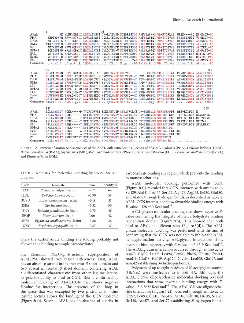
3.2. AFAL Model Structure. Phaseolus vulgaris agglutinin(PHA) showed high max score in the BLAST alignment,but other proteins present also acceptable identities as canbe seen in the multiple alignment in Figure 1. The AFALsequence analysis showed the highest identity among plantlectins with PHA (64%). However, the molecular modelingof AFAL structure does not show favorable results when themodel chosen was PHA, with the Z-score of −3.7 (Table 1).Low quality model is expected to have strongly negative Z-scores (−1 to −4) featuring the model as unworkable. Thisapparent discrepancy occurs because the ideal template hashigh sequence identity, secondary structure similarity, andstructural domain assignment.
New templates with favorable result of the BLAST align-ment were tested. Lectins of Dolichos biflorus (58%), Buteamonosperma (51%),Glycinemax (50%),Robinia pseudoacacia(48%),Pisum sativum (42%),Erythrina crista-galli (38%), andErythrina corallodendron (37%) were identified as the bestones.
All similar proteins were used as templates for molecularmodeling by SWISS-MODEL program, but only the chain Aof Pisum sativum lectin (PSL) (PDB code 2BQP) [35] showedhighZ-scorewhich converges to a statistically valid structuralmodel (Table 1). Dimeric PSL is also a mannose/glucosespecific metalloprotein and its structure provided the bestmodeling template at 1.90 A resolutionwith sequence identityof 42% [36].
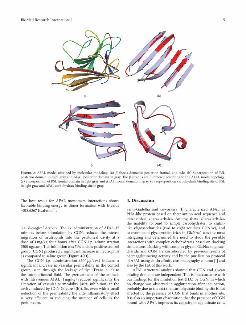
The AFAL structure model is folded as a 𝛽 sandwich(thirteen𝛽 strands), as previously observed in legume lectins.Themonomer model shows three 𝛽 sheets domains: a frontalone, a posterior one, and another positioned at the side(Figure 2(a)). The model differs from the PSL template,in loops regions, number of 𝛽 strands (sixteen in PSL),and carbohydrate binding site. The AFAL model structurecontains five 𝛽 strands in the posterior 𝛽 sheet domain(Figure 2(b)). The common canonical oligomeric structuresin legume lectins show six𝛽 strands. In addition, two𝛽 standsaremissing in the frontal𝛽 sheets domain of the AFALmodel(Figure 2(c)).
The formation of dimer canonical structures is commonin legume lectins, but the absence of a 𝛽 strand in canonicaldomain (posterior 𝛽 sheets) prevents the possible formationof the conventional canonical dimer in AFAL model. Theabsence of the two helixes in theAFAL structure (Figure 2(d))
4 BioMed Research International
WREK----IQ EPNKAGP-ADFATSFTFNIQ
QPYTRPLPADQPYTRPLPAD
VPNNAGP-ADWATSFTANIF APNKSSS-ADFATSFRFTIY APNIATI-ADFAASFNFTFY APDTKRL-ADFVTSFTFIIQ APNPATT-ADFETRFSFSIEFETRFSFSIEFVTSFTFVIN APNSYNV-ADf.tsf.f.I. aPn.a...AD
91 180AFAL GMAFALVPVG SEPKDKGAGL LGLFDE--YD SNRHPVAVEF DTCYNL---- -----EHDPK ERHSIRSIAT PRWDFPNGEN AEVLITYDEEPHA GLAFALVPVG SQPKD-KGGF LGLFDGS--N SNFHTVAVEF DTLYNKD-WD PTE--RHIGI DVNSIRSIKT TRWDFVNGEN AEVLITYDSSDB58 GIAFALVPVG SEPKS-NSGF LGVFDSDVYD NSAQTVAVEF DTFSNTD-WD PTS--RHIGI DVNSIKSIRT ASWGLANGQN AEILITYNAABMA GLAFFLAPVS SPPKA-GAGF LGLFDSAVFN SSYQTVAVEF DTYENTVFLD PPD--THIGI DVNSIKSIKT VKWDLANGEA AKVLITYDSSSBL GLAFFLAPID TKPQT-HAGY
TQPLD-LGGM
TKPQT-GGGY
LGLFNEN--E SGDQVVAVEF DTFRNS--WD PPN--PHIGI NVNSIRSIKT TSWDLANNKV AKVLITYDASRPbAI GLAFFLAPVD LGIFKDGYFN KSNQIVAVEF DTFSNGD-WD PKG--RHLGI NVNSIESIKT VPWNWTNGEV ANVFISYEASECL GLVFFMGPTK SKPAQ-GYGY LGVFNNSKQD NSYQTLAVEF DTFSNP--WD PPQV-PHIGI DVNSIRSIKT QPFQLDNGQV ANVVIKYDASEcorL GLVFFMGPTK SKPAQ-GGGY LGIFNNSKQD NSYQTLGVEF DTFSNQ--WD PPQV-PHIGI DVNSIRSIKT QPFQLDNGQV ANVVIKYDASPSL GFTFFIAPVD LGVFNSAEYD KTTQTVAVEF DTFYNAA-WD PSNRDRHIGI DVNSIKSVNT KSWKLQNGEE ANVVIAFNAAConsensus
AFAL PHA DB58 BMA SBLRPbAI ECL EcorL PSL Consensus
AFAL PHA DB58 BMA SBLRPbAI ECL EcorL PSL Consensus
GlaFfl.Pv. s.P...g.G. LG.F.....# ...qtvAVEF DTf.N.. wd p.....Higi #vnSIrS!kT ..w.l.NG#. A.VlIt%#as
181 268LQLLVASLVY PGER-----P YYLPSDRVEI EDELPEYVIP GFSATRGLNE G--ETHDVLS WSFASKMPDE QES-EGLDLA E TNLLVASLVY PSQK-----T SFIVSDTVDL KSVLPEWVSV GFSATTGINK GNVETNDVLS WSFASKLSDE TTS-EGLNLA NLVLNKILTSLLVASLVH PSRR-----T SYIVSERVDI TNELPEYVSI GFSATTGLSE GYTETHDVLS WSFASKLPDD STT-EPLDIA SYLVRNVLAKLLVAALVY PSSK-----T SFILSDVVDL KSVLPEWVSI GFSAATGASS GYIETHDVFS WSFASKLSFX XXXXX-LDLA SFLVAN TSLLVASLVY PSQR-----T SNILSDVVDL KTSLPEWVRI GFSAATGLDI PG-ESHDVLS WSFASNLPHA SSNIDPLDLT SFVLHEAITKSLTASLVY PSLE-----T SFIIDAIVDV KIVLPEWVRF GFSATTGIDK GYVQTNDVLS WSFESNLPGSKILLAVLVY PSSG-----A KQVLPEWVDV GLSGATGAQR WSFHASLPETSKILHAVLVY PSSG-----A IYTIAEIVDV
IYTIAEIVDVKQVLPEWVDV GLSGATGAQR DAAETHDVYS
DAAETHDVYSWSFQASLPET ND
ND
TNVLTVSLTY PNNSLEEEVT SYTLSDVVSL KDVVPEWVRI GFSATTGAEY A---AHEVLS WSFHSELSt.lLvasLvY Ps.......t s%..sd.Vd. k.vlPEwV.. GfSattG... g..eth#VlS WSFas.$p.. ......l... ........
FKSNDIYFNFQADIQSFSFKTTNTDSFTFSAETVSFSWNTGSLSFSFPVETISFSFSVETISFSFSTETTSFLIT..t.sf.f.
NLNFNEQSLIRF--NETNLINF--NSSSFIKFKPNQPNLKKFVPKQPNMIKFAPNQPYLIEFEPGNNDLTEFEPGNDNLTKFSPDQQNLI.f.pn#..$i
LQGDATVSSTLQRDASVSSSLQGDATVSSSKQGDATVTSSLQGDAIVTSSNQGDALVTSTLQGAAIITQSLQGASLITQSFQGDGYTTKElQGda.vts.
G--RLTNVVDGQLRLTNLNG-KLRLTKVKGGTLQLTKVDKGKLQLNKVDEGVLQLTNV-VGVLQLTKINQGVLQLTKINQ-KLTLTKAVKg.l.LTkv..
NGEPRTSSLGNGEPRVGSLGNGLPTLSSLGNGVPDPKSLGNGTPKPSSLGNGVPSSKSLGNGMPAWSDTGNGMPAWSDTGN------TVGNg.p...slG
RAFYSAP--IRAFYSAPIQIRAFYSSPIQIRALYASPINIRALYSTPIHIRALYAAPFQIRTLYTKPVHIRTLYAKPVHIRALYSSPIHIRalYs.P..I
WDKPTGRLASWDNTTGTVASYDKSTGAVASWDSKTGVVASWDKETGSVASWDSTTGNVASWDMTTGTVASWDMTTGTVASWDRETGNVANWD..TG.vAs
311
Figure 1: Alignment of amino acid sequences of the AFAL with some lectins. Lectins of Phaseolus vulgaris (PHA), Dolichos biflorus (DB58),Buteamonosperma (BMA),Glycine max (SBL), Robinia pseudoacacia (RPbAI), Erythrina crista-galli (ECL), Erythrina corallodendron (EcorL)and Pisum sativum (PSL).
Table 1: Templates for molecular modeling by SWISS-MODELprogram.
Code Template Score Identity %1FAT Phaseolus vulgaris lectin −3.7 641LUL Dolichos biflorus lectin −3.99 583USU Butea monosperma lectin −3.18 512SBA Glycine max lectin −3.32 501FNY Robinia pseudoacacia lectin −3.75 482BQP Pisum sativum lectin 0.00 423N35 Erythrina corallodendron lectin −3.86 381UZY Erythrina crystagalli lectin −3.87 37
alters the carbohydrate binding site folding probably notallowing the binding to simple carbohydrates.
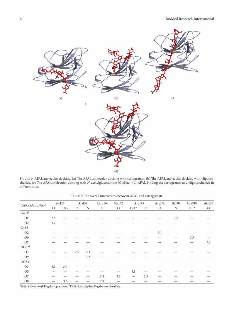
3.3. Molecular Docking. Structural superposition ofAFAL/PSL showed two major differences. First, AFALhas an absent 𝛽 strand in the posterior 𝛽 sheet domain andtwo absent in frontal 𝛽 sheet domain, conferring AFALa differentiated characteristic from other legume lectins:its possible ability to bind to CGN. This is confirmed bymolecular docking of AFAL CGN that shows negativeE-value for interactions. The presence of the loop inthe space that was occupied by two 𝛽 strands in otherlegume lectins allows the binding of the CGN molecule(Figure 3(a)). Second, AFAL has an absence of a helix in
carbohydrate binding site region, which prevents the bindingto monosaccharides.
AFAL molecular docking, performed with CGN,(Figure 3(a)) revealed that CGN interacts with amino acidsSer129, Ala131, Leu156, Ser172, Asp173, Arg174, Ile130, Glu180,and Ala108 through hydrogen bonds, as described in Table 2.AFAL CGN interactions show favorable binding energy with𝐸-value −328.430 Kcal⋅mol−1.
AFAL glycan molecular docking also shows negative 𝐸-value confirming the integrity of the carbohydrate bindingrecognition domain (Figure 3(b)). This showed that bothbind to AFAL on different sites (Figure 3(d)). The AFALglycan molecular docking was performed with the aim ofconfirming that the CGN was not able to inhibit the AFALhemagglutination activity. AFL glycan interactions showfavorable binding energy with𝐸-value −445.4719 Kcal⋅mol−1.The AFAL glycan interaction occurred through amino acidsAsp73, Gly92, Leu93, Leu94, Leu96, Phe97, Glu110, Cys114,Asn116, Glu118, His119, Asp120, Gly194, Leu195, Glu197, andAsn512 establishing 34 hydrogen bonds.
Polymers of up to eight residues of N-acetylglucosamine(GlcNac) were ineffective to inhibit HA. Although, theAFAL GlcNac oligosaccharide molecular docking revealedinteractions that show favorable binding energy with 𝐸-value −313.5632Kcal⋅mol−1. The AFAL GlcNac oligosaccha-ride interaction (Figure 3(c)) occurred through amino acidsGly92, Leu93, Glu110, Asp112, Asn116, Glu118, His119, Ser129,Ile 130, Asp173, and Ser172 establishing 21 hydrogen bonds.
BioMed Research International 5
(a) (b)
(c) (d)
Figure 2: AFAL model obtained by molecular modeling. (a) 𝛽 sheets domains: posterior, frontal, and side. (b) Superposition of PSLposterior domain in light gray and AFAL posterior domain in gray. The 𝛽 strands are numbered according to the AFAL model topology.(c) Superposition of PSL frontal domain in light gray and AFAL frontal domain in gray. (d) Superposition carbohydrate binding site of PSLin light gray and AFAL carbohydrate binding site in gray.
The best result for AFAL monomers interactions showsfavorable binding energy to dimer formation with 𝐸-value−518.6367 Kcal⋅mol−1.
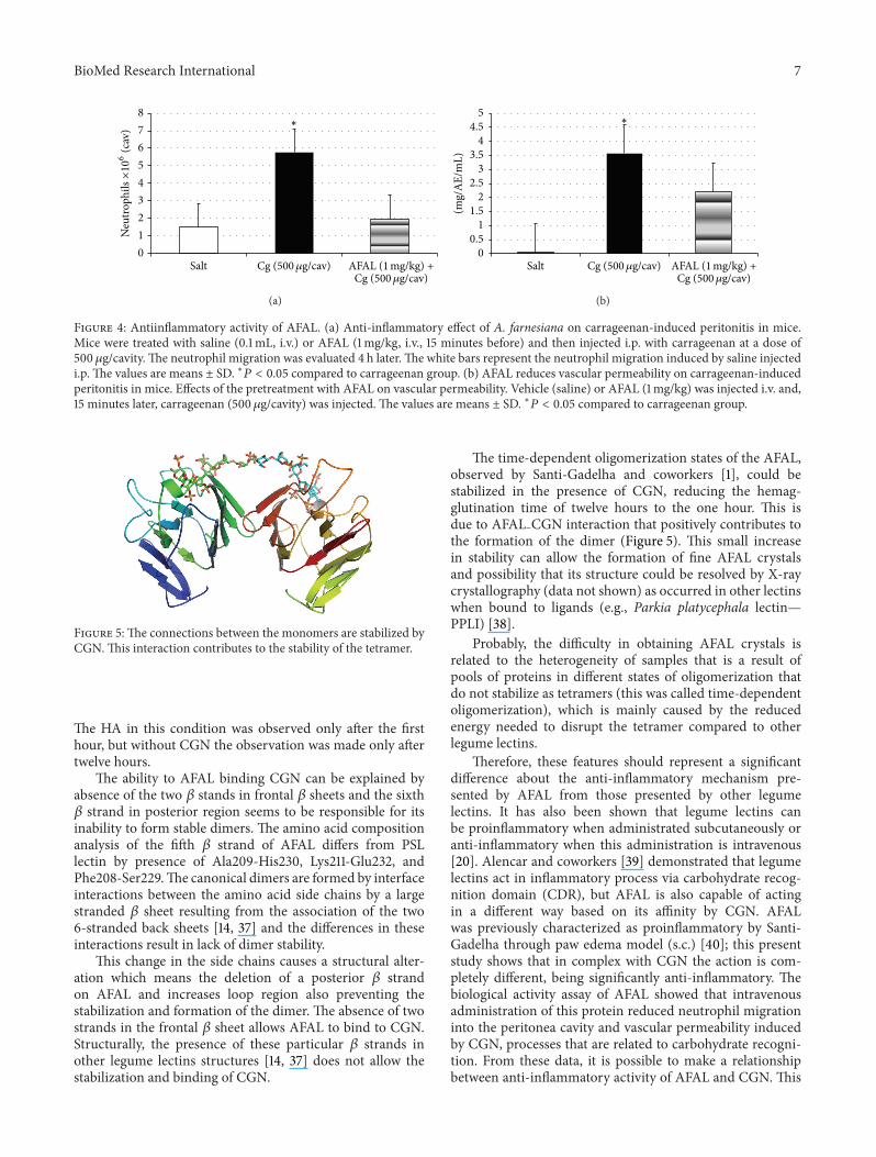
3.4. Biological Activity. The i.v. administration of AFAL, 15minutes before stimulation by CGN, reduced the intensemigration of neutrophils into the peritoneal cavity at adose of 1mg/kg four hours after CGN i.p. administration(500𝜇g/cav.).This inhibitionwas 71% and the positive controlgroup (CGN) produced a significant increase in neutrophilsas compared to saline group (Figure 4(a)).
The CGN i.p. administration (500 𝜇g/cav.) induced asignificant increase in vascular permeability in the controlgroup, seen through the leakage of dye (Evans blue) tothe intraperitoneal fluid. The pretreatment of the animalswith intravenous AFAL (1mg/kg) reduced significantly thealteration of vascular permeability (40% inhibition) in thecavity induced by CGN (Figure 4(b)). So, even with a smallreduction of the permeability the anti-inflammatory effectis very efficient in reducing the number of cells in theperitoneum.
4. Discussion
Santi-Gadelha and coworkers [1] characterized AFAL asPHA-like protein based on their amino acid sequence andbiochemical characteristics. Among these characteristics,the inability to bind to simple carbohydrates, to chitin-like oligosaccharides (two to eight residues GlcNAc), andto ovomucoid glycoprotein (rich in GlcNAc) was the mostintriguing and determined the need to study the possibleinteractions with complex carbohydrates based on dockingsimulations. Docking with complex glycan, GlcNac oligosac-charide and CGN are corroborated by previous results ofhaemagglutinating activity and by the purification protocolof AFAL using chitin affinity chromatography column [1] andalso by the HA of this work.
AFAL structural analysis showed that CGN and glycanbinding domains are independent.This is in accordance withour findings for the inhibition test (HA) by CGN, in whichno change was observed in agglutination after incubation,probably due to the fact that carbohydrate binding site is notaffected by the presence of CGN that binds in another site.It is also an important observation that the presence of CGNbound with AFAL improves its capacity to agglutinate cells.
6 BioMed Research International
(a) (b) (c)
(d)
Figure 3: AFAL molecular docking. (a) The AFAL molecular docking with carrageenan. (b) The AFAL molecular docking with oligosac-charide. (c) The AFAL molecular docking with N-acetylglucosamine (GlcNac). (d) AFAL binding the carrageenan and oligosaccharide indifferent sites.
Table 2: The overall interactions between AFAL and carrageenan.
CARRAGEENAN Ser129 Ala131 Leu156 Ser172 Asp173 Arg174 Ile130 Glu180 Ala108O OG O N O O OD2 O O N OE2 O
G4S31
O1 2.8 — — — — — — — — 3.2 — —O2 3.2 — — — — — — — — — — —
G4S5O2 — — — — — — — — 3.1 — — —O6 — — — — — — — — — — 3.3 —O7 — — — — — — — — — — — 3.2
DGS22
O7 — — 2.5 2.3 — — — — — — — —O9 — — — 3.2 — — — — — — — —
DGS4O3 3.3 3.0 — — — — — — — — — —O5 — — — — — — 3.1 — — — — —O7 — — — — 2.8 3.2 — 2.5 — — — —O8 — 3.3 — — 2.9 — — — — — — —
1G4S: 4-O-sulfo-𝛽-D-galactopyranose; 2DGS: 3,6-anhydro-D-galactose-2-sulfate.
BioMed Research International 7
012345678
Salt
∗
Cg (500𝜇g/cav)Cg (500𝜇g/cav) AFAL (1mg/kg) +
Neu
troph
ils×106
(cav
)
(a)
00.5
11.5
22.5
33.5
44.5
5
(mg/
AE/
mL)
SaltCg (500𝜇g/cav)
Cg (500𝜇g/cav) AFAL (1mg/kg) +
∗
(b)
Figure 4: Antiinflammatory activity of AFAL. (a) Anti-inflammatory effect of A. farnesiana on carrageenan-induced peritonitis in mice.Mice were treated with saline (0.1mL, i.v.) or AFAL (1mg/kg, i.v., 15 minutes before) and then injected i.p. with carrageenan at a dose of500 𝜇g/cavity. The neutrophil migration was evaluated 4 h later. The white bars represent the neutrophil migration induced by saline injectedi.p. The values are means ± SD. ∗𝑃 < 0.05 compared to carrageenan group. (b) AFAL reduces vascular permeability on carrageenan-inducedperitonitis in mice. Effects of the pretreatment with AFAL on vascular permeability. Vehicle (saline) or AFAL (1mg/kg) was injected i.v. and,15 minutes later, carrageenan (500 𝜇g/cavity) was injected. The values are means ± SD. ∗𝑃 < 0.05 compared to carrageenan group.
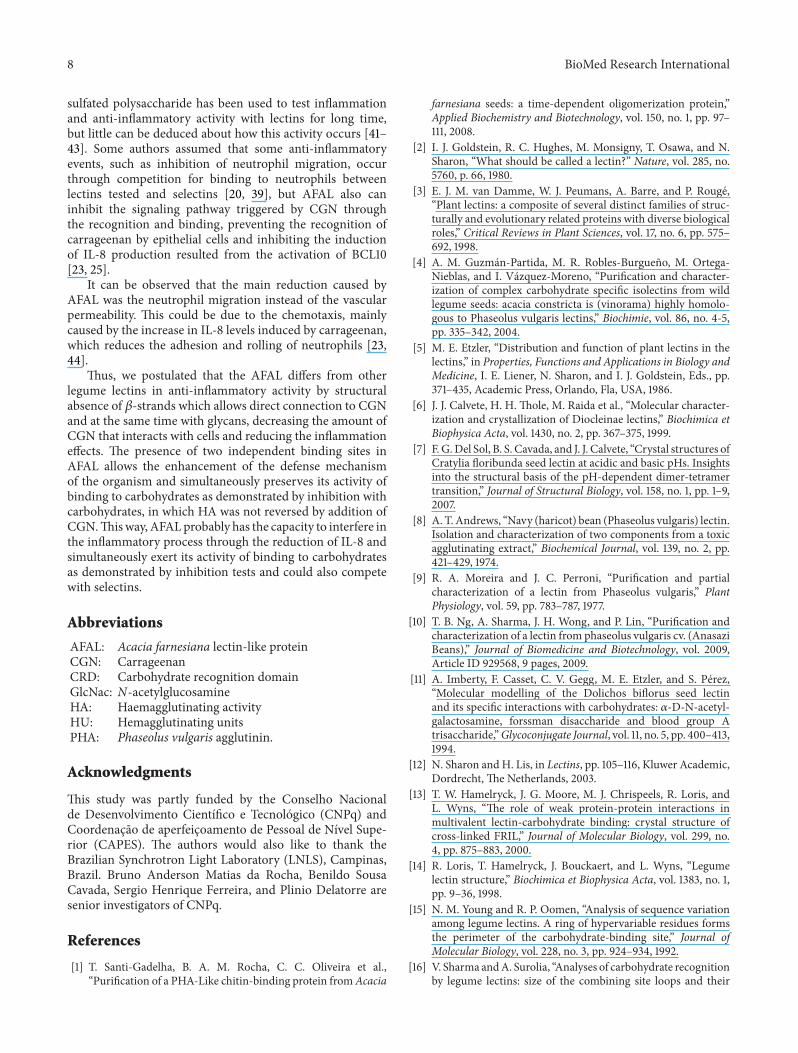
Figure 5:The connections between the monomers are stabilized byCGN.This interaction contributes to the stability of the tetramer.
The HA in this condition was observed only after the firsthour, but without CGN the observation was made only aftertwelve hours.
The ability to AFAL binding CGN can be explained byabsence of the two 𝛽 stands in frontal 𝛽 sheets and the sixth𝛽 strand in posterior region seems to be responsible for itsinability to form stable dimers. The amino acid compositionanalysis of the fifth 𝛽 strand of AFAL differs from PSLlectin by presence of Ala209-His230, Lys211-Glu232, andPhe208-Ser229.The canonical dimers are formed by interfaceinteractions between the amino acid side chains by a largestranded 𝛽 sheet resulting from the association of the two6-stranded back sheets [14, 37] and the differences in theseinteractions result in lack of dimer stability.
This change in the side chains causes a structural alter-ation which means the deletion of a posterior 𝛽 strandon AFAL and increases loop region also preventing thestabilization and formation of the dimer. The absence of twostrands in the frontal 𝛽 sheet allows AFAL to bind to CGN.Structurally, the presence of these particular 𝛽 strands inother legume lectins structures [14, 37] does not allow thestabilization and binding of CGN.
The time-dependent oligomerization states of the AFAL,observed by Santi-Gadelha and coworkers [1], could bestabilized in the presence of CGN, reducing the hemag-glutination time of twelve hours to the one hour. This isdue to AFAL CGN interaction that positively contributes tothe formation of the dimer (Figure 5). This small increasein stability can allow the formation of fine AFAL crystalsand possibility that its structure could be resolved by X-raycrystallography (data not shown) as occurred in other lectinswhen bound to ligands (e.g., Parkia platycephala lectin—PPLI) [38].
Probably, the difficulty in obtaining AFAL crystals isrelated to the heterogeneity of samples that is a result ofpools of proteins in different states of oligomerization thatdo not stabilize as tetramers (this was called time-dependentoligomerization), which is mainly caused by the reducedenergy needed to disrupt the tetramer compared to otherlegume lectins.
Therefore, these features should represent a significantdifference about the anti-inflammatory mechanism pre-sented by AFAL from those presented by other legumelectins. It has also been shown that legume lectins canbe proinflammatory when administrated subcutaneously oranti-inflammatory when this administration is intravenous[20]. Alencar and coworkers [39] demonstrated that legumelectins act in inflammatory process via carbohydrate recog-nition domain (CDR), but AFAL is also capable of actingin a different way based on its affinity by CGN. AFALwas previously characterized as proinflammatory by Santi-Gadelha through paw edema model (s.c.) [40]; this presentstudy shows that in complex with CGN the action is com-pletely different, being significantly anti-inflammatory. Thebiological activity assay of AFAL showed that intravenousadministration of this protein reduced neutrophil migrationinto the peritonea cavity and vascular permeability inducedby CGN, processes that are related to carbohydrate recogni-tion. From these data, it is possible to make a relationshipbetween anti-inflammatory activity of AFAL and CGN. This
8 BioMed Research International
sulfated polysaccharide has been used to test inflammationand anti-inflammatory activity with lectins for long time,but little can be deduced about how this activity occurs [41–43]. Some authors assumed that some anti-inflammatoryevents, such as inhibition of neutrophil migration, occurthrough competition for binding to neutrophils betweenlectins tested and selectins [20, 39], but AFAL also caninhibit the signaling pathway triggered by CGN throughthe recognition and binding, preventing the recognition ofcarrageenan by epithelial cells and inhibiting the inductionof IL-8 production resulted from the activation of BCL10[23, 25].
It can be observed that the main reduction caused byAFAL was the neutrophil migration instead of the vascularpermeability. This could be due to the chemotaxis, mainlycaused by the increase in IL-8 levels induced by carrageenan,which reduces the adhesion and rolling of neutrophils [23,44].
Thus, we postulated that the AFAL differs from otherlegume lectins in anti-inflammatory activity by structuralabsence of 𝛽-strands which allows direct connection to CGNand at the same time with glycans, decreasing the amount ofCGN that interacts with cells and reducing the inflammationeffects. The presence of two independent binding sites inAFAL allows the enhancement of the defense mechanismof the organism and simultaneously preserves its activity ofbinding to carbohydrates as demonstrated by inhibition withcarbohydrates, in which HA was not reversed by addition ofCGN.Thisway,AFALprobably has the capacity to interfere inthe inflammatory process through the reduction of IL-8 andsimultaneously exert its activity of binding to carbohydratesas demonstrated by inhibition tests and could also competewith selectins.
Abbreviations
AFAL: Acacia farnesiana lectin-like proteinCGN: CarrageenanCRD: Carbohydrate recognition domainGlcNac: N-acetylglucosamineHA: Haemagglutinating activityHU: Hemagglutinating unitsPHA: Phaseolus vulgaris agglutinin.
Acknowledgments
This study was partly funded by the Conselho Nacionalde Desenvolvimento Cientıfico e Tecnologico (CNPq) andCoordenacao de aperfeicoamento de Pessoal de Nıvel Supe-rior (CAPES). The authors would also like to thank theBrazilian Synchrotron Light Laboratory (LNLS), Campinas,Brazil. Bruno Anderson Matias da Rocha, Benildo SousaCavada, Sergio Henrique Ferreira, and Plinio Delatorre aresenior investigators of CNPq.
References
[1] T. Santi-Gadelha, B. A. M. Rocha, C. C. Oliveira et al.,“Purification of a PHA-Like chitin-binding protein fromAcacia
farnesiana seeds: a time-dependent oligomerization protein,”Applied Biochemistry and Biotechnology, vol. 150, no. 1, pp. 97–111, 2008.
[2] I. J. Goldstein, R. C. Hughes, M. Monsigny, T. Osawa, and N.Sharon, “What should be called a lectin?” Nature, vol. 285, no.5760, p. 66, 1980.
[3] E. J. M. van Damme, W. J. Peumans, A. Barre, and P. Rouge,“Plant lectins: a composite of several distinct families of struc-turally and evolutionary related proteins with diverse biologicalroles,” Critical Reviews in Plant Sciences, vol. 17, no. 6, pp. 575–692, 1998.
[4] A. M. Guzman-Partida, M. R. Robles-Burgueno, M. Ortega-Nieblas, and I. Vazquez-Moreno, “Purification and character-ization of complex carbohydrate specific isolectins from wildlegume seeds: acacia constricta is (vinorama) highly homolo-gous to Phaseolus vulgaris lectins,” Biochimie, vol. 86, no. 4-5,pp. 335–342, 2004.
[5] M. E. Etzler, “Distribution and function of plant lectins in thelectins,” in Properties, Functions and Applications in Biology andMedicine, I. E. Liener, N. Sharon, and I. J. Goldstein, Eds., pp.371–435, Academic Press, Orlando, Fla, USA, 1986.
[6] J. J. Calvete, H. H. Thole, M. Raida et al., “Molecular character-ization and crystallization of Diocleinae lectins,” Biochimica etBiophysica Acta, vol. 1430, no. 2, pp. 367–375, 1999.
[7] F. G.Del Sol, B. S. Cavada, and J. J. Calvete, “Crystal structures ofCratylia floribunda seed lectin at acidic and basic pHs. Insightsinto the structural basis of the pH-dependent dimer-tetramertransition,” Journal of Structural Biology, vol. 158, no. 1, pp. 1–9,2007.
[8] A. T. Andrews, “Navy (haricot) bean (Phaseolus vulgaris) lectin.Isolation and characterization of two components from a toxicagglutinating extract,” Biochemical Journal, vol. 139, no. 2, pp.421–429, 1974.
[9] R. A. Moreira and J. C. Perroni, “Purification and partialcharacterization of a lectin from Phaseolus vulgaris,” PlantPhysiology, vol. 59, pp. 783–787, 1977.
[10] T. B. Ng, A. Sharma, J. H. Wong, and P. Lin, “Purification andcharacterization of a lectin from phaseolus vulgaris cv. (AnasaziBeans),” Journal of Biomedicine and Biotechnology, vol. 2009,Article ID 929568, 9 pages, 2009.
[11] A. Imberty, F. Casset, C. V. Gegg, M. E. Etzler, and S. Perez,“Molecular modelling of the Dolichos biflorus seed lectinand its specific interactions with carbohydrates: 𝛼-D-N-acetyl-galactosamine, forssman disaccharide and blood group Atrisaccharide,”Glycoconjugate Journal, vol. 11, no. 5, pp. 400–413,1994.
[12] N. Sharon and H. Lis, in Lectins, pp. 105–116, Kluwer Academic,Dordrecht, The Netherlands, 2003.
[13] T. W. Hamelryck, J. G. Moore, M. J. Chrispeels, R. Loris, andL. Wyns, “The role of weak protein-protein interactions inmultivalent lectin-carbohydrate binding: crystal structure ofcross-linked FRIL,” Journal of Molecular Biology, vol. 299, no.4, pp. 875–883, 2000.
[14] R. Loris, T. Hamelryck, J. Bouckaert, and L. Wyns, “Legumelectin structure,” Biochimica et Biophysica Acta, vol. 1383, no. 1,pp. 9–36, 1998.
[15] N. M. Young and R. P. Oomen, “Analysis of sequence variationamong legume lectins. A ring of hypervariable residues formsthe perimeter of the carbohydrate-binding site,” Journal ofMolecular Biology, vol. 228, no. 3, pp. 924–934, 1992.
[16] V. Sharma andA. Surolia, “Analyses of carbohydrate recognitionby legume lectins: size of the combining site loops and their
BioMed Research International 9
primary specificity,” Journal of Molecular Biology, vol. 267, no.2, pp. 433–445, 1997.
[17] J. Bouckaert, R. Loris, F. Poortmans, and L. Wyns, “Crys-tallographic structure of metal-free concanavalin A at 2.5 Aresolution,” Proteins, vol. 23, no. 4, pp. 510–524, 1995.
[18] J. Bouckaert, Y. Dewallef, F. Poortmans, L. Wyns, and R.Loris, “The structural features of concanavalin A governingnon-proline peptide isomerization,” The Journal of BiologicalChemistry, vol. 275, no. 26, pp. 19778–19787, 2000.
[19] J. Lescar, R. Loris, E. Mitchell et al., “Isolectins I-A and I-B of Griffonia (Bandeiraea) simplicifolia. Crystal structure ofmetal-free GS I-B4 and molecular basis for metal binding andmonosaccharide specificity,” The Journal of Biological Chem-istry, vol. 277, no. 8, pp. 6608–6614, 2002.
[20] B. A. M. Rocha, P. Delatorre, T. M. Oliveira et al., “Structuralbasis for both pro- and anti-inflammatory response inducedby mannose-specific legume lectin from Cymbosema roseum,”Biochimie, vol. 93, no. 5, pp. 806–816, 2011.
[21] U. K. Mazumder, M. Gupta, L. Manikandan, S. Bhattacharya, P.K. Haldar, and S. Roy, “Evaluation of anti-inflammatory activityof Vernonia cinerea Less. Extract in rats,” Phytomedicine, vol. 10,no. 2-3, pp. 185–188, 2003.
[22] S. Bhattacharyya, H. Liu, Z. Zhang et al., “Carrageenan-inducedinnate immune response is modified by enzymes that hydrolyzedistinct galactosidic bonds,” Journal of Nutritional Biochemistry,vol. 21, no. 10, pp. 906–913, 2010.
[23] A. Borthakur, S. Bhattacharyya, P. K. Dudeja, and J. K. Tobac-man, “Carrageenan induces interleukin-8 production throughdistinct Bcl10 pathway in normal human colonic epithelialcells,”American Journal of Physiology: Gastrointestinal and LiverPhysiology, vol. 292, no. 3, pp. G829–G838, 2007.
[24] S. Bhattacharyya, R. Gill, L. C. Mei et al., “Toll-like receptor 4mediates induction of the Bcl10-NF𝜅B- interleukin-8 inflam-matory pathway by carrageenan in human intestinal epithelialcells,” The Journal of Biological Chemistry, vol. 283, no. 16, pp.10550–10558, 2008.
[25] A. Borthakur, S. Bhattacharyya, A.N.Anbazhagan, A. Kumar, P.K. Dudeja, and J. K. Tobacman, “Prolongation of carrageenan-induced inflammation in human colonic epithelial cells byactivation of an NF𝜅B-BCL10 loop,” Biochimica et BiophysicaActa, vol. 1822, no. 8, pp. 1300–1307, 2012.
[26] S. F. Altschul,W. Gish,W.Miller, E.W.Myers, and D. J. Lipman,“Basic local alignment search tool,” Journal ofMolecular Biology,vol. 215, no. 3, pp. 403–410, 1990.
[27] F. Corpet, “Multiple sequence alignment with hierarchicalclustering,” Nucleic Acids Research, vol. 16, no. 22, pp. 10881–10890, 1988.
[28] K. Arnold, L. Bordoli, J. Kopp, and T. Schwede, “The SWISS-MODEL workspace: a web-based environment for proteinstructure homology modelling,” Bioinformatics, vol. 22, no. 2,pp. 195–201, 2006.
[29] D. W. Ritchie and G. J. L. Kemp, “Protein docking usingspherical polar Fourier correlations,” Proteins, vol. 39, pp. 178–194, 2000.
[30] Y. Bourne, J. Mazurier, D. Legrand et al., “Structures of alegume lectin complexed with the human lactotransferrin N2fragment, and with an isolated biantennary glycopeptide: roleof the fucose moiety,” Structure, vol. 2, no. 3, pp. 209–219, 1994.
[31] S. Arnott, W. E. Scott, D. A. Rees, and C. G. A. McNab, “Iota-carrageenanmolecular structure and packing of polysaccharidedouble helices in oriented fibres of divalent cation salts,” Journalof Molecular Biology, vol. 90, no. 2, pp. 253–267, 1974.
[32] A. W. Schuttelkopf and D. M. F. van Aalten, “PRODRG:a tool for high-throughput crystallography of protein-ligandcomplexes,” Acta Crystallographica D, vol. 60, no. 8, pp. 1355–1363, 2004.
[33] G. E. P. De Souza and S. H. Ferreira, “Blockade by anti-macrophage serum of the migration of PMN neutrophils intothe inflamed peritoneal cavity,”Agents and Actions, vol. 17, no. 1,pp. 97–103, 1985.
[34] G. Thurston, P. Baluk, and D. M. McDonald, “Determinants ofendothelial cell phenotype in venules,” Microcirculation, vol. 7,no. 1, pp. 67–80, 2000.
[35] V. Z. Pletnev, S. N. Ruzheinikov, I. N. Tsygannik et al., “Thestructure of pea lectin-D-glucopyranose complex at a 1.9 Aresolution,” Russian Journal of Bioorganic Chemistry, vol. 23, no.6, pp. 469–478, 1997.
[36] D. Sen and D. K.Mandal, “Pea lectin unfolding reveals a uniquemolten globule fragment chain,” Biochimie, vol. 93, no. 3, pp.409–417, 2011.
[37] K. V. Brinda, N. Mitra, A. Surolia, and S. Vishveshwara,“Determinants of quaternary association in legume lectins,”Protein Science, vol. 13, no. 7, pp. 1735–1749, 2004.
[38] F. Gallego Del Sol, C. Nagano, B. S. Cavada, and J. J. Calvete,“The first crystal structure of a Mimosoideae lectin reveals anovel quaternary arrangement of awidespread domain,” Journalof Molecular Biology, vol. 353, no. 3, pp. 574–583, 2005.
[39] N. M. N. Alencar, E. H. Teixeira, A. M. S. Assreuy, B. S. Cavada,C. A. Flores, and R. A. Ribeiro, “Leguminous lectins as tools forstudying the role of sugar residues in leukocyte recruitment,”Mediators of Inflammation, vol. 8, no. 2, pp. 107–113, 1999.
[40] T. Santi-Gadelha, Isolamento, purificacao, caracterizacao e ativi-dade biologica da lectina da fracao das albuminas da semente deAcacia farnesiana [Ph.D. thesis], Universidade Federal doCeara,Ceara, Brazil, 2005.
[41] J. F. M. Leite, A. M. S. Assreuy, M. R. L. Mota et al., “Antinoci-ceptive and anti-inflammatory effects of a lectin-like substancefrom Clitoria fairchildiana R. Howard seeds,”Molecules, vol. 17,no. 3, pp. 3277–3290, 2012.
[42] T. B. A. Rangel, B. A. M. Rocha, G. A. Bezerra et al., “Crystalstructure of a pro-inflammatory lectin from the seeds ofDiocleawilsonii Standl,” Biochimie, vol. 94, no. 2, pp. 525–532, 2012.
[43] A. M. S. Assreuy, M. D. Shibuya, G. J. Martins et al., “Anti-inflammatory effect of glucose-mannose binding lectins iso-lated from Brazilian beans,” Mediators of Inflammation, vol. 6,no. 3, pp. 201–210, 1997.
[44] S. Bhattacharyya, P. K. Dudeja, and J. K. Tobacman,“Carrageenan-induced NF𝜅B activation depends on distinctpathways mediated by reactive oxygen species and Hsp27 orby Bcl10,” Biochimica et Biophysica Acta, vol. 1780, no. 7-8, pp.973–982, 2008.

Submit your manuscripts athttp://www.hindawi.com
Hindawi Publishing Corporationhttp://www.hindawi.com Volume 2014
Anatomy Research International
PeptidesInternational Journal of
Hindawi Publishing Corporationhttp://www.hindawi.com Volume 2014
Hindawi Publishing Corporation http://www.hindawi.com
International Journal of
Volume 2014
Zoology
Hindawi Publishing Corporationhttp://www.hindawi.com Volume 2014
Molecular Biology International
GenomicsInternational Journal of
Hindawi Publishing Corporationhttp://www.hindawi.com Volume 2014
The Scientific World JournalHindawi Publishing Corporation http://www.hindawi.com Volume 2014
Hindawi Publishing Corporationhttp://www.hindawi.com Volume 2014
BioinformaticsAdvances in
Marine BiologyJournal of
Hindawi Publishing Corporationhttp://www.hindawi.com Volume 2014
Hindawi Publishing Corporationhttp://www.hindawi.com Volume 2014
Signal TransductionJournal of
Hindawi Publishing Corporationhttp://www.hindawi.com Volume 2014
BioMed Research International
Evolutionary BiologyInternational Journal of
Hindawi Publishing Corporationhttp://www.hindawi.com Volume 2014
Hindawi Publishing Corporationhttp://www.hindawi.com Volume 2014
Biochemistry Research International
ArchaeaHindawi Publishing Corporationhttp://www.hindawi.com Volume 2014
Hindawi Publishing Corporationhttp://www.hindawi.com Volume 2014
Genetics Research International
Hindawi Publishing Corporationhttp://www.hindawi.com Volume 2014
Advances in
Virolog y
Hindawi Publishing Corporationhttp://www.hindawi.com
Nucleic AcidsJournal of
Volume 2014
Stem CellsInternational
Hindawi Publishing Corporationhttp://www.hindawi.com Volume 2014
Hindawi Publishing Corporationhttp://www.hindawi.com Volume 2014
Enzyme Research
Hindawi Publishing Corporationhttp://www.hindawi.com Volume 2014
International Journal of
Microbiology


