Embed Size (px)
Citation preview
Quantitative Imaging Biomarker Ontology (QIBO) for KnowledgeRepresentation of Biomedical Imaging Biomarkers
Andrew J. Buckler & M. Ouellette & J. Danagoulian &
G. Wernsing & Tiffany Ting Liu & Erica Savig &
Baris E. Suzek & Daniel L. Rubin & David Paik
Published online: 16 April 2013# Society for Imaging Informatics in Medicine 2013
Abstract A widening array of novel imaging biomarkersis being developed using ever more powerful clinicaland preclinical imaging modalities. These biomarkershave demonstrated effectiveness in quantifying biologi-cal processes as they occur in vivo and in the earlyprediction of therapeutic outcomes. However, quantita-tive imaging biomarker data and knowledge are notstandardized, representing a critical barrier to accumu-lating medical knowledge based on quantitative imagingdata. We use an ontology to represent, integrate, andharmonize heterogeneous knowledge across the domainof imaging biomarkers. This advances the goal of de-veloping applications to (1) improve precision and recallof storage and retrieval of quantitative imaging-related
data using standardized terminology; (2) streamline thediscovery and development of novel imaging biomarkersby normalizing knowledge across heterogeneous re-sources; (3) effectively annotate imaging experimentsthus aiding comprehension, re-use, and reproducibility;and (4) provide validation frameworks through rigorousspecification as a basis for testable hypotheses andcompliance tests. We have developed the QuantitativeImaging Biomarker Ontology (QIBO), which currentlyconsists of 488 terms spanning the following upperclasses: experimental subject, biological intervention,imaging agent, imaging instrument, image post-processing algorithm, biological target, indicated biolo-gy, and biomarker application. We have demonstratedthat QIBO can be used to annotate imaging experimentswith standardized terms in the ontology and to generatehypotheses for novel imaging biomarker–disease associa-tions. Our results established the utility of QIBO in enablingintegrated analysis of quantitative imaging data.
Keywords Imaging biomarker . Ontology development .
Quantitative imaging
Introduction
Imaging, by CT and MR, has been ranked by physicians asthe single most important medical innovation [1]. In addi-tion to common clinical imaging modalities, biomedicalresearch studies use many other rich and diverse types ofimaging data, including high-resolution microscopy images,fluorescence imaging, and recently developed nanoparticleimaging. The biological significance of these imaging stud-ies often involves identifying imaging biomarkers that are
Andrew J. Buckler and Tiffany Ting Liu contributed equally to thisstudy.
A. J. Buckler (*) :M. Ouellette : J. Danagoulian :G. WernsingBBMSC, 225 Main Street, Suite 15,Wenham, MA 01984, USAe-mail: [email protected]: www.bbmsc.com
T. T. Liu :D. L. Rubin :D. PaikBiomedical Informatics Training Program,Stanford, CA 94305, USA
T. T. Liu :D. L. Rubin :D. PaikDepartment of Radiology,225 Main Street, Stanford, CA 94305, USA
E. SavigCancer Biology Program, Stanford University,Stanford, CA 94305, USA
B. E. SuzekGeorgetown University, Washington, DC 20007, USA
J Digit Imaging (2013) 26:630–641DOI 10.1007/s10278-013-9599-2
indicators of the underlying biology. Currently, quantitativeimaging suffers from the lack of a standardized representa-tion of quantitative image features and content [2–5].
As one resource that depends on this, the concept of“image biobanking” as an analog to tissue biobanking hasgreat promise [6, 7]. Tools have become available for han-dling the complexity of genotype [8–12], and similar ad-vancements are needed to describe phenotype, especially asderived from imaging [4, 13–20]. Publicly accessible re-sources that support large image archives support file shar-ing and have so far not yet merged into a framework thatsupports the collaborative work needed to meet the potentialof quantitative image analysis. With the availability of toolsfor automatic ontology-based annotation of datasets withterms from biomedical ontologies, coupled with image ar-chives and the means for batch selection and processing ofimage and clinical data, imaging will go through a similarincrease in capability analogous to what advanced sequenc-ing techniques have gone through in molecular biology.
Quantitative imaging biomarkers, which are the focus ofthis work, provide a numerical characterization of the un-derlying biology or pathology as opposed to using textual orcategorical descriptions of an observer’s subjective visualinterpretation. Accelerated by advances in imaging tech-niques, a wide array of novel quantitative imaging bio-markers have been developed and have demonstratedeffectiveness in quantifying biological processes and inclinical use. For example, imaging biomarkers, such aschange in tumor uptake measured by the standardizeduptake value (SUV) of [18F]-FDG positron emission to-mography (PET), can be used in monitoring diseaseprogression, prediction of response to treatment, as wellas drug development [21].
Fully describing a quantitative imaging biomarker andhow it is used involves specifying a series of heterogeneousconcepts that span the fields of imaging physics, contrastagent or probe chemistry, biology, and quantitation tech-niques. Interpretation of data used in the development andvalidation of quantitative imaging biomarkers requires thesedisparate concepts to be related together with scientificallyrigorous epistemology. We posit that this motivates an im-mediate need for an ontology to represent the complex andheterogeneous imaging biomarker data and knowledge withsufficient coverage of these fields [22]. In this endeavor, wedeveloped an ontology that represents the knowledge do-main of quantitative imaging biomarkers and we have ex-plored applications enabled by it.
Imaging biomarker research
The Biomarkers Definitions Working Group at NationalInstitutes of Health defines biomarkers as characteristicsthat are objectively measured as indicators of normal
biological processes, pathological changes, or pharmaco-logic responses to a therapeutic intervention [2, 23]. Forexample, the measurement of the serum concentration of theglycoprotein CA125 assayed by ELISA can be used tomonitor therapy during treatment for ovarian cancer. Whenbiomarkers are obtained from biomedical images, they arereferred to as imaging biomarkers. While the term imagingbiomarker is sometimes used to refer directly to the exoge-nous imaging agent or its molecular target, we use the termhere in a broader context to represent the measurement ofthe characteristic obtained through imaging. There is muchresearch interest in imaging biomarkers, and several relatedefforts have been initiated in imaging biomarker research inthe scientific community.
The Radiological Society of North America has formedthe Quantitative Imaging Biomarker Alliance (QIBA) toadvance quantitative imaging in clinical care and facilitatingimaging as a biomarker in clinical trials [24, 25]. Whileengaging many stakeholders across academia, government,and industry, the effort is limited to the most mature bio-markers. Also, they are not addressing biomarkers from aknowledge engineering perspective and have only limitedactivity associated with formal verification activities.
The Center for Biomarkers in Imaging at the Massachu-setts General Hospital has developed a large imaging bio-marker catalog with about 350 imaging biomarkers [26].Although extensive, this cataloging effort is not focusedon a structured and standardized representation of quantita-tive imaging biomarkers.
Biomedical Imaging Research
Imaging techniques allow non-invasive interrogation of thebiology. This has fundamental utility in characterizing tis-sues and biological processes in the context of a livingorganism. The non-invasive nature also allows studies tobe carried out serially, and thus enables characterization ofbiological changes over time.
Recently, molecular imaging has attracted considerableinterest among researchers in the imaging sciences becauseit enables quantification of biological activities at the cellu-lar and molecular levels [27]. For example, changes inactivities of the receptor tyrosine kinase EGFR can bevisualized and quantified through the optical imaging ofreconstituted luciferase [8]. PET enables detection and quan-tification of molecular processes such as glucose metabo-lism, angiogenesis, apoptosis, and necrosis [28, 29].Radiolabeled annexin V uptake by apoptotic and necroticcells is used to measure apoptosis, necrosis, and other dis-ease processes using PET [30, 31]. Chelated gadoliniumattached to small peptides recognizes cell receptors andquantify receptor activities using magnetic imaging tech-niques. Similarly, microbubbles and nanobubbles attached
J Digit Imaging (2013) 26:630–641 631
to antibodies such as anti-P-selectin may be used to imagetargeted molecules associated with inflammation, angiogen-esis, intravascular thrombus, and tumors [32].
Despite the obvious advantages of imaging, there aremany obstacles to the integration of biomedical images fromdifferent studies to conduct integrative analyses. First, thereis significant heterogeneity and variability in imaging-basedmeasurements. Unlike DNA sequence analysis or gene ex-pression analysis whose data alphabets and fold changemeasurements are readily standardized, imaging is oftenviewed as being both a fundamentally subjective mediumand significantly more complex due to the assay methodsused. In particular, postprocessing, analysis, and interpre-tation are generally highly idiosyncratic to the particularstudy. Even apparently simple steps can have significantvariations.
For example, imaging experts identify lesions and drawregions of interests (ROIs) around the lesions using imageprocessing software. Previous studies have shown that thereis significant variability across experts as well as acrossimage processing algorithms implemented in different plat-forms [3, 4, 33]. There are many approaches to extractmeasurements from ROIs. For example, the SUV used inPET measures relative cellular uptake of an imaging probe.The most commonly used PET imaging probe in cancerdiagnosis is [18F]-FDG that is used to measure glucosemetabolisms. This single parameter has several variations:max SUV (SUVmax), mean SUV (SUVmean), and relativeSUVs. SUVmax and SUVmean are absolute values whereasrSUV is a ratio of the SUV value of one anatomical region tothat of another. The multiple ways of quantifying lesionSUV make it difficult to compare analyses across differentstudies [5].
As another motivating example, the rich data in biomed-ical images are not reused. The integration of imaging datafrom various experiments in order to promote new knowl-edge has not been effectively achieved. The biomedicalimaging community requires a bioinformatics infrastructurethat would enable researchers to search, access, and analyzethis large amount of imaging data. One of the main chal-lenges may be that the complexity and diversity of imagingdata far exceeds genomic and proteomic data.
Despite these difficulties, significant efforts have beenmade to build imaging resources and databases. The Na-tional Biomedical Imaging Archive (NBIA) and The CancerImaging Archive developed by National Cancer Institute arelarge in vivo image repositories [34]. Images are publiclyavailable to researchers in the biomedical research commu-nity for many purposes including lesion detection softwaredevelopment and the quantitative imaging assessment ofdrug responses [34]. Researchers are able to query anddownload images from 15 major imaging modalities and18 anatomical sites for different tumor types. Early efforts to
publish primary data along with analyzed results in thecontext of peer-reviewed journals have begun as well [35].
Another imaging-related database is the Molecular Imag-ing and Contrast Agent Database (MICAD) created by theNational Center for Biotechnology Information [20]. Insteadof images, the MICAD database focuses on molecular im-aging agents and has 1,373 molecular imaging agents listedas of February 2013. In addition, there are thousands morein “pending” status. The imaging agents in MICAD encom-pass radioactive labeled small molecules [15], nanoparticles[16], proteins such as antibodies [36] and fluorescent taggedproteins [18], and labeled cells such as stem cells for track-ing homing to tumors [19]. Moreover, there are 170 diversebiological applications corresponding to the imagingagents, ranging from biological transporter imaging, celltracking, angiogenesis imaging to drug resistance anddisease detection [20].
Because the emphasis of the MICAD database is onmolecular imaging, many imaging agents have specificmolecular targets. In total, there are unique 350 moleculartargets, including mRNA, integrins, growth factors, insulinreceptor, stem cells, etc. There are redundancies in thesetarget annotations as a target can be used to annotate else-where using its synonyms. However, there is no formalizedknowledge framework for this rich database that is publiclyavailable.
Ontologies and their applications in biomedical research
An ontology is a framework that represents knowledgeentities in a specific domain as well as relationships betweenentities [22]. The ontological structure is an ideal frameworkdue to its ability to integrate heterogeneous and complexknowledge. It can be used to discover underlying associa-tions in a knowledge domain.
It consists of three main components: terms/classes,properties/attributes, and instances. Terms describe impor-tant entities in a knowledge domain. Classes are structuredin an IS-A hierarchy where each subclass is a more specifictype of its superclass. Properties (or attributes) of each termdescribe the characteristics of classes. Instances are specificexamples of terms in the ontology.
Also, by formally defining terms and synonyms of termsin a domain, an ontology facilitates the elimination of ter-minology variation and ambiguity, and thus can be used tolink data and knowledge from different sources. For exam-ple, Gene Ontology is a major bioinformatics initiative thatprovides a controlled vocabulary for annotations of geneproducts [37]. It covers three areas: cellular component,molecular function, and biological process. Gene ontology(GO) is now widely used in annotations of gene expression[37]. For example, in microarray analysis, up- anddownregulated genes are annotated using GO to represent
632 J Digit Imaging (2013) 26:630–641
common molecular functions or biological processes toidentify the common theme among differentially regulatedgenes [38]. For example, enrichment of differentiallyexpressed genes in a tumor reveals growth factor activity.In an area where data mining is tedious, ontologies allowsfor intelligent data mining.
Ontologies are reusable and can be linked with otherontologies for specific applications [39]. For example, theFoundational Model of Anatomy (FMA) is a domain ontol-ogy for correlating different views of anatomy. Washingtonet al. integrated FMA and other ontologies to associatehuman diseases with animal models [40].
Other widely used biomedical ontologies include NCIthesaurus (NCIt) and medical subject headings (MeSH)[41]. NCI thesaurus is a controlled terminology developedby the National Cancer Institute in collaboration with manypartners, focusing on enabling the communication of infor-mation in cancer research. MeSH is the National Library ofMedicine-controlled vocabulary thesaurus used for indexingarticles in PubMed. Because of its comprehensiveness, it isused as a reference terminology in other applications [42].
Informatics in Imaging Biomarker Research
RadLex (https://www.rsna.org/RadLex.aspx) is a unified lan-guage of radiology terms for standardized indexing and re-trieval of radiology information resources [43]. With morethan 30,000 terms, RadLex satisfies the needs of softwaredevelopers, system vendors, and radiology users by adoptingthe best features of existing terminology systems while pro-ducing new terms to fill critical gaps. For example, researchersand clinicians can use the RadLex terminology to annotateradiological images [44]. It unifies and supplements otherlexicons and standards, such as SNOMED-CT and DICOM.
Annotation and Image Markup (AIM) is an informationmodel focused on clinical imaging that provides humanobservers with the ability to record explanatory or descrip-tive information about their observations and to attach thisinformation to specific locations within an image [45].Using RadLex terms, it provides a standardized informationmodel compatible with DICOM-SR, XML, and HL7 for-mats for such observations.
Formal Definition as Basis for Validation and QualificationFramework
The lack of consensus methods and carefully characterizedperformance impedes the widespread availability of urgent-ly needed quantitative imaging techniques in medicine. Aprecondition for use is the demonstration of performanceaccording to recognized descriptive statistics computed in adefined patient population with a specific biological phe-nomenon associated with a known disease state, supported
by evidence in large patient populations, and externallyvalidated. Not yet merged into a framework that supportsthe collaborative work needed to meet the potential ofquantitative imaging analysis are the application of ad-vanced statistical techniques, the development of controlledvocabularies and service-oriented architecture for process-ing large image archives. With the availability of tools forthe automatic ontology-based annotation of datasets withterms from biomedical ontologies such as those made pos-sible by the Quantitative Imaging Biomarker Ontology(QIBO), coupled with imaging archives and a means forthe batch selection and processing of imaging and clinicaldata, we believe that imaging will go through a similarincrease in capability analogous to the gains advanced se-quencing techniques have brought to molecular biology.
To address the need for informatics methods in imagingresearch, validation, and qualification, the goal of this projectis to develop a structured knowledge representation using anontology to integrate heterogeneous knowledge in imagingbiomarkers. The domain of our ontology is defined as imagingbiomarkers for both preclinical and clinical applications.
Materials and Methods
We have created the QIBO to provide a basis for standard-izing semantics inclusive of the terms as well as the relation-ships among them.
Initial Curation to Collect Terms
To gather terms, relationships, and properties of the terms inthe imaging biomarker ontology, we started with a literaturereview of the journal Molecular Imaging and Biology. Wesampled 22 articles published since 2006. The inclusioncriteria was that articles made nontrivial use of quantitativeimaging measurements, which was not true of manychemistry/probe development papers. Articles were ran-domly sampled and added until it was subjectively deter-mined that the scope of the concepts had begun to elucidateenough structure such that an ontology could be designedaround it. This work was by two individuals with 23 yearsof imaging research experience in total with all workreviewed by both and the more senior arbitrating conflicts.Further, we also distilled terms associated with 95 putativebiomarkers in oncology indications from 43 articles in suchjournals as JNM, JMRI, Cell, Molecular Imaging and Biol-ogy, and Transgenic Research; 47 putative biomarkers incardiovascular indications from 30 articles in such journalsas JNM, Molecular Therapy, Stroke, and Circulation; 52putative biomarkers in neurology indications from 25journals such as Nuclear Medicine and Biology,NeuroImage, and Nuclear Instruments & Methods in
J Digit Imaging (2013) 26:630–641 633
Physics Research; 22 putative biomarkers in musculoskele-tal indications from 6 articles in such journals as EJNM andMolecular Imaging; 5 putative biomarkers in endocrinologyindications from 3 articles in Proceedings of the NationalAcademy of Sciences and Vanderbilt University Institute ofImaging Science conference proceedings; and 4 putativebiomarkers from 19 articles in pulmonary indications from19 articles in such journals as European Respiratory Jour-nal, Proceedings of American Thoracic Society, MedicalPhysics, and Thoracic Imaging. We examined the methodsand results of experiments reported in the papers, focusingon abstracting terms and relationships that characterize andannotate an imaging biomarker. This approach allows us toexamine specific instances of biomarkers and to be able tobuild the ontology from real world examples.
Reusing Other Publicly Available Ontologies
To build the imaging biomarker ontology, we reviewed a rangeof publicly available ontologies and terminologies, includingMeSH [41], NCI thesaurus [46], GO [37], FMA [39], DiseaseOntology [47], and BIRNLex (a controlled terminology forBiomedical Informatics Research Network) [48] to adopt oradapt them wherever possible. For example, entities of anatom-ical structurewere imported fromFMAand entities of biologicalprocess were extracted from GO, MeSH, and NCI thesaurus.
Our solution to integrating multiple ontologies is to minimal-ly and unambiguously reference the external class from withinQIBO. This is achieved by specifying the external source on-tology, the external source class from the source ontology andthe QIBO class that references the external source class. In ourcase, most of the ontologies we reuse in QIBO are fairly stable.We coded the minimal references directly in the ontology asannotation properties in Web Ontology Language (OWL).
Design Decisions
The domain of the ontology includes imaging biomarkers forboth preclinical and clinical applications featuring molecularimaging because of its richness and biological specificity.
We have built the OWL-based ontology using Protégé, awidely used and freely available ontology editing tool [49].Protégé supports two ontology modeling paradigms: OWLand frames. Despite many similarities between the two lan-guages, OWL provides description logic reasoning capabilitywith high expressive power [50]. Classes can be asserteddirectly in the ontology in both OWL and frames, describingnecessary conditions. Only necessary and sufficient condi-tions can be defined in OWL to specify new classes wherean OWL classifier can be run to generate inferred hierarchy.Inferred hierarchy is particularly suitable for representing theheterogeneous knowledge in imaging biomarkers. It allowsthe defining of newly discovered biomarkers. OWL enables
powerful knowledge reasoning. It is capable of conveyingcomplexity and richness in imaging biomarker research.
Based on our curation efforts, we first identified top-levelterms that capture major entities appearing in an imagingbiomarker experiment. We also created synonyms and defini-tions for classes in the ontology. To ensure consistency, Is–Arelationship (subsumption) is strictly conserved in every branchof the ontology. Other relationships that organize the terms in astructured and meaningful way are defined as properties, suchas the relationships between the first level classes.
We anticipate that QIBO will be extended to relate and linkto other established ontologies such as FMA [51], GO [52],SNOMED [53], and RadLex [54] as well as other standard-ized domain ontologies such as from the Open Biological andBiomedical Ontologies Foundry [55], leveraging the BasicFormal Ontology (BFO) [56] upper ontology for alignmentthrough a shared abstract level. It also incorporates NBIA [57]and AIM UML models, associated common data elementsand underlying NCIt and other ontology concepts.
Results
Upper Level Terms and Their Relations
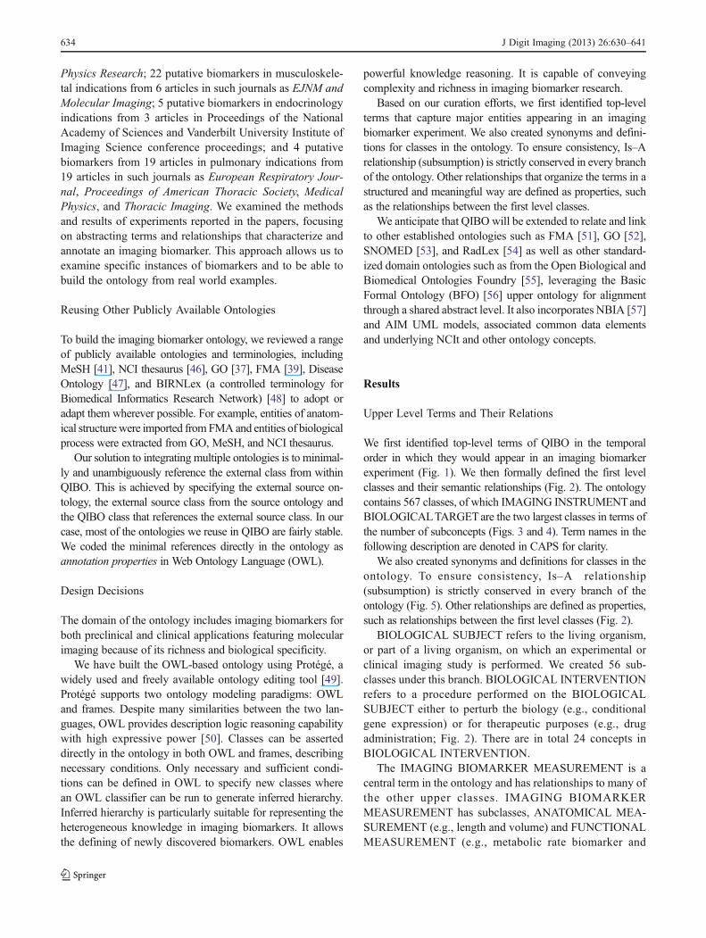
We first identified top-level terms of QIBO in the temporalorder in which they would appear in an imaging biomarkerexperiment (Fig. 1). We then formally defined the first levelclasses and their semantic relationships (Fig. 2). The ontologycontains 567 classes, of which IMAGING INSTRUMENTandBIOLOGICALTARGETare the two largest classes in terms ofthe number of subconcepts (Figs. 3 and 4). Term names in thefollowing description are denoted in CAPS for clarity.
We also created synonyms and definitions for classes in theontology. To ensure consistency, Is–A relationship(subsumption) is strictly conserved in every branch of theontology (Fig. 5). Other relationships are defined as properties,such as relationships between the first level classes (Fig. 2).
BIOLOGICAL SUBJECT refers to the living organism,or part of a living organism, on which an experimental orclinical imaging study is performed. We created 56 sub-classes under this branch. BIOLOGICAL INTERVENTIONrefers to a procedure performed on the BIOLOGICALSUBJECT either to perturb the biology (e.g., conditionalgene expression) or for therapeutic purposes (e.g., drugadministration; Fig. 2). There are in total 24 concepts inBIOLOGICAL INTERVENTION.
The IMAGING BIOMARKER MEASUREMENT is acentral term in the ontology and has relationships to many ofthe other upper classes. IMAGING BIOMARKERMEASUREMENT has subclasses, ANATOMICAL MEA-SUREMENT (e.g., length and volume) and FUNCTIONALMEASUREMENT (e.g., metabolic rate biomarker and
634 J Digit Imaging (2013) 26:630–641
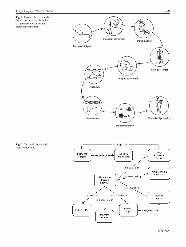
Fig. 1 First level classes in theQIBO, organized by the orderof appearance in an imagingbiomarker experiment
Fig. 2 Top level classes andtheir relationships
J Digit Imaging (2013) 26:630–641 635

permeability biomarker), which are the super classes for 28and 18 concepts, respectively.
IMAGING INSTRUMENT, as the largest branch with 115concepts in the ontology, includes conventional anatomicalimaging instruments (e.g., CT and MRI) as well as molecularand functional imaging instruments (e.g., microscope and bio-luminescence imaging). ALGORITHM describes image pro-cessing, analytic techniques and/or modeling methods used toextract the IMAGING BIOMARKER MEASUREMENT.There are 20 different algorithm concepts in the branch. Al-though this term often is not mentioned with the IMAGING
BIOMARKER MEASUREMENT, it is an indispensable stepin the acquisition of the measurement. IMAGING AGENT isan exogenous material (e.g., contrast agent or molecular probe)optionally administered to the BIOLOGICAL SUBJECT thatis used to visualize some component or process within thesubject. IMAGING AGENT contains 57 subclasses.
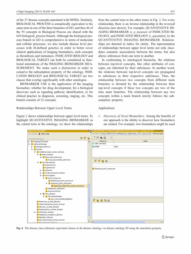
BIOLOGICAL TARGET, as the second largest branchwith 113 concepts in the ontology, is the part of the biolog-ical subject that is optionally targeted by the IMAGINGAGENT; it is visualized in the image and measured in orderto create the imaging biomarker. BIOLOGICAL TARGETrefers to biological components such as biomolecules, cells,or anatomical structure/space. The subclasses are populatedusing terms from an existing ontology NCI thesaurus, aswell as our own terms. For example, subclasses of enzymein molecular target are shared with those of enzyme in NCIthesaurus; multicellular organ level target shares subclasseswith skeletal system part in NCI thesaurus. INDICATEDBIOLOGY includes 55 biological process and 37 diseaseconcepts, which are populated using terms from GO, MeSH,and Disease Ontology. For example, terms under DISEASEreference equivalent classes in the Disease Ontology viaDOID using the annotation property (Fig. 6). There are 24
Fig. 3 Distribution of classes in each of the top-level terms in theQuantitative Imaging Biomarker Ontology: postprocessing algorithm(20), biological intervention (24 concepts), biomarker use (32 con-cepts), imaging agent (57 concepts), biological subject (57 concepts),quantitative imaging biomarker (57 concepts), indicated biology (92concepts), biological target (113 concepts), and imaging instrument(115 concepts)
Fig. 4 Circular graph of the QIBO to get an overview of the nineclasses. Upper classes are highlighted in yellow and each node is aclass
Fig. 5 A partial view of the QIBO in Protégé
636 J Digit Imaging (2013) 26:630–641
of the 37 disease concepts annotated with DOIDs. Similarly,BIOLOGICAL PROCESS is semantically equivalent to thesame term in one of the three branches of GO, and thus 46 ofthe 55 concepts in Biological Process are shared with theGO biological_process branch. Although the biological pro-cess branch in GO is comprehensive in terms of molecularand cellular processes, we also include disease level pro-cesses with ill-defined genetics in order to better coverclinical applications of imaging biomarkers, such conceptsas thrombosis and metastasis. INDICATED BIOLOGY andBIOLOGICAL TARGET can both be considered as func-tional annotations of the IMAGING BIOMARKER MEA-SUREMENT. We make such a distinction in order toconserve the subsumption property of the ontology. INDI-CATED BIOLOGY and BIOLOGICAL TARGET are twoclasses that overlap significantly with other ontologies.
BIOMARKER USE is the application of the imagingbiomarker, whether for drug development, for a biologicaldiscovery such as signaling pathway identification, or forclinical practice in diagnosis, screening, staging, etc. Thisbranch consists of 32 concepts.
Relationships Between Upper Level Terms
Figure 2 shows relationships between upper level terms. Tohighlight QUANTITATIVE IMAGING BIOMARKER asthe central term in the ontology, we show the relationships
from the central term to the other terms in Fig. 2. For everyrelationship, there is an inverse relationship in the reverseddirection (not shown). For example, QUANTITATIVE IM-AGING BIOMARKER is_a_measure of INDICATED BI-OLOGY, and INDICATED BIOLOGY is_quantified_by theQUANTITATIVE IMAGING BIOMARKER. Relation-ships are denoted in italics for clarity. The representationof relationships between upper level terms not only eluci-dates semantic associations between the terms, but alsoallows inferences from one term to another.
In conforming to ontological hierarchy, the relationsbetween top-level concepts, like other attributes of con-cepts, are inherited by their subclasses. In another word,the relations between top-level concepts are propagatedto subclasses in their respective subclasses. Thus, therelationship between two concepts from different mainbranches is dictated by the relationship between theirtop-level concepts if these two concepts are two of thenine main branches. The relationship between any twoconcepts within a main branch strictly follows the sub-sumption property.
Applications
1. Discovery of Novel Biomarkers. Among the benefits ofour approach is the ability to discover how biomarkersare related. For example, two biomarkers might be used
Fig. 6 The disease class references equivalent classes in the disease ontology via disease ontology ID using the annotation property
J Digit Imaging (2013) 26:630–641 637
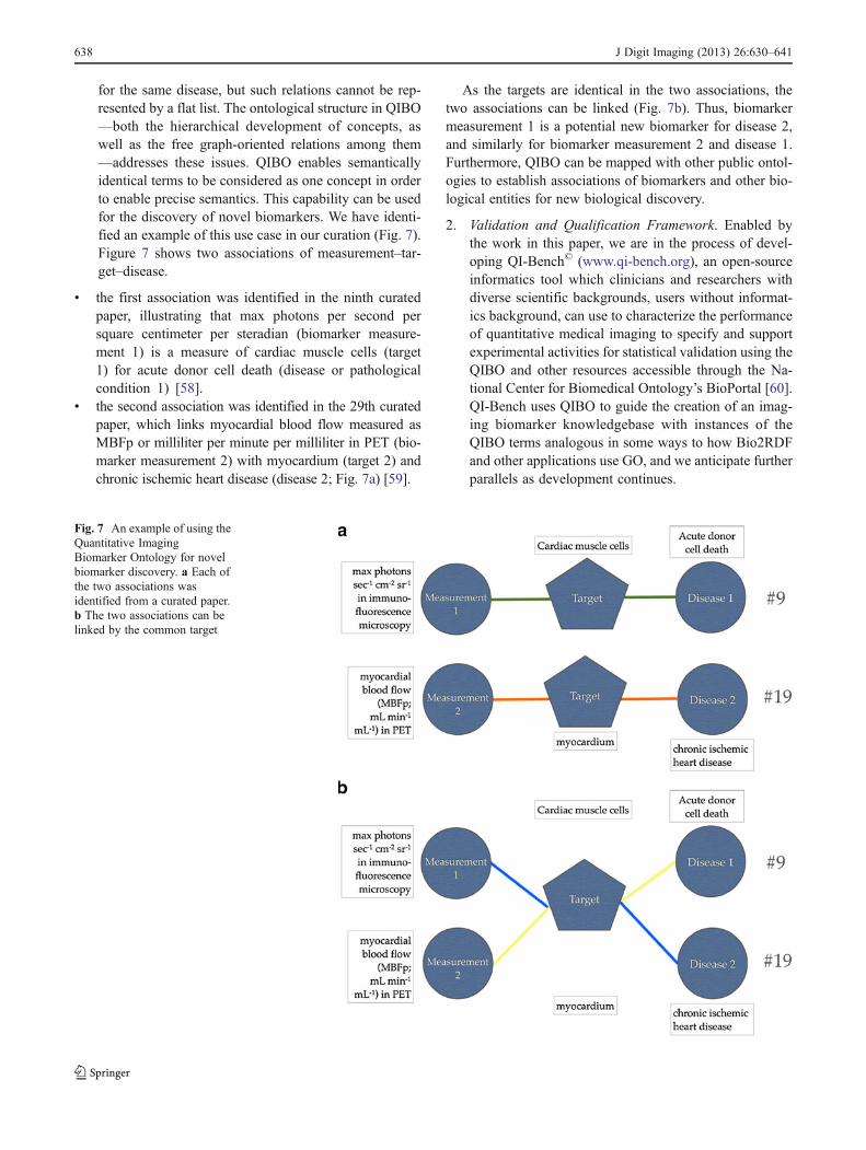
for the same disease, but such relations cannot be rep-resented by a flat list. The ontological structure in QIBO—both the hierarchical development of concepts, aswell as the free graph-oriented relations among them—addresses these issues. QIBO enables semanticallyidentical terms to be considered as one concept in orderto enable precise semantics. This capability can be usedfor the discovery of novel biomarkers. We have identi-fied an example of this use case in our curation (Fig. 7).Figure 7 shows two associations of measurement–tar-get–disease.
& the first association was identified in the ninth curatedpaper, illustrating that max photons per second persquare centimeter per steradian (biomarker measure-ment 1) is a measure of cardiac muscle cells (target1) for acute donor cell death (disease or pathologicalcondition 1) [58].
& the second association was identified in the 29th curatedpaper, which links myocardial blood flow measured asMBFp or milliliter per minute per milliliter in PET (bio-marker measurement 2) with myocardium (target 2) andchronic ischemic heart disease (disease 2; Fig. 7a) [59].
As the targets are identical in the two associations, thetwo associations can be linked (Fig. 7b). Thus, biomarkermeasurement 1 is a potential new biomarker for disease 2,and similarly for biomarker measurement 2 and disease 1.Furthermore, QIBO can be mapped with other public ontol-ogies to establish associations of biomarkers and other bio-logical entities for new biological discovery.
2. Validation and Qualification Framework. Enabled bythe work in this paper, we are in the process of devel-oping QI-Bench© (www.qi-bench.org), an open-sourceinformatics tool which clinicians and researchers withdiverse scientific backgrounds, users without informat-ics background, can use to characterize the performanceof quantitative medical imaging to specify and supportexperimental activities for statistical validation using theQIBO and other resources accessible through the Na-tional Center for Biomedical Ontology’s BioPortal [60].QI-Bench uses QIBO to guide the creation of an imag-ing biomarker knowledgebase with instances of theQIBO terms analogous in some ways to how Bio2RDFand other applications use GO, and we anticipate furtherparallels as development continues.
Fig. 7 An example of using theQuantitative ImagingBiomarker Ontology for novelbiomarker discovery. a Each ofthe two associations wasidentified from a curated paper.b The two associations can belinked by the common target
638 J Digit Imaging (2013) 26:630–641
Discussion
This work presented here has several limitations. First, upperontologies such as BFO can provide clear philosophical dis-tinctions between things such as continuants and occurrents andcan enable ontological alignment. Currently, QIBO does notderive from a standardized upper ontology because of theobfuscation of the specific terms that are of actual use toimaging researchers. Second, reusing other ontologiesavoids duplicating efforts and allows for integration be-tween different ontologies as well as interoperability ofapplications using them. However, the actual implementa-tion of seamlessly merging existing ontologies into a devel-oping ontology is still an unsolved challenge and ourapproach may not be ideal. Third, since ontology develop-ment is a continual process, a more formalized structuremust be developed for the ongoing development and main-tenance of the ontology by a larger community. For in-stance, the addition of concepts and level of detail ofrelations should be arbitrated in a fair and consistent man-ner and unintentional duplication must be avoided. QIBO isstill in its early stages but its structure, content, and docu-mentation follow and is evaluated against terminology re-view principles [61].
The Quantitative Imaging Biomarker Ontology we havedeveloped integrates knowledge from multiple fields, span-ning the context for use, and assay methods. Context for useis represented by biology of interest (biological target, indi-cated biology, and biomarker application). The ontologyalso includes both clinical terms (e.g., DISEASE) and termsused in basic biological research (e.g., BIOLOGICAL PRO-CESS). Assay methods are represented by upper classes,including image technique (e.g., IMAGING AGENT andIMAGING INSTRUMENT), and method of quantitation( e . g . , IMAGE PROCESSING and ANALYSISALGORITHM).
Integration of these different fields will facilitate bio-marker discovery and accelerate the translation of imag-ing biomarkers from research to clinical use. Inaddition, we reuse existing ontologies whenever possi-ble. The complete QIBO is available to the public onthe BioPortal website [62].
The subsumption (IS–A) relationship and other typesof relationships between terms maintained in the ontol-ogy enable semantically meaningful retrievals of relatedterms in QIBO. For example, the ontology may be usedto annotate instances of imaging biomarkers. Similarbiomarker instances can be retrieved based on theirsemantic similarity.
To our knowledge, this is the first ontological represen-tation of knowledge in imaging biomarker research.Tulipano et al. developed a terminology in molecular imag-ing, focusing on bridging the imaging domain with gene
product function, process and location described in GO [63,64]. However, the top level terms in this ontology onlyencompass imaging (IMAGING INSTRUMENT, AMPLI-FICATION TECHNIQUE, IMAGING PROBE), BIOLOG-I CAL TARGET ( IMAG ING TARGET ) , a n dMOLECULARE IMAGING ENTITY, which is too broadto specify and differentiate different imaging biomarkers.
RadLex and QIBO are two closely related knowledgerepresentations in imaging, but each with a differentfocus. The RadLex annotations are semantic descriptorsof qualitative interpretation on images, whereas QIBOsemantically captures quantitative features that are com-putationally derived.
One promising use of the ontology is to bridge the QIBOterms according to their relationships and create statementsrepresented as RDF triples and store them in an RDF storesuch as Bio2RDF. We have begun this work as one aspect ofQI-Bench. By doing so, we are building semantically richspecifications for linked data. This representation will pro-vide a direct benefit in allowing integrated knowledgeacross imaging and non-imaging data sets, as well as en-abling applications to assemble/transform the set of RDFtriples to SPARQL queries.
Data integration on the conceptual level enabled by thismethod abstracts out implementation details to increase theaccessibility of data. Data integration could be performedacross the genomic, gene expression, clinical phenotype,and imaging data, using federated SPARQL queries andinferencing to formulate testable hypotheses and associateddatasets for the validation of a new imaging biomarkerbased on a linked data specification of the biomarker usingterms from QIBO.
Conclusions
We have developed QIBO to support imaging biomarkerresearch. It integrates heterogeneous knowledge in the fieldof quantitative imaging and bridges preclinical and clinicalimaging biomarker research. Presently, we validated theontology using published imaging biomarker data. We havedemonstrated its utility in various applications associatedwith the QI-Bench program such as data retrieval, miningnew information from the literature and discovering novelimaging biomarkers.
Acknowledgments These activities have been supported in part withfederal funds from the National Institute of Standards and Technology,Department of Commerce, under Cooperative Agreement No.70NANB10H223 (which partially funds the QI-Bench program). Wewould also like to acknowledge Yi Liu of the Biomedical InformaticsProgram at Stanford University annotating linked terms in QIBOshared with Disease Ontology.
J Digit Imaging (2013) 26:630–641 639
References
1. Fuchs VR, Sox Jr, HC: Physicians' views of the relative impor-tance of thirty medical innovations. Health Aff (Millwood)20(5):30–42, 2001
2. Atkinson AJ, et al: Biomarkers and surrogate endpoints: preferreddefinitions and conceptual framework. Clinical pharmacology andtherapeutics 69(3):89–95, 2001
3. Zhao B, James LP, Moskowitz CS, Guo P, et al: Evaluatingvariability in tumor measurements from same-day repeat CT scansof patients with non-small cell lung cancer. Radiology 252(1):263–272, 2009
4. Sheikh HR, Sabir MF, Bovik AC: A statistical evaluation of recentfull reference image quality assessment algorithms. IEEE Trans-actions on Image Processing: a Publication of the IEEE SignalProcessing Society 15(11):3440–3451, 2006
5. Buckler AJ, Boellaard R: Standardization of quantitative imaging:the time is right, and 18F-FDG PET/CT is a good place to start.Journal of Nuclear Medicine: Official Publication, Society of Nu-clear Medicine 52(2):171–172, 2011
6. Wong D: Liaison Committee Discusses Possible Radiotracer SharingClearinghouse, in American College of Neuropsychopharmacology,2006
7. Wong DF: Imaging in drug discovery, preclinical, and early clin-ical development. Journal of Nuclear Medicine: Official Publica-tion, Society of Nuclear Medicine 49(6):26N–28N, 2008
8. Gene Ontology Consortium: Creating the gene ontology resource:design and implementation. Genome Res 11(8):1425–1433, 2001
9. Ashburner M, Ball CA, Blake JA, Botstein D, et al: Gene ontol-ogy: tool for the unification of biology. The Gene Ontology Con-sortium. Nat Genet 25(1):25–29, 2000
10. Romero-Zaliz RC, Rubio-Escudero C, Cobb JP, Herrera F, CordonO, Zwir I: A multiobjective evolutionary conceptual clusteringmethodology for gene annotation within structural databases: acase of study on the Gene Ontology Database. IEEE Transactionson Evolutionary Computation 12(6):679–701, 2008
11. Brazma A, Hingamp P, Quackenbush J, Sherlock G, et al: Minimuminformation about a microarray experiment (MIAME)-toward stan-dards for microarray data. Nat Genet 29(4):365–371, 2001
12. Sirota M, Dudley JT, Kim J, Chiang AP, Morgan AA, Sweet-Cordero A, Sage J, Butte AJ: Discovery and preclinical validationof drug indications using compendia of public gene expressiondata. Science translational medicine 3(96):96ra77, 2011
13. Brown MS, Shah SK, Pais RC, Lee YZ, McNitt-Gray MF, GoldinJG, Cardenas AF, Aberle DR: Database design and implementationfor quantitative image analysis research. IEEE Trans Inf TechnolBiomed 9(1):99–108, 2005
14. Maier D, Kalus W, Wolff M, Kalko SG, et al: Knowledge man-agement for systems biology a general and visually driven frame-work applied to translational medicine. BMC Syst Biol 5:38, 2011
15. Toyohara J, Kumata K, Fukushi K, Irie T, Suzuki K: Evaluation of4'-[methyl-14C]thiothymidine for in vivo DNA synthesis imaging.Journal of Nuclear Medicine: Official Publication, Society of Nu-clear Medicine 47(10):1717–1722, 2006
16. Yuk SH, Oh KS, Cho SH, Lee BS, Kim SY, Kwak BK, Kim K,Kwon IC: Glycol chitosan/heparin immobilized iron oxidenanoparticles with a tumor-targeting characteristic for magneticresonance imaging. Biomacromolecules 12(6):2335–2343, 2011
17. Veenendaal LM, Jin H, Ran S, Cheung L, Navone N, Marks JW,Waltenberger J, Thorpe P, Rosenblum MG: In vitro and in vivostudies of a VEGF121/rGelonin chimeric fusion toxin targeting theneovasculature of solid tumors. Proc Natl Acad Sci U S A99(12):7866–7871, 2002
18. Wen X, Lyu MA, Zhang R, Lu W, Huang Q, Liang D, RosenblumMG, Li C: Biodistribution, pharmacokinetics, and nuclear imaging
studies of 111In-labeled rGel/BLyS fusion toxin in SCID micebearing B cell lymphoma. Molecular Imaging and Biology: MIB:the Official Publication of the Academy of Molecular Imaging13(4):721–729, 2011
19. Wang HH, Wang YX, Leung KC, Au DW, et al: Durable mesen-chymal stem cell labelling by using polyhedral superparamagneticiron oxide nanoparticles. Chemistry 15(45):12417–12425, 2009
20. http://www.ncbi.nlm.nih.gov/books/NBK5330/, Molecular Imag-ing and Contrast Agent Database (MICAD). 2011
21. Quon A, Gambhir SS: FDG-PET and beyond: molecular breastcancer imaging. Journal of Clinical Oncology: Official Journal ofthe American Society of Clinical Oncology 23(8):1664–1673,2005
22. Noy NFM, DL: Ontology development 101: a guide to creatingyour first ontology. Stanford Knowledge Systems Laboratory tech-nical report KSL-01-05 and Stanford Medical Informatics techni-cal report SMI-2001-0880, 2001
23. Jaffer FA, Weissleder R: Molecular imaging in the clinical arena.JAMA: The Journal of the American Medical Association293(7):855–862, 2005
24. Buckler AJ, Bresolin L, Dunnick NR, Sullivan DC: A collabora-tive enterprise for multi-stakeholder participation in the advance-ment of quantitative imaging. Radiology 258(3):906–914, 2011
25. Buckler AJ, Bresolin L, Dunnick NR, Sullivan DC, et al: Quanti-tative imaging test approval and biomarker qualification: interre-lated but distinct activities. Radiology 259(3):875–884, 2011
26. Smith JJ, Sorensen AG, Thrall JH: Biomarkers in imaging: realiz-ing radiology's future. Radiology 227(3):633–638, 2003
27. Li W, Li F, Huang Q, Frederick B, Bao S, Li CY: Noninvasiveimaging and quantification of epidermal growth factor receptorkinase activation in vivo. Cancer research 68(13):4990–4997,2008
28. West C, Charnley N: The potential of PET to increase understand-ing of the biological basis of tumour and normal tissue response toradiotherapy. Br J Radiol November 2005 Supplement_28:50–54;doi:10.1259/bjr/83746792, 2005
29. Rudin M, Weissleder R: Molecular imaging in drug discovery anddevelopment. Nat Rev Drug Discov 2(2):123–131, 2003
30. Toretsky J, Levenson A, Weinberg IN, Tait JF, Uren A, Mease RC:Preparation of F-18 labeled annexin V: a potential PET radiophar-maceutical for imaging cell death. Nuclear medicine and biology31(6):747–752, 2004
31. Zijlstra S, Gunawan J, Burchert W: Synthesis and evaluation of a18F-labelled recombinant annexin-V derivative, for identificationand quantification of apoptotic cells with PET. Applied Radiationand Isotopes: Including data, Instrumentation and Methods for usein agriculture, industry and medicine 58(2):201–207, 2003
32. Klibanov AL: Ligand-carrying gas-filled microbubbles: ultrasoundcontrast agents for targeted molecular imaging. BioconjugateChemistry 16(1):9–17, 2005
33. Habte F, B.S., Keren S, Doyle TC, Levin CS, Paik DS: In situstudy of the impact of inter- and intra-reader variability on regionof interest (ROI) analysis in preclinical molecular imaging. Amer-ican Journal of Nuclear Medicine and Molecular Imaging, 2013 (inpress)
34. Archive NBI: https://cabig.nci.nih.gov/tools/NCIA, 201135. Buckler AJ SL, Petrick N, McNitt-Gray M, Zhao B, Fenimore C,
Reeves AP, Mozley PD, Avila RS: Data sets for the qualification ofCT as a quantitative imaging biomarker in lung cancer. Opticsexpress 18(14):16, 2010
36. Veenendaal LM, Jin H, Ran S, Cheung L, Navone N, Marks JW,Waltenberger J, Thorpe P, Rosenblum MG: In vitro and in vivostudies of a VEGF121/rGelonin chimeric fusion toxin targeting theneovasculature of solid tumors. Proceedings of the National Acad-emy of Sciences of the United States of America 99(12):7866–7871, 2002
640 J Digit Imaging (2013) 26:630–641
37. Ashburner M, Ball CA, Blake JA, Botstein D, et al: Gene ontol-ogy: tool for the unification of biology, The Gene Ontology Con-sortium. Nature genetics 25(1):25–29, 2000
38. Subramanian A, Tamayo P, Mootha VK, Mukherjee S, et al: Geneset enrichment analysis: a knowledge-based approach forinterpreting genome-wide expression profiles. Proceedings of theNational Academy of Sciences of the United States of America102(43):15545–15550, 2005
39. Rosse C, Mejino Jr, JL: A reference ontology for biomedicalinformatics: the Foundational Model of Anatomy. Journal of bio-medical informatics 36(6):478–500, 2003
40. Washington NL, Haendel MA, Mungall CJ, Ashburner M,Westerfield M, Lewis SE: Linking human diseases to animalmodels using ontology-based phenotype annotation. PLoS biology7(11):e1000247, 2009
41. Lowe HJ, Barnett GO: Understanding and using the medicalsubject headings (MeSH) vocabulary to perform literaturesearches. JAMA: The Journal of the American Medical Associa-tion 271(14):1103–1108, 1994
42. Swanson DR: N.R.S.a.V.I.T., Ranking indirect connections inliterature-based discovery: The role of Medical Subject Headings(MeSH). Journal of the American Society for Information Scienceand Technology 57(11):1427–1439, 2006
43. Rubin DL: Creating and curating a terminology for radiology:ontology modeling and analysis. Journal of Digital Imaging: theOfficial Journal of the Society for Computer Applications in Ra-diology 21(4):355–362, 2008
44. Rubin DL, Rodriguez C, Shah P, Beaulieu C: iPad: semanticannotation and markup of radiological images. AMIA. AnnualSymposium proceedings/AMIA Symposium. AMIA Symposium,2008, pp 626–30
45. Rubin DL, M.P., Kleper V, Supekar K, Channin DS: Medicalimaging on the semantic web: annotation and image Markup, inAAAI Spring Symposium Series, Semantic Scientific KnowledgeIntegration 2008: Stanford University
46. Thesaurus N: https://cabig.nci.nih.gov/tools/NCI_Thesaurus, 201147. Schriml LM, Arze C, Nadendla S, Chang YW, Mazaitis M, Felix V,
Feng G, KibbeWA: Disease Ontology: a backbone for disease seman-tic integration. Nucleic Acids Res, 2011. doi:10.1093/nar/gkr972
48. Bug WJ, Ascoli GA, Grethe JS, Gupta A, et al: The NIFSTD andBIRNLex vocabularies: building comprehensive ontologies forneuroscience. Neuroinformatics 6(3):175–194, 2008
49. Rubin DL, Noy NF, Musen MA: Protege: a tool for managing andusing terminology in radiology applications. Journal of Digital
Imaging: the Official Journal of the Society for Computer Appli-cations in Radiology 20(Suppl 1):34–46, 2007
50. Wang H, Rector A, Drummond N, Horridge M. et al: Frames andOWL Side by Side. 9th International Protégé Conference,Stanford, California, USA, 2006
51. The Foundational Model of Anatomy ontology (FMA). Availablefrom: http://sig.biostr.washington.edu/projects/fm/AboutFM.html.Accessed 27 November 2011
52. The Gene Ontology project . Avai lable from: ht tp: / /www.geneontology.org/, accessed 27 November 2011
53. SNOMED Clinical Terms® (SNOMED CT®). Available from:http://www.nlm.nih.gov/research/umls/Snomed/snomed_main.html. Accessed 27 November 2011
54. RSNA RadLex. Available from: http://www.rsna.org/informatics/radlex.cfm. Accessed 27 November 2011
55. The Open Biological and Biomedical Ontologies. Available from:http://www.obofoundry.org/. Accessed 27 November 2011
56. BFO Basic Formal Ontology. Available from: http:/ /www.ifomis.org/bfo. Accessed 27 November 2011
57. National Biomedical Imaging Archive. 2011; Available from:https://cabig.nci.nih.gov/tools/NCIA. Accessed 27 November2011
58. Gheysens O, Lin S, Cao F, Wang D, Chen IY, Rodriguez-Porcel M,Min JJ, Gambhir SS, Wu JC: Noninvasive evaluation of immuno-suppressive drug efficacy on acute donor cell survival. Molecularimaging and biology: MIB: the official publication of the Academyof Molecular Imaging 8(3):163–170, 2006
59. Knaapen P, Bondarenko O, Beek AM, Gotte MJ, et al: Impact ofscar on water-perfusable tissue index in chronic ischemic heartdisease: Evaluation with PET and contrast-enhanced MRI. Molec-ular imaging and biology: MIB: the official publication of theAcademy of Molecular Imaging 8(4):245–251, 2006
60. Bench Q: (www.qi-bench.org)61. Cimino JJ, Hayamizu TF, Bodenreider O, Davis B, Stafford GA,
Ringwald M: The caBIG terminology review process. Journal ofbiomedical informatics 42(3):571–580, 2009
62. Bioportal Qo: http://bioportal.bioontology.org/ontologies/46348?p=terms
63. Tulipano PK, Millar WS, Cimino JJ: Linking molecular imagingterminology to the gene ontology (GO). Pac Symp Biocomput p.613–23, 2003
64. Tulipano PK, Tao Y, Millar WS, Zanzonico P, et al: Naturallanguage processing and visualization in the molecular imagingdomain. Journal of biomedical informatics 40(3):270–281, 2007
J Digit Imaging (2013) 26:630–641 641


