Embed Size (px)
Citation preview

Physica Medica (2013) 29, 65e74
Available online at www.sciencedirect.com
journal homepage: http: / / int l .e lsevierhealth.com/journals /ejmp
ORIGINAL PAPER
Radio-protective effects of melatonin againstirradiation-induced oxidative damage in ratperipheral blood
Alireza Shirazi a,b,*, Ehsan Mihandoost b, Mehran Mohseni c,Mahmoud Ghazi-Khansari d, Seied Rabie Mahdavi a
aDepartment of Medical Physics and Biomedical Engineering, Faculty of Medicine, Tehran University of Medical Sciences,Keshavarz Blvd., Poursina Ave., Tehran 468, IranbDepartment of Medical Radiation Engineering, Science and Research Branch, Islamic Azad University, Tehran, IrancDepartment of Radiology and Medical Physics, Faculty of Paramedicine, Kashan University of Medical Sciences, Kashan,IrandDepartment of Pharmacology, Faculty of Medicine, Tehran University of Medical Sciences, Tehran, Iran
Received 16 August 2011; received in revised form 15 November 2011; accepted 25 November 2011Available online 15 December 2011
KEYWORDSMelatonin;Ionizing radiation;Oxidative damage;Antioxidant enzymes
* Corresponding author. DepartmentSciences, Keshavarz Blvd., Poursina A
E-mail addresses: [email protected]
1120-1797/$ - see front matter ª 201doi:10.1016/j.ejmp.2011.11.007
Abstract During radiotherapy, ionizing irradiation interacts with biological systems to producefree radicals, which attacks various cellular components. The hematopoietic system is well-known to be radiosensitive and its damage may be life-threatening. Melatonin synergisticallyacts as an immunostimulator and antioxidant. In this studywe used a total of 120 rats with 20 ratsin each group. Group 1 did not receivemelatonin or irradiation (Control group), Group 2 receivedonly 10 mg/kg melatonin (Mel group), Group 3 exposed to dose of 2 Gy irradiation (2 Gy Radgroup), Group 4 exposed to 8 Gy irradiation (8 Gy Rad group), Group 5 received 2 Gy irradiationplus 10 mg/kg melatonin (Mel þ2 Gy Rad group) and Group 6 received 8 Gy irradiation plus10 mg/kg melatonin (Melþ8 Gy Rad group). Following exposure to radiation, five rats from eachgroup were sacrificed at 4, 24, 48 and 72 h. Exposure to different doses of irradiation resulted ina dose-dependent decline in the antioxidant enzymes activity and lymphocyte count (LC) and anincrease in the nitric oxide (NO) levels of the serum. Pre-treatment with melatonin (10 mg/kg)ameliorates harmful effects of 2 and 8 Gy irradiation by increasing lymphocyte count(LC) as wellas antioxidant enzymes activity and decreasing NO levels at all time-points. In conclusion10 mg/kg melatonin is likely to be a threshold concentration for significant protection against
of Medical Physics and Biomedical Engineering, Faculty of Medicine, Tehran University of Medicalve., Tehran 468, Iran. Tel.: þ98 2188973661; fax: þ98 21 66482654.c.ir, [email protected] (A. Shirazi).
1 Associazione Italiana di Fisica Medica. Published by Elsevier Ltd. All rights reserved.

66 A. Shirazi et al.
lower dose of 2 Gy gamma irradiation compared to higher dose of 8 Gy. Therefore, it seems thatradio-protective effects of melatonin are dose-dependent.ª 2011 Associazione Italiana di Fisica Medica. Published by Elsevier Ltd. All rights reserved.
[20].Melatonin has been reported to inhibit the activity of
IntroductionRecently application of radiation science in differentsettings (e.g., radiotherapy, biomedical research, militaryand space research) is increased and therefore protectinghumans against the harmful effects of radiation is a majorchallenge that needs an urgent solution. During radio-therapy, ionizing irradiation interacts with biologicalsystems to produce free radicals or reactive oxygen species(ROS), which attack various cellular components includingDNA, proteins and membrane lipids, leading to seriouscellular damage [1].
To control the flux of ROS, aerobic cells have developedtheir own defense system, the antioxidant system, whichincludes enzymatic and non-enzymatic components [2].The antioxidant system consists of low-molecular-weightantioxidant molecules, such as glutathione (GSH), mela-tonin and various antioxidant enzymes [2]. For instance theantioxidant enzymes, superoxide dismutase (SOD), the firstline of defense against oxygen-derived free radicals,catalyses the dismutation of the superoxide anion ðO$�
2 Þinto H2O2.Hydrogen peroxide can be transformed into H2Oand O2 by catalase when it is present in peroxisomes ofeukaryotic cells. Glutathione peroxidase (GSH-Px) isa selenoprotein, which reduces lipidic or nonlipidic hydroperoxides as well as H2O2 while oxidizing GSH [3,4].
There is increasing evidence that nitric oxide (NO), aswell as its derivatives, may play a role in multistagecarcinogenesis [5]. Nitric oxide, together with reactiveoxygen species (ROS), is known to induce cytotoxicity andcytostasis. Various studies using NO and H2O2 inducedoxidative damage have shown to induce similar cytotoxicity[6]. NO reacts rapidly with the superoxide anion ðO$�
2 Þ toform peroxynitrite (ONOO�), which in itself is cytotoxic andreadily decomposes into the highly reactive and toxichydroxyl radical (�OH) and nitrogen dioxide (NO2) [5].
The hematopoietic system is well-known to be radiosen-sitive and its damage may be life-threatening [2]. Hence,agents which protect the hematopoietic system and lymphoidcells from radiation-induced damage need to be identified.Moreover, it has been observed that the protection of normaltissues may provide an increase in tumor control by allowingfor an increase in the radiation dose [7,8]. Various chemicalcompounds such as amifostine and other sulfhydrylcompounds have been investigated as potential radio-protective agents [9]. However, the inherent toxicity ofthese agents at the radio-protective doses warranted furthersearch for safer and more effective radioprotectors [9,10].
Melatonin synergistically acts as an immunostimulator[11,12] and antioxidant [13e17]. Moreover, due to small sizeand high lipophilicity, melatonin crosses biologicalmembranes and reaches to all compartments of the cell [18].
Melatonin has been shown to be a direct free radicalscavenger and indirect antioxidant via its stimulatoryactions on antioxidant enzymes activity [13,16,19] andinhibitory actions on pro-oxidative enzymes activity
nitric oxide synthase(NOS) [20e22],the enzyme catalyzingthe formation of NO [20]. Thus, melatonin, by inhibitingNOS activity, decreases the formation of NO and theproduct of its interaction with O$�
2 , ONOO� [20].In this study, we investigated the possible radio-protective
effects of pre-treatment withmelatonin against whole-body-irradiation-induced oxidative damage on peripheral blood ofrats at different time-points after exposure.
Material and method
The experimental protocol was in accordance with theguidelines for care and use of laboratory animals as adop-ted by the Ethics Committee of the School of Medicine,Tehran University of Medical Sciences, Tehran, Iran.
Animal care and maintenance
Eight - to ten-week-old male Wistar rats, each weighing180e220 g, were obtained from Department of Pharma-cology Experimental Animal Laboratory, School of Medicine,Tehran University of Medical Sciences. They were housed inanimal cages, with room temperature maintained at 20e22�
C, relative humidity of 50e70% and an airflow rate of 15exchange/h. Also, a time-controlled system provided08:00e20:00 h light and 20:00e08:00 h dark cycles. All ratswere given standard rodent chow diet and water from sani-tized bottle fitted with stopper and sipper tubes.
Experimental design
After acclimatization for a week to laboratory conditions,the rats were divided into six different groups. Half an hourbefore the start of experiment, all rats were transferred toa laboratory near the Cobalt 60-gamma irradiator (Thera-tron 780, Atomic energy of Canada limited, Canada)facility. In this study we used a total of 120 rats with 20 ratsin each group. Group 1 did not receive melatonin or irra-diation (Control group) but received both 5% absoluteethanol in 0.5 ml phosphate-buffered saline (PBS), asa vehicle, intraperitoneally (IP) and sham-irradiation.Group 2 received only 10 mg/kg body weight melatonin(Mel group) plus sham-irradiation. Group 3 exposed to doseof 2 Gy whole-body gamma irradiation (2 Gy Rad group) plus5% absolute ethanol in 0.5 ml phosphate-buffered saline IP.Group 4 exposed to dose of 8 Gy whole-body gamma irra-diation (8 Gy Rad group) plus 5% absolute ethanol in 0.5 mlphosphate-buffered saline IP. Group 5 received 2 Gy whole-body gamma irradiation plus 10 mg/kg melatonin (Melþ2 Gy Rad group). Group 6 received 8 Gy whole-bodygamma irradiation plus 10 mg/kg melatonin (Melþ8 GyRad group). Rats in groups 2, 5and 6 were given anintraperitoneal injection of freshly prepared melatonin(SigmaeAldrich Co., St. Louis, MO, USA) in 0.5 mL of 5%absolute ethanol solution. Melatonin was first dissolved in

Oxidative damage in rat peripheral blood 67
a small amount of absolute ethanol (25 mL) and then dilutedwith phosphate-buffered saline (PBS) in final ethanolconcentration 5%. Half an hour after the injections, all ofthe rats were anesthetized with an intraperitoneal injec-tion of ketamin (60 mg/kg) and xylazin (20 mg/kg), andthen the rats in groups 3, 4, 5 and 6 were exposed toa whole-body gamma irradiation doses of 2 and 8 Gy ata dose rate of 101 cGy/min with a source surface distance(SSD) of 80 cm and fixed field size of 10 � 10 cm2 at roomtemperature (22 � 2 �C).
Following exposure to gamma irradiation, five rats fromeach group were sacrificed at 4, 24, 48 and 72 h. Blood wascollected from heart puncture under ether anesthesia inheparinized tube for lymphocyte collection and non-heparinized tube for serum collection. Each blood samplewas divided into two parts. One part was used forlymphocyte count and another part was used formeasurement of antioxidant enzymes activities and nitricoxide levels in serum. Serums were frozen at �20 �C for thefollowing measurements.
Lymphocyte count (LC)
Lymphocytes were isolated from each blood sample usingFicolleHistopaque density gradients (Sigma, St. Louis, MO,USA) with modification. Blood was diluted 1:3 with phos-phate-buffered saline (PBS) and layered on to the Histo-paque in the ratio of 2:1 (blood þ PBS: Histopaque). Theblood was centrifuged at 400 � g for 20 min at roomtemperature. The lymphocytes layer was removed and thenwashed twice in PBS at 250 � g for 10 min each. Liquid layerwas removed and then added 1 ml of PBS to sediment layer(lymphocytes layer) as a final sample. A thin layer of finalsample was prepared on a glass slide and number oflymphocytes was counted by microscope (Olympus OpticalCo. Ltd, Japan). LC was also expressed as 10 6 cells/mL.
Biochemical parameters
All of the parameters assessments were operated accordingto instructions of BioVision assay kits (980 Linda VistaAvenue, Mountain View, CA 94043 USA) and determined bya colorimetric method with ILISA Microplate Reader (BioTek Instruments, Inc, USA).
Superoxide dismutase (SOD) assay kit, briefly, utilizesWST-1 that produces a water-soluble formazan dye uponreduction with superoxide anion. The rate of the reductionwith a superoxide anion is linearly related to the xanthineoxidase (XO) activity, and is inhibited by SOD. Therefore,the activity of SOD can be determined by absorbance at450 nm using a microplate reader. The SOD activity was alsoexpressed as percent of inhibition rate (inhibition rate %).Glutathione peroxidase (GSH-Px) Assay Kit, briefly,measures glutathione peroxidase (GSH-Px) activity througha coupled reaction with glutathione reductase (GR). In theassay, GSH-Px reduces Cumene Hydroperoxide, andoxidizes GSH to GSSG. The generated GSSG is reduced toGSH with consumption of NADPH by GR. The decrease ofNADPH is proportionally to GSH-Px activity in the reactions.The decrease of NADPH can be measured by absorbance at340 nm. The GSH-Px activities were expressed as mU/mL
(One unit is defined as the amount of enzyme that willcause the oxidation of 1.0 mmol of NADPH to NADPþ underthe assay kit condition per minute at 25 �C). Catalase(CAT), briefly, can be determined by this manner, catalasefirst reacts with H2O2 to produce water and oxygen, theunconverted H2O2 reacts with OxiRed� probe to producea product, which can be measured at 570 nm. Catalaseactivity is reversely proportional to the signal. The CATactivity was also expressed as mU/mL (One unit of catalaseis the amount of catalase decomposes 1.0 mmol of H2O2 permin at pH 4.5 at 25 �C). Nitric Oxide (NO) Colorimetric AssayKit provides a measure of total nitrate or nitrite in two-stepprocess. The first step converts nitrate to nitrite utilizingnitrate reductase. The second step uses Griess Reagents toconvert nitrite to a deep purple azo compound. The amountof the azo chromophore accurately reflects nitric oxideamount in samples. Nitric oxide level can be determined asa function of nitrate concentration by absorbance at540 nm. The NO level was expressed as nitrate nmol/mL.
Statistical analysis
Each data point represents mean � standard error on themean (SEM) of at least five animals per group. A one-wayanalysis of variance (ANOVA) was performed to comparedifferent groups, followed by Tukey’s multiple comparisontests. P < 0.05 was considered to represent a statisticallysignificant difference.
Results
The lymphocyte count, antioxidant enzymes activities andnitric oxide levels of six groups at all time-points are givenin Figs. 1e5.
Lymphocyte count (LC)
As can be seen in Fig. 1, LC in 2 and 8 Gy Rad groups wassignificantly decreased when compared with control group atall time-points of post-irradiation (p < 0.05). The values for8GyRadgroupwere less than2GyRadgroupatall time-pointsand was statistically significant only at 4 h post-irradiation.pre-treatment with melatonin (10 mg/kg) ameliorates theharmful effects of irradiation. The LC in Melþ 2 or 8 Gy Radgroup was higher compared to 2 or 8 Gy Rad exposed group.The values were statistically significant at all time-points of2 Gy post-irradiation and only at 4 h of 8 Gy post-irradiation.
Superoxide dismutase (SOD) activity
As shown in Fig. 2, SOD activity in the irradiated groups(2 and 8 Gy Rad) decreased significantly when comparedwith control group at all time-points of post-irradiation(p < 0.05) except at 24 h exposure to 2 Gy. SOD activityof 8 Gy Rad group was significantly lower than 2 Gy Radgroup at all time-points (p < 0.05). Pre-treatment withmelatonin (10 mg/kg) increases the SOD activity of 2 and8 Gy irradiation at all time-points. The values were,however, statistically significant at the sampling times 4and 24 h of both 2 and 8 Gy.

a
b
Figure 1 Effect of melatonin pre-treatment (10 mg/kg) on lymphocyte count (LC) of peripheral blood at 4, 24, 48 and 72 h afterexposure to (a) 2 Gy and (b) 8 Gy irradiation. Vertical bars represent meanþ SEM, nZ 5 for each group. Con, Control; Mel, Melatoninonly; 2GyRad, 2Gy Irradiation only;Melþ2GyRad,Melatonin treatment and2Gy irradiation; 8GyRad, 8Gy Irradiation only;Melþ8GyRad, Melatonin treatment and 8 Gy irradiation. yp < 0.05 when compared with their respective control groups, *p < 0.05 whencompared with their respective 2 Gy Rad groups and **p < 0.05 when compared with their respective 8 Gy Rad groups.
68 A. Shirazi et al.
Glutathione peroxidase (GSH-Px) activity
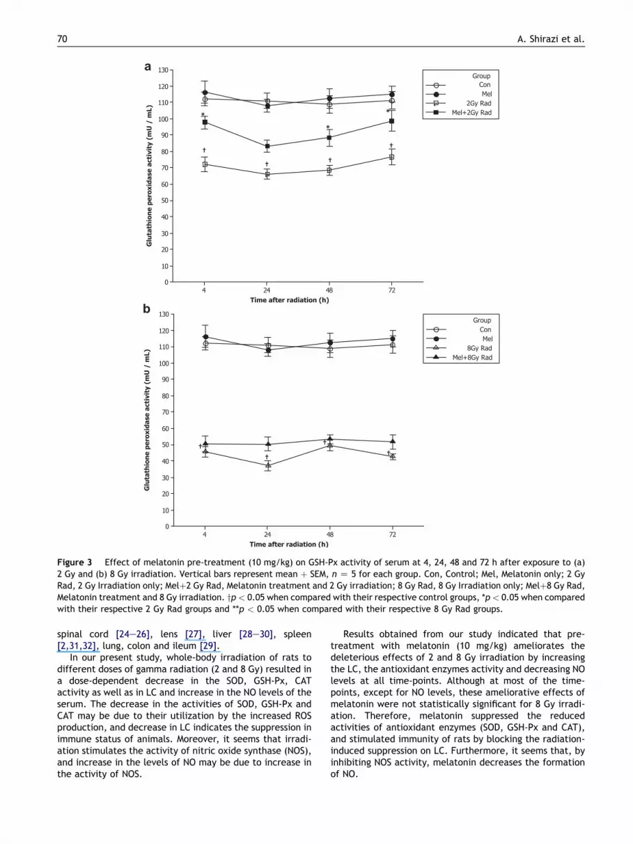
As shown in Fig. 3, GSH-Px activity in the irradiated groups(2 and 8 Gy Rad) decreased significantly when comparedwith control group at all time-points of post-irradiation(p < 0.05). GSH-Px activity for 8 Gy Rad group wassignificantly lower than 2 Gy Rad group at all time-points(p < 0.05) except at 48 h post-irradiation. Results ob-tained from Fig. 3 indicated that pre-treatment withmelatonin (10 mg/kg) ameliorates the deleterious effectsof 2 and 8 Gy irradiation by increasing GSH-Px activity atall time-points. However, this increase was not significant
at any of the time-points of 8 Gy post-irradiation. Pre-treatment with melatonin (10 mg/kg) significantlyincreased the GSH-Px activity when compared with 2 GyRad group at all time-points other than 24 h post-irradiation (p < 0.05).
Catalase (CAT) activity
As shown in Fig. 4, CAT activity of irradiated groups (2 and8 Gy Rad) decreased significantly when compared withcontrol group at all time-points of post-irradiation

a
b
Figure 2 Effect of melatonin pre-treatment (10 mg/kg) on SOD activity of serum at 4, 24, 48 and 72 h after exposure to (a) 2 Gyand (b) 8 Gy irradiation. Vertical bars represent mean þ SEM, n Z 5 for each group. Con, Control; Mel, Melatonin only; 2 Gy Rad,2 Gy Irradiation only; Melþ2 Gy Rad, Melatonin treatment and 2 Gy irradiation; 8 Gy Rad, 8 Gy Irradiation only; Melþ8 Gy Rad,Melatonin treatment and 8 Gy irradiation. yp < 0.05 when compared with their respective control groups, *p < 0.05 when comparedwith their respective 2 Gy Rad groups and **p < 0.05 when compared with their respective 8 Gy Rad groups.
Oxidative damage in rat peripheral blood 69
(p < 0.05). CAT activity for 8 Gy Rad group was significantlylower than 2 Gy Rad group at all time-points. Pre-treatmentwith melatonin (10 mg/kg) ameliorates the injurious effectsof 2 and 8 Gy irradiation by increasing the CAT activity at alltime-points. However, majority of these increases were notstatistically significant except for 8 Gy at 24 h post-irradiation (p < 0.05).
Nitric oxide (NO) level
As shown in Fig. 5, pre-treatment with melatonin (10 mg/kg)ameliorates the deleterious effects of 2 and 8 Gy irradiationby significant decrease in NO levels at all time-points
(p < 0.05). Moreover, NO levels of irradiated groups (2 and8 Gy Rad) were significantly high (p < 0.05) when comparedwith control group at all time-points of post-irradiation.A maximum value was observed at 4 h after exposure to8 Gy irradiation. NO levels for 8 Gy Rad group were signifi-cantly higher than 2 Gy Rad group at all time points(p < 0.05).
Discussion
Several studies demonstrated that melatonin by antioxi-dant properties, appeared to ameliorate irradiation-induced injury in various organs including brain [23],
a
b
Figure 3 Effect of melatonin pre-treatment (10 mg/kg) on GSH-Px activity of serum at 4, 24, 48 and 72 h after exposure to (a)2 Gy and (b) 8 Gy irradiation. Vertical bars represent mean þ SEM, n Z 5 for each group. Con, Control; Mel, Melatonin only; 2 GyRad, 2 Gy Irradiation only; Melþ2 Gy Rad, Melatonin treatment and 2 Gy irradiation; 8 Gy Rad, 8 Gy Irradiation only; Melþ8 Gy Rad,Melatonin treatment and 8 Gy irradiation. yp < 0.05 when compared with their respective control groups, *p < 0.05 when comparedwith their respective 2 Gy Rad groups and **p < 0.05 when compared with their respective 8 Gy Rad groups.
70 A. Shirazi et al.
spinal cord [24e26], lens [27], liver [28e30], spleen[2,31,32], lung, colon and ileum [29].
In our present study, whole-body irradiation of rats todifferent doses of gamma radiation (2 and 8 Gy) resulted ina dose-dependent decrease in the SOD, GSH-Px, CATactivity as well as in LC and increase in the NO levels of theserum. The decrease in the activities of SOD, GSH-Px andCAT may be due to their utilization by the increased ROSproduction, and decrease in LC indicates the suppression inimmune status of animals. Moreover, it seems that irradi-ation stimulates the activity of nitric oxide synthase (NOS),and increase in the levels of NO may be due to increase inthe activity of NOS.
Results obtained from our study indicated that pre-treatment with melatonin (10 mg/kg) ameliorates thedeleterious effects of 2 and 8 Gy irradiation by increasingthe LC, the antioxidant enzymes activity and decreasing NOlevels at all time-points. Although at most of the time-points, except for NO levels, these ameliorative effects ofmelatonin were not statistically significant for 8 Gy irradi-ation. Therefore, melatonin suppressed the reducedactivities of antioxidant enzymes (SOD, GSH-Px and CAT),and stimulated immunity of rats by blocking the radiation-induced suppression on LC. Furthermore, it seems that, byinhibiting NOS activity, melatonin decreases the formationof NO.

a
b
Figure 4 Effect of melatonin pre-treatment (10 mg/kg) on CAT activity of serum at 4, 24, 48 and 72 h after exposure to (a) 2 Gyand (b) 8 Gy irradiation. Vertical bars represent mean þ SEM, n Z 5 for each group. Con, Control; Mel, Melatonin only; 2 Gy Rad,2 Gy Irradiation only; Melþ2 Gy Rad, Melatonin treatment and 2 Gy irradiation; 8 Gy Rad, 8 Gy Irradiation only; Melþ8 Gy Rad,Melatonin treatment and 8 Gy irradiation. yp < 0.05 when compared with their respective control groups, *p < 0.05 when comparedwith their respective 2 Gy Rad groups and **p < 0.05 when compared with their respective 8 Gy Rad groups.
Oxidative damage in rat peripheral blood 71
On the other hand, melatonin treatment maintained LC,antioxidant enzymes activity and NO levels close to controlgroups and showed insignificant changes compared tocontrol groups except at 4 h for lymphocyte count and NOlevel. Thus, administration of melatonin (10 mg/kg) did notinduce serious side effects and acute toxicity.
Koc et al. [33] investigated the antioxidant role ofmelatonin (at 5 and 10 mg/kg) in the liver tissue againsttotal-body gamma irradiation-induced oxidative damagewith a single dose of 6.0 Gy. The results demonstratedthat in irradiated rats, that were pretreated with melatonin(5 or 10 mg/kg), malondialdehyde (MDA) levels, as an endproduct of lipid peroxidation in the liver tissue, were
significantly lowered, whereas the SOD and GSH-Px activi-ties were significantly increased. They concluded thatpre-treatment with melatonin may prevent irradiation-induced liver damage [33].
After exposure to 6 Gy Whole-body irradiation, livermalondialdehyde (MDA) and nitric oxide (NO) levels weremeasured by Taysi et al. [30]. Gamma irradiation causeda significant increase in liver MDA and NO levels. HepaticMDA and NO levels in irradiated rats that were pretreatedwith melatonin (5 or 10 mg/kg) were significantlydecreased [30]. Furthermore, the antioxidant property ofmelatonin (5 mg/kg, administrated daily for 10 days beforeirradiation) against a single dose of 5 Gy total-cranium

a
b
Figure 5 Effect of melatonin pre-treatment (10 mg/kg) on NO levels of serum at 4, 24, 48 and 72 h after exposure to (a) 2 Gy and(b) 8 Gy irradiation. Vertical bars represent mean þ SEM, n Z 5 for each group. Con, Control; Mel, Melatonin only; 2 Gy Rad, 2 GyIrradiation only; Melþ2 Gy Rad, Melatonin treatment and 2 Gy irradiation; 8 Gy Rad, 8 Gy Irradiation only; Melþ8 Gy Rad, Melatonintreatment and 8 Gy irradiation. yp < 0.05 when compared with their respective control groups, *p < 0.05 when compared with theirrespective 2 Gy Rad groups and **p < 0.05 when compared with their respective 8 Gy Rad groups.
72 A. Shirazi et al.
irradiation-induced cataract in the lens of rats was inves-tigated [27]. Irradiation significantly increased the MDAlevel and also significantly decreased SOD and GSH-Pxactivity, emphasizing the generation of increased oxida-tive stress. Melatonin supplementation with irradiationsignificantly increased the activity of SOD and GSH-Pxenzymes and significantly decreased the MDA level [27].
El-Missiry et al. [28] showed that treatmentwith 10mg/kgmelatonin for 4 days (daily) before acute irradiation(2 and4 Gy) significantly reduced radiation-induced increases inMDA and protein carbonyl levels(the oxidative stressmarkers) in the liver and significantly maintained hepaticglutathione (GSH) content, glutathione-S-transferase (GST),
and catalase(CAT) activities close to the control groupvalues.
Sharma et al. showed that, due to its antioxidant prop-erties, melatonin increased the immunity in squirrels, byprotecting their hematopoietic system and lymphoid organsagainst 2.06 Gy X-ray-induced cellular toxicity [31]. In theirstudy, Total leukocyte and lymphocyte counts (TLC and LC)in the peripheral blood and lipid peroxidation (LPO) status,superoxide dismutase (SOD) activities and total antioxidantstatus (TAS) were measured in the spleens of squirrels. pre-treatment with melatonin prior to the irradiation signifi-cantly increased LC, TLC, SOD activity and TAS statuscompared to irradiation exposed groups whereas LPO status

Oxidative damage in rat peripheral blood 73
was decreased [31]. In another study, a radio-protectiveeffect of melatonin against 5 Gy gamma irradiation duringthe reproductively active and inactive phases (RAP and RIP)of Indian palm squirrels was evaluated. Results showed thatmelatonin pre-treatment significantly increased the LC andincreased SOD activity in the spleen of squirrels comparedwith irradiation groups[32].
The results of treatment with melatonin followed byradiation exposed groups were similar to the results of thereported studies, where pre-treatment with melatoninincreased SOD, GSH-Px and CAT activity and decreased thelevels of NO [27,28,30e33]. Our results of LCare in agreementwith that of Sharma et al. [2,31,32] studies when reportedthat pre-treatment with melatonin increased lymphocytecount in irradiated groups. Thus our results support the find-ings of previously published literature. Melatonin pre-treatment increased LC, the activity of the antioxidantenzymes and decreased the levels of nitric oxide. Further-more, toour knowledge, therehavebeennopublished studiesinvestigating possible radio-protective effects of melatoninon peripheral blood (serum) by measuring activities of theseantioxidant enzymes (SOD, GSH-Px and CAT)and levels ofnitric oxide (NO) at different time-points after exposure todifferent doses of whole-body gamma irradiation.
Despite the lack of clinical and experimental studies theresult of the present study, our previous data [24e26,34],and findings of other investigators, suggest that adminis-tration of this agent may enable the use of higher doses ofirradiation during radiotherapy and may be beneficial inalleviating the complications of cancer treatment.
Conclusion
Based on our results, ionizing radiation causes dose-dependent oxidative damage on the hematopoietic systemand melatonin, due to its immune stimulatory and anti-oxidative properties, ameliorates radiation-induced injuryto this system. In conclusion 10mg/kgmelatonin is likely to bean adequate concentration for offering significant protectionagainst lower dose of gamma irradiation (2 Gy) than higherdose of 8 Gy. Long-term administration of this drug mayproduce more protection against higher doses of irradiation,because lower dose of melatonin might not be enough toscavenge all the free radicals generated by higher dose ofirradiation and stimulate cellular antioxidant defenses.Therefore, it seems that radio-protective effects of mela-tonin are dose-dependent.However, further experiments andclinical trials on this subject are still necessary to validate it.
Acknowledgment
This study was supported by grant number 10444 from thevice chancellor of research at Tehran University of MedicalSciences. There is no conflict of interest in this study.
References
[1] Mansour HH, Hafez HF, Fahmy NM, Hanafi N. Protective effectof N-acetylcysteine against radiation induced DNA damage
and hepatic toxicity in rats. Biochemical Pharmacology 2008;75:773e80.
[2] Sharma S, Haldar C. Melatonin prevents X-ray irradiationinduced oxidative damagein peripheral blood and spleen ofthe seasonally breeding rodent, Funambulus pennanti duringreproductively active phase. International Journal of Radia-tion Biology 2006;82:411e9.
[3] Michiels C, Raes M, Toussaint O, Remacle J. Importance of Se-glutathione peroxidase, catalase, and Cu/Zn-SOD for cellsurvival against oxidative stress. Free Radical Biology andMedicine 1994;17:235e48.
[4] Taysi S, Polat F, Gul M, Sari R, Bakan E. Lipid peroxidation,some extracellular antioxidants, and antioxidant enzymes inserum of patients with rheumatoid arthritis. RheumatologyInternational 2002;21:200e4.
[5] Ohshima H, Bartsch H. Chronic infections and inflammatoryprocesses as cancer risk factors: possible role of nitric oxide incarcinogenesis. Mutation Research/Fundamental and Molec-ular Mechanisms of Mutagenesis 1994;305:253e64.
[6] Abu-Shakra A, McQueen ET, Cunningham ML. Rapid analysis ofbase-pair substitutions induced by mutagenic drugs throughtheir oxygen radical or epoxide derivatives. MutationResearch/Genetic Toxicology and Environmental Mutagenesis2000;470:11e8.
[7] Weiss J. Pharmacologic approaches to protection againstradiation-induced lethality and other damage. EnvironmentalHealth Perspectives 1997;105:1473.
[8] Weiss JF, Landauer MR. Radioprotection by antioxidantsa.Annals of the New York Academy of Sciences 2000;899:44e60.
[9] Nair C, Parida D, Nomura T. Radioprotectors in radiotherapy.Journal of Radiation Research 2001;42:21e37.
[10] Wu W, Abraham L, Ogony J, Matthews R, Goldstein G, Ercal N.Effects of N-acetylcysteine amide (NACA), a thiol antioxidanton radiation-induced cytotoxicity in Chinese hamster ovarycells. Life Sciences 2008;82:1122e30.
[11] Maestroni GJM. The immunoneuroendocrine role of mela-tonin. Journal of Pineal Research 1993;14:1e10.
[12] Bubenik GA. Review: gastrointestinal melatonin: localization,function, and clinical relevance. Digestive Diseases andSciences 2002;47:2336e48.
[13] Tan D, Chen L, Poeggeler B, Manchester L, Reiter R. Mela-tonin: a potent, endogenous hydroxyl radical scavenger.Endocrine Journal 1993;1:57e60.
[14] Hardeland R, Reiter R, Poeggeler B, Tan DX. The significanceof the metabolism of the neurohormone melatonin: anti-oxidative protection and formation of bioactive substances.Neuroscience & Biobehavioral Reviews 1993;17:347e57.
[15] Reiter R, Tan D, Osuna C, Gitto E. Actions of melatonin in thereduction of oxidative stress. Journal of Biomedical Science2000;7:444e58.
[16] Rodriguez C, Mayo JC, Sainz RM, Antolin I, Herrera F, Martin V,et al. Regulation of antioxidant enzymes: a significant role formelatonin. Journal of Pineal Research 2004;36:1e9.
[17] Vijayalaxmi RRJ, Tan D, Herman T, Thomas Jr C. Melatonin asa radioprotective agent: a review. International Journal ofRadiation Oncology Biology Physics 2004;59:639e53.
[18] Reiter RJ, Tan DX, Cabrera J, D’arpa D, Sainz RM, Mayo JC,et al. The oxidant/antioxidant network: role of melatonin.Neurosignals 2000;8:56e63.
[19] Shirazi A, Ghobadi G, Ghazi-Khansari M. A radiobiologicalreview on melatonin: a novel radioprotector. Journal ofRadiation Research 2007;48:263e72.
[20] Karbownik M, Reiter R. Antioxidative effects of melatonin inprotection against cellular damage caused by ionizing radia-tion. Proceedings of the Society for Experimental Biology andMedicine 2000;225:9e22.
[21] Bettahi I, Pozo D, Osuna C, Reiter R, Acuna-Castroviejo D,Guerrero J. Physiological concentrations of melatonin inhibit

74 A. Shirazi et al.
nitric oxide synthase activity in the rat hypothalamus. Journalof Pineal Research 1996;20:205e10.
[22] Pozo D, Reiter RJ, Calvo JR, Guerrero JM. Inhibition of cere-bellar nitric oxide synthase and cyclic GMP production bymelatonin via complex formation with calmodulin. Journal ofCellular Biochemistry 1997;65:430e42.
[23] Erol FS, Topsakal C, Ozveren MF, Kaplan M, Ilhan N,Ozercan IH, et al. Protective effects of melatonin and vitaminE in brain damage due to gamma radiation. NeurosurgicalReview 2004;27:65e9.
[24] Shirazi A, Haddadi G, Ghazi-Khansari M, Abolhassani F,Mahdavi S, Eshraghyan M. Evaluation of melatonin forprevention of radiation myelopathy in irradiated cervicalspinal cord. Yakhteh Medical Journal 2009;11:43e8.
[25] Aghazadeh S, Azarnia M, Shirazi A, Mahdavi S, Zangii B.Melatonin as a protective agent in spinal cord damage aftergamma irradiation. Reports of Practical Oncology & Radio-therapy 2007;12:95e9.
[26] Shirazi A, Haddadi G, Minaee B, Sepehrizadeh Z,Mahdavi S, Jaberi E, et al. Evaluation of melatonin formodulation of apoptosis-related genes in irradiatedcervical spinal cord. International Journal of Low Radia-tion 2010;7:436e45.
[27] Karslioglu I, Ertekin MV, Taysi S, Koer I, Sezen O,Gepdiremen A, et al. Radioprotective effects of melatonin onradiation-induced cataract. Journal of Radiation Research2005;46:277e82.
[28] El-Missiry M, Fayed T, El-Sawy M, El-Sayed A. Ameliorativeeffect of melatonin against gamma-irradiation-inducedoxidative stress and tissue injury. Ecotoxicology and Environ-mental Safety 2007;66:278e86.
[29] Sener G, Jahovic N, Tosun O, Atasoy BM, BC Yegen. Melatoninameliorates ionizing radiation-induced oxidative organdamage in rats. Life Sciences 2003;74:563e72.
[30] Taysi S, Koc M, Buyukokuro lu ME, Alt nkaynak K, ahin YN.Melatonin reduces lipid peroxidation and nitric oxide duringirradiation induced oxidative injury in the rat liver. Journal ofPineal Research 2003;34:173e7.
[31] Sharma S, Haldar C, Chaube SK. Effect of exogenous mela-tonin on X-ray induced cellular toxicity in lymphatic tissue ofIndian tropical male squirrel, Funambulus pennanti. Interna-tional Journal of Radiation Biology 2008;84:363e74.
[32] Sharma S, Haldar C, Chaube S, Laxmi T, Singh S. Long-termmelatonin administration attenuates low-LET {gamma}-radia-tion-induced lymphatic tissue injury during the reproductivelyactive and inactive phases of Indian palm squirrels (Funambuluspennanti). British Journal of Radiology 2010;83:137.
[33] Koc M, Taysi S, Buyukokuroglu ME, Bakan N. Melatoninprotects rat liver against irradiation-induced oxidative injury.Journal of Radiation Research 2003;44:211e5.
[34] Shirazi A, Haddadi G, Asadi-Amoli F, Sakhaee S, Ghazi-Khansari M, Avand A. Radioprotective effect of melatonin inreducing oxidative stress in rat lenses. Cell Journal (Yakhteh)2011;13:79e82.