Embed Size (px)
Citation preview
EUKARYOTIC CELL, June 2004, p. 646–662 Vol. 3, No. 31535-9778/04/$08.00�0 DOI: 10.1128/EC.3.3.646–662.2004Copyright © 2004, American Society for Microbiology. All Rights Reserved.
RasC Plays a Role in Transduction of Temporal Gradient Informationin the Cyclic-AMP Wave of Dictyostelium discoideum
Deborah Wessels,1 Rebecca Brincks,1 Spencer Kuhl,1 Vesna Stepanovic,1 Karla J. Daniels,1Gerald Weeks,2 Chinten J. Lim,2 George Spiegelman,2 Danny Fuller,3 Negin Iranfar,3
William F. Loomis,3 and David R. Soll1*W. M. Keck Dynamic Image Analysis Facility, Department of Biological Sciences, The University of Iowa, Iowa City,
Iowa 522421; Department of Microbiology and Immunology, The University of British Columbia, Vancouver,British Columbia2; and Department of Biology, University of California, San Diego,
La Jolla, California 920393
Received 8 October 2003/Accepted 8 January 2004
To define the role that RasC plays in motility and chemotaxis, the behavior of a rasC null mutant, rasC�, inbuffer and in response to the individual spatial, temporal, and concentration components of a natural cyclicAMP (cAMP) wave was analyzed by using computer-assisted two-dimensional and three-dimensional motionanalysis systems. These quantitative studies revealed that rasC� cells translocate at the same velocity andexhibit chemotaxis up spatial gradients of cAMP with the same efficiency as control cells. However, rasC� cellsexhibit defects in maintaining anterior-posterior polarity along the substratum and a single anterior pseudo-pod when translocating in buffer in the absence of an attractant. rasC� cells also exhibit defects in theirresponses to both the increasing and decreasing temporal gradients of cAMP in the front and the back of awave. These defects result in the inability of rasC� cells to exhibit chemotaxis in a natural wave of cAMP. Theinability to respond normally to temporal gradients of cAMP results in defects in the organization of thecytoskeleton, most notably in the failure of both F actin and myosin II to exit the cortex in response to thedecreasing temporal gradient of cAMP in the back of the wave. While the behavioral defect in the front of thewave is similar to that of the myoA�/myoF� myosin I double mutant, the behavioral and cytoskeletal defectsin the back of the wave are similar to those of the S13A myosin II regulatory light-chain phosphorylationmutant. Expression array data support the premise that the behavioral defects exhibited by the rasC� mutantare the immediate result of the absence of RasC function.
The Ras GTPases function as molecular switches in theregulation of a variety of responses to extracellular signals (3,21, 35, 36). Dictyostelium discoideum, like higher eukaryotes,contains a number of Ras GTPases (6, 21, 22). Because of itsattributes, Dictyostelium provides a unique experimental sys-tem for exploring the roles played by the Ras GTPases in cellmotility and chemotaxis (6, 21, 49). First, because it is haploid,null mutations are readily generated and rescued (16, 18).Second, because the behavior of Dictyostelium amoebae inbuffer and in response to the temporal, spatial, and concentra-tion components of the natural chemotactic wave has beencharacterized in detail by computer-assisted methods (33, 34),a unique contextual framework exists for identifying specificbehavioral defects manifested in mutants (29) and for deduc-ing from them the specific role played by mutated genes inmotility and/or chemotaxis (4, 8, 10, 42, 45, 50, 51).
In a previous study, it was demonstrated that rasC� cellscould not progress through the early stages of development orform aggregates unless they were pulsed with the chemoattrac-tant cyclic AMP (cAMP), indicating that RasC was necessaryfor signaling (20). rasC� cells artificially pulsed with cAMPwere then capable of forming aggregates on filter pads. Whenmixed with a majority of normal cells, they could also enter
aggregates and exhibit chemotaxis towards the source of aspatial gradient of cAMP emitted by a micropipette (20). Al-though these results suggested that exogenously added cAMPrendered rasC� cells aggregation competent and that RasCwas not required for chemotaxis in spatial gradients of theattractant, experiments were not performed to test whetherRasC played a direct role in the basic motile behavior of a cellin the absence of a chemotactic signal or in the complex cel-lular responses to the temporal and concentration componentsof the natural cAMP wave (29, 46).
Employing a set of experimental protocols developed toanalyze the basic motile behavior of amoebae in the absence ofattractant and their capacity to respond to the individual spa-tial, temporal, and concentration components of the chemo-tactic wave (29), we demonstrate that rasC� cells can efficientlyexhibit chemotaxis up a spatial gradient of cAMP, the mech-anism regulating orientation at the onset of the front of anatural wave. However, rasC� cells exhibit a number of dis-crete behavioral defects, including fragmentation and z-axisextension of the anterior pseudopod in buffer, absence of avelocity surge in response to the increasing temporal gradientof cAMP in the front of a wave, and abnormal polarization inresponse to the decreasing temporal gradient of cAMP in theback of a wave. The behavioral defects in the front and theback of the wave are accompanied by abnormalities in thelocalization of F actin and myosin in the cortex. Because ourexperimental protocols allowed us to correlate regulatory path-ways with specific phases of the natural wave and associated
* Corresponding author. Mailing address: 302 BBE, Department ofBiological Sciences, The University of Iowa, Iowa City, IA 52242.Phone: (319) 335-1117. Fax: (319) 335-2772. E-mail: [email protected].
646
on June 23, 2016 by guesthttp://ec.asm
.org/D
ownloaded from
behavioral responses, we formulated a working model in whichRasC plays roles both in the response of cells to the increasingtemporal gradient of cAMP in the front of the wave, upstreamof MyoA/MyoF, and in the response of cells to the decreasingtemporal gradient of cAMP in the back of the wave, upstreamof the phosphorylation of the myosin II regulatory light chain.
MATERIALS AND METHODS
Origin and maintenance of control, rasC�, and rescued strains. The parentalAx2 strain (control), the rasC� null strain, and the rasC� rescued strain, referredto here as rasC�/rasC�, have been described previously in detail (20). Briefly, therasC� mutant was generated by targeted gene replacement via homologousrecombination with a rasC� construct that was disrupted by a blasticidin Sresistance-selectable marker. An ectopically encoded RasC expression vectorwas constructed by directionally ligating the rasC cDNA to the rasC promoterthat was cloned by PCR from Ax2 genomic DNA. Transformation of this vectorinto the rasC� mutant resulted in the rasC�/rasC� strain. RasC expression in therescued strain was verified by Western blotting against a RasC-specific rabbitpolyclonal antibody. rasC� and rasC�/rasC� cells were grown in HL-5 mediumsupplemented with 10 �g of blasticidin S/ml and 10 �g of G418/ml, respectively.Cells were grown on plates since attachment promoted a higher proportion ofmononuclear cells. For experimental purposes, cells were harvested from platesat the confluent-monolayer stage.
Obtaining behaviorially equivalent cells. Cells were harvested from growthcultures, washed with buffered salts solution (BSS; 20 mM KCl, 2.5 mM MgCl2,20 mM KH2PO4 [pH 6.4]), and distributed on filter pads saturated with BSS ata cell density of 5 � 106 per cm2 (30). Cells were washed from the filter pads atthe ripple stage, which represents the onset of aggregation (30) and the time atwhich cells achieve maximum velocity (40). To obtain rasC� cells that hadachieved maximum velocity during development equivalent to that of controlcells, a number of protocols were tested and the following one was selected (seeResults). rasC� cells were harvested from growth cultures, washed with BSS,resuspended in BSS at a cell density of 5 � 106 per ml and maintained insuspension on a rotary water bath shaker at 22°C. After 1 h, cells were pulsedevery 6 min with 50 nM cAMP for 6 h. Cells were then washed in BSS anddispersed on a filter pad as described for control cells. rasC� cells were thenharvested from filter pads after 2 h of incubation, at which time cells hadachieved maximum velocity (see Results).
Microarray analysis. The methods used have been described in detail else-where (14, 15, 24). At various time points, cells were collected from cAMP-pulsed suspension cultures, pelleted, and dissolved in Trizol reagent (Gibco-BRL, Gaithersburg, Md.) for preparation of RNA. For analysis, Corning slideswere microarrayed with 6,345 cDNA and genomic targets by using a MolecularDynamics GenIII robot in the BioGEM facility, University of California, SanDiego, as previously described. Inserts from 5,655 cDNAs were generously pro-vided by the Japanese EST Project. A list of genes is available at http://www.biology.ucsd.edu/loomis-cgi/microarray/rasC-array.html. All genes in this studywere sequence verified. Probes were prepared from total RNA collected at 2-hintervals during wild-type and mutant development as well as from time-aver-aged reference RNA (15). Superscript II DNA polymerase (Invitrogen, Carls-bad, Calif.) was used to incorporate either Cy-5- or Cy-3-conjugated dCTP(Amersham, St. Louis, Mo.) into DNA. Following incubation at 42°C for 3 h,unincorporated dyes were removed using Microcon-30 columns (Millipore, Bur-lington, Mass.) and three washes with 450 �l of Tris-EDTA buffer, before dryingand resuspension in a solution containing 5� SSC (1� SSC is 0.15 M NaCl plus0.015 M sodium citrate), 0.3% sodium dodecyl sulfate, and 25% formamide.Labeled probes from the time points and the time-averaged RNA were mixedand hybridized at 42°C to microarrays for 6 to 12 h. Probed microarrays wereanalyzed in an Axon Genepix 4000B scanner (Axon Instruments, Foster City,Calif.), and the measurements were processed with the associated software. TotalCy3 signal was normalized to total Cy5 signal after background subtraction toallow independent slides to be compared. The Cy3/Cy5 ratios for individualgenes were then calculated at each time point for subsequent analyses. Theexpression patterns of rasC� cells were compared to those of strain Ax4, whichare indistinguishable from those of strain Ax2 (37). The expression of each genewas based on measurements taken at 13 independent time points during thedevelopmental time course. Each analysis was repeated at least twice. Theselected cutoff at a threefold increase was based on the reproducibility of thedata from 10 independent time course experiments.
Basic motile behavior. Cells were washed from filter pads and distributed onthe glass wall of a Sykes-Moore perfusion chamber (Bellco Glass, Vineland, N.J.)
positioned on the stage of an upright microscope with a long-distance workingcondenser and a �25 bright-field objective as previously described (8, 48, 50, 51).After 5 min of incubation, the chamber was perfused with BSS at a rate thatturned over 1 chamber volume equivalent every 15 s.
Spatial gradient of cAMP. Cells were dispersed either on the bridge of aPlexiglas chemotaxis chamber (38) modeled after that of Zigmond (52) forbright-field analysis or on the bridge of a quartz chamber developed for differ-ential interference contrast (DIC) microscopy (27, 28). BSS alone was added toone of the two wells bordering the bridge, and BSS containing 10�6 M cAMP wasadded to the other. Cells were incubated for 5 min to facilitate cell adhesion andallow for the genesis of a steep gradient of chemoattractant across the bridge (27,28).
Simulated temporal waves of cAMP. Cell behavior was analyzed in a series offour temporal waves of cAMP generated in the absence of established spatialgradients in a Sykes-Moore perfusion chamber as previously described (8, 32, 33,39, 41, 50, 51) by using NE-1000 Multi-Phaser programmable syringe pumps(New Era Pump Systems, Wantagh, N.Y.) (11). The shapes of the cAMP waveswithin the chamber were verified using xylene cyanol FF dye (Molecular Probes,Eugene, Oreg.) (11).
Rapid addition of 10�6 M cAMP. Cells were distributed on the wall of aSykes-Moore chamber and perfused with BSS for 10 min as described above. Theperfusion solution was then switched to BSS containing 10�6 M cAMP (40).Previous analyses with fluorescent dye demonstrated that the concentration ofcAMP increases from 0 to 10�6 M in approximately 12 s (40).
Natural cAMP waves. To test the ability of rasC� cells to respond to naturalwaves of cAMP generated by wild-type cells, mixing experiments were performedaccording to previously described methods (50, 51) with modifications. Growth-phase rasC� cells were incubated overnight in HL-5 medium containing 5 �10�5 M DiI (Molecular Probes). Labeled cells were then washed in BSS, resus-pended to a final concentration of 5 � 106 cells per ml, maintained in suspensionfor 1 h, and then pulsed every 6 min for 5 h with 50 nM cAMP. rasC� cells werethen mixed with unlabeled control cells at a ratio of 1:9. Control cells wereprestarved for 3 h before mixing so that the developmental timings of the twostrains were comparable (see Results). The mixed population was plated on a35-mm-diameter petri dish at a density of 2.4 � 106 cells per ml in 2 ml, the dishwas placed on the stage of an Axiovert 100STV Zeiss microscope, and the samplewas examined with a NORAN laser scanning confocal microscope (LSCM).Transmitted and fluorescent images were simultaneously collected every 20 s,averaged using Intervision software, and saved on a hard drive in Silicon Graph-ics format (SGI Inc., Mountain View, Calif.). These movies were then convertedto Quick Time format, and the motion of labeled rasC� and unlabeled neigh-boring control cells was analyzed.
2D computer-assisted analysis. 2D-DIAS software was used for computer-assisted two-dimensional (2D) analyses (32, 33). Video images of all prepara-tions were digitized onto the hard drive of a Macintosh computer at a rate of 15frames per min (4-s intervals) with a frame-grabber board (Data Translation Inc.,Marlboro, Mass.). Cell perimeters from the digitized movies were automaticallyoutlined using the grayscale threshold algorithm of 2D-DIAS. Outlines wereconverted to beta-spline replacement images. Motility parameters were com-puted from the centroid positions and dynamic morphology parameters werecomputed from contours of the replacement images according to formulas dis-cussed elsewhere in detail (32, 33). To compute the instantaneous velocity of acell in frame n, a line was drawn from the centroid in frame n � 1 to the centroidin frame n � 1 and the length of that line was divided by twice the time intervalbetween frames. Directional change was computed as the direction in the inter-val (n �1, n) minus the direction in the interval (n, n � 1). Directional changevalues greater than 180° were subtracted from 360°, resulting in a positive valuebetween 0° and 180°. Maximum length was defined as the longest chord betweenany two points along the cell perimeter. Maximum width was defined as thelongest chord perpendicular to the maximum length chord. Roundness wascalculated as (4� � area/perimeter squared) � 100. Convexity was defined as theabsolute value of the sum of positive turn angles, in degrees, of line segmentsconnecting the vertices of a cell’s shape. The chemotactic index (CI) was calcu-lated as the net distance traveled directly towards the source of chemoattractantin a spatial gradient chamber divided by the total distance traveled in that timeperiod. The percent positive chemotaxis was the proportion of cells exhibiting apositive CI in a spatial gradient of cAMP over the period of analysis. Differenceimages were generated by superimposing the perimeter outline of the cell inframe n onto the perimeter outline in frame n � 1. Expansion zones, color codedgreen, were demarcated as regions of the cell in frame n not overlapping theoutlined cell in frame n � 1. Contraction zones, color coded red, were demar-cated as regions of the cell in frame n � 1 not overlapping the outlined cell inframe n.
VOL. 3, 2004 RasC AND DICTYOSTELIUM CHEMOTAXIS 647
on June 23, 2016 by guesthttp://ec.asm
.org/D
ownloaded from
3D computer-assisted analysis. The 3D-DIAS software program was used forcomputer-assisted three-dimensional (3D) reconstruction and motion analysis(13, 32, 33, 34, 43, 47). For 3D reconstructions in buffer and in temporal wavesof cAMP, 350 �l of a dilute suspension of cells was inoculated into a Dvorak-Stotler chamber (Lucas-Highland, Inc., Chantilly, Va.). The chamber was placedon the stage of a Zeiss ICM 405 inverted microscope equipped with DIC opticsand a Planapo �63 oil immersion objective. The chamber was either perfusedwith BSS or treated with four temporal waves of cAMP as described above. For3D reconstructions in spatial gradients of cAMP, cells were dispersed on thebridge of a quartz chemotaxis chamber (28). To obtain optical sections of a cell,the coarse focus knob of the microscope was connected to a stepper-motorprogrammed for 60 optical sections in 2 s in the z axis at 0.3-�m increments. Thisentire process was repeated at 5-s intervals. Images were recorded through anOptronics cooled charge-coupled device camera directly into a Macintosh iMACcomputer at 30 frames per s by using iMovie software. After the movie wascompressed into the DIAS format, 3D-DIAS software in the newly developedJAVA-based DIAS 4.0 platform (E. Voss and D. R. Soll, unpublished data)automatically outlined the perimeter of the in-focus portion of the image in eachoptical section by using a pixel complexity algorithm (33). The interface betweenthe particulate cytoplasm of the main cell body and the nonparticulate cytoplasmof the pseudopodial extensions was readily identified in DIC images of opticalsections. The distal nonparticulate zones of pseudopodial regions were manuallyoutlined in the in-focus portions of each optical section to generate a faceted 3Dreconstruction of pseudopods, which were color coded red and inserted into thefaceted cell image.
The construction of a 3D faceted image of the cell surface has been describedelsewhere in detail (13, 33, 43, 47). For computation of 3D parameters, the 3Dposition of the centroid of the cell was computed by averaging the x, y, and zcoordinates of all points interior to the 3D faceted cell image. Volume and heightwere computed as the mathematical volume of the faceted cell and as themaximum height of the faceted image in the z axis, respectively. The overhangparameter represented the percent cell area in the x and y axes not in contactwith the substratum.
F-actin and myosin II staining. Cells were fixed with 4% paraformaldehyde in10 mM 2(N-morpholino)ethanesulfonic acid (MES; Sigma, St. Louis, Mo.) buffer(pH 6.1) containing 138 mM KCl, 3 mM MgCl2, and 2 mM EGTA. To localizeF actin, cells were stained with Oregon green-phalloidin (Molecular Probes, Inc.)according to methods previously described (50, 51). Prior to immunostaining formyosin II, antigen retrieval was performed by processing cells in target retrievalsolution (DAKO Corp., Carpinteria, Calif.) in a steamer for 20 min at 90°Caccording to the manufacturer’s recommendations. The solution and coverslipswere removed from heat and allowed to cool to room temperature prior to beingrinsed in Tris-HCl-buffered saline (50 mM Tris-HCl–150 mM NaCl [pH 7.4]).Nonspecific binding was blocked with 10% normal goat serum (Sigma) in Dul-becco’s phosphate-buffered saline (PBS; pH 7.1; Gibco, Grand Island, N.Y.). Tolocalize myosin II, cells were incubated with rabbit anti-myosin II antibody(1/1,000), a generous gift from Arturo De Lozanne (University of Texas, Austin),in PBS for 45 min at 37°C. Following extensive rinsing with PBS, cells werestained with fluorescein isothiocyanate-labeled goat anti-rabbit antibody (1/200;Molecular Probes, Inc.) for 30 min at room temperature. Coverslips containingthe F-actin- or myosin II-stained preparations were rinsed and mounted usingGelvatol supplemented with 0.6% N-propylgallate (Sigma). Cells were thenscanned with a Bio-Rad Radiance 2100MP LSCM at 0.2-�m increments in thez axis, and the z series was projected as a single image (projection image). Toobtain a ratio of the intensity of either F-actin or myosin II staining in the cortexto that in the cytoplasm, a stained Ax2 cell was scanned 1 �m above thesubstratum and the image was optimized. The optimized LSCM parameters forthis reference cell (laser power, iris aperture, gain, and scan rate) were then usedto obtain scans 1 �m above the substratum for subsequent control and rasC�
cells. Line profiles of intensity through the cortex and cytoplasm were obtainedjust posterior to the cell nucleus by using Bio-Rad Laser Sharp 2000 5.0 software.Digital images of xy planes, line profiles, and projected z series image stacks wereprocessed using Adobe Photoshop software.
RESULTS
Developmental regulation of motility. The increase in cellmotility associated with the acquisition of chemotactic respon-siveness to cAMP and the onset of aggregation (40) provides adevelopmental landmark for comparison of strains (8, 31, 40,50, 51). Because rasC� cells fail to aggregate in homogeneous
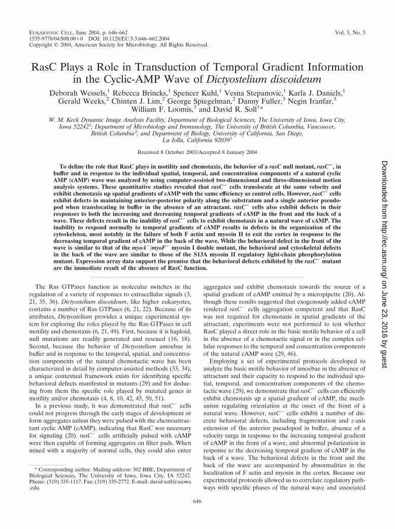
monolayers due to their inability to relay a cAMP signal (20),it was first necessary to identify conditions that producedrasC� cells that were comparable to aggregation-competentcontrol (Ax2) cells. We therefore analyzed motility under avariety of conditions, including starvation on filter pads, star-vation in suspension with or without cAMP pulses, and star-vation in suspension with cAMP pulses, followed by plating onpads. In the case of the parental control strain (Ax2), singlecells achieved maximum velocity between 5 and 6 h of starva-tion on development pads (Fig. 1A), coincident with the ripplestage, which represents the onset of aggregation (30). Similarresults were obtained with the rasC�/rasC� strain (data notshown). In the case of the rasC� strain, however, cells attainedsimilar peak velocity when they were pulsed with 50 nM cAMPat 6-min intervals during the final 5 h of a 6-h period in shakingcultures and then starved for 2 subsequent h on a filter pad(Fig. 1B).
Developmental regulation of gene expression. To be surethat the defects in motility and chemotaxis identified in rasC�
cells were due directly to the lack of RasC function in regula-tory pathways, Lim et al. (20) previously demonstrated byNorthern analysis that cAMP-pulsed rasC� cells expressed thedevelopmentally regulated cAMP receptor CAR1 and the cou-pled heterotrimeric G protein G�2. To further explore geneexpression, microarray analyses were performed with rasC�
cells by using slides that carried 6,345 cDNA and genomictargets and the results were compared with those for Ax4 cellsdeveloped in the same manner (14, 15, 24). It has previouslybeen demonstrated that Ax2 (the control for rasC�) and Ax4cells exhibit similar changes in gene expression during devel-opment (37). Only mRNAs whose expression increased three-fold or more in cAMP-pulsed control cells were compared with
FIG. 1. Developmental regulation of motility in rasC� cells. Thedevelopmental regulation of cell motility was assessed by plotting sin-gle-cell velocity as a function of development time. (A) For control(Ax2) cells, the peak velocity (arrow) occurred 5 to 6 h after cellswashed free of growth medium were plated on a development padsaturated with BSS. (B) For rasC� cells, cells washed free of growthmedium were suspended in buffer and pulsed with cAMP every 6 minbetween 1 and 6 h of incubation. Cells were then plated on a devel-opment pad saturated with BSS. The peak velocity (arrow) occurred2 h after rasC� cells were plated on the development pad. The meaninstantaneous velocity at each time point was computed from theaverage instantaneous velocities of 20 amoebae, each analyzed for 10min.
648 WESSELS ET AL. EUKARYOT. CELL
on June 23, 2016 by guesthttp://ec.asm
.org/D
ownloaded from
those in cAMP-pulsed rasC� cells in order to set a stringentcondition for identifying genes that may not be expressed inthe latter.
All of the preaggregation genes, including those for CAR1,G�2, the secreted cAMP-phosphodiesterase and its inhibitor,the adenylyl cyclase ACA, and the internal phosphodiesteraseRegA, began accumulating immediately upon induction of de-velopment and reached peak levels at the same time in rasC�
and Ax4 cells (data available at http://www.biology.ucsd/loomis-cgi/microarray/rasC-array.html). Likewise, mRNAs for thecell-cell adhesion proteins gp80 (CsaA) and gp150 (LagC)accumulated to peak values within 12 h of development inmutant and control cells. All of the postaggregative mRNAsthat accumulated at least threefold in wild type, includingthose encoding the spore coat proteins, accumulated at leastthreefold in rasC� cells. While genes that did not give clearsignals on the microarrays could have been affected in rasC�
cells, the transcriptional profiles that characterize developmen-tal stages through aggregation were not significantly perturbedby the loss of RasC as long as the cells were pulsed with cAMP(see http://www.biology.ucsd/loomis-cgi/microarray/rasC-array.html).
Basic motile behavior is defective in rasC� cells. Cells dis-tributed at low density on the glass wall of a chamber wereperfused continuously with BSS to ensure that they did notcondition their soluble microenvironment. The mean instanta-neous velocity of rasC� cells was statistically indistinguishablefrom that of control Ax2 and rasC�/rasC� cells (Tables 1 and2). However, a histogram of the instantaneous velocities ofindividual cells suggested a broader distribution for rasC� cellsthan for control cells at both low and high velocities (data notshown). The broadened range of instantaneous velocities ofrasC� cells was accompanied by other behavioral abnormali-ties. First, perimeter tracks of rasC� cells possessed more sideprojections, which upon further analysis proved to be due toinstability at the anterior ends of cells (data not shown), sug-gesting either a more dynamic anterior pseudopod or less
secure attachment of the anterior end to the substratum. Sec-ond, the directional change parameter of rasC� cells was sig-nificantly higher than that of control or rasC�/rasC� cells (Ta-bles 1 and 2), again suggesting less persistent translocation.Third, both the mean roundness and mean convexity parame-ters of rasC� cells were significantly higher than those of con-trol cells (Table 1). Together, these results indicated abnor-malities in rasC� behavior in the absence of attractant.
To investigate further the aberrant behavior in buffer, con-trol and rasC� cells were reconstructed in three dimensions at4-s intervals using 3D-DIAS software (13, 33, 47). The tworepresentative control cells shown in Fig. 2A and B were elon-gate and extended a single dominant anterior pseudopod (la-beled “a”) along the substratum. In both cases, lateral pseu-dopods (labeled “l”) that had formed prior to reconstructionwere retracted back into the cell bodies. Intermittently, thedominant anterior pseudopod lifted off the substratum andthen returned to the substratum during the period of analysis(Fig. 2A and B). Eight additional control and 10 rasC�/rasC�
cells reconstructed in three dimensions behaved in a similarfashion (data not shown).
In contrast to those of control or rasC�/rasC� cells, theanterior ends of the two representative rasC� cells shown inFig. 2C and D frequently extended off the substratum in the zaxis. When extending their anterior ends in the z axis, rasC�
cells remained attached to the substratum at their posteriorends (Fig. 2C and D). The posterior end, or uropod, wasidentified in those cells by assessing the direction of transloca-tion from earlier frames. Frequently, either the anterior pseu-dopod of rasC� cells bifurcated or multiple anterior pseudo-pods extended from the general anterior half of the cell (Fig.2C and D). In some cases, it was difficult to keep track of thedominant anterior pseudopod, as is evident during z-axis ex-tension of the cell in Fig. 2D. Eight additional rasC� cellsreconstructed in three dimensions exhibited similar behavior(data not shown). Qualitative 3D analyses of vegetative rasC�
cells migrating on a glass surface revealed defects in the ante-
TABLE 1. Quantitative 2D-DIAS analysis of the basic motile behavior of rasC� cells in buffer reveals subtle defectsa
Cells
Mean � SD
Instantaneousvelocity (�m/min)
Directional change(°/4 s)
Maximum length(�m)
Maximum width(�m) Roundness (%) Convexity (°)
Ax2 (control) 13.5 � 4.6 28.6 � 11.0 20.0 � 2.4 8.1 � 1.4 42.1 � 7.3 647 � 71rasC� 12.3 � 7.9 35.6 � 10.4 19.8 � 4.3 8.2 � 1.2 55.8 � 12.8 799 � 52rasC�/rasC� 11.7 � 3.3 27.7 � 9.9 19.8 � 4.1 7.7 � 1.6 47.6 � 7.8 606 � 69
a A total of 50 cells of each strain were examined. P values were computed by the Student t test; a P value of �0.05 was considered nonsignificant. P values for allcomparisons of Ax2 with rasC� cells and of Ax2 with rasC�/rasC� cells were nonsignificant, except for P values for comparisons of Ax2 with rasC� cells for directionalchange (P 5 � 10�3), roundness (P 10�5), and convexity (P 10�5).
TABLE 2. Quantitative 3D-DIAS analysis of the basic motile behavior of rasC� cells in buffer reveals subtle defectsa
Cells
Mean � SD
Instantaneousvelocity (�m/min)
Directional change(°/4 s) Vol (�m3) Surface area
(�m2) Ht (�m) Overhang(%)
Ax2 (control) 19.1 � 3.9 30.5 � 2.6 678 � 164 721 � 133 9.3 � 1.7 18.3 � 4.3rasC� 19.8 � 8.1 37.2 � 5.7 732 � 304 808 � 226 12.7 � 2.2 40.9 � 12.0
a A total of 10 cells of each strain were examined. P values were computed by the Student t test; a P value of �0.05 was considered nonsignificant. P values for allcomparisons of Ax2 with rasC� cells were nonsignificant, except for P values for comparisons of Ax2 with rasC� cells for height (P 0.020) and overhang (P 0.004).
VOL. 3, 2004 RasC AND DICTYOSTELIUM CHEMOTAXIS 649
on June 23, 2016 by guesthttp://ec.asm
.org/D
ownloaded from
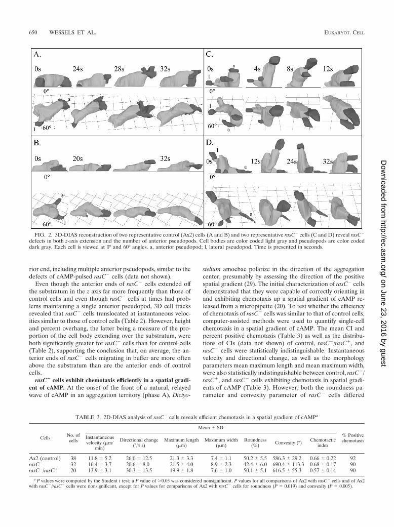
rior end, including multiple anterior pseudopods, similar to thedefects of cAMP-pulsed rasC� cells (data not shown).
Even though the anterior ends of rasC� cells extended offthe substratum in the z axis far more frequently than those ofcontrol cells and even though rasC� cells at times had prob-lems maintaining a single anterior pseudopod, 3D cell tracksrevealed that rasC� cells translocated at instantaneous veloc-ities similar to those of control cells (Table 2). However, heightand percent overhang, the latter being a measure of the pro-portion of the cell body extending over the substratum, wereboth significantly greater for rasC� cells than for control cells(Table 2), supporting the conclusion that, on average, the an-terior ends of rasC� cells migrating in buffer are more oftenabove the substratum than are the anterior ends of controlcells.
rasC� cells exhibit chemotaxis efficiently in a spatial gradi-ent of cAMP. At the onset of the front of a natural, relayedwave of cAMP in an aggregation territory (phase A), Dictyo-
stelium amoebae polarize in the direction of the aggregationcenter, presumably by assessing the direction of the positivespatial gradient (29). The initial characterization of rasC� cellsdemonstrated that they were capable of correctly orienting inand exhibiting chemotaxis up a spatial gradient of cAMP re-leased from a micropipette (20). To test whether the efficiencyof chemotaxis of rasC� cells was similar to that of control cells,computer-assisted methods were used to quantify single-cellchemotaxis in a spatial gradient of cAMP. The mean CI andpercent positive chemotaxis (Table 3) as well as the distribu-tions of CIs (data not shown) of control, rasC�/rasC�, andrasC� cells were statistically indistinguishable. Instantaneousvelocity and directional change, as well as the morphologyparameters mean maximum length and mean maximum width,were also statistically indistinguishable between control, rasC�/rasC�, and rasC� cells exhibiting chemotaxis in spatial gradi-ents of cAMP (Table 3). However, both the roundness pa-rameter and convexity parameter of rasC� cells differed
FIG. 2. 3D-DIAS reconstruction of two representative control (Ax2) cells (A and B) and two representative rasC� cells (C and D) reveal rasC�
defects in both z-axis extension and the number of anterior pseudopods. Cell bodies are color coded light gray and pseudopods are color codeddark gray. Each cell is viewed at 0° and 60° angles. a, anterior pseudopod; l, lateral pseudopod. Time is presented in seconds.
TABLE 3. 2D-DIAS analysis of rasC� cells reveals efficient chemotaxis in a spatial gradient of cAMPa
Cells No. ofcells
Mean � SD
% Positivechemotaxis
Instantaneousvelocity (�m/
min)
Directional change(°/4 s)
Maximum length(�m)
Maximum width(�m)
Roundness(%) Convexity (°) Chemotactic
index
Ax2 (control) 38 11.8 � 5.2 26.0 � 12.5 21.3 � 3.3 7.4 � 1.1 50.2 � 5.5 586.3 � 29.2 0.66 � 0.22 92rasC� 32 16.4 � 3.7 20.6 � 8.0 21.5 � 4.0 8.9 � 2.3 42.4 � 6.0 690.4 � 113.3 0.68 � 0.17 90rasC�/rasC� 20 13.9 � 3.1 30.3 � 13.5 19.9 � 1.8 7.6 � 1.0 50.1 � 5.1 616.5 � 55.3 0.57 � 0.14 90
a P values were computed by the Student t test; a P value of �0.05 was considered nonsignificant. P values for all comparisons of Ax2 with rasC� cells and of Ax2with rasC�/rasC� cells were nonsignificant, except for P values for comparisons of Ax2 with rasC� cells for roundness (P 0.019) and convexity (P 0.005).
650 WESSELS ET AL. EUKARYOT. CELL
on June 23, 2016 by guesthttp://ec.asm
.org/D
ownloaded from
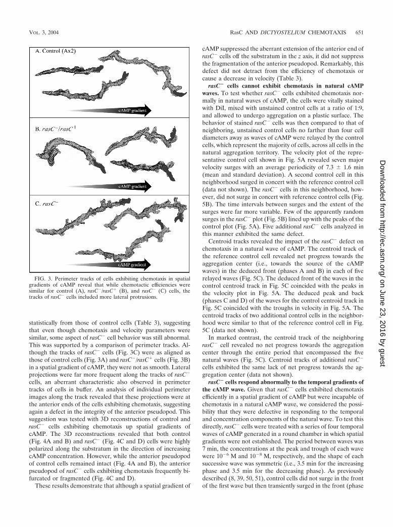
statistically from those of control cells (Table 3), suggestingthat even though chemotaxis and velocity parameters weresimilar, some aspect of rasC� cell behavior was still abnormal.This was supported by a comparison of perimeter tracks. Al-though the tracks of rasC� cells (Fig. 3C) were as aligned asthose of control cells (Fig. 3A) and rasC�/rasC� cells (Fig. 3B)in a spatial gradient of cAMP, they were not as smooth. Lateralprojections were far more frequent along the tracks of rasC�
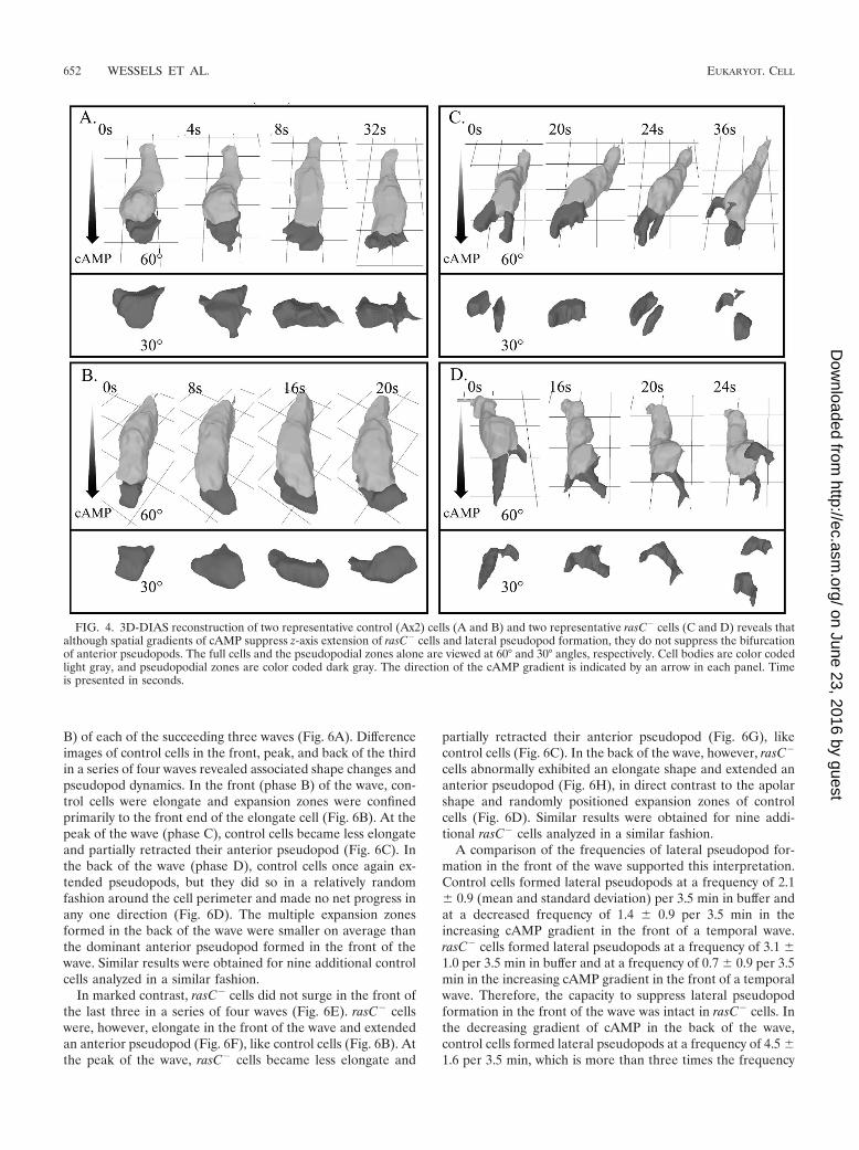
cells, an aberrant characteristic also observed in perimetertracks of cells in buffer. An analysis of individual perimeterimages along the track revealed that these projections were atthe anterior ends of the cells exhibiting chemotaxis, suggestingagain a defect in the integrity of the anterior pseudopod. Thissuggestion was tested with 3D reconstructions of control andrasC� cells exhibiting chemotaxis up spatial gradients ofcAMP. The 3D reconstructions revealed that both control(Fig. 4A and B) and rasC� (Fig. 4C and D) cells were highlypolarized along the substratum in the direction of increasingcAMP concentration. However, while the anterior pseudopodof control cells remained intact (Fig. 4A and B), the anteriorpseudopod of rasC� cells exhibiting chemotaxis frequently bi-furcated or fragmented (Fig. 4C and D).
These results demonstrate that although a spatial gradient of
cAMP suppressed the aberrant extension of the anterior end ofrasC� cells off the substratum in the z axis, it did not suppressthe fragmentation of the anterior pseudopod. Remarkably, thisdefect did not detract from the efficiency of chemotaxis orcause a decrease in velocity (Table 3).
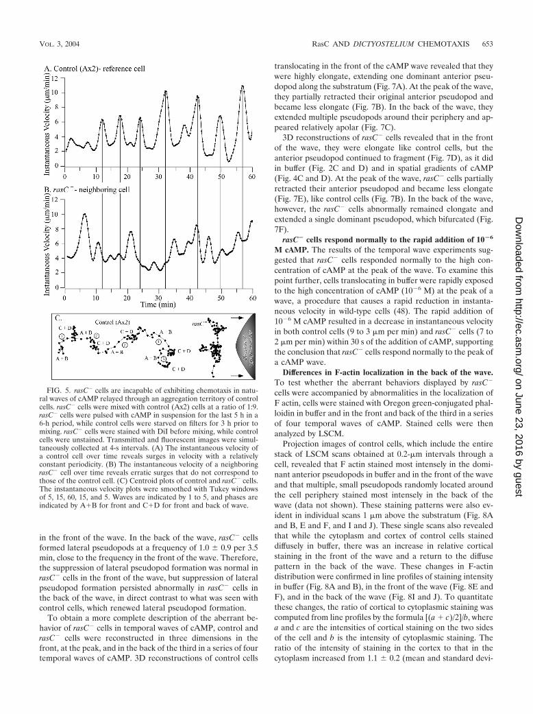
rasC� cells cannot exhibit chemotaxis in natural cAMPwaves. To test whether rasC� cells exhibited chemotaxis nor-mally in natural waves of cAMP, the cells were vitally stainedwith DiI, mixed with unstained control cells at a ratio of 1:9,and allowed to undergo aggregation on a plastic surface. Thebehavior of stained rasC� cells was then compared to that ofneighboring, unstained control cells no farther than four celldiameters away as waves of cAMP were relayed by the controlcells, which represent the majority of cells, across all cells in thenatural aggregation territory. The velocity plot of the repre-sentative control cell shown in Fig. 5A revealed seven majorvelocity surges with an average periodicity of 7.3 � 1.6 min(mean and standard deviation). A second control cell in thisneighborhood surged in concert with the reference control cell(data not shown). The rasC� cells in this neighborhood, how-ever, did not surge in concert with reference control cells (Fig.5B). The time intervals between surges and the extent of thesurges were far more variable. Few of the apparently randomsurges in the rasC� plot (Fig. 5B) lined up with the peaks of thecontrol plot (Fig. 5A). Five additional rasC� cells analyzed inthis manner exhibited the same defect.
Centroid tracks revealed the impact of the rasC� defect onchemotaxis in a natural wave of cAMP. The centroid track ofthe reference control cell revealed net progress towards theaggregation center (i.e., towards the source of the cAMPwaves) in the deduced front (phases A and B) in each of fiverelayed waves (Fig. 5C). The deduced front of the waves in thecontrol centroid track in Fig. 5C coincided with the peaks inthe velocity plot in Fig. 5A. The deduced peak and back(phases C and D) of the waves for the control centroid track inFig. 5C coincided with the troughs in velocity in Fig. 5A. Thecentroid tracks of two additional control cells in the neighbor-hood were similar to that of the reference control cell in Fig.5C (data not shown).
In marked contrast, the centroid track of the neighboringrasC� cell revealed no net progress towards the aggregationcenter through the entire period that encompassed the fivenatural waves (Fig. 5C). Centroid tracks of additional rasC�
cells exhibited the same lack of net progress towards the ag-gregation center (data not shown).
rasC� cells respond abnormally to the temporal gradients ofthe cAMP wave. Given that rasC� cells exhibited chemotaxisefficiently in a spatial gradient of cAMP but were incapable ofchemotaxis in a natural cAMP wave, we considered the possi-bility that they were defective in responding to the temporaland concentration components of the natural wave. To test thisdirectly, rasC� cells were treated with a series of four temporalwaves of cAMP generated in a round chamber in which spatialgradients were not established. The period between waves was7 min, the concentrations at the peak and trough of each wavewere 10�6 M and 10�8 M, respectively, and the shape of eachsuccessive wave was symmetric (i.e., 3.5 min for the increasingphase and 3.5 min for the decreasing phase). As previouslydescribed (8, 39, 50, 51), control cells did not surge in the frontof the first wave but then transiently surged in the front (phase
FIG. 3. Perimeter tracks of cells exhibiting chemotaxis in spatialgradients of cAMP reveal that while chemotactic efficiencies weresimilar for control (A), rasC�/rasC� (B), and rasC� (C) cells, thetracks of rasC� cells included more lateral protrusions.
VOL. 3, 2004 RasC AND DICTYOSTELIUM CHEMOTAXIS 651
on June 23, 2016 by guesthttp://ec.asm
.org/D
ownloaded from
B) of each of the succeeding three waves (Fig. 6A). Differenceimages of control cells in the front, peak, and back of the thirdin a series of four waves revealed associated shape changes andpseudopod dynamics. In the front (phase B) of the wave, con-trol cells were elongate and expansion zones were confinedprimarily to the front end of the elongate cell (Fig. 6B). At thepeak of the wave (phase C), control cells became less elongateand partially retracted their anterior pseudopod (Fig. 6C). Inthe back of the wave (phase D), control cells once again ex-tended pseudopods, but they did so in a relatively randomfashion around the cell perimeter and made no net progress inany one direction (Fig. 6D). The multiple expansion zonesformed in the back of the wave were smaller on average thanthe dominant anterior pseudopod formed in the front of thewave. Similar results were obtained for nine additional controlcells analyzed in a similar fashion.
In marked contrast, rasC� cells did not surge in the front ofthe last three in a series of four waves (Fig. 6E). rasC� cellswere, however, elongate in the front of the wave and extendedan anterior pseudopod (Fig. 6F), like control cells (Fig. 6B). Atthe peak of the wave, rasC� cells became less elongate and
partially retracted their anterior pseudopod (Fig. 6G), likecontrol cells (Fig. 6C). In the back of the wave, however, rasC�
cells abnormally exhibited an elongate shape and extended ananterior pseudopod (Fig. 6H), in direct contrast to the apolarshape and randomly positioned expansion zones of controlcells (Fig. 6D). Similar results were obtained for nine addi-tional rasC� cells analyzed in a similar fashion.
A comparison of the frequencies of lateral pseudopod for-mation in the front of the wave supported this interpretation.Control cells formed lateral pseudopods at a frequency of 2.1� 0.9 (mean and standard deviation) per 3.5 min in buffer andat a decreased frequency of 1.4 � 0.9 per 3.5 min in theincreasing cAMP gradient in the front of a temporal wave.rasC� cells formed lateral pseudopods at a frequency of 3.1 �1.0 per 3.5 min in buffer and at a frequency of 0.7 � 0.9 per 3.5min in the increasing cAMP gradient in the front of a temporalwave. Therefore, the capacity to suppress lateral pseudopodformation in the front of the wave was intact in rasC� cells. Inthe decreasing gradient of cAMP in the back of the wave,control cells formed lateral pseudopods at a frequency of 4.5 �1.6 per 3.5 min, which is more than three times the frequency
FIG. 4. 3D-DIAS reconstruction of two representative control (Ax2) cells (A and B) and two representative rasC� cells (C and D) reveals thatalthough spatial gradients of cAMP suppress z-axis extension of rasC� cells and lateral pseudopod formation, they do not suppress the bifurcationof anterior pseudopods. The full cells and the pseudopodial zones alone are viewed at 60° and 30° angles, respectively. Cell bodies are color codedlight gray, and pseudopodial zones are color coded dark gray. The direction of the cAMP gradient is indicated by an arrow in each panel. Timeis presented in seconds.
652 WESSELS ET AL. EUKARYOT. CELL
on June 23, 2016 by guesthttp://ec.asm
.org/D
ownloaded from
in the front of the wave. In the back of the wave, rasC� cellsformed lateral pseudopods at a frequency of 1.0 � 0.9 per 3.5min, close to the frequency in the front of the wave. Therefore,the suppression of lateral pseudopod formation was normal inrasC� cells in the front of the wave, but suppression of lateralpseudopod formation persisted abnormally in rasC� cells inthe back of the wave, in direct contrast to what was seen withcontrol cells, which renewed lateral pseudopod formation.
To obtain a more complete description of the aberrant be-havior of rasC� cells in temporal waves of cAMP, control andrasC� cells were reconstructed in three dimensions in thefront, at the peak, and in the back of the third in a series of fourtemporal waves of cAMP. 3D reconstructions of control cells
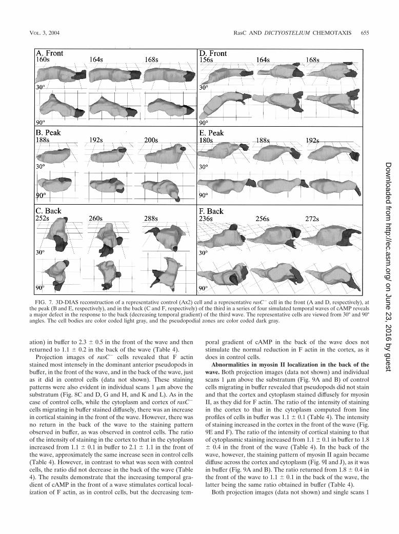
translocating in the front of the cAMP wave revealed that theywere highly elongate, extending one dominant anterior pseu-dopod along the substratum (Fig. 7A). At the peak of the wave,they partially retracted their original anterior pseudopod andbecame less elongate (Fig. 7B). In the back of the wave, theyextended multiple pseudopods around their periphery and ap-peared relatively apolar (Fig. 7C).
3D reconstructions of rasC� cells revealed that in the frontof the wave, they were elongate like control cells, but theanterior pseudopod continued to fragment (Fig. 7D), as it didin buffer (Fig. 2C and D) and in spatial gradients of cAMP(Fig. 4C and D). At the peak of the wave, rasC� cells partiallyretracted their anterior pseudopod and became less elongate(Fig. 7E), like control cells (Fig. 7B). In the back of the wave,however, the rasC� cells abnormally remained elongate andextended a single dominant pseudopod, which bifurcated (Fig.7F).
rasC� cells respond normally to the rapid addition of 10�6
M cAMP. The results of the temporal wave experiments sug-gested that rasC� cells responded normally to the high con-centration of cAMP at the peak of the wave. To examine thispoint further, cells translocating in buffer were rapidly exposedto the high concentration of cAMP (10�6 M) at the peak of awave, a procedure that causes a rapid reduction in instanta-neous velocity in wild-type cells (48). The rapid addition of10�6 M cAMP resulted in a decrease in instantaneous velocityin both control cells (9 to 3 �m per min) and rasC� cells (7 to2 �m per min) within 30 s of the addition of cAMP, supportingthe conclusion that rasC� cells respond normally to the peak ofa cAMP wave.
Differences in F-actin localization in the back of the wave.To test whether the aberrant behaviors displayed by rasC�
cells were accompanied by abnormalities in the localization ofF actin, cells were stained with Oregon green-conjugated phal-loidin in buffer and in the front and back of the third in a seriesof four temporal waves of cAMP. Stained cells were thenanalyzed by LSCM.
Projection images of control cells, which include the entirestack of LSCM scans obtained at 0.2-�m intervals through acell, revealed that F actin stained most intensely in the domi-nant anterior pseudopods in buffer and in the front of the waveand that multiple, small pseudopods randomly located aroundthe cell periphery stained most intensely in the back of thewave (data not shown). These staining patterns were also ev-ident in individual scans 1 �m above the substratum (Fig. 8Aand B, E and F, and I and J). These single scans also revealedthat while the cytoplasm and cortex of control cells staineddiffusely in buffer, there was an increase in relative corticalstaining in the front of the wave and a return to the diffusepattern in the back of the wave. These changes in F-actindistribution were confirmed in line profiles of staining intensityin buffer (Fig. 8A and B), in the front of the wave (Fig. 8E andF), and in the back of the wave (Fig. 8I and J). To quantitatethese changes, the ratio of cortical to cytoplasmic staining wascomputed from line profiles by the formula [(a � c)/2]/b, wherea and c are the intensities of cortical staining on the two sidesof the cell and b is the intensity of cytoplasmic staining. Theratio of the intensity of staining in the cortex to that in thecytoplasm increased from 1.1 � 0.2 (mean and standard devi-
FIG. 5. rasC� cells are incapable of exhibiting chemotaxis in natu-ral waves of cAMP relayed through an aggregation territory of controlcells. rasC� cells were mixed with control (Ax2) cells at a ratio of 1:9.rasC� cells were pulsed with cAMP in suspension for the last 5 h in a6-h period, while control cells were starved on filters for 3 h prior tomixing. rasC� cells were stained with DiI before mixing, while controlcells were unstained. Transmitted and fluorescent images were simul-taneously collected at 4-s intervals. (A) The instantaneous velocity ofa control cell over time reveals surges in velocity with a relativelyconstant periodicity. (B) The instantaneous velocity of a neighboringrasC� cell over time reveals erratic surges that do not correspond tothose of the control cell. (C) Centroid plots of control and rasC� cells.The instantaneous velocity plots were smoothed with Tukey windowsof 5, 15, 60, 15, and 5. Waves are indicated by 1 to 5, and phases areindicated by A�B for front and C�D for front and back of wave.
VOL. 3, 2004 RasC AND DICTYOSTELIUM CHEMOTAXIS 653
on June 23, 2016 by guesthttp://ec.asm
.org/D
ownloaded from
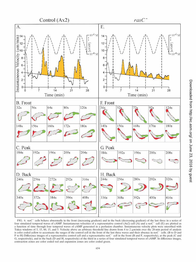
FIG. 6. rasC� cells behave abnormally in the front (increasing gradient) and in the back (decreasing gradient) of the last three in a series offour simulated temporal waves of cAMP. Instantaneous velocities of a representative control (Ax2) cell (A) and a rasC� cell (E) are plotted asa function of time through four temporal waves of cAMP generated in a perfusion chamber. Instantaneous velocity plots were smoothed withTukey windows of 5, 15, 60, 15, and 5. Velocity above an arbitrary threshold line drawn from 4 to 2 �m/min over the 28-min period of analysisis color coded yellow to accentuate the surges of the control cell in the front of the last three waves and their absence in rasC� cells. (B to D andF to H) Difference images of a representative control cell and a representative rasC� cell in the front (B and F, respectively), at the peak (C andG, respectively), and in the back (D and H, respectively) of the third in a series of four simulated temporal waves of cAMP. In difference images,contraction zones are color coded red and expansion zones are color coded green.
654
on June 23, 2016 by guesthttp://ec.asm
.org/D
ownloaded from
ation) in buffer to 2.3 � 0.5 in the front of the wave and thenreturned to 1.1 � 0.2 in the back of the wave (Table 4).
Projection images of rasC� cells revealed that F actinstained most intensely in the dominant anterior pseudopods inbuffer, in the front of the wave, and in the back of the wave, justas it did in control cells (data not shown). These stainingpatterns were also evident in individual scans 1 �m above thesubstratum (Fig. 8C and D, G and H, and K and L). As in thecase of control cells, while the cytoplasm and cortex of rasC�
cells migrating in buffer stained diffusely, there was an increasein cortical staining in the front of the wave. However, there wasno return in the back of the wave to the staining patternobserved in buffer, as was observed in control cells. The ratioof the intensity of staining in the cortex to that in the cytoplasmincreased from 1.1 � 0.1 in buffer to 2.1 � 1.1 in the front ofthe wave, approximately the same increase seen in control cells(Table 4). However, in contrast to what was seen with controlcells, the ratio did not decrease in the back of the wave (Table4). The results demonstrate that the increasing temporal gra-dient of cAMP in the front of a wave stimulates cortical local-ization of F actin, as in control cells, but the decreasing tem-
poral gradient of cAMP in the back of the wave does notstimulate the normal reduction in F actin in the cortex, as itdoes in control cells.
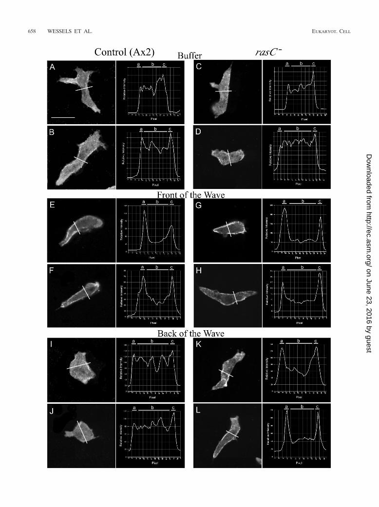
Abnormalities in myosin II localization in the back of thewave. Both projection images (data not shown) and individualscans 1 �m above the substratum (Fig. 9A and B) of controlcells migrating in buffer revealed that pseudopods did not stainand that the cortex and cytoplasm stained diffusely for myosinII, as they did for F actin. The ratio of the intensity of stainingin the cortex to that in the cytoplasm computed from lineprofiles of cells in buffer was 1.1 � 0.1 (Table 4). The intensityof staining increased in the cortex in the front of the wave (Fig.9E and F). The ratio of the intensity of cortical staining to thatof cytoplasmic staining increased from 1.1 � 0.1 in buffer to 1.8� 0.4 in the front of the wave (Table 4). In the back of thewave, however, the staining pattern of myosin II again becamediffuse across the cortex and cytoplasm (Fig. 9I and J), as it wasin buffer (Fig. 9A and B). The ratio returned from 1.8 � 0.4 inthe front of the wave to 1.1 � 0.1 in the back of the wave, thelatter being the same ratio obtained in buffer (Table 4).
Both projection images (data not shown) and single scans 1
FIG. 7. 3D-DIAS reconstruction of a representative control (Ax2) cell and a representative rasC� cell in the front (A and D, respectively), atthe peak (B and E, respectively), and in the back (C and F, respectively) of the third in a series of four simulated temporal waves of cAMP revealsa major defect in the response to the back (decreasing temporal gradient) of the third wave. The representative cells are viewed from 30° and 90°angles. The cell bodies are color coded light gray, and the pseudopodial zones are color coded dark gray.
VOL. 3, 2004 RasC AND DICTYOSTELIUM CHEMOTAXIS 655
on June 23, 2016 by guesthttp://ec.asm
.org/D
ownloaded from
656 WESSELS ET AL. EUKARYOT. CELL
on June 23, 2016 by guesthttp://ec.asm
.org/D
ownloaded from
�m above the substratum of rasC� cells in buffer (Fig. 9C andD) and in the front of the wave (Fig. 9G and H) revealedstaining patterns similar to those for control cells (Fig. 9A andB and E and F, respectively). The ratio of the intensity ofcortical staining to that of cytoplasmic staining for rasC� cellsincreased from 1.1 � 0.2 in buffer to 1.7 � 0.5 in the front ofthe wave, values close to those for control cells under therespective conditions (Table 4). However, in the back of thewave, myosin II was abnormally retained in the cortex (Fig. 9Kand L), in direct contrast to what was seen with control cells(Fig. 9I and J). While the staining intensity ratio of control cellsdecreased from 1.8 � 0.4 in the front of the wave to 1.1 � 0.1in the back of the wave, the ratios of rasC� cells stayed ap-proximately the same, 1.7 � 0.5 and 1.6 � 0.3, respectively, inthe front and back of the wave (Table 4). These results dem-onstrate that while in control cells myosin II exits the cortex inresponse to the decreasing temporal gradient of cAMP in theback of the wave, myosin II, like F actin, is abnormally retainedin the cortex of rasC� cells.
DISCUSSION
Guanine nucleotide exchange factors activate Ras proteinsby catalyzing the exchange of GDP with GTP (2). GTPase-activating proteins inactivate Ras proteins by catalyzing GTPhydrolysis (2). Receptor-coupled Ras proteins are responsiblefor the activation of a variety of signaling pathways upon re-ceptor occupancy (3, 21, 35, 36). In Dictyostelium, RasC, whichis expressed throughout the life cycle (6), is involved in theactivation of adenylate cyclase and Akt/PKB, the latter possiblyresulting from the activation of phosphatidylinositol 3-kinase(PI3K), which catalyzes the phosphorylation of PIP2 to PIP3(5). Both PI3K1 and PI3K2, potential targets of RasC, have
been shown to play roles in chemotaxis (9, 23). The originalcharacterization revealed that while rasC� cells that had beenartificially pulsed with cAMP could not signal, they could ex-hibit chemotaxis up a spatial gradient of cAMP released froma micropipette. Pulsed rasC� cells were also capable of form-ing aggregates at high density and of entering aggregatesformed by control cells (20). Although these experiments sug-gested that RasC was not involved in the behavioral responsesof cells to a chemoattractant, they did not test whether RasCwas involved in the basic motile behavior of a cell in theabsence of an attractant or in single-cell responses to the tem-poral and concentration components of a natural wave.
RasC plays a role in basic motile behavior. Although rasC�
cells moved at approximately the same average velocity as didcontrol cells, they had problems maintaining their front end onthe substratum and in maintaining an intact anterior pseudo-pod. The front end of rasC� cells frequently extended off thesubstratum in a manner similar to that seen with the nullmutant for the vasodilator-stimulated phosphoprotein gene(vasp�) (12) and the null mutant for clathrin (chc�) (47). SincerasC� cells exhibited these defects in the absence of chemoat-tractant, it seems likely that its role in maintaining elongatecells on a substratum and in maintaining an intact anteriorpseudopod is independent of cAMP receptor (CAR1) occu-pancy. Observations of vegetative cell behavior revealed simi-lar abnormal behavior in buffer (data not shown), supportingthis conclusion. Wessels et al. (44) have demonstrated thatwhen lateral pseudopods contact the substratum, they are sta-bilized. Hence, RasC may function immediately downstreamof adhesion sites or mechanoreceptors involved in pseudopodstabilization during migration in buffer as well as downstreamof the cAMP receptor CAR1 during chemotaxis.
RasC is not necessary for chemotaxis in a spatial gradient ofa chemoattractant. In the original analysis, it was demon-strated that rasC� cells exhibited chemotaxis up a spatial gra-dient of cAMP released from a micropipette (20). Here, weperformed a quantitative analysis of the efficiency of chemo-taxis as well as a detailed analysis of the behavior of individualcells during chemotaxis. Our results demonstrate that a spatialgradient of cAMP normalizes the z-axis extension defect inbuffer but not the tendency of rasC� pseudopods to bifurcateor fragment. Our results demonstrate, however, that RasC isnot essential for cell polarization or efficient chemotaxis in aspatial gradient of cAMP. Therefore, it is unlikely that RasCplays a role in those pathways that regulate cell polarization inresponse to a chemotactic signal (e.g., the pathway that in-cludes PAKa) (4, 5).
RasC plays an essential role in responding to temporalgradients of cAMP. The temporal gradients of cAMP in thefront and back of a natural wave play fundamental roles inregulating Dictyostelium chemotaxis in a natural aggregation
TABLE 4. Ratio of F-actin staining in the cortex and the cytoplasmand ratio of myosin II staining in the cortex and the cytoplasm of
representative cells in buffer in relation to the temporal wavea
Staining Condition
Ax2 Cells (control) rasC� cells
No. ofcells Ratio No. of
cells Ratio
F actin Buffer 10 1.1 � 0.2 6 1.1 � 0.1Wave front 9 2.3 � 0.5 9 2.1 � 1.1Wave back 10 1.1 � 0.2 8 2.0 � 0.6
Myosin II Buffer 6 1.1 � 0.1 12 1.1 � 0.2Wave front 12 1.8 � 0.4 14 1.7 � 0.5Wave back 12 1.1 � 0.1 15 1.6 � 0.3
a The ratios of the staining intensity of cortical regions (a and c) to that of thecytoplasm (b) were computed by the formula [(a � c)/2]/b. The intensities in thecortical regions at the two ends of the scan (approximately six pixels) wereaveraged. The intensities in the regions between the cortical regions were con-sidered cytoplasmic and were averaged.
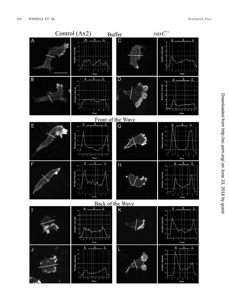
FIG. 8. Line profiles of the intensity of F-actin staining in the cortex and cytoplasm reveal subtle differences between control (Ax2) and rasC�
cells in buffer and in the front of the wave. In each panel, the position of the line profile across the stained cell image, just posterior to the nucleus,and a plot of the line profile are presented. The analyzed optical section in each case was 1 �m above the substratum. In each line profile, relativeintensity is plotted as a function of distance in pixels. Cells were analyzed in the third in a series of four simulated temporal waves of cAMP. Theaverage ratios of the intensity of staining in the cortex to that in the cytoplasm are presented in Table 4. a and c, cortical zones; b, cytoplasmic zone.Scale bar, 5 �m.
VOL. 3, 2004 RasC AND DICTYOSTELIUM CHEMOTAXIS 657
on June 23, 2016 by guesthttp://ec.asm
.org/D
ownloaded from
658 WESSELS ET AL. EUKARYOT. CELL
on June 23, 2016 by guesthttp://ec.asm
.org/D
ownloaded from
territory (29). In response to the increasing temporal gradientof cAMP in the front of a simulated temporal wave, rasC� cellssuppressed lateral pseudopod formation like control cells butthey did not surge (i.e., exhibit a transient increase in velocity).This response was opposite to that of the regA� mutant (45).RegA is an intracellular phosphodiesterase (19). Therefore,regA� cells should have abnormally increased levels of intra-cellular cAMP, while rasC� cells would be expected to haveabnormally reduced levels of intracellular cAMP, a possibleexplanation for the opposite phenotypes exhibited in the frontof the wave. Hence, one might also expect cells with constitu-tively active protein kinase A activity (pkAR�) (51) to behavein the same manner as regA� cells and opposite that of rasC�
cells. The finding that pkAR� cells were constitutively ovoidunder all conditions, affecting their behavior in the front of awave (51), precludes this comparison. However, it is just aslikely that RegA, which carries a two-component responseregulator domain, and RasC, which regulates pathways otherthan those involving cAMP, also affect behavior through path-ways independent of cAMP. What is clear is that regA and rasCdeletion mutants affect different components of the behavioralresponse to the increasing temporal gradient of cAMP in thefront of the wave. Hence, we separated RegA and RasC intotwo independent pathways emanating from the front of thewave and ending in distinctly different behavioral responses inthe working model shown in Fig. 10. Since ERK2 has beenpostulated to be a negative regulator of RegA (19), an erkB-null mutant should respond abnormally to the front of thewave in the same manner as rasC� cells and opposite that ofregA� cells. It would therefore be of considerable interest toanalyze the detailed chemotactic behavior of erkB� cells (25)to see whether they exhibit defects in the front of the wavesimilar to those exhibited by rasC� cells. In contrast to regA�,cells of the myoA�/myoF� double mutant do not surge inresponse to an increasing temporal gradient of cAMP (8), aresponse similar to that of rasC�. For that reason, we verytentatively placed MyoA/MyoF downstream of RasC in theRasC-dependent pathway emanating from the front of thewave in the model in Fig. 10.
Although partially defective in their response to the increas-ing temporal gradient in the front of the wave, rasC� cellsresponded normally to the high concentration of cAMP at thepeak of the wave. However, rasC� cells then responded ab-normally to the decreasing temporal gradient of cAMP in theback of the wave. Rather than becoming apolar and extendingsmall pseudopods in random directions as control cells do,rasC� cells once again extended a dominant anterior pseudo-pod, usually from the original front end of the cell, and exhib-ited a relatively elongate, polar morphology along the substra-tum. The abnormal retention of polarity and the abnormalsuppression of lateral pseudopod formation in the back of thewave were the same defects exhibited by the myosin II regu-
latory light chain phosphorylation mutant S13A (50), suggest-ing that RasC and the phosphorylation of RLC function alonga common independent pathway emanating from the back ofthe wave, as in the model in Fig. 10. S13A mutant cells respondnormally to the front of the wave, supporting the independenceof the RasC pathway in the back of the wave.
Previous qualitative results (20) suggested that rasC� cellscould aggregate. In chimeric mixes, rasC� cells were observedto be late in entering streams and aggregates of normal cells(20), suggesting a fundamental defect in behavioral responsesto relayed cAMP waves. The mixing experiments performedhere demonstrate that rasC� cells do not surge or orient inresponse to a natural wave and therefore do not make netprogress towards an aggregation center in an aggregation ter-ritory. We conclude that it is the defective responses to tem-poral gradients of cAMP in the front and back of a wave thatpreclude chemotaxis in a natural wave of cAMP.
RasC and cytoskeletal reorganization. Staining experimentsrevealed a major defect in the localization of F actin andmyosin II in the cortex in response to the decreasing temporalgradient of cAMP in the back of a wave in association withabnormal maintenance of an elongate cell morphology. Myo-sin II has been demonstrated to play a fundamental role inregulating the shape of a Dictyostelium amoeba (26, 42). Ourresults indicate that the normal loss of anterior-posterior po-larity and the random extension of pseudopods in control cellsin the back of a wave are dependent on RasC activity and areassociated with the reduction of F actin and myosin II in thecortex. Although one might assume that this reduction of Factin and myosin II is the direct result of myosin heavy-chain(MHC) phosphorylation based on previous in vitro (17) and invivo (7) studies, the retention of myosin II in the cortex ofrasC� cells and maintenance of an elongate shape are alsocharacteristic of the S13A mutant, in which the myosin regu-latory light chain cannot be phosphorylated (50). This may beexplained by the observations of Berlot et al. (1) that theaddition of cAMP to aggregation-competent Dictyosteliumamoebae suspended in buffer results in an increase in thephosphorylation of both the MHC and regulatory light chain,suggesting coordinate phosphorylation and function. Interest-ingly, both in vitro (17) and in vivo (7) studies have demon-strated that phosphorylation of the MHC results in the depo-lymerization of myosin II and its movement from the cortex tocytoplasm. The phenotype of the S13A mutant suggests thatthe myosin II regulatory light chain must also be phosphory-lated for the reduction in F actin and myosin II in the cellcortex in the back of the wave. Therefore, the model in Fig. 10may eventually be expanded to include both myosin regulatorylight chain and MHC phosphorylation as downstream targetsof the RasC pathway emanating from the back of the wave.Analyses of the 3XALA mutant, in which MHC was engi-neered to mimic a permanently unphosphorylated state, and
FIG. 9. Line profiles of the intensity of myosin II staining in the cortex and the cytoplasm reveal a major difference between control (Ax2) andrasC� cells in the back of the wave. In each panel, the position of the line profile across the cell, just posterior to the nucleus, and a plot of theline profile are presented. The analyzed optical section in each case was 1 �m above the substratum. Cells were analyzed in the third of a seriesof four simulated temporal waves of cAMP. The average ratios of the intensity of staining in the cortex to that in the cytoplasm are presented inTable 4. a and c, cortical zones; b, cytoplasmic zone. Scale bar, 5 �m.
VOL. 3, 2004 RasC AND DICTYOSTELIUM CHEMOTAXIS 659
on June 23, 2016 by guesthttp://ec.asm
.org/D
ownloaded from

the 3XASP mutant, in which MHC was engineered to mimic apermanently phosphorylated state (7), are now in progress totest this possibility.
An emerging model. The model that has begun to emergeincludes independent pathways emanating from the differentphases of the chemotactic wave (Fig. 10). Two parallel path-ways, one dependent on RegA (45, 51) and another dependenton RasC, emanate from the front of the wave and are inducedby increasing receptor occupancy with time (i.e., a positivetemporal gradient of cAMP). The parallel pathway dependenton the activation of PKA is activated by the very high concen-tration of cAMP at the peak of the wave, presumably the resultof full receptor occupancy (51). Finally, a second RasC-depen-
dent pathway emanates from the back of the wave and isinduced by decreasing receptor occupancy with time (i.e., anegative temporal gradient of cAMP). Each pathway is respon-sible for a phase-specific behavior; in sequence, the behaviorsrepresent the complex response to the natural wave (Fig. 10).One interesting, albeit tentative, aspect of this model is that atleast two downstream targets of the regulatory pathways so fardelineated are myosins, both myosin II and myosin I. In thecase of myosin II, localization is effected by phosphorylation-dephosphorylation of both the heavy chain (7; P. Heid, D.Wessels, K. Daniels, H. Zhang, and D. R. Soll, unpublisheddata) and light chain (50). As more mutants, including bothregulatory and cytoskeletal, are analyzed within the contextual
FIG. 10. Model of regulation of the sequence of behaviors associated with the four phases of the wave, based on data obtained here for therasC� mutant and in the following studies of other mutants that used the same contextual framework and experimental protocols (31) foridentifying the exact behavioral defects in the different components of the natural wave: regA� (45), myoA�/myoF� (7), pkaR� (47), S13A (48).PKA, protein kinase A; Rlc, regulatory light chain; Rlc-P, phosphorylated regulatory light chain.
660 WESSELS ET AL. EUKARYOT. CELL
on June 23, 2016 by guesthttp://ec.asm
.org/D
ownloaded from
framework developed to distinguish parallel pathways emanat-ing from the different phases of the wave (29), we may find thatonly a few cytoskeletal components, including the myosins,serve as the targets of regulatory cascades for effecting thephase-specific behaviors that in sequence represent the com-plex chemotactic response to the natural wave. Because of thecomplexity of each response, it is likely that each cascade mayhave more than one target.
ACKNOWLEDGMENTS
This research was supported by National Institutes of Health grantsHD-18577 to D.R.S. and GM60447 and GM62350 to W.F.L. and agrant from the MRC of Canada to G.W.
REFERENCES
1. Berlot, C. H., J. A. Spudich, and P. N. Devreotes. 1985. Chemoattractant-elicited increases in myosin phosphorylation in Dictyostelium. Cell 43:307–314.
2. Boguski, M., and F. McCormick. 1993. Proteins regulating Ras and itsrelatives. Nature 366:643–654.
3. Campbell, S., R. Khosravi-Far, K. L. Rossman, G. J. Clark, and C. J. Der.1998. Increasing complexity of Ras signaling. Oncogene 17:1395–1413.
4. Chung, C., and R. A. Firtel. 1999. PAKa, a putative PAK family member, isrequired for cytokinesis and the regulation of the cytoskeleton in Dictyoste-lium discoideum cells during chemotaxis. J. Cell Biol. 174:559–575.
5. Chung, C. Y., G. Potikyan, and R. A. Firtel. 2001. Control of cell polarity andchemotaxis by Akt/PKB and PI3 kinase through the regulation of PAKa.Mol. Cell 7:937–947.
6. Daniel, J., G. Spiegelman, and G. Weeks. 1995. Dictyostelium ras genes, p.100–104. In M. Serial and L. Huber (ed.), Guidebook to the small GTPase.Oxford University Press, New York, N.Y.
7. Egelhoff, T. T., R. J. Lee, and J. A. Spudich. 1993. Dictyostelium myosin heavychain phosphorylation sites regulate myosin filament assembly and localiza-tion in vivo. Cell 75:363–371.
8. Falk, D. L., D. Wessels, L. Jenkins, T. Pham, S. Kuhl, M. A. Titus, and D. R.Soll. 2003. Shared, unique and redundant functions of three members of theclass I myosins (MyoA, MyoB and MyoF) in motility and chemotaxis inDictyostelium. J. Cell Sci. 116:3985–3999.
9. Funamoto, S., K. Milan, R. Meili, and R. A. Firtel. 2001. Role of phospha-tidylinositol 3� kinase and a downstream pleckstrin homology domain-con-taining protein in controlling chemotaxis in Dictyostelium. J. Cell Biol. 153:795–810.
10. Funamoto, S., R. Meili, S. Lee, L. Parry, and R. Firtel. 2002. Spatial andtemporal regulation of 3-phosphoinositides by PI3-kinase and PTEN medi-ates chemotaxis. Cell 109:611–623.
11. Geiger, J., D. Wessels, and D. R. Soll. 2003. Human PMNs respond totemporal waves of chemoattractant like Dictyostelium. Cell Motil. Cytoske-let. 56:27–44.
12. Han, Y., C. Chung, D. Wessels, S. Stephens, M. A. Titus, D. R. Soll, and R. A.Firtel. 2002. Requirement of VASP for cell adhesion, filopodia formation,and chemotaxis. J. Biol. Chem. 277:49877–49887.
13. Heid, P., E. Voss, and D. R. Soll. 2002. 3D-DIASemb: a computer-assistedsystem for reconstructing and motion analyzing in 4D every cell and nucleusin a developing embryo. Dev. Biol. 245:329–347.
14. Iranfar, N., D. Fuller, and W. F. Loomis. 2003. Genome-wide expressionanalyses of gene regulation during early development of Dictyostelium dis-coideum. Eukaryot. Cell 2:664–670.
15. Iranfar, N., D. Fuller, R. Sasik, T. Hwa, M. Laub, and W. F. Loomis. 2001.Expression patterns of cell-type-specific genes in Dictyostelium. Mol. Biol.Cell 12:2590–2600.
16. Kessin, R. H. 2001. Dictyostelium: evolution, cell biology and the develop-ment of multicellularity. Cambridge University Press, Cambridge, UnitedKingdom.
17. Kuczmarski, E. R., and J. A. Spudich. 1980. Regulation of myosin self-assembly: phosphorylation of heavy chain inhibits formation of thick fila-ments. Proc. Natl. Acad. Sci. USA 77:7292–7296.
18. Kuspa, A., and W. Loomis. 1994. Transformation of Dicytostelium—genedisruptions, insertional mutagenesis, and promoter traps. Methods Mol.Genet. 3:3–21.
19. Laub, M. T., and W. F. Loomis. 1998. A molecular network that producesspontaneous oscillations in excitable cells of Dictyostelium. Mol. Biol. Cell9:3521–3532.
20. Lim, C. J., G. B. Spiegelman, and G. Weeks. 2001. RasC is required foroptional activation of adenylyl cyclase and Akt/PKB during aggregation.EMBO J. 20:4490–4499.
21. Lim, C. J., G. B. Spiegelman, and G. Weeks. 2002. Cytoskeletal regulation byDictyostelium Ras subfamily proteins. J. Muscle Res. Cell Motil. 23:729–736.
22. Reymond, C. D., R. H. Gomer, M. C. Medhy, and R. A. Firtel. 1984. Devel-opmental regulation of a Dicytostelium gene encoding a protein homologousto mammalian Ras protein. Cell 39:141–148.
23. Rickert, P., O. D,.Weiner, F. Wang, H. R. Bourne, and G. Servant. 2000.Leukocytes navigate by compass: roles of PI3Kgamma and its lipid products.Trends Cell Biol. 10:466–473.
24. Sasik, R., N. Iranfar, T. Hwa, and W. F. Loomis. 2002. Extracting transcrip-tional events from temporal gene expression patterns during Dictyosteliumdevelopment. Bioinformatics 18:61–66.
25. Segall, J. E., A. Kuspa, G. Shaulsky, M. Ecke, M. Maeda, C. Gaskins, R. A.Firtel, and W. F. Loomis. 1995. A MAP kinase necessary for receptor-mediated activation of adenylyl cyclase in Dictyostelium. J. Cell Biol. 128:405–413.
26. Sheldon, E., and D. A. Knecht. 1996. Dictyostelium cell shape generationrequires myosin II. Cell Motil. Cytoskelet. 35:59–67.
27. Shutt, D., and D. R. Soll. 1999. HIV-induced T-cell syncytia release a twocomponent T-helper cell chemoattractant composed of Nef and Tat. J. CellSci. 112:3931–3941.
28. Shutt, D. C., L. M. Jenkins, E. Carolan, J. Stapleton, K. Daniels, R.Kennedy, and D. R. Soll. 1998. T cell syncytia induced by HIV release T cellchemoattractants: demonstration with a newly developed single cell chemo-taxis chamber. J. Cell Sci. 111:99–109.
29. Soll, D. R., D. Wessels, P. Heid, and H. Zhang. 2002. A contextual frame-work for characterizing motility and chemotaxis mutants in Dictyosteliumdiscoideum. J. Muscle Res. Cell Motil. 23:659–672.
30. Soll, D. R. 1979. Timers in developing systems. Science 203:841–849.31. Soll, D. R. 1987. Methods for manipulating and investigating developmental
timing in Dictyostelium discoideum. Methods Cell Biol. 28:413–431.32. Soll, D. R. 1995. The use of computers in understanding how animal cells
crawl. Int. Rev. Cytol. 163:43–104.33. Soll, D. R., and E. Voss. 1998. Two and three dimensional computer systems
for analyzing how cells crawl, p. 25–52. In D. R. Soll and D. Wessels (ed.),Motion analysis of living cells. John Wiley, Inc., New York, N.Y.
34. Soll, D. R., E. Voss, O. Johnson, and D. J. Wessels. 2000. Three-dimensionalreconstruction and motion analysis of living crawling cells. Scanning 22:249–257.
35. Suzuki, J., Y. Yamazaki, G. Li, Y. Kaziro, and H. Koide. 2000. Involvementof Ras and Ra1 in chemotactic migration of skeletal myoblasts. Mol. Cell.Biol. 20:4658–4665.
36. Takai, Y., T. Sasaki, and T. Matozaki. 2001. Small GTP-binding proteins.Physiol. Rev. 81:153–208.
37. Van Driessche, N., C. Shaw, M. Katoh, T. Morio, R. Sucgang, M. Ibarra, H.Kuwayama, T. Saito, H. Urushihara, M. Maeda, I. Takeuchi, H. Ochiai, W.Eaton, J. Tollett, J. Halter, A. Uspa, Y. Tanaka, and G. Shaulsky. 2002. Atranscriptional profile of multicellular development in Dictyostelium discoi-deum. Development 129:1543–1552.
38. Varnum, B., and D. R. Soll. 1984. Effect of cAMP on single cell motility inDictyostelium. J. Cell Biol. 99:1151–1155.
39. Varnum, B., K. Edwards, and D. R. Soll. 1985. Dictyostelium amoebae altermotility differently in response to increasing versus decreasing temporalgradients of cAMP. J. Cell Biol. 101:1–5.
40. Varnum, B., K. Edwards, and D. R. Soll. 1986. The developmental regulationof single cell motility in Dictyostelium discoideum. Dev. Biol. 113:218–227.
41. Varnum-Finney, B., K. Edwards, E. Voss, and D. R. Soll. 1987. Amoebae ofDictyostelium discoideum respond to an increasing temporal gradient of thechemoattractant cAMP with a reduced frequency of turning: evidence for atemporal mechanism in amoeboid chemotaxis. Cell Motil. Cytoskelet. 8:7–17.
42. Wessels, D., D. R. Soll, D. Knecht, W. F. Loomis, A. DeLozanne, and J.Spudich. 1988. Cell motility and chemotaxis in Dictyostelium amoebae lack-ing myosin heavy chain. Dev. Biol. 128:164–177.
43. Wessels, D., E. Voss, N. Von Bergen, R. Burns, J. Stites, and D. R. Soll. 1998.A computer-assisted system for reconstructing and interpreting the dynamicthree-dimensional relationships of the outer surface, nucleus and pseudo-pods of crawling cells. Cell Motil. Cytoskelet. 41:225–246.
44. Wessels, D., H. Vawter-Hugart, J. Murray, and D. R. Soll. 1994. Threedimensional dynamics of pseudopod formation and the regulation of turningduring the motility cycle of Dictyostelium. Cell Motil. Cytoskelet. 27:1–12.
45. Wessels, D., H. Zhang, J. Reynolds, K. Daniels, P. Heid, S. Liu, A. Kuspa, G.Shaulsky, W. F. Loomis, and D. R. Soll. 2000. The internal phosphodiester-ase RegA is essential for the suppression of lateral pseudopods during Dic-tyostelium chemotaxis. Mol. Biol. Cell 11:2803–2820.
46. Wessels, D., J. Murray, and D. R. Soll. 1992. Behavior of Dictyosteliumamoebae is regulated primarily by the temporal dynamics of the naturalcAMP wave. Cell Motil. Cytoskelet. 23:145–156.
47. Wessels, D., J. Reynolds, O. Johnson, E. Voss, R., Burns, K. Daniels, E.Garrard, T. O’Hallaran and D. R. Soll. 2000. Clathrin plays a novel role inthe regulation of cell polarity, pseudopod formation, uropod stability andmotility in Dictyostelium. J. Cell Sci. 113:26–36.
VOL. 3, 2004 RasC AND DICTYOSTELIUM CHEMOTAXIS 661
on June 23, 2016 by guesthttp://ec.asm
.org/D
ownloaded from
48. Wessels, D., N. Schroeder, E., Voss, A. Hall, J. Condeelis, and D. R. Soll.1989. cAMP mediated inhibition of intracellular particle movement andactin reorganization in Dictyostelium. J. Cell Biol. 109:2841–2851.
49. Wilkins, A., and R. H. Insall. 2001. Small GTPase in Dictyostelium. TrendsGenet. 17:41–48.
50. Zhang, H., D. Wessels, P. Fey, K. Daniels, R. Chisholm, and D. R. Soll. 2002.Phosphorylation of the myosin regulatory light chain plays a role in cell
motility and polarity during Dictyostelium chemotaxis. J. Cell Sci. 115:1733–1747.
51. Zhang, H., P. Heid, D. Wessels, K. Daniels, T. Pham, W. F. Loomis, andD. R. Soll. 2003. Constitutively active protein kinase A disrupts motility andchemotaxis in Dictyostelium discoideum. Eukaryot. Cell 2:62–75.
52. Zigmond, S. H. 1977. Ability of polymorphonuclear leukocytes to orient ingradients of chemotactic factor. J. Cell Biol. 75:606–616.
662 WESSELS ET AL. EUKARYOT. CELL
on June 23, 2016 by guesthttp://ec.asm
.org/D
ownloaded from


