Embed Size (px)
Citation preview

Ratios of activated matrix metalloproteinase-9 to tissueinhibitor of matrix metalloproteinase-1 in wound fluidsare inversely correlated with healing of pressure ulcers
GLENN P. LADWIG, JD, MSa; MARTIN C. ROBSON, MDb; RAN LIU, MDa; M. ANN KUHN, MDb;DAVID F. MUIR, PhDc; GREGORY S. SCHULTZ, PhDa
Previous analyses of fluids collected from chronic, nonhealing wounds found elevated levels of inflammatorycytokines, elevated levels of proteinases, and low levels of growth factor activity compared with fluids collected fromacute, healing wounds. This led to the general hypothesis that chronic inflammation in acute wounds produceselevated levels of proteinases that destroy essential growth factors, receptors, and extracellular matrix proteins,which ultimately prevent wounds from healing. To test this hypothesis further, pro- and activated matrixmetalloproteinases (MMP-2 and MMP-9), tissue inhibitors of metalloproteinases (TIMP-1 and TIMP-2), and the ratiosof MMPs/TIMPs were assayed in fluids and biopsies collected from 56 patients with chronic pressure ulcers. Specimensincluded ulcers treated for 0, 10, and 36 days with conventional therapy or with exogenous cytokine therapies.Quantitative assay data were correlated with the amount of healing. The average MMP-9/TIMP-1 ratio in fluids from56 ulcers decreased significantly as the chronic pressure ulcers healed. Furthermore, the average MMP-9/TIMP-1 ratiowas significantly lower for fluids collected on day 0 from wounds that ultimately healed well (‡85% reduction in initialwound volume) compared with wounds that healed poorly (<50% wound volume reduction). These data show thatthe ratio of MMP-9/TIMP-1 levels is a predictor of healing in pressure ulcers and they provide additional support for thehypothesis that high levels of MMP activity and low levels of MMP inhibitor impair wound healing in chronic pressureulcers. (WOUND REP REG 2002;10:26–37)
Healing of skin wounds normally occurs in a predictable
sequence of phases including hemostasis, inflammation,
mitosis, angiogenesis, and synthesis of extracellular matrix
(ECM) followed by remodeling of the scar matrix.1–3 These
processes are regulated by numerous molecules including
growth factors, cytokines, proteinases, and their inhibitors.
Chronic failure of an acute wound to progress through the
phases of healing is probably due to interference with the
normal interactions of these molecules. Previous studies
that analyzed fluids collected from chronic wounds found
elevated levels of inflammatory cytokines, elevated levels
of proteinases, and low levels of growth factor activ-
ity compared with acute, healing wounds.4–20 These
APMA p-Aminophenyl mercuric acetate
bFGF basic Fibroblast growth factor
ECM Extracellular matrix
ELISA Enzyme-linked immunosorbent assay
GM-CSF Granulocyte macrophage-colony stimula
ting factor
MMP Matrix metalloproteinase
TIMP-1 Tissue inhibitor of metalloproteinase-1
TNF-a Tumor necrosis factor-aFrom the Institute for Wound Researcha, Department of
Obstetrics and Gynecology and Department ofNeurosciencec, University of Florida, Gainesville;and the Institute for Tissue Regeneration, Repairand Rehabilitationb, Bay Pines VAMC, Bay Pines,Florida.
Reprint Requests: Gregory S. Schultz, PhD, Institute forWound Research, Department of Obstetricsand Gynecology, 1600 SW Archer Road,Gainesville, FL 32610-0294. Fax: (352) 392-6994;Email: [email protected].
Copyright � 2002 by the Wound Healing Society.ISSN: 1067-1927 $15.00 + 0
26

observations led to the hypothesis that chronic wounds
develop because of prolonged inflammation in acute
wounds which produces elevated levels of proteinases
that destroy growth factors, receptors, and ECM proteins
that are essential for healing.21,22 If this hypothesis is
correct, it follows that elevated levels of inflammatory
cytokines and proteinases should decrease as chronic
wounds begin to heal. This study examined levels of matrix
metalloproteinase (MMP)-2, MMP-9 and tissue inhibitor of
metalloproteinase-1 (TIMP-1) in sequential wound fluids
and biopsies collected from 56 patients with chronic
pressure ulcers and correlated the levels of these MMPs
and inhibitor with the extent of ulcer healing.
MATERIALS AND METHODSWound fluid and tissue samples were collected from
patients enrolled in a four-arm, blinded, prospective,
randomized placebo-controlled pressure ulcer clinical trial
comparing topical administration of granulocyte macro-
phage-colony stimulating factor (GM-CSF), basic fibroblast
growth factor (bFGF), and sequential treatment with
GM-CSF followed by bFGF over a 35-day period. Details
of the clinical trial and results have been published
previously.23 Briefly, ulcers were between 10 and 200 cm3
and of least 8 weeks duration. Wound biopsies and fluid
samples were taken from the ulcers at day 0 (pretreat-
ment), day 10, and day 36. Measurements of the pressure
ulcer volume were performed on day 0 and weekly for 5
weeks using planimetry of the ulcer opening and volume
determination using alginate molds.24 Five previous trials of
130 patients with pressure ulcers reported a mean healing
response of a 70% ± 2% decrease in ulcer volume over a
4-week period for patients treated with placebo.25 There-
fore, in this study, a response of at least an 85% decrease in
wound volume over 35 days was chosen as indicative of
the good healers, intermediate healers were defined as a
50–85% decrease in volume, and poor healers were defined
as a decrease in wound volume of less than 50%.
Collection and processing of wound fluidsand biopsiesWound fluids were collected from the ulcers using
porous, inert hydrophilic dextranomer beads.26 Briefly,
beads were placed in each pressure ulcer forming a layer
approximately ¼ inch thick. The ulcer was dressed with
an occlusive dressing and covered with an adherent
elastic wrap. The beads remained in the ulcers for
24 hours then the beads were collected, and 1 gram of
the saturated beads was mixed with 1 ml of 100 mM
sodium phosphate, pH 7.4 for 12 hours in a vertical
shaker at 4 �C to elute the wound fluid protein from
the dextran beads. The beads were then centrifuged
at 1000 · g for 5 minutes at 4 �C and the supernatant
solution were stored at )80 �C until analyzed.
A 4-mm punch biopsy of tissues was collected at either
the center or the edge of the pressure ulcer from 13
patients and stored at )80 �C. Frozen punch biopsies were
weighed, cut into small pieces, and then homogenized
using a 1-ml frosted glass-on-glass homogenizer (Wheaton,
Milville, NJ) in buffer (20 mM Tris, 0.1% Triton X-100,
pH 7.4) at a ratio of 100 mg tissue/ml of homogenizing
buffer. The homogenates were centrifuged at 14,000 · g
for 5 minutes to remove particulate matter, and the
supernatant was stored )80 �C until analyzed.
Quantitative gelatin zymographyLevels of the pro and activated forms of the two
gelatinases, MMP-2 (gelatinase A) and MMP-9 (gelatinase
B), were measured in fluids and homogenized biopsies
from pressure ulcers using quantitative gelatin zymogra-
phy.10,27 Briefly, 15 ll of appropriately diluted wound fluid
or biopsy homogenate supernatant were mixed with an
equal volume of sample buffer (63 mM Tris-HCl, pH 6.8,
10% glycerol, 2% sodium dodecyl sulfate and 0.0025%
bromophenol blue; Novex, San Diego, CA) and incubated
at room temperature for 10 minutes. Twenty ll of the
equilibrated sample were loaded into a well of a 15-well
precast gelatin zymogram gel (Novex, San Diego, CA). To
measure levels of pro and activated MMP-9, samples
were electrophoresed at 4 �C at 95 V until the brom-
ophenol blue tracking dye had migrated through the
stacking gel and then were electrophoresed at 125 V until
the tracking dye reached the bottom of the gel (approxi-
mately 2.5 hours). To measure levels of pro and activated
MMP-2, gels were run an additional hour after the
tracking dye reached the bottom of the gel (approxi-
mately 3.5 hours). Gels were then immersed in renatur-
ing buffer (2.7% Triton X-100, w/v) and placed on a rotary
shaker at 30 r.p.m. for 30 minutes at 37 �C. The gels
were then placed in developing buffer (50 mM Tris-HCl,
200 mM NaCl, 5 mM CaCl2, 0.2% Brij 35, w/v) on a rotary
shaker at 37 �C for 24 hours to allow the MMPs to digest
the gelatin substrate. After digestion, the gels were
stained with Coomassie Rapid Stain (Diversified Biotech,
Boston, MA) and destained with 12.5% trichloroacetic
acid. Gels were photographed with a Kodak KD120
digital camera (Eastman, NY) and the relative pixel
density of each band was measured using Kodak Digital
Science image analysis software.28,29 Pre-stained molecu-
lar weight standards (Novex, San Diego, CA) and one
sample of pro and activated MMP-9 and MMP-2 were run
on each gel.
WOUND REPAIR AND REGENERATIONVOL. 10, NO. 1 LADWIG, ET AL. 27

Standard curves for pro and activated formsof MMP-2 and MMP-9Levels of pro and activated MMP-2 and MMP-9 in samples
were calculated from standard curves generated with
recombinant pro and activated MMP-2 and MMP-9 (Onco-
gene Research Products, Cambridge, MA). Briefly, serial
twofold dilutions of the pro and activated forms of MMP-2
and MMP-9 were subjected to gelatin zymography as
described above. Bands were digitized and the amount of
recombinant protein was plotted versus the band intensity.
Standard curves (Figure 1, panels A–D) were generated for
each enzyme form. The ranges of MMP standards and
correlation coefficients were: 39–10,000 pg of pro MMP-2
(r2 ¼ 0.98); 39–5000 pg of activated MMP-2 (r2 ¼ 0.99);
5–1250 pg of pro MMP-9 (r2 ¼ 0.98); and 5–625 pg of
activated MMP-9 (r2 ¼ 0.99). Levels of pro and activated
MMP-2 and MMP-9 were expressed as ng of MMP/ml of
wound fluid or as ng of MMP/gm of biopsy.
It was observed that there were relatively higher
concentrations of MMP-9 than MMP-2 in most wound fluid
samples. Therefore, different dilutions of the samples were
made to generate band intensities for the MMPs that fell in
the optimal ranges of their standard curves. Typically,
wound fluid samples were diluted 150-fold in phosphate
buffer to measure levels of pro and activated MMP-2, while
wound fluid samples were diluted 500-fold to measure
levels of pro and activated MMP-9.
Gelatin zymography of samples treated with aninhibitor and an activator of MMPsTo confirm that the bands detected in the gelatin
zymograms were produced by MMP activities, replicate
gelatin zymogram gels were developed in the presence of
the MMMP inhibitor 1,10-phenanthroline (5 mM). In addi-
tion, an experiment was performed to activate the latent
MMP-9 recombinant standard using the organomercurial
reagent, p-aminophenyl mercuric acetate (APMA; Sigma
Chemical Co., St. Louis, MO). APMA was added to native
samples to a final concentration of 1 mM and incubated for
4 hours at 37 �C. Then the sample was applied to the
gelatin zymogram.30
Western blot analysis of MMP-2 and MMP-9To further characterize the bands observed by zymogra-
phy, Western blots were performed using modifications of
a previously reported technique.14 Briefly, MMP standards
(25 ng), wound fluids, and biopsy homogenates samples
(10 ll of undiluted samples) were electroblotted from gels
FIGURE 1. Quantitative gelatin zymog-
raphy for pro and activated MMP-2 and
MMP-9. Standard curves were gener-
ated for pro MMP-2 (panel A), activated
MMP-2 (panel B), pro MMP-9 (panel C),
and activated MMP-9 (panel D) using
the amounts of recombinant MMPs
indicated above each lane of the gel-
atin zymograms. The quadratic equa-
tions and correlation coefficients are
shown for each curve.
WOUND REPAIR AND REGENERATIONJANUARY–FEBRUARY 200228 LADWIG, ET AL.

to nitrocellulose membranes (Millipore, Bedford, MA),
rinsed briefly in de-ionized water, and fixed in 25%
isopropanol/10% acetic acid/65% water for 30 minutes at
room temperature with agitation. After rinsing in water for
10 minutes, the blot was equilibrated for 10 minutes in
washing buffer (50 mM Tris-HCl, pH 7.4, 1.5% NaCl and
0.1% Triton X-100) then blocked for 1 hour in washing
buffer containing 5% (w/v) nonfat dry milk. The blot was
incubated for 2 hours with the primary antibodies, which
were diluted to 1 lg/ml in blocking buffer. Primary
antibodies were mouse monoclonal anti-human MMP-2
and anti-human MMP-9, which bind both the pro and
activated forms of the MMPs (Oncogene Research Prod-
ucts, Cambridge, MA). After incubation with the primary
antibodies, the blots were washed and then incubated with
a peroxidase-conjugated secondary antibody for 2 hours
(porcine anti-mouse IgG-horseradish peroxidase, IgG-
HRP, Dako, Carpinteria, CA) at a 1:1000 dilution with
blocking buffer. The blot was washed and subjected to a
chemiluminescence detection system (Pierce, Rockford,
IL) as described by the manufacturer’s protocol.
TIMP-1 enzyme-linked immunosorbent assayLevels of TIMP-1 were measured in wound fluids and
tissue biopsies using a commercially available enzyme-
linked immunosorbent assay (ELISA; Amersham Pharma-
cia Biotech, Piscataway, NJ). The assay has a sensitivity of
1.25 ng/ml and recognizes total human TIMP-1, i.e., free
TIMP-1 and TIMP-1 in complexes with MMP-1, MMP-2,
MMP-3, MMP-9 and pro MMP-9. The TIMP-1 ELISA does
not cross-react with TIMP-2, TIMP-3, or TIMP-4. Levels of
TIMPs in samples were expressed as ng/ml of wound fluid
and ng/gm of biopsy tissue.
Statistical analysisThe data were categorized into treatment groups and
clinical response groups. Because there were insufficient
frozen biopsy samples for a statistical comparison, only
gelatinolytic profiles between wound fluids and biopsy
homogenates were qualitatively compared. All zymogram
and ELISA data were statistically analyzed using a
multivariate analysis of variance (MANOVA) with Statis-
tica software (StatSoft Inc., Tulsa, OK) to determine
significant differences between treatment groups and
clinical response groups.
All sequential data obtained on gelatinase activity,
TIMP-1 activity, and ulcer measurements were evaluated
for possible correlation using the Spearman Rank Order
Correlation. With this test, pairs of variables with positive
correlation coefficients and p-values <0.05 tend to
increase together. For pairs of variables with negative
correlation coefficients and p-values <0.05, one variable
tends to decrease while the other increases. The Spearman
Rank Order Correlation was performed on these data
comparing the percent decrease in wound volume and
relative change in gelatinase and TIMP-1 activity. The data
were then categorized by treatment group and clinical
response group and another Spearman Rank Order
Correlation was performed. In addition, a two-sample
t-test assuming unequal variances was performed. Statis-
tical significance was accepted at p < 0.05.
RESULTSThe standard curves for pro MMP-2, activated MMP-2, pro
MMP-9, and activated MMP-9 are shown in Figure 1,
panels A–D, respectively. Separation of each of the lysis
bands corresponding to the pro and activated MMP-2 and
MMP-9 standards was sufficient to exclude contributions
from other forms of MMPs. Similar to previous reports, the
integrated densities of the lysis bands for the four MMP
standards are nearly linear over a substantial portion of the
concentration range for the standard MMPs.10,29 However,
at high levels of the MMP standards, the intensities of the
lysis bands began to generate a curvilinear plot. Quadratic
equations were used to describe the best fit curve to the
data with correlation coefficients (r2) ‡ 0.98 for all stand-
ard curves.
Figure 2, panels A and B show the relative sensitivities
of the gelatin zymograms to the pro and activated forms of
MMP-2 and MMP-9. As is evident by the different x-inter-
cepts, as well as the slopes of the lines, the gelatin
zymography was approximately eight times more sensitive
in detecting MMP-9 than MMP-2. Stated differently, the limit
of detection (sensitivity) for the pro and activated forms of
MMP-9 was approximately eight times lower than the limit
of detection for the pro and activated forms of MMP-2.
MMP levels in wound fluid and biopsiesThe greater sensitivity of gelatin zymograms for MMP-9
compared with MMP-2 produces an important, and
potentially misleading, visual effect when samples of
wound fluids and biopsies are analyzed. For example,
visual evaluation of the gelatinolytic band patterns
observed in Figure 3, panel A, would suggest that biopsy
samples may contain slightly higher amounts of MMP-9
than MMP-2 proteins because the band intensities and
areas appear slightly larger for MMP-9 than for MMP-2.
However, as shown in Figure 4, panel A, the average
amount of pro MMP-2 protein is approximately 1.5-fold
higher than the average amount of pro MMP-9 in biopsies,
and the visual impression of the relative levels of MMPs is
incorrect owing to the greater sensitivity of gelatin
zymograms to MMP-9 gelatinolytic activity.
WOUND REPAIR AND REGENERATIONVOL. 10, NO. 1 LADWIG, ET AL. 29

Visual evaluation of the gelatinolytic band patterns
observed in Figure 3, panel B, suggests that samples of
wound fluids contain abundant amounts of pro MMP-9 and
activated MMP-9 and have extremely low or undetectable
levels of pro MMP-2 and activated MMP-2. However, as
shown in Figure 4, panel B, because of the greater
sensitivity of gelatin zymograms for MMP-9 gelatinolytic
activity, wound fluid samples actually contain only about
four times more MMP-9 than MMP-2 when the levels of
MMPs are calculated using the standard curves and
appropriate dilutions of wound fluids are analyzed.
As shown in Figure 3, panel A, 9 of 10 homogenates of
wound biopsies (lanes 6–15) contained bands that comi-
grated at 72 kDa with the pro MMP-2 standard and at
66 kDa with the activated MMP-2 standard. Furthermore,
the relative intensity of the pro MMP-2 band was greater
than the intensity of the activated MMP-2 band in eight of
the nine homogenates. The trends that were observed in
these 10 representative biopsy homogenates were repea-
ted when all 37 biopsy samples were analyzed. For
example, a band corresponding to pro-MMP-2 was
observed in 35 of the 37 biopsies (95%) and a band
corresponding to activated MMP-2 was observed in 34 of
37 biopsies (92%). In addition, the pro-MMP-2 band was
more intense than the activated MMP-2 band by at least
1.5-fold in 28 of the 35 biopsies (80%) that had MMP-2
bands. As shown in panel A of Figure 4, the mean level in
wound fluids of pro MMP-2 (90,000 ng/ml ± 11,000 ng/ml)
was 4.2-fold higher than the level of activated MMP-2
(21,000 ng/ml ± 3000 ng/ml) (p ¼ 0.03).
In contrast to the very high percentage of biopsies
containing detectable pro MMP-2 or activated MMP-2
bands, none of 10 representative wound fluid samples
contained detectable pro MMP-2 or activated MMP-2 bands
when 5 ll of 500-fold diluted wound fluids were assayed
(Figure 3, panel B, lanes 6–15). This observation also was
confirmed when 5 ll of 200-fold dilutions of wound fluids
FIGURE 2. Comparison of the sensitivities of gelatin zymography
between MMP-2 and MMP-9. The data generated by the
standard curves presented in Figure 1 for pro MMP-2 and pro
MMP-9 are plotted in panel A, and the data for activated MMP-2
and activated MMP-9 are plotted in panel B. As indicated by the
slopes and intercepts, gelatin zymography is approximately
eightfold more sensitive to MMP-9 than to MMP-2.
FIGURE 3. Representative gelatin zymograms of biopsy homo-
genates and wound fluids. Gelatin zymograms containing
representative samples of biopsies (panel A) and wound fluids
(panel B) are presented. In both gels, lane 1 contained molecu
lar weight standards, and lanes 2 through 5 contained purified
pro and activated MMP-2 (72 kDa, 66 kDa) and pro and acti-
vated MMP-9 (92 kDa, 86 kDa) standards, respectively. Lanes 6
through 15 contained biopsy homogenates (panel A) or wound
fluids (panel B). Homogenates of biopsies (panel A) consistently
produced both pro and activated MMP-2 and pro MMP-9
bands. In contrast, wound fluids (panel B) produced MMP-9
bands but no detectable MMP-2 bands at this dilution.
WOUND REPAIR AND REGENERATIONJANUARY–FEBRUARY 200230 LADWIG, ET AL.

were assayed. Only 20 of 125 (16%) wound fluid samples
had a detectable pro MMP-2 band, and only 4 of the 20
fluids (3% overall) also had a detectable activated MMP-2
band. Also, the activated MMP-2 band was less than half
the intensity of the pro MMP-2 band in the four wound fluid
samples that had both pro and activated MMP-2 bands. As
shown in panel B of Figure 4, the mean level of pro MMP-2
in biopsies was 1800 ± 200 ng/ml and the mean level of
activated MMP-2 was 100 ± 50 ng/ml) (p ¼ 0.03).
Analysis of the pro MMP-9 and activated MMP-9 bands
in biopsies also revealed interesting general patterns. As
seen in Figure 3, panel B, 9 of 10 wound fluids (lanes 6–15)
contained a clearly identifiable band that migrated at
86 kDa, which coincided with the migration of the acti-
vated MMP-9 standard (lane 5). Furthermore, the intensity
of the activated MMP-9 band was greater than or equal to
the intensity of the pro MMP-9 band in seven of the nine
wound fluid samples that had MMP-9 bands. As shown in
panel A of Figure 4, the mean level of pro MMP-9 in
biopsies was 55,000 ± 20,000 ng/gm and the mean level of
activated MMP-9 was 800 ± 300 ng/gm) (p ¼ 0.03).
Extending these trends to 125 wound fluid samples,
114 (91%) of the wound fluids contained a detectable
activated MMP-9 band, and 89 (71%) contained a detectable
pro MMP-9 band when 5 ll of 200-fold dilutions of wound
fluids were assayed. Also, the relative intensity of the
activated MMP-9 band was equal to or greater than the
intensity of the pro MMP-9 band in 94 of the 125 (75%)
wound fluid samples. Finally, none of the 125 wound fluids
had only a pro MMP-9 band and no detectable activated
MMP-9 band. As shown in panel B of Figure 4, the mean
level of pro MMP-9 in ulcer fluids was 8100 ± 3000 ng/ml
and the mean level of activated MMP-9 was
4800 ± 1300 ng/ml) (p ¼ 0.03).
These data lead to the general conclusions that in
chronic wound fluids there is relatively little pro MMP-2 or
activated MMP-2 compared with levels of MMP-9, and
most of the MMP-9 in wound fluid samples is activated
MMP-9. This implies that pro MMP-9 molecules are rapidly
converted to activated MMP-9 in the chronic wound
environment as opposed to being retained or complexed
as an inactivated zymogen.
Inhibition by 1, 10-phenanthroline confirm MMPactivitiesAs shown in Figure 5, the pro MMP-9 standard (lane 1)
migrated at the predicted 92-kDa size and the activated
MMP-9 standard (lane 2) migrated at the expected 86-kDa
size. Lane 3 contains biopsy, and lanes 4 and 5 contain
wound fluids. The typical gelatinolytic patterns were
generated showing substantial levels of MMP-2 and
MMP-9 in the biopsy sample and only detectable levels of
pro and activated MMP-9 in the wound fluid samples.
As shown in lanes 7–9 of Figure 5, addition of
1,10-phenanthroline (a known inhibitor of MMPs) to the
zymography incubation buffer resulted in complete inhibi-
tion of all gelatinase activities detected in the biopsy
sample from lane 3 and the wound fluid samples from lanes
4 and 5. These data confirm that the proteinase bands
detected in the wound fluids and biopsies were attributed
to MMP activities.
Western blot of wound samples identifies MMP-2and MMP-9Immunoblots were performed to further correlate the
gelatinolytic bands observed by zymography with the
MMP-2 and MMP-9 standards. As shown in panel A of
Figure 6, 25 ng of pro MMP-2 standard (72 kDa, lane 2) and
25 ng of activated MMP-2 standard (66 kDa, lane 3) were
both readily detected by the anti-MMP-2 antibody. The
biopsy sample (lane 5) contained two immunoreactive
FIGURE 4. MMP-2, MMP-9 and TIMP-1 levels in biopsy homo-
genates and wound fluids at day 0. Levels of pro and activated
MMP-2, MMP-9 and TIMP-1 (mean ± SEM) were measured in
biopsies (panel A) and wound fluids (panel B) using quantitative
gelatin zymography and enzyme-linked immunosorbent assay
(ELISA). Biopsies typically contained pro MMP-2, activated
MMP-2, and pro MMP-9 but not activated MMP-9. In contrast,
wound fluids typically contained pro MMP-2, pro MMP-9, and
activated MMP-9 but not activated MMP-2. Both biopsies and
wound fluids contained TIMP-1.
WOUND REPAIR AND REGENERATIONVOL. 10, NO. 1 LADWIG, ET AL. 31

bands that comigrated with the pro MMP-2 and activated
MMP-2 standards. This agrees with the results obtained
from the gelatin zymograms, which consistently detected
both pro MMP-2 and activated MMP-2 bands in biopsy
samples. Very faint immunoreactive MMP-2 bands were
detected in the wound fluid sample (lane 4). This is also
consistent with the observation from gelatin zymograms
that wound fluids typically contain extremely low levels of
MMP-2 compared with biopsy samples.
As seen in panel B of Figure 6, 25 ng of pro MMP-9
standard (lane 2; 92 kDa) was readily detected in the
Western blot. However, 25 ng of activated MMP-9 (lane 3;
86 kDa) produced only a weak band. This suggests that the
monoclonal antibody detects pro MMP-9 protein much
more readily than activated MMP-9 protein. A weak band
corresponding to pro MMP-9 protein was generated in the
wound fluid sample (lane 4). In addition, diffuse higher-
molecular-weight bands of immunoreactive MMP-9 protein
were evident in lane 4. Similar higher-molecular-weight
gelatinolytic bands were visible by zymography as well
(Figure 3, panels A and B), which may consist of
aggregates of MMP-9 protein or TIMP/MMP-9 complexes
that are recognized by the MMP-9 antibody. Very faint
MMP-9 immunoreactive bands were detected in the biopsy
sample (lane 5), which probably reflects the relatively low
levels of MMP-9 protein detected in biopsy samples by
gelatin zymography and the relatively poor recognition of
activated MMP-9 by this antibody.
Relationship between MMP and TIMP levels andcytokine treatmentThere were no statistically significant differences detected
between the average levels of MMP-2, MMP-9, and TIMP-1,
on any of the three sampling days and any of the cytokine
treatment arms (GM-CSF, bFGF, or sequential treatment
with GM-CSF followed by bFGF). However, MMP and
TIMP analysis based on clinical response did produce
significant differences, as discussed below.
Patients can be clustered into three groupsbased on their healing responseThe clinical description of the percentage of wound closure
for each of the 61 patients at the end of the 36-day study has
been reported previously.23 Samples from 56 of the 61
patients were used in the present study. Based on the extent
of wound closure, the patients were clustered into three
groups, regardless of cytokine treatment. As shown in
Figures 7, 13 patients were designated as good healers
FIGURE 5. Gelatin zymogram of activated and inhibited biopsy
and wound fluid samples. Gelatin zymography was performed
on samples of recombinant pro MMP-9 (lane 1) and activated
MMP-9 (lane 2). Lane 3 contained a biopsy homogenate, and
lanes 4 and 5 contained two different wound fluid samples.
Lane 6 contained pro and activated MMP-9, and lanes 7
through 9 contained the same samples as lanes 3 through 5 but
were incubated with 5 mM 1,10-phenanthroline during devel-
opment. The gelatinolytic bands corresponding to pro MMP-9
and activated MMP-9 were present in the biopsy and wound
fluid samples (lanes 3–5). Incubation with 1,10-phenanthroline
during development inhibited gelatinase activity, indicating
that the bands in lanes 3–5 were produced by MMPs.
FIGURE 6. Western blots of MMP-2 and MMP-9 in wound fluid
and biopsy samples. Immunoreactive MMP-2 (panel A) and
MMP-9 (panel B) were visualized in biopsy and wound fluid
samples by Western blotting. Panel A contained recombinant
pro MMP-2 (lane 2), activated MMP-2 (lane 3), and panel B
contained pro MMP-9 (lane 2) and activated MMP-9 (lane 3). A
wound fluid (lane 4), a biopsy (lane 5), and molecular weight
standards (left lanes) were on both panels A and B. Immuno-
reactive pro MMP-2 and activated MMP-2 were readily
detected in the biopsy sample (lane 5, panel A) but not in the
wound fluid sample (lane 4). In panel B, recombinant pro MMP-9
(lane B) was detected more readily than activated MMP-9
(lane 5). Pro MMP-9 and higher-molecular-weight bands were
readily detected In the wound fluid sample (lane 4) but not in
the biopsy sample (lane 5).
WOUND REPAIR AND REGENERATIONJANUARY–FEBRUARY 200232 LADWIG, ET AL.

(their pressure ulcer healed >85%), 37 patients were
designated as intermediate healers because their pressure
ulcers healed <85% but >45%, and eight patients were
designated as poor healers because their ulcer healed <45%.
MMP-9 levels in wound fluids were significantlyhigher at day 0 in patients who healed poorlyAs shown in Figure 8, levels of activated MMP-9 in
wound fluids were significantly (p < 0.05) higher at day 0
(pretreatment) in those patients who healed poorly
compared with those patients who healed well (good
healers). On day 10, both pro MMP-9 and activated MMP-9
exhibited a significant (Spearman’s Rank Correlation,
p < 0.05) inverse relationship with percent closure of the
wound. On day 36, only activated MMP-9 exhibited an
inverse relationship with percent wound closure
(p < 0.05). Overall, the general impression is that levels
of activated MMP-9 tend to decrease as healing proceeds
on days 10 and 36 for intermediate and poor healers,
while they remain at a constant low level in fluids of
ulcers that healed well.
As shown in Figure 9, the difference in levels of pro-
MMP-2 in wound fluids at day 0 between good healers
and poor healers approached statistical significance
(P ¼ 0.077). The levels of MMP-2 did not progressively
decrease as healing proceeded on days 10 and 36 but
remained nearly constant. This might indicate that the
expression of MMP-2 in skin is more constitutive and is
regulated independently and to a lesser extent than
MMP-9.19,31
TIMP-1 levels in wound fluids were lowerat day 0 in patients who healed poorlyAs seen in Figure 10, levels of TIMP-1 at day 0 were
significantly lower in patients who healed poorly compared
with patients who healed well (p ¼ 0.05). Also, the levels of
FIGURE 7. Distribution of clinical outcomes of patients treated in
the companion study. A clinical response rate of 85% wound
closure or greater after 35 days was chosen as indicative of a
‘‘good healer.’’ A clinical response rate of less than 50% wound
closure was indicative of a ‘‘poor healer.’’ A clinical response
rate of between 51% and 84% wound closure was indicative of
an ‘‘intermediate healer.’’
FIGURE 8. Levels of activated MMP-9 in wound fluids categor-
ized by clinical response group. The average level of activated
MMP-9 in wound fluids from good healers was significantly lower
than levels of activated MMP-9 in poor responders and poor
healers at day 0 (pretreatment) as measured by quantitative
zymography (p < 0.05). Values are the mean ± SEM. Good
healers: n ¼ 12; poor healers: n ¼ 8; intermediate healers:
n ¼ 36).
FIGURE 9. Levels of pro MMP-2 in wound fluids categorized by
clinical response group. There was a trend toward lower levels
of pro MMP-2 in wound fluids from good healers compared with
poor healers at day 0 (pretreatment) as measured by quanti-
tative zymography (p < 0.08). Values are the mean ± SEM.
(Good healers: n ¼ 12; poor healers: n ¼ 8; intermediate heal-
ers: n ¼ 36).
WOUND REPAIR AND REGENERATIONVOL. 10, NO. 1 LADWIG, ET AL. 33
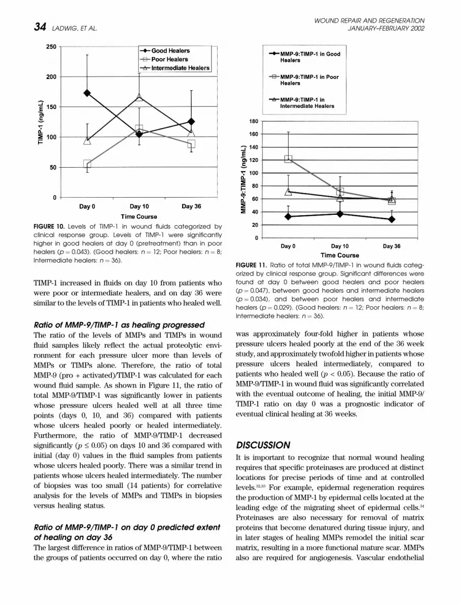
TIMP-1 increased in fluids on day 10 from patients who
were poor or intermediate healers, and on day 36 were
similar to the levels of TIMP-1 in patients who healed well.
Ratio of MMP-9/TIMP-1 as healing progressedThe ratio of the levels of MMPs and TIMPs in wound
fluid samples likely reflect the actual proteolytic envi-
ronment for each pressure ulcer more than levels of
MMPs or TIMPs alone. Therefore, the ratio of total
MMP-9 (pro + activated)/TIMP-1 was calculated for each
wound fluid sample. As shown in Figure 11, the ratio of
total MMP-9/TIMP-1 was significantly lower in patients
whose pressure ulcers healed well at all three time
points (days 0, 10, and 36) compared with patients
whose ulcers healed poorly or healed intermediately.
Furthermore, the ratio of MMP-9/TIMP-1 decreased
significantly (p £ 0.05) on days 10 and 36 compared with
initial (day 0) values in the fluid samples from patients
whose ulcers healed poorly. There was a similar trend in
patients whose ulcers healed intermediately. The number
of biopsies was too small (14 patients) for correlative
analysis for the levels of MMPs and TIMPs in biopsies
versus healing status.
Ratio of MMP-9/TIMP-1 on day 0 predicted extentof healing on day 36The largest difference in ratios of MMP-9/TIMP-1 between
the groups of patients occurred on day 0, where the ratio
was approximately four-fold higher in patients whose
pressure ulcers healed poorly at the end of the 36 week
study, and approximately twofold higher in patients whose
pressure ulcers healed intermediately, compared to
patients who healed well (p < 0.05). Because the ratio of
MMP-9/TIMP-1 in wound fluid was significantly correlated
with the eventual outcome of healing, the initial MMP-9/
TIMP-1 ratio on day 0 was a prognostic indicator of
eventual clinical healing at 36 weeks.
DISCUSSIONIt is important to recognize that normal wound healing
requires that specific proteinases are produced at distinct
locations for precise periods of time and at controlled
levels.32,33 For example, epidermal regeneration requires
the production of MMP-1 by epidermal cells located at the
leading edge of the migrating sheet of epidermal cells.34
Proteinases are also necessary for removal of matrix
proteins that become denatured during tissue injury, and
in later stages of healing MMPs remodel the initial scar
matrix, resulting in a more functional mature scar. MMPs
also are required for angiogenesis. Vascular endothelial
FIGURE 10. Levels of TIMP-1 in wound fluids categorized by
clinical response group. Levels of TIMP-1 were significantly
higher in good healers at day 0 (pretreatment) than in poor
healers (p ¼ 0.043). (Good healers: n ¼ 12; Poor healers: n ¼ 8;
Intermediate healers: n ¼ 36).FIGURE 11. Ratio of total MMP-9/TIMP-1 in wound fluids categ-
orized by clinical response group. Significant differences were
found at day 0 between good healers and poor healers
(p ¼ 0.047), between good healers and intermediate healers
(p ¼ 0.034), and between poor healers and intermediate
healers (p ¼ 0.029). (Good healers: n ¼ 12; Poor healers: n ¼ 8;
Intermediate healers: n ¼ 36).
WOUND REPAIR AND REGENERATIONJANUARY–FEBRUARY 200234 LADWIG, ET AL.

cells secrete MMPs that degrade the basement membrane
and enable new capillary loops to emerge.35 Other
proteinases are required for activation of latent growth
factors and for conversion of procollagen molecules to
tropocollagen molecules that can associate into collagen
fibers. However, prolonged, elevated levels of proteinases
appear to have detrimental effects on wound healing.
Furthermore, a reduction in MMP activities in fluids
collected from chronic venous ulcers was associated with
the initiation of healing in the ulcers,4 and a reduction in
plasminogen activator was associated with initiation of
healing in a pressure ulcer.20 At present there are no
reports correlating the rates of healing of chronic pressure
ulcers with the levels of MMPs and TIMPs in fluids. This
study measured levels of MMPs and TIMPs in sequential
fluids and biopsies from a series of 56 patients with
chronic pressure ulcers that were treated with conven-
tional therapy or with exogenous cytokine therapies and
found a strong correlation between a low ratio of
MMP-9/TIMP-1 and healing of the ulcers.
Previous reports provided data supporting the hypo-
thesis that elevated levels of cytokines and proteinases,
especially MMPs, contribute to the failure of wounds to
heal. For example, Tarnuzzer and Schultz36 reported that
proteinase levels (measured using Azocoll assay) in fluids
from chronic wounds were significantly elevated relative
to acute wound fluids. Similar results were reported by
Wysocki and colleagures6 and by Yager and colleagues10 by
gelatin zymography. Trengove et al.4 extended these
observations by showing that proteinase levels (measured
by Azocoll assay) in wound fluids from 15 chronic venous
ulcers significantly decreased 2 weeks after the ulcers
began to heal. The present study provides the strongest
data yet supporting this hypothesis by showing a strong
correlation between good healing and low ratios of
MMP-9/TIMP-1 in a large series of patients with chronic
pressure ulcers treated in a prospective, randomized,
masked study.23 However, this study, as well as the
previous studies, provides correlative data and does not
directly test the hypothesis that elevated proteinases
actively cause wounds to fail to heal. To answer that
question, future clinical studies need to be performed to
treat chronic wounds with selective proteinase inhibitors
and assess correlations in healing.
Previous studies provided important information
regarding proteinases and chronic wounds, yet several
important questions remain. For example, it is not known if
fluid samples and biopsies from chronic wounds contain
similar profiles and relative levels of MMPs and TIMPs, or
if data generated from analysis of wound fluids or biopsies
allow a better assessment of the molecular environment of
chronic wounds. Specifically, no direct comparisons of the
levels of MMPs and TIMPs in fluids and biopsies collected
from the same wounds have been reported. This study is
the first to compare gelatin zymogram profiles and
proteinase levels in both chronic wound fluids and biopsies
from pressure ulcer patients. As indicated by the gelatin
zymograms in Figure 3, both biopsies (panel A) and wound
fluids (panel B) typically contain intense gelatinolytic
bands corresponding to pro MMP-9 and activated MMP-9.
In contrast, wound fluids (panel B) typically have very faint
lysis bands corresponding to pro MMP-2 and activated
MMP-2 compared with the intensities of pro MMP-2 and
activated MMP-2 bands in biopsies (panel A). This band
pattern is reinforced by the quantitative data presented in
Figure 4, which show that the average levels of pro and
activated MMP-2 are significantly higher than the average
levels of pro and activated MMP-9 in the biopsies, while the
opposite relationship is found in wound fluids. Thus,
samples of wound fluids and biopsies do not have the same
profiles of MMP-2 and MMP-9.
Another important distinction between MMP profiles
of biopsies and wound fluid samples involves the activa-
tion of the latent forms of MMP-2 and MMP-9. Specifically,
as shown in Figures 3 and 4, biopsy samples contained
extremely low levels of activated MMP-9 in marked
contrast to wound fluid samples, in which more than
50% of the total MMP-9 was activated. The opposite trend
was observed for latent and activated MMP-2, with
substantial amounts of activated MMP-2 present in biopsy
samples (about 25% of total MMP-2 was activated MMP-2)
and very low levels of activated MMP-2 in wound fluid
samples. These data indicate that there are major differ-
ences between the MMP-2 and MMP-9 compositions of
wound fluids and biopsies. The explanations for these
differences are not known. However, if pro and activated
MMP-2 bind more tightly to ECM components than pro
and activated MMP-9, then their relative levels in biopsies
should be higher than in wound fluids. Extraction of the
biopsies with nonionic detergent may dissociate the pro
and activated MMP-2 from the matrix proteins, which
would increase their concentration in the biopsy extracts.
Interestingly, immunohistochemical staining for MMP-2
tends to be substantially more intense than for MMP-9 in
the ECM and fibroblasts for most biopsies (unpublished
observations).
Previous studies have used different assays to meas-
ure proteinase activities in wound fluid samples. We used
the Azocoll assay to measure proteinase activity in 40
wound fluid samples collected from various types of
chronic wounds and in 22 samples of acute wound fluids
collected from mastectomy drains.4 The average level of
proteinase activity in the chronic wound fluids was
60–71 lg/ml (median, 23 lg/ml), which was 30-fold higher
WOUND REPAIR AND REGENERATIONVOL. 10, NO. 1 LADWIG, ET AL. 35

than the average level of 0.8–0.3 lg/ml in the 22 acute
wound fluid samples (median, 0.8 lg/ml). The Azocoll
proteinase assay measures the combined activity of all
proteinases that have the ability to degrade ECM, using
bovine hide as the substrate. Thus, the Azocoll assay
preferentially measures gelatinase activity, but other
proteinases, such as neutrophil elastase, are able to
degrade the substrate. In addition, wound fluids were
collected in our previous study by covering the ulcers with
an occlusive dressing and allowing fluids to spontaneously
accumulate for 1 hour. In this study, porous, inert,
hydrophilic dextranomer beads were used to adsorb the
wound fluids over a 24-hour period. Nevertheless, the
combined levels of activated MMP-2 and activated MMP-9
activities reported in this current study using gelatin
zymography (approximately 5 lg/ml) are relatively similar
to the levels of proteolytic activity measured by Azocoll
assay in our previous study (60 lg/ml).
Yager and colleagues10 also used quantitative gelatin
zymography to analyze five samples of wound fluid
obtained from patients with pressure ulcers and five
samples of fluids collected from patients with acute
mastectomy wounds. The chronic wound fluids were
collected under an occlusive dressing over a 4–6-hour
period, and the average level of pro MMP-9 was
186 ± 59 ng/100 lg protein. Assuming the average protein
concentration of wound fluids collected under occlusive
dressings is approximately 40 mg/ml,37 the pro MMP-9
activities would equate to 74 ± 24 lg/100 lg protein, which
is similar to the level of 10 lg pro MMP-9/ml detected in our
current experiments. In addition, Yager and colleagues10
reported the average level of pro MMP-9 activity was
approximately 50-fold higher than the average level of pro
MMP-2 activity in the chronic wound fluids, which agrees
with our finding of about eightfold higher MMP-9 activity
than MMP-2 activity in the pressure ulcer wound fluids.
We previously proposed the hypothesis that the
presence of elevated levels of MMPs in chronic wounds
contributed to their failure to heal.21 Analysis of the data in
this study showed a statistically significant correlation
between elevated levels of activated MMP-9 and poor
healing, regardless of the treatment regimen that patients
received. Furthermore, combining information on the
levels of TIMP-1 and MMP-9 on wound fluids revealed
that the ratio of the TIMP and MMP-9 also showed a
statistically significant correlation between poor healing
and an elevated ratio of MMP-9/TIMP-1. Thus, the data
generated in this study provide strong additional support
for the concept that high levels of proteolytic activity and
low levels of MMP inhibitors are detrimental to healing.
However, these data are merely correlative and did not
provide direct evidence for a cause-and-effect relationship
between high levels of proteinases and poor wound
healing. Thus, there is a need to conduct the next stage
of investigation of this hypothesis. This could involve a
clinical study evaluating the effect of topical application of
an MMP inhibitor on healing of chronic wounds. Several
candidate inhibitors can be suggested, including the very
potent synthetic dipeptide MMP inhibitor, Ilomostat, or the
less potent but readily available antibiotic, doxycycline. In
addition to its antibiotic activity, doxycycline is a compet-
itive inhibitor of most MMPs.38 Ilomostat and doxycycline
(unpublished data) also inhibit the tumor necrosis factor-a(TNF-a)-converting enzyme, which is a metalloproteinase,
and prevent TNF-a release from cultures of inflammatory
cells that are stimulated by endotoxin.39 Thus, Ilomostat or
doxycycline could potentially act to reduce the levels of
both the pro-inflammatory cytokine, TNF-a and the activity
of MMPs if applied topically to chronic wounds.14 The data
generated in these series of experiments justify further
evaluation of these new strategies and could eventually
result in an extensive adjuvant therapy that could promote
healing of chronic wounds.
References1. Bennett NT, Schultz GS. Growth factors and wound healing: bio-
chemical properties of growth factors and their receptors. Am J Surg
1993;165:728–37.
2. Bennett NT, Schultz GS. Growth factors and wound healing. Part II.
Role in normal and chronic wound healing. Am J Surg 1993;166:74–81.
3. Nwomeh BC, Yager DR, Cohen IK. Physiology of the chronic wound.
Clin Plast Surg 1998;25:341–56.
4. Trengove NJ, Stacey MC, Macauley S, Bennett N, Gibson J, Burslem F,
Murphy G, Schultz G. Analysis of the acute and chronic wound envi-
ronments: the role of proteases and their inhibitors. Wound Rep Reg
1999;7:442–52.
5. Wysocki AB, Grinnell F. Fibronectin profiles in normal and chronic
wound fluid. Lab Invest 1990;63:825–31.
6. Wysocki AB, Staiano-Coico L, Grinnell F. Wound fluid from chronic
leg ulcers contains elevated levels of metalloproteinases MMP-2 and
MMP-9. J Invest Dermatol 1993;101:64–8.
7. Yager DR, Chen SM, Ward SI, Olutoye OO, Diegelmann RF, Cohen IK.
Ability of chronic wound fluids to degrade peptide growth factors is
associated with increased levels of elastase activity and diminished
levels of proteinase inhibitors. Wound Rep Reg 1997;5:23–32.
8. Harris IR, Yee KC, Walters CE, Cunliffe WJ, Kearney JN, Wood
EJ, Ingham E. Cytokine and protease levels in healing and
non-healing chronic venous leg ulcers. Exp Dermatol 1995;4: 342–
9.
9. Nwomeh BC, Liang HX, Cohen IK, Yager DR. MMP-8 is the predom-
inant collagenase in healing wounds and nonhealing ulcers. J Surg Res
1999;81:189–95.
10. Yager DR, Zhang LY, Liang HX, Diegelmann RF, Cohen IK. Wound
fluids from human pressure ulcers contain elevated matrix metallo-
proteinase levels and activity compared to surgical wound fluids.
J Invest Dermatol 1996;107:743–8.
11. Trengove NJ, Bielefeldt-Ohmann H, Stacey MC. Mitogenic activity and
cytokine levels in non-healing and healing chronic leg ulcers. Wound
Rep Reg 2000;8:13–25.
12. Bucalo B, Eaglstein WH, Falanga V. Inhibition of cell proliferation by
chronic wound fluid. Wound Rep Reg 1993;1:181–6.
WOUND REPAIR AND REGENERATIONJANUARY–FEBRUARY 200236 LADWIG, ET AL.

13. Wlaschek M, Pes D, Achterberg V, Meyer-Ingold W, Scharfetter-
Kochanek K. Protease inhibitors protect growth factor activity in
chronic wounds. Br J Dermatol 1997;137:646–7.
14. Weckroth M, Vaheri A, Lauharanta J, Sorsa T, Konttinen YT. Matrix
metalloproteinases, gelatinase, and collagenase in chronic leg ulcers.
J Invest Dermatol 1996;106:1119–24.
15. Rao CN, Ladin DA, Liu YY, Chilukuri K, Hou ZZ, Woodley DT.
a1-Antitrypsin is degraded and non-functional in chronic wounds but
intact and functional in acute wounds: the inhibitor protects fibro-
nectin from degradation by chronic wound fluid enzymes. J Invest
Dermatol 1995;105:572–8.
16. Bullen EC, Longaker MT, Updike DL, Benton R, Ladin D, Hou Z.
Tissue inhibitor of metalloproteinases-1 is decreased and activated
gelatinases are increased in chronic wounds. J Invest Dermatol
1995;104:236–40.
17. Rogers AA, Burnett S, Moore JC, Shakespeare PG, Chen WYJ.
Involvement of proeolytic enzymes, plasminogen activators and
matrix metalloproteinases in the pathophysiology of pressure ulcers.
Wound Rep Reg 1995;3:273–83.
18. Barone EJ, Yager DR, Pozez AL, Olutoye OO, Crossland MC,
Diegelmann RF, Cohen IK. Interleukin-1alpha and collagenase
activity are elevated in chronic wounds. Plastic Reconstr Surg
1998;102:1023–7.
19. Tarlton JF, Vickery CJ, Leaper DJ, Bailey AJ. Postsurgical wound
progression monitored by temporal changes in the expession of
matrix metalloproteinase-9. Br J Dermatol 1997;137:506–16.
20. Wysocki AB, Kusakabe AO, Chang S, Tuan TL. Temporal expression
of urokinase plasminogen activator, plasminogen activator inhibitor
and gelatinase-B in chronic wound fluid switches from a chronic to
acute wound profile with progression to healing. Wound Rep Reg
1999;7:154–65.
21. Mast BA, Schultz GS. Interactions of cytokines, growth factors, and
proteases inacuteandchronicwounds.WoundRepReg1996;4:411–20.
22. Yager DR, Nwomeh BC. The proteolytic environment of chronic
wounds. Wound Rep Reg 1999;7:433–41.
23. Robson MC, Hill DP, Smith PD, Wang X, Meyer-Siegler K, Ko F,
VandeBerg JS, Payne WG, Ochs D, Robson LE. Sequential cytokine
therapy for pressure ulcers: clinical and mechanistic response. Ann
Surg 2000;231:600–11.
24. Resch CS, Kerner E, Robson MC, Heggers JP, Scherer M, Boertman
JA, Schileru R. Pressure sore volume measurement. A technique
to document and record wound healing. J Am Geriatr Soc 1988;36:
444–6.
25. Cooper DM, Robson MC. In the evaluation of potential wound healing
agents a placebo arm is not nontreatment. Wound Rep Reg
1996;4:A128. [Abstract].
26. Matsuoka J, Grotendorst GR. Two peptides related to platelet-derived
growth factor are present in human wound fluid. Proc Natl Acad Sci
USA 1989;86:4416–20.
27. Fini ME, Girard MT. The pattern of metalloproteinase expression by
corneal fibroblasts is altered by passage in cell culture. J Cell Sci
1990;97(Pt. 2):373–83.
28. Hibbs MS, Hasty KA, Seyer JM, Kang AH, Mainardi CL. Biochemical
and immunological characterization of the secreted forms of human
neutrophil gelatinase. J Biol Chem 1985;260:2493–500.
29. Kleiner DE, Stetler-Stevenson WG. Quantitative zymography: detec-
tion of picogram quantities of gelatinases. Anal Biochem
1994;218:325–9.
30. Todor DR, Lewis I, Bruno G, Chyatte D. Identification of a serum
gelatinase associated with the occurrence of cerebral aneurysms as
pro-matrix metalloproteinase-2. Stroke 1998;29:1580–3.
31. Mauviel A. Cytokine regulation of metalloproteinase gene expression.
J Cell Biochem 1993;53:288–95.
32. Parks WC. Matrix metalloproteinases in repair. Wound Rep Reg
1999;7:423–32.
33. Barrick B, Campbell EJ, Owen CA. Leukocyte proteinases in wound
healing: roles in physiologic and pathologic processes. Wound Rep
Reg 1999;7:410–22.
34. Parks WC. The production, role, and regulation of matrix metallo-
proteinsases in the healing epidermis. Wounds 1995;7:23A–37A.
35. Fisher C, Gilbertson-Beadling S, Powers EA, Petzold G, Poorman R,
Mitchell MA. Interstitial collagenase is required for angiogenesis in
vitro. Dev Biol 1994;162:499–510.
36. Tarnuzzer RW, Schultz GS. Biochemical analysis of acute and chronic
wound environments. Wound Rep Reg 1996;4:321–5.
37. Trengove NJ, Langton SR, Stacey MC. Biochemical analysis of wound
fluid from nonhealing and healing chronic leg ulcers. Wound Rep Reg
1996;4:234–9.
38. Smith GN Jr, Mickler EA, Hasty KA, Brandt KD. Specificity of inhibi-
tion of matrix metalloproteinase activity by doxycycline: relationship
to structure of the enzyme. Arthritis Rheum 1999;42:1140–6.
39. Solorzano CC, Ksontini R, Pruitt JH, Auffenberg T, Tannahill C,
Galardy RE, Schultz GP, MacKay SL, Copeland EM III, Moldawer LL. A
matrix metalloproteinase inhibitor prevents processing of tumor
necrosis factor alpha (TNF alpha) and abrogates endotoxin-induced
lethality. Shock 1997;7:427–31.
WOUND REPAIR AND REGENERATIONVOL. 10, NO. 1 LADWIG, ET AL. 37


