Embed Size (px)
Citation preview

Biochemical Characterization of the CellularGlycosylphosphatidylinositol-linked Membrane Type-6Matrix Metalloproteinase*
Received for publication, January 22, 2010, and in revised form, March 2, 2010 Published, JBC Papers in Press, March 22, 2010, DOI 10.1074/jbc.M110.107094
Ilian A. Radichev, Albert G. Remacle, Sergey A. Shiryaev, Angela N. Purves, Sherida L. Johnson, Maurizio Pellecchia,and Alex Y. Strongin1
From the Sanford-Burnham Medical Research Institute, La Jolla, California 92037
Ubiquitously expressed membrane type-1 matrix metallo-proteinase (MT1-MMP), an archetype member of the MMPfamily, binds tissue inhibitor of metalloproteinases-2 (TIMP-2), activates matrix metalloproteinase-2 (MMP-2), and stimu-lates cell migration in various cell types. In contrast with MT1-MMP, the structurally similar MT6-MMP associates with thelipid raft compartment of the plasma membrane using a GPIanchor. As a result, MT6-MMP is functionally distinct fromMT1-MMP.MT6-MMP is insufficiently characterized as yet. Inaddition, a number of its biochemical features are both conflict-ing and controversial. To reassess the biochemical features ofMT6-MMP,wehave expressed theMT6-MMPconstruct taggedwith a FLAG tag in breast carcinoma MCF-7 and fibrosarcomaHT1080 cells.We thenusedphosphatidylinositol-specific phos-pholipase C to release MT6-MMP from the cell surface andcharacterized the solubilizedMT6-MMP fractions.We now areconfident that cellularMT6-MMPpartially exists in its complexwith TIMP-2. Both TIMP-1 and TIMP-2 are capable of inhibit-ing the proteolytic activity of MT6-MMP. MT6-MMP does notstimulate cell migration. MT6-MMP, however, generates a sig-nificant level of gelatinolysis of the fluorescein isothiocyanate-labeled gelatin and exhibits an intrinsic, albeit low, ability toactivate MMP-2. As a result, it is exceedingly difficult to recordthe activation of MMP-2 by cellular MT6-MMP. Because of itslipid raft localization, cellular MT6-MMP is inefficiently inter-nalized. MT6-MMP is predominantly localized in the cell-to-cell junctions. Because MT6-MMP has been suggested to play arole in disease, including cancer and autoimmunemultiple scle-rosis, the identity of its physiologically relevant cleavage targetsremains to be determined.
The members of the matrix metalloproteinase (MMP)2 fam-ily degrade a wide spectrum of extracellular matrix proteins,
growth factors, and cell receptors and play important roles inmultiple diseases (1–3). Inmalignancy,MMPs, especiallymem-brane-type MMPs (MT-MMPs), have been proposed to playkey roles in tumor invasion and metastasis (4). As a result, it isnow accepted that in depth mechanistic understanding of theMT-MMP functionality will ultimately lead to novel and effec-tive therapies against invasive, metastatic malignancies (5).The MT-MMP subfamily includes six members, named
MT1-MMP (MMP-14), MT2-MMP (MMP-15), MT3-MMP(MMP-16), MT4-MMP (MMP-17), MT5-MMP (MMP-24),and MT6-MMP (MMP-25). MT1-MMP, MT2-MMP, MT3-MMP, and MT5-MMP are anchored to the plasma membranevia a transmembrane domain (1, 4). In contrast, the attachmentof MT4-MMP and MT6-MMP to the plasma membrane takesplace via a glycosylphosphatidylinositol (GPI) moiety thatdirects these proteinases to the caveola-enriched lipid raft com-partment (6). Although there is a significant level of knowledgeof the structural-functional relationships and regulation ofMT1-MMP, an archetype member of the subfamily, exceed-ingly little is known about the biochemical and cellular proper-ties ofGPI-linkedMT4-MMPand especiallyMT6-MMP.Orig-inally, MT6-MMP was cloned from a fetal liver cDNA libraryand from leukocytes (7–9). Because of its abundance in leuko-cytes, the protease has also been called leukolysin. MT6-MMPwas also found to be abnormally expressed by colon, prostate,urothelial, and brain tumors, suggesting its implication in thediversified malignancies (10). MT6-MMP is a membrane pro-teinase with an extracellular prodomain followed by the cata-lytic domain, the hinge region, the hemopexin domain, the stalkregion, and a GPI anchor attached to the carboxyl end of thestalk. Similar to MT1-MMP, MT6-MMP is synthesized as aninactive precursor that is transformed into the functionallyactive proteinase by the cleavage action of furin-like proproteinconvertases (1, 6).The regulationmechanisms, the functional role, and the rep-
ertoire of physiologically relevant cleavage targets of MT6-MMP remain largely unknown. In addition to a limited numberof extracellular matrix components, including fibronectin, gel-atin, type IV collagen, and chondroitin and dermatan sulfate
* This work was supported, in whole or in part, by National Institutes of HealthGrants CA83017 and CA77470 (to A. Y. S.).
1 To whom correspondence should be addressed. E-mail: [email protected].
2 The abbreviations used are: MMP, matrix metalloproteinase; AAT, �1-an-titrypsin; GPI, glycosylphosphatidylinositol; DAPI, 4�,6-diamidino-2-phenylindole; EZ-link sulfo-NHS-SS-biotin, sulfosuccinimidyl-2-(bio-tinamido)ethyl-1,3-dithiopropionate; HT cells, human fibrosarcomaHT1080 cells; Mca-PLGL-Dpa-AR-NH2, methoxycoumarin-4-yl)-acetyl-Pro-Leu-Gly-Leu-(3-[2,4-dinitrophenyl]-l-2,3-diaminopropionyl)-Ala-Arg-NH2; MCF7 cells, human breast carcinoma MCF-7 cells; MESNA, 2-mercap-toethane sulfonic acid; MT-MMP, membrane-type MMP; MT1-MMP andMT6-MMP, membrane type-1 and membrane type-6 MMP, respectively;
MT1CAT and MT6CAT, the individual catalytic domain of MT1-MMP andMT6-MMP, respectively; MT6F, MT6-MMP tagged with a FLAG tag; PLC,phosphatidylinositol-specific phospholipase C; TIMP, tissue inhibitor ofmetalloproteinases; DMEM, Dulbecco’s modified Eagle’s medium; LC, liq-uid chromatography; MS/MS, tandem mass spectrometry; FITC, fluores-cein isothiocyanate; RIPA, radioimmune precipitation assay.
THE JOURNAL OF BIOLOGICAL CHEMISTRY VOL. 285, NO. 21, pp. 16076 –16086, May 21, 2010© 2010 by The American Society for Biochemistry and Molecular Biology, Inc. Printed in the U.S.A.
16076 JOURNAL OF BIOLOGICAL CHEMISTRY VOLUME 285 • NUMBER 21 • MAY 21, 2010
by guest on January 25, 2016http://w
ww
.jbc.org/D
ownloaded from

proteoglycan, the catalytic domain of MT6-MMP has beenreported to cleave galectin-3, urokinase plasminogen activatorreceptor and myelin basic protein (6, 11, 12). The relevance ofMT6-MMP proteolysis of these target proteins to the enzymefunctionality in both normal development and disease progres-sion, however, is not entirely clear as of now.It is widely accepted that tissue inhibitors of MMPs (TIMPs)
play an important role in the regulation of the net proteolyticactivity of MMPs (13). Four individual TIMPs, includingTIMP-1 andTIMP-2, are known in humans (14). There are twodomains in both TIMP-1 andTIMP-2. The inhibitoryN-termi-nal domain directly interacts, albeit with different affinity, withthe active site of the MMP catalytic domain. The C-terminaldomain of TIMP-1 forms a stoichiometric complex with thehemopexin domain of theMMP-9 proenzyme. TIMP-2 forms asimilar complex with the MMP-2 proenzyme. TIMP-2 per-forms not only as an inhibitor but also as an essential compo-nent of theMT1-MMP-dependent activation pathway, leadingto the MMP-2 mature enzyme (4, 15–18).The existing data suggest that both TIMP-1 and TIMP-2
inhibit the proteolytic activity of the individual catalyticdomain of MT6-MMP (6, 10, 19). There are conflicting resultsthat point to the potential role of MT6-MMP in the mecha-nisms of MMP-2 activation (6, 20). Cellular MT6-MMP, how-ever, was not observed in a complex with either TIMP-1 orTIMP-2, thus raising a question of how the activity of MT6-MMP is regulated (6, 21). To date, all of the biochemical studieshave employed the individual catalytic domain rather than thefull-length MT6-MMP enzyme or its short C-terminal trunca-tions (6, 9, 10, 12, 19). As a result, the question of whether otherdomains of MT6-MMP do or do not affect the functionality ofthe proteinase remains to be answered.To understand better the main parameters of the MT6-
MMP functionality, we biochemically characterized the MT6-MMP samples directly isolated from the cells and reexaminedthe ability of cellular MT6-MMP to associate with TIMP-2, toactivate MMP-2, and to function as an invasion-promoting,matrix-degrading proteinase. Because the current knowledgeofMT6-MMP is exceedingly limited, we believe that themech-anistic observations we generated shed additional light on thefunction of the lipid raft-associated, GPI-linked MT6-MMP incancer.
MATERIALS AND METHODS
General Reagents and Antibodies—All reagents werepurchased from Sigma unless indicated otherwise. EZ-linksulfo-NHS-SS-biotin was from Pierce. (7-Methoxycoumarin-4-yl)-acetyl-Pro-Leu-Gly-Leu-(3-[2,4-dinitrophenyl]-l-2,3-diaminopropionyl)-Ala-Arg-NH2 (Mca-PLGL-Dpa-AR-NH2)was obtained from R&D Systems. GM6001 (a potent, widerange hydroxamate inhibitor of MMPs, including MT1-MMP)and the rabbit MT1-MMP (anti-hinge) antibody were pur-chased fromChemicon.Amurinemonoclonal E-cadherin anti-body (catalog no. 610181) was purchased from BD Biosciences.Purified TIMP-1 and a rabbit MT6-MMP antibody were a kindgift from Dr. Rafael Fridman (Wayne State University, Detroit,MI). Recombinant TIMP-2 and the TIMP-2-free MMP-2proenzyme were isolated from the conditioned medium of
CHO cells and p2AHT2A72 cells, respectively. p2AHT2A72cells were derived from the fibrosarcoma HT1080 cell linesequentially transfected with E1A and MMP-2 cDNAs,respectively (22, 23). The sheep TIMP-2 antibody was fromAbcam. The secondary antibodies were purchased from Jack-son ImmunoResearch. The secondary species-specific antibod-ies conjugatedwithAlexa Fluor 594 (red) and greenAlexa Fluor488 (green) were obtained at Molecular Probes. Human �1-an-titrypsin (AAT) and a protease inhibitor mixture set III werefrom Calbiochem.Recombinant MMPs—The individual catalytic domain of
MT6-MMP (MT6CAT) and MT1-MMP (MT1CAT) were ex-pressed in Escherichia coli, purified from the inclusion bodies,and refolded to restore its catalytic activity (11). The recombi-nant pro-forms of the catalytic domain of MMP-2 andMMP-9were purified from the serum-free medium conditioned bystably transfected HEK293 cells and then activated using4-aminophenylmercuric acetate (24). The MMP activity wasdetermined using the fluorescent Mca-PLGL-Dpa-AR-NH2peptide substrate. The concentrations of the catalytically activeMT6CAT, MT1CAT, MMP-2, andMMP-9 were measured bytitration against a standard GM6001 solution of a knownconcentration (11, 12, 25).Cloning of FLAG-tagged MT6-MMP (MT6F)—The full-
length human MT6-MMP cDNA gene (GenBankTM NM_022468) was a gift from Dr. Rafael Fridman (Wayne State Uni-versity). The sequence coding for a FLAG tag was insertedbetweenGly-514 and Pro-515 in the stalk region ofMT6-MMPto generate theMT6F construct. The authenticity of the recom-binant constructs was confirmed by DNA sequencing. MT6Fwas recloned into the pcDNA3.1D/V5-His-TOPO-neo plasmid.Cells—Human breast carcinoma MCF-7 cells (MCF7 cells)
and human fibrosarcoma HT1080 cells (HT cells) were ob-tained from the ATCC (Manassas, VA). HT and MCF7 cellsstably transfected with MT6F in the pcDNA3.1D/V5-His-TOPO-neo plasmid (MCF7-MT6F and HT-MT6F cells) andcontrol cells transfected with the original pcDNA3.1D/V5-His-neo plasmid (MCF7-mock and HT-mock cells) were ob-tained as described earlier (26). To avoid clonal effects, 5–6neomycin-resistant MT6F cell clones were combined and usedin our further studies. HT and MCF7 cells stably transfectedwith MT1-MMP (HT-MT1 and MCF7-MT1 cells, respec-tively) were isolated and characterized earlier (27). Cells wereroutinely cultured in high glucose DMEM supplemented with10% fetal bovine serum, 100 units/ml penicillin, and 100 �g/mlstreptomycin.Fractionation of Cellular MT6-MMP—Confluent cells (1 �
108) were detached using an enzyme-free cell dissociation solu-tion (Chemicon). Cells were suspended in serum-free DMEMand incubated for 2 h at 37 °C with 0.25 units/ml phosphatidy-linositol-specific phospholipase C (PLC). Cells were collectedby centrifugation at 300 � g for 5 min. The supernatant (PLCfraction) was clarified by centrifugation at 20,000 � g, 20 min.The following steps were performed at 4 °C. The collected cellswere lysed in 10ml of 20 mMTris-HCl, pH 7.9, 150 mMNaCl, 5mM MgCl2, 10% glycerol, a protease inhibitor mixture set III, 1mM phenylmethylsulfonyl fluoride, and 0.5% Nonidet P-40(Nonidet P-40 buffer). The extract was centrifuged (40 min;
Characterization of MT6-MMP
MAY 21, 2010 • VOLUME 285 • NUMBER 21 JOURNAL OF BIOLOGICAL CHEMISTRY 16077
by guest on January 25, 2016http://w
ww
.jbc.org/D
ownloaded from

20,000 � g). The supernatant (Nonidet P-40 fraction; 20–40mg of total protein) and the PLC fractionwere incubated for 4 hwith a 50% FLAG M2 antibody-bead slurry (40 �l). The beadswere collected by centrifugation and washed six times in theabove buffer but with 0.1% Nonidet P-40 instead of 0.5% Non-idet P-40. The beads were next incubated for 1 h with 20 �l ofFLAG peptide (0.2 mg/ml) to elute the FLAG-containing con-structs. The beads were removed by centrifugation. The super-natant samples were analyzed further using SDS-PAGE fol-lowed by silver staining and LC/MS/MS; by Western blottingwith the FLAG M2 (dilution 1:4,000), MT6-MMP (dilution1:1,500), and TIMP-2 (dilution 1:1,000) antibodies; and by theenzymatic activity assays. The species-specific peroxidase-conju-gated secondary antibodies (dilution 1:3,000) and a SuperSignalWest Dura extended duration substrate Kit (Pierce) were usedfor the detection of the immunopositive protein bands.Mass Spectrometry—Following SDS-PAGE, the MT6F-PLC
and MT6F-Nonidet P-40 bands were excised and subjected toan in-gel tryptic digest. The digest peptides were extracted andthen identified by LC/MS/MS using an LTQ XL Linear IonTrap mass spectrometer (Thermo Scientific). MS/MS spectrawere searched against the Swiss-Prot database using theSEQUEST Sorcerer software. The search led to the multiplepeptides the sequence of which permitted an unambiguousidentification of MT6-MMP.Cell Surface Biotinylation and 2-Mercaptoethane Sulfonic
Acid (MESNA) Treatment—Cells were surface-biotinylatedusing membrane-impermeable EZ-Link sulfo-NHS-SS-biotin(0.3 mg/ml), lysed in 0.5% Nonidet P-40 or RIPA buffer (20 mM
Tris-HCl, pH 7.4, containing 150 mM NaCl, 0.1% SDS, 1% Tri-ton X-100 and 1% sodium deoxycholate). The biotin-labeledplasma membrane proteins were pulled down using streptavi-din-beads (27). The precipitates were dissolved in SDS samplebuffer (0.125 M Tris-HCl, pH 6.8, 20% glycerol, 2% SDS, 0.005%bromphenol blue, and 5% 2-mercaptoethanol) and analyzed byWestern blotting with the MT1-MMP and MT6-MMP anti-body, followed by species-specific secondary antibodies conju-gated with horseradish peroxidase and a TMB/M substrate.For the uptake experiments, immediately following the com-
pletion of the biotinylation procedure, the biotinylated cellswere incubated for 30–60 min at 37 °C in serum-free DMEMsupplemented with 1% insulin/transferrin/selenium to allowthe internalization of biotin-labeled MT1-MMP (27, 28). Toremove the residual cell surface biotin, cells were incubated for25 min on ice in Sorenson phosphate buffer (14.7 mMKH2PO4,2 mM Na2HPO4, 120 mM sorbitol, pH 7.8) containing mem-brane-impermeable MESNA (150 mM). Cells were next exten-sively washed and lysed, and the lysates were precipitated usingstreptavidin-beads and analyzed as above.Gelatin Zymography—Cells (2� 105) were seeded for 24 h in
DMEM plus 10% fetal bovine serum in wells of a 12-well plate.The medium was then replaced with serum-free DMEM (0.35ml/well) supplemented with pro-MMP-2 (0.5 nM). When indi-cated, TIMP-1 or TIMP-2 (0.5–100 nM each) or GM6001 (50�M) was added. In 18 h, the medium aliquots (25 �l) were ana-lyzed by gelatin zymography in 0.1% gelatin, 10% acrylamidegels (18). 15% gelatin gels were used to detect the gelatinolyticactivity of the purified 25-kDa MT6CAT construct.
In the in vitro assays, pro-MMP-2 (15 nM) was co-incubatedfor 2 h at 37 °C with the purified MT1CAT andMT6CAT con-structs (0.15–15 nM) in 50 mM HEPES, pH 7.5, containing 10mM CaCl2 and 50 �M ZnCl2. Where indicated, GM6001 (5 �M)was added to the reactions. The digest aliquots were then ana-lyzed by gelatin zymography.Cleavage of AAT—The cleavage reactions (22 �l each) were
performed in 50 mM HEPES, pH 7.5, containing 10 mM CaCl2and 50 �M ZnCl2. AAT (1.6 �M) was co-incubated for 3 h at37 °C with MT1CAT and MT6CAT (16 nM each; 1:100enzyme/substrate molar ratio), and cellular MT6F was isolatedfrom the PLC fraction (16–80 nM; 1:20–1:100 enzyme/sub-stratemolar ratio). The cleavage reactions were stopped using a5� SDS sample buffer and analyzed by SDS-PAGE followed byCoomassie staining. Where indicated, GM6001 (5 �M) wasadded to the reactions to inhibit MMP activity.Enzymatic Assay—MMP activity was measured in wells of a
96-well plate in 0.2 ml of 50 mM HEPES, pH 7.5, containing 10mM CaCl2 and 50 �M ZnCl2. Mca-PLGL-Dpa-AR-NH2 (5 �M)was used as a fluorescent substrate. The concentration ofMMPs in the reactions was 20–600 fmol. The steady-state rateof substrate hydrolysis was monitored continuously (�ex � 320nm and �em � 400 nm) at 37 °C for 3–75 min using a fluores-cence spectrophotometer. Where indicated, TIMP-1 (2.5–50nM), TIMP-2 (2.5–25 nM), and GM6001 (1 �M) were co-incu-bated for 30 min at ambient temperature with the MMP sam-ples prior to the addition of the substrate. The samples weremeasured in triplicate. The results were highly reproduciblewithout any significant day to day variations.A Library of the Potential MMP Inhibitors—Our prototype
CFL-1 (chelator fragment library-1) was described earlier(29). Our extended library now includes �500 potential MMPinhibitors.Determination of the IC50 Values of Inhibitors—MT1CAT,
MT6CAT, MMP-2, and MMP-9 (10 nM each) were preincu-bated for 30 min at ambient temperature with increasing con-centrations of the individual compounds from theMMP inhib-itor library. The residual activity of MMPs was then measuredusing Mca-PLGL-Dpa-AR-NH2. IC50 values of the inhibitorswere calculated using GraphPad Prism as a fitting software.Synthesis of BI-92G11 and BI-102C8—A solution of the
corresponding acid (0.200 g, 0.933 mmol) and dimethylfor-mamide (0.053 g, 0.933 mmol) in CH2Cl2 (5 ml) was cooledto 0 °C. Oxalyl chloride (2.05 mmol) was then added slowly.Vigorous gas evolution was observed. After stirring for 2 h at0 °C, this solution was added to a solution of hydroxylaminehydrochloride (0.259 g, 3.73 mmol) and triethylamine (0.566g, 5.60 mmol) in tetrahydrofuran (5 ml)/H2O (1 ml). Afterstirring for an additional 4 h at ambient temperature, themixture was poured into 2 N HCl and extracted with CH2Cl2.The organic phase was dried over Na2SO4 and evaporated invacuo. The residue was recrystallized from aqueous ethanolor purified via flash chromatography (30). BI-92G11: 1HNMR (500 MHz, CDCl3) �10.56 (s, 1H), 8.89 (brs, 1H), 7.89(d, 2H, J � 7.0 Hz), 7.75 (t, 1H, J � 7.0 Hz), 7.66 (t, 2H, J � 7.0Hz), 3.49 (m, 2H), 3.03 (m, 2H). HRMS e/z (electrospray ioni-zation time-of-flight). Found 184.0423 [MH]�, C8H9NO2Srequires 184.0423. BI-102C8: 1H NMR (600 MHz, (CD3)2SO)
Characterization of MT6-MMP
16078 JOURNAL OF BIOLOGICAL CHEMISTRY VOLUME 285 • NUMBER 21 • MAY 21, 2010
by guest on January 25, 2016http://w
ww
.jbc.org/D
ownloaded from
�10.72 (s, 1H), 8.95 (s, 1H), 7.48 (t, 1H, J � 7.5), 7.37–7.33 (m,5H), 6.95–6.93 (d, 2H, J � 8.5), 4.07–4.04 (q, 2H, J � 6.9),1.36–1.34 (t, 3H, J � 6.9).CellMigration—Cellmigration experimentswere performed
in wells of a 24-well, 8-�m pore size, Transwell plate (CorningCostar). A 6.5-mm insert membrane was coated with 100 �l oftype I collagen (100 �g/ml in DMEM) and then dried for 16 h.The collagen coating was rehydrated in 0.5 ml of DMEM for 30min before the experiments. The inner chamber containedDMEM supplemented with 10% fetal bovine serum as che-moattractant. Cells (1� 104) were seeded in the outer chamberin serum-free DMEM. GM6001 (25 �M) or DMSO alone(0.01%) were added to both inner and outer chambers 15 minbefore plating the cells. Cells were allowed to migrate for 3.5 hand then stained with 20% methanol, 0.2% Crystal Violet. Cellsfrom the upper membrane surface were removed with a cottonswab. Incorporated dye from the migrated cells was extractedusing 0.25 ml of 1% SDS. The A570 value of the extract wasmeasured. Data aremeans� S.E. from several individual exper-iments performed in triplicate. Statistical analysis was per-formed by the two-tailed unpaired t test.In SituGelatin ZymographyUsing FITC-gelatin—Toprepare
FITC-gelatin (31), gelatin (2 mg/ml) was conjugated for 2 h atambient temperature to 100 �g/ml FITC in 0.1 M carbonate-bicarbonate buffer, pH 9.1. Unreacted dye was removed by gelfiltration through a G-25 SephadexM column (GEHealthcare)equilibrated in PBS. The A280 and A494 values of the fractionswere determined. The fractions with an FITC/protein molarratio of �3 were used further (32). Sterilized 15-mm glass cov-erslips were coated for 2 h at 37 °C with 100 �l of FITC-gelatin
(100 �g/ml). The excess of gelatinwas removed by washing with pre-warmed DMEM. Cells (2 � 104)were seeded onto the gelatin-coatedcoverslips and incubated for 16 h at37 °C in DMEM with or withoutGM6001 (50 �M). The cells werethen fixed with 4% paraformalde-hyde for 10 min andmounted in theVectashieldmedium (Vector Labora-tories) containing 4�,6-diamidino-2-phenylindole (DAPI) for the nuclearstaining. The slides were then ana-lyzed using an Olympus BX51 fluo-rescentmicroscope equipped with aMagnaFire digital camera.Immunostaining of Cells—Cells
were fixed for 10min in4%paraform-aldehyde, permeabilized using 0.1%TritonX-100, washedwith PBS con-taining 0.1% Tween 20, and blockedfor 1 h in 1% casein. Cells werethen stained for 1 h at ambienttemperature using the rabbit anti-body toMT6-MMP (dilution 1:250)and the monoclonal murine anti-body to E-cadherin (dilution 1:1,000),followed by incubation for 1 h with
the secondary antibodies (dilution 1:200) conjugated withAlexa Fluor 594 or Alexa Fluor 488, respectively. The slideswere mounted in the Vectashield medium with DAPI. Imageswere acquired at an original magnification of �600 using anOlympus BX51 fluorescence microscope equipped with aMagnaFire digital camera.
RESULTS
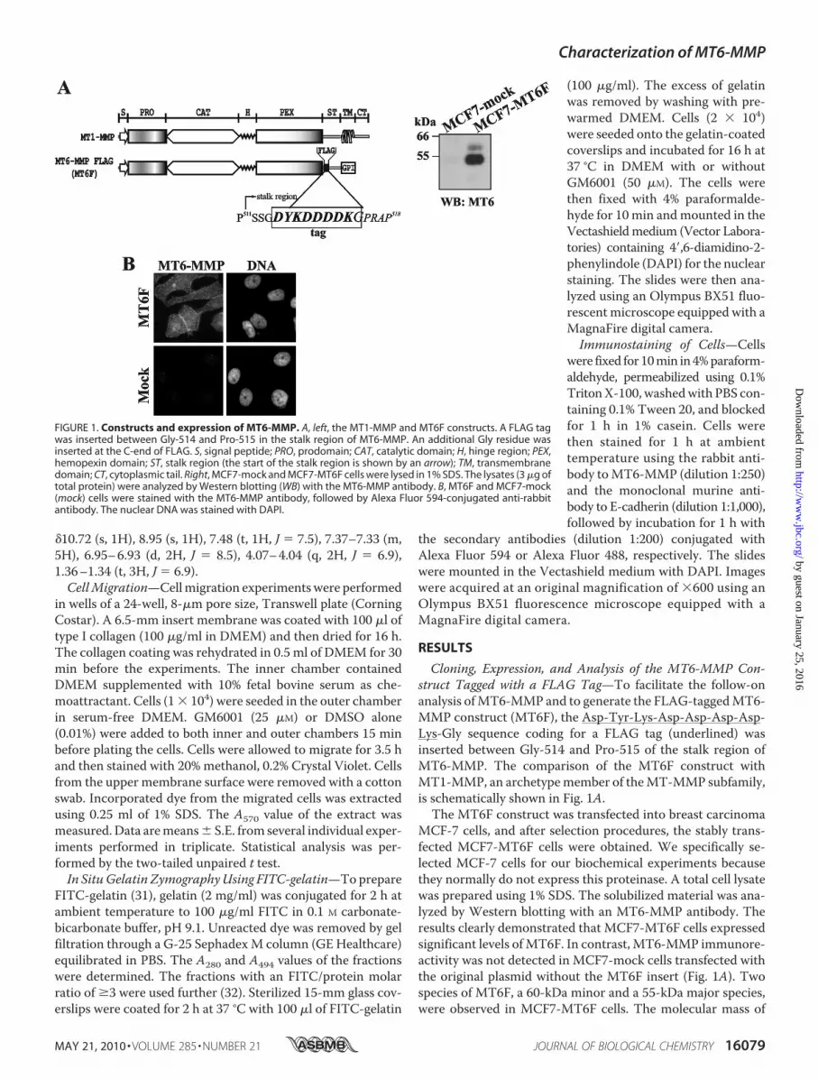
Cloning, Expression, and Analysis of the MT6-MMP Con-struct Tagged with a FLAG Tag—To facilitate the follow-onanalysis ofMT6-MMP and to generate the FLAG-taggedMT6-MMP construct (MT6F), the Asp-Tyr-Lys-Asp-Asp-Asp-Asp-Lys-Gly sequence coding for a FLAG tag (underlined) wasinserted between Gly-514 and Pro-515 of the stalk region ofMT6-MMP. The comparison of the MT6F construct withMT1-MMP, an archetypemember of theMT-MMP subfamily,is schematically shown in Fig. 1A.The MT6F construct was transfected into breast carcinoma
MCF-7 cells, and after selection procedures, the stably trans-fected MCF7-MT6F cells were obtained. We specifically se-lected MCF-7 cells for our biochemical experiments becausethey normally do not express this proteinase. A total cell lysatewas prepared using 1% SDS. The solubilized material was ana-lyzed by Western blotting with an MT6-MMP antibody. Theresults clearly demonstrated that MCF7-MT6F cells expressedsignificant levels ofMT6F. In contrast, MT6-MMP immunore-activity was not detected in MCF7-mock cells transfected withthe original plasmid without the MT6F insert (Fig. 1A). Twospecies of MT6F, a 60-kDa minor and a 55-kDa major species,were observed in MCF7-MT6F cells. The molecular mass of
FIGURE 1. Constructs and expression of MT6-MMP. A, left, the MT1-MMP and MT6F constructs. A FLAG tagwas inserted between Gly-514 and Pro-515 in the stalk region of MT6-MMP. An additional Gly residue wasinserted at the C-end of FLAG. S, signal peptide; PRO, prodomain; CAT, catalytic domain; H, hinge region; PEX,hemopexin domain; ST, stalk region (the start of the stalk region is shown by an arrow); TM, transmembranedomain; CT, cytoplasmic tail. Right, MCF7-mock and MCF7-MT6F cells were lysed in 1% SDS. The lysates (3 �g oftotal protein) were analyzed by Western blotting (WB) with the MT6-MMP antibody. B, MT6F and MCF7-mock(mock) cells were stained with the MT6-MMP antibody, followed by Alexa Fluor 594-conjugated anti-rabbitantibody. The nuclear DNA was stained with DAPI.
Characterization of MT6-MMP
MAY 21, 2010 • VOLUME 285 • NUMBER 21 JOURNAL OF BIOLOGICAL CHEMISTRY 16079
by guest on January 25, 2016http://w
ww
.jbc.org/D
ownloaded from

these species correlated well with the expected size of theproenzyme and the mature enzyme of MT6F, suggesting thatthe cellular furin-like proprotein convertases processed the denovo synthesized MT6F construct during its trafficking to thecell surface. MT6-MMP immunoreactivity was predominantlylocalized in the cell-cell contact regions (Fig. 1B).To support the predominant presence of MT6-MMP at the
cell-cell contact regions, we then performed immunostainingof MCF7-MT6F cells using the antibodies to MT6-MMP andE-cadherin (the classic homophilic adhesion molecule that isnormally present in cell-cell junctions) (33, 34). There was aclear co-localization ofMT6F with E-cadherin in cell-cell junc-tion regions in MCF7-MT6F cells (Fig. 2). The functional sig-nificance of the predominant association of MT6-MMP withthe cell-cell junctions remains to be identified.Fractionation of Cellular MT6-MMP—To analyze cell com-
partmentation of MT6F in more detail, we used several pull-down and detection procedures. Thus, cells were surface-biotinylated and then extracted using the Nonidet P-40buffer and, alternatively, the RIPA buffer. The concentrationof 0.5% Nonidet P-40 in the Nonidet P-40 buffer was insuffi-cient to solubilize the lipid rafts and to release the GPI-an-
chored cellular MT6F. In turn, theRIPA buffer solubilized well thelipid raft-associated MT6F. The ex-tracted Nonidet P-40 and RIPAsamples were precipitated using theFLAG M2 antibody and streptavi-din-beads, respectively.MCF7-MT6F cells were also co-
incubated with PLC to destroy theGPI linker and to liberate cell sur-face-associated MT6F. The solubi-lized fraction and the residual cellswere separatedbycentrifugation.Thecell samples were then extractedwiththe Nonidet P-40 buffer. Both theNonidet P-40 extract and the PLC-solubilized samples were precipi-tated using the FLAG M2 antibody-beads. The samples were thenanalyzed usingWestern blotting withFLAG M2 and MT6-MMP antibod-ies.MCF-7 cells thatwere transfectedwith MT1-MMP (MCF7-MT1 cells)were used as an additional control.Because the mature MT6-MMP
enzyme was associated with the lipidrafts, we expected that the NonidetP-40 extraction procedure wouldpredominantly liberate the intracel-lular pool of MT6-MMP. The anal-ysis of both the PLC and the Non-idet P-40 samples supported thissuggestion. Thus, theWestern blot-ting analysis with the FLAG M2antibody detected the presence ofthe 55-kDa mature enzyme and
40-kDa degraded forms in the PLC-extracted samples. In turn,the 60-kDa proenzyme was detected in the Nonidet P-40 sam-ples in addition to the 55-kDa enzyme and the minor amountsof the 40-kDa proteolyzed forms of MT6-MMP. The level ofdegradedMT6-MMPwas minor relative to that of MT1-MMPin both HT1080 and MCF-7 cells (Fig. 3).Overall, our results imply that, as MT1-MMP, MT6-MMP
is activated by the furin-like proprotein convertases duringits trafficking to the cell surface. As a result, MT6-MMP ispredominantly represented at the cell surface by the lipidraft-associated mature enzyme and its proteolyzed forms,whereas the Nonidet P-40 extracted material included theresidual amounts of the intracellular pool of the MT6-MMPproenzyme.TIMP-2 Is Associated with Cellular MT6-MMP—To
determine also if TIMP-2 was present in the cellular MT6Fsamples, MCF7-MT6F, MCF7-MT1, and MCF7-mock cellswere extracted using the Nonidet P-40 buffer. The extractswere immunoprecipitated using the FLAGM2 antibody-beads.The precipitated MT6F samples clearly displayed the presenceof TIMP-2, suggesting that cellular MT6F can form a complexwith this inhibitor. In contrast, neither FLAG nor TIMP-2
FIGURE 2. Co-localization of MT6-MMP with E-cadherin in MCF7-MT6F cells. Cells were stained with theMT6-MMP and E-cadherin antibodies (red and green, respectively). The nuclei were stained with DAPI. Thearrows point to the cell-cell junction regions, in which MT6-MMP and E-cadherin are co-localized. The slideswere observed using a fluorescent microscope (magnification, �600).
FIGURE 3. Analysis of cellular MT6-MMP. Left, MCF7-MT6F cells were treated with PLC followed by NonidetP-40 extraction of the cells. The PLC and Nonidet P-40 fractions were precipitated (IP) using the FLAG M2antibody-beads. The precipitates were analyzed by Western blot (WB) with the FLAG M2 antibody. Middle leftpanel, MCF7-mock, MCF7-MT6F, and MCF7-MT1 cells were surface-biotinylated. Cells were lysed in RIPA buffer.The biotin-labeled proteins were pulled down using streptavidin-beads. The samples were analyzed usingWestern blot with the MT6-MMP antibody. Middle right panels, MCF7-mock, MCF7-MT6F, and MCF7-MT1 cellswere extracted with Nonidet P-40. The extracts were precipitated using the FLAG M2 antibody-beads. Theprecipitates were analyzed by WB with the MT6-MMP (MT6; top) and TIMP-2 (bottom) antibodies. Right panel,HT-mock, HT-MT1, MCF7-mock, and MCF7-MT1 cells were surface-biotinylated. Cells were lysed in RIPA buffer.The biotin-labeled proteins were pulled down using streptavidin-beads. The samples were analyzed usingWestern blot with the MT1-MMP antibody.
Characterization of MT6-MMP
16080 JOURNAL OF BIOLOGICAL CHEMISTRY VOLUME 285 • NUMBER 21 • MAY 21, 2010
by guest on January 25, 2016http://w
ww
.jbc.org/D
ownloaded from
immunoreactivity was observed if theMCF7-mock andMCF7-MT1 samples were analyzed (Fig. 3).Mass Spectrometry Analysis of the Isolated MT6-MMP
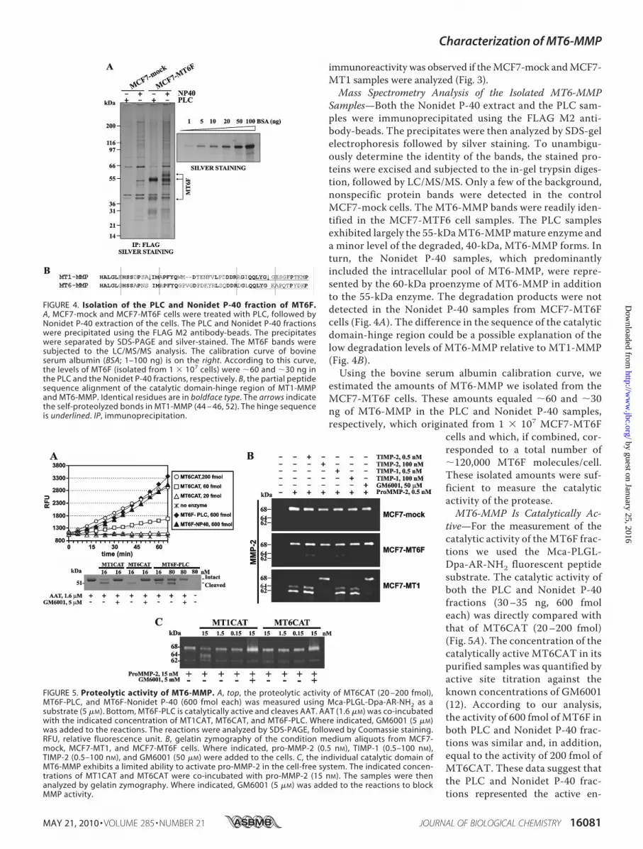
Samples—Both the Nonidet P-40 extract and the PLC sam-ples were immunoprecipitated using the FLAG M2 anti-body-beads. The precipitates were then analyzed by SDS-gelelectrophoresis followed by silver staining. To unambigu-ously determine the identity of the bands, the stained pro-teins were excised and subjected to the in-gel trypsin diges-tion, followed by LC/MS/MS. Only a few of the background,nonspecific protein bands were detected in the controlMCF7-mock cells. The MT6-MMP bands were readily iden-tified in the MCF7-MTF6 cell samples. The PLC samplesexhibited largely the 55-kDaMT6-MMPmature enzyme anda minor level of the degraded, 40-kDa, MT6-MMP forms. Inturn, the Nonidet P-40 samples, which predominantlyincluded the intracellular pool of MT6-MMP, were repre-sented by the 60-kDa proenzyme of MT6-MMP in additionto the 55-kDa enzyme. The degradation products were notdetected in the Nonidet P-40 samples from MCF7-MT6Fcells (Fig. 4A). The difference in the sequence of the catalyticdomain-hinge region could be a possible explanation of thelow degradation levels of MT6-MMP relative to MT1-MMP(Fig. 4B).Using the bovine serum albumin calibration curve, we
estimated the amounts of MT6-MMP we isolated from theMCF7-MT6F cells. These amounts equaled �60 and �30ng of MT6-MMP in the PLC and Nonidet P-40 samples,respectively, which originated from 1 � 107 MCF7-MT6F
cells and which, if combined, cor-responded to a total number of�120,000 MT6F molecules/cell.These isolated amounts were suf-ficient to measure the catalyticactivity of the protease.MT6-MMP Is Catalytically Ac-
tive—For the measurement of thecatalytic activity of theMT6F frac-tions we used the Mca-PLGL-Dpa-AR-NH2 fluorescent peptidesubstrate. The catalytic activity ofboth the PLC and Nonidet P-40fractions (30–35 ng, 600 fmoleach) was directly compared withthat of MT6CAT (20–200 fmol)(Fig. 5A). The concentration of thecatalytically active MT6CAT in itspurified samples was quantified byactive site titration against theknown concentrations of GM6001(12). According to our analysis,the activity of 600 fmol of MT6F inboth PLC and Nonidet P-40 frac-tions was similar and, in addition,equal to the activity of 200 fmol ofMT6CAT. These data suggest thatthe PLC and Nonidet P-40 frac-tions represented the active en-
FIGURE 4. Isolation of the PLC and Nonidet P-40 fraction of MT6F.A, MCF7-mock and MCF7-MT6F cells were treated with PLC, followed byNonidet P-40 extraction of the cells. The PLC and Nonidet P-40 fractionswere precipitated using the FLAG M2 antibody-beads. The precipitateswere separated by SDS-PAGE and silver-stained. The MT6F bands weresubjected to the LC/MS/MS analysis. The calibration curve of bovineserum albumin (BSA; 1–100 ng) is on the right. According to this curve,the levels of MT6F (isolated from 1 � 107 cells) were �60 and �30 ng inthe PLC and the Nonidet P-40 fractions, respectively. B, the partial peptidesequence alignment of the catalytic domain-hinge region of MT1-MMPand MT6-MMP. Identical residues are in boldface type. The arrows indicatethe self-proteolyzed bonds in MT1-MMP (44 – 46, 52). The hinge sequenceis underlined. IP, immunoprecipitation.
FIGURE 5. Proteolytic activity of MT6-MMP. A, top, the proteolytic activity of MT6CAT (20 –200 fmol),MT6F-PLC, and MT6F-Nonidet P-40 (600 fmol each) was measured using Mca-PLGL-Dpa-AR-NH2 as asubstrate (5 �M). Bottom, MT6F-PLC is catalytically active and cleaves AAT. AAT (1.6 �M) was co-incubatedwith the indicated concentration of MT1CAT, MT6CAT, and MT6F-PLC. Where indicated, GM6001 (5 �M)was added to the reactions. The reactions were analyzed by SDS-PAGE, followed by Coomassie staining.RFU, relative fluorescence unit. B, gelatin zymography of the condition medium aliquots from MCF7-mock, MCF7-MT1, and MCF7-MT6F cells. Where indicated, pro-MMP-2 (0.5 nM), TIMP-1 (0.5–100 nM),TIMP-2 (0.5–100 nM), and GM6001 (50 �M) were added to the cells. C, the individual catalytic domain ofMT6-MMP exhibits a limited ability to activate pro-MMP-2 in the cell-free system. The indicated concen-trations of MT1CAT and MT6CAT were co-incubated with pro-MMP-2 (15 nM). The samples were thenanalyzed by gelatin zymography. Where indicated, GM6001 (5 �M) was added to the reactions to blockMMP activity.
Characterization of MT6-MMP
MAY 21, 2010 • VOLUME 285 • NUMBER 21 JOURNAL OF BIOLOGICAL CHEMISTRY 16081
by guest on January 25, 2016http://w
ww
.jbc.org/D
ownloaded from

zyme ofMT6-MMP and that�30% of the isolatedmaterial wasfully catalytically potent.To strengthen our results further, we tested if MT6F-PLC
was capable of cleaving AAT, a common and convenient sub-strate for testing the functional activity of the individualMMPsin vitro (35). Several individual MMPs, including MMP-1,MMP-3, MMP-7, MMP-9, MMP-26, and MT1-MMP, havebeen reported to cleave AAT and to destroy its serpin activity(35–40). The individual MMPs cleave 55-kDa AAT near the Cterminus and generate the 51-kDa N-terminal fragment as wellas a C-terminal fragment of �4 kDa (38). In agreement withthese data, MT1CAT, MT6CAT, and MT6F-PLC cleaved the55-kDa AAT and generated, as a result, the 51-kDa cleavageproduct (Fig. 5).MT6CATwas superior in these tests relative toMT1CAT and especially MT6F-PLC. The latter, however,clearly demonstrated its ability to specifically cleave AAT.MT6F-PLC activity against AAT was severalfold lower com-pared with that of MT6CAT, thus confirming the data weobtained with the fluorescent peptide substrate.Cellular MT6-MMP Does Not Activate MMP-2 Efficiently—
Cells were incubated with 30 ng/ml (0.5 nM) purified pro-MMP-2, and then gelatin zymography of the medium aliquotswas used to identify the status of pro-MMP-2. As expected,MCF7-MT1 cells readily activated the 68-kDa MMP-2 proen-zyme and generated, as a result, the 64-kDa intermediate (theminor band) and the 62-kDa mature enzyme (the majorband). GM6001 and TIMP-2 fully blocked the activation ofpro-MMP-2 by MCF7-MT1 cells, whereas TIMP-1 did notdemonstrate any significant effect. In contrast with MCF7-
MT1 cells, both MCF7-mock andMCF7-MT6F cells did not activatepro-MMP-2 (Fig. 5B).To determine if MT6-MMP
exhibits an intrinsic capacity toprocess and activate the MMP-2proenzyme, we co-incubated pro-MMP-2 with increasing concentra-tions of MT1CAT and MT6CAT.As expected, MT1CAT readily gen-erated the 64-kDa activation inter-mediate of MMP-2 in the cleavagereactions. These results are consis-tent with the well established abilityof MT1-MMP to proteolyticallycleave the prodomain region of theMMP-2 proenzyme in both the cellsystem and the cell-free system (41,42). In turn, the ability of MT6CATto process pro-MMP-2 was low,and as a result, insignificant levelsof the processed, 64-kDa, MMP-2species were observed in the sam-ples. GM6001 inhibited the pro-cessing of pro-MMP-2 byMT1CATand MT6CAT (Fig. 5C). Based onthese data, we conclude that MT6-MMP has a low ability, especiallywhen compared with MT1-MMP,
to accomplish the activation of the MMP-2 proenzyme.Because of this low intrinsic capability of the individual cata-lytic domain ofMT6-MMP to activateMMP-2, it is exceedinglydifficult to observe any meaningful levels of pro-MMP-2 acti-vation using the cells that express MT6-MMP.Gelatinolytic Activity of Purified MT6-MMP—To determine
if cellular MT6F isolated from the PLC and Nonidet P-40 frac-tions was catalytically active, we compared their gelatinolyticactivities with those of MT6CAT and MMP-2. The resultsshowed that both the PLC and Nonidet P-40 factions of MT6Fwere capable of gelatin hydrolysis. The specific gelatinolyticactivity ofMT6F in these fractions was comparable with that ofMT6CAT. It became clear, however, that both cellular MT6Ffractions and MT6CAT were at least 1,000-fold less active ingelatin zymography tests compared with MMP-2 (Fig. 6A).Cellular MT6-MMP Does Not Stimulate Cell Migration—
Because MCF-7 cells do not efficiently migrate, we used highlymigratory HT1080 cells to assess a potential effect of MT6-MMP on cell locomotion. For this purpose, HT1080 cells werestably transfected with the MT6F construct (HT-MT6F cells).The migration efficiency of HT-MT6F cells was compared inthe presence and absence of GM6001 with that of HT1080-mock cells (HT-mock cells) transfected with the original plas-mid and HT1080 cells transfected with MT1-MMP (HT-MT1cells). There was an�30% reduction of themigration efficiencyof HT-MT6F cells compared with HT-mock cells. In contrastwith HT1080-mock and HT1080-MT1, GM6001 had no effecton migration of HT-MT6F cells (Fig. 6B). Based on these tests,we concluded that MT6-MMP does not stimulate cell migra-
FIGURE 6. Gelatinolytically active MT6-MMP does not support cell migration. A, gelatin zymography ofMT6F-PLC and MT6F-Nonidet P-40 (0.5–2.5 pmol), MT6CAT (0.05–2 pmol), and pro-MMP-2 (0.1–1 fmol).B, migration assay. HT-mock, HT-MT1, and HT-MT6F cells (1 � 104) were allowed to migrate through type Icollagen-coated Transwell inserts. Where indicated, GM6001 (50 �M) was added to the cells. The migrationefficiency was calculated relative to HT-mock cells (100%). C, the uptake of MT6-MMP by cells. To preventproteolysis of cellular MT1-MMP and MT6-MMP, HT and MCF7-MT6F cells (the top and bottom panels, respec-tively) were co-incubated with GM6001 (50 �M) for 16 h. The cells were then surface-biotinylated using mem-brane-impermeable, cleavable EZ-Link NHS-SS biotin and incubated for 30 – 60 min at 37 °C to stimulate theuptake of biotin-labeled plasma membrane proteins by the cells. Biotin-labeled protein was captured onstreptavidin-beads, and the captured material was analyzed by Western blotting with the MT1-MMP andMT6-MMP antibodies. Where indicated, MESNA was used to release a biotin moiety from the cell surface-associated proteins. One representative experiment is shown. Multiple additional experiments generatedsimilar results. IP, immunoprecipitation; WB, Western blot.
Characterization of MT6-MMP
16082 JOURNAL OF BIOLOGICAL CHEMISTRY VOLUME 285 • NUMBER 21 • MAY 21, 2010
by guest on January 25, 2016http://w
ww
.jbc.org/D
ownloaded from

tion. From the migration perspectives, lipid raft-associatedMT6-MMP performs similarly to the tailless MT1-MMP lack-ing the cytoplasmic tail domain. In contrast to the invasion-promoting wild-type MT1-MMP, the tailless MT1-MMP con-struct is primarily associated with the lipid raft compartmentand does not stimulate cell migration (43).The Uptake Rate of Cellular MT1-MMP and MT6-MMP—
Todetermine if the uptake rate of cellularMT6-MMPaffects itsability to support cell migration, we compared the internaliza-tion rate of cellular MT1-MMP and MT6-MMP. For this pur-pose, we used HT-mock andMCF7-MT6F cells. The cells weresurface-biotinylated with membrane-impermeable, cleavable,EZ-Link NHS-SS-biotin. Biotinylation was followed by incuba-tion of the cells at 37 °C to initiate protein uptake. Cells werenext transferred on ice to arrest protein trafficking and thentreated with MESNA to release the biotin moiety from theresidual cell surface-associated MT1-MMP and MT6-MMPmolecules. The biotin-labeled internalized MMPs were pro-tected fromMESNA. The labeled MT1-MMP andMT6-MMPpools were then captured on streptavidin-beads, and the cap-tured material was analyzed by Western blotting. These testsdemonstrated that a major portion of cell surface-associatedMT1-MMP was already internalized following a 30-min incu-bation.After 60min, the levels of the biotin-labeledMT1-MMPwere lower in HT-mock cells. In contrast, only a small fractionofMT6-MMPwas protected fromMESNA at 30–60min, thus
suggesting that MT6-MMP was in-efficiently internalized, especiallywhen compared with MT1-MMP(Fig. 6C). Because of its associationwith the lipid rafts, the bulk of cellsurfaceMT6-MMPwas still presenton the cell surface following a30–60-min incubation. As a result,we conclude that the observed lowinternalization rate cannot contrib-ute to the inability of MT6-MMP tosupport cell migration.TIMP-1 and TIMP-2 Efficiently
Inhibit MT6-MMP—We next com-pared the inhibitory efficiency ofTIMP-1 and TIMP-2 againstMT6CAT relative to that of MMP-2.For these purposes, MMP-2 andMT6CAT were co-incubated for 30min with the indicated amounts ofthe inhibitors. The residual activityof MMP-2 and MT6CAT was thenmeasuredusingMca-PLGL-Dpa-AR-NH2 as a substrate. Under our exper-imental conditions, we recorded anearly complete andcomplete inhibi-tion of MT6CAT at the enzyme/TIMP-1 molar ratio of 1:2 and 1:5,respectively, whereas no significantinhibition of MMP-2 was observedat the MMP-2/TIMP-1 molar ratioof 1:20 (Fig. 7A). Our data correlate
well with the earlier observations by others whose reports indi-cated that TIMP-1 was a more potent inhibitor of MT6-MMPcompared with MMP-2 (10). In turn, TIMP-2 was equally effi-cient in inhibiting MT6CAT and MMP-2. Indeed, a completeinhibition of both enzymes was observed at a 5–10-molarexcess of TIMP-2 (Fig. 7A).Similar results were obtained when the efficiency of TIMP-1
and TIMP-2 were measured using the PLC fraction of cellularMT6F. Thus, a nearly complete inhibition of the purifiedMT6Fconstruct was observed at a 5–10-fold molar excess of bothTIMP-1 and TIMP-2 (Fig. 7B). Overall, we conclude thatMT6-MMP is similarly sensitive to the inhibition by both TIMP-1and TIMP-2. These parameters discriminate MT6-MMP fromMT1-MMP, which is highly sensitive to TIMP-2 inhibition butinsensitive to TIMP-1 (42).Selective Inhibitors of MT6-MMP—To get a clearer idea of
the inhibitor profile of MT6-MMP compared with otherMMPs, we screened the our inhibitor library of the poten-tial MMP inhibitors using MT1CAT, MT6CAT, MMP-2,andMMP-9.Mca-PLGL-Dpa-AR-NH2was used as a substrate.The inhibitory kinetic parameters, including the IC50 values, ofthe identified hits were then determined. The secondary screenemploying the cleavage of AAT was used to confirm the inhib-itory efficiency of the hit compounds (not shown). We readilyidentified a number of hits, from which BI-102C8 and BI-92G11 were the most selective against MT6-MMP when com-
FIGURE 7. TIMP-1 and TIMP-2 inhibit MT6CAT and MT6F. A, MMP-2 (20 ng) and MT6CAT (40 ng) wereco-incubated with TIMP-1 and TIMP-2 at the indicated enzyme/inhibitor molar ratio. The residual activity wasmeasured using Mca-PLGL-Dpa-AR-NH2 as a substrate. B, MT6F-PLC was co-incubated with TIMP-1 and TIMP-2at the indicated enzyme/inhibitor molar ratio. The residual activity was measured using Mca-PLGL-Dpa-AR-NH2 as a substrate. RFU, relative fluorescence units.
Characterization of MT6-MMP
MAY 21, 2010 • VOLUME 285 • NUMBER 21 JOURNAL OF BIOLOGICAL CHEMISTRY 16083
by guest on January 25, 2016http://w
ww
.jbc.org/D
ownloaded from

pared with MT1-MMP, MMP-2, and MMP-9 (Table 1). Thesedata emphasize the structural differences existing between thecatalytic domain ofMT6-MMP and otherMMPs. Our findingsalso suggest that the selective MT6-MMP inhibitors we identi-fied and especially BI-92G11, a submicromolar range inhibitorof MT6-MMP, can be used both as valuable molecular tools inthe MMP studies and as a valid starting point for further itera-tive optimization leading to the pharmacological inhibitors ofMT6-MMP in disease, including cancer and multiple sclerosis(6, 11, 12).Cellular MT6-MMP Degrades Gelatin—To test if cellular
MT6-MMP degrades gelatin, we used in situ zymography per-formed with FITC-conjugated gelatin. For this purpose, weplated MCF7-mock, MCF7-MT6F, and MCF7-MT1 cells onthe FITC-labeled gelatin. MMP activity caused digestion of theFITC-gelatin, which was visualized as dark zones without fluo-rescence. MCF7-mock did not cause any noticeable hydrolysisof the FITC-gelatin. In contrast,MCF7-MT6F andMCF7-MT1cells were comparably active in the cleavage of the FITC-gelatin(Fig. 8). GM6001 completely abolished gelatinolytic activity ofMCF7-MT6F and MCF7-MT1 cells, thus confirming that theMMP activity was directly involved in this gelatin cleavage.
DISCUSSION
GPI-linked MT6-MMP is one of the least studied membersof the MMP family. Because of the GPI anchor, MT6-MMP isdirectly associated with the lipid rafts in the plasma mem-branes. The association with this specific compartment affectsthe functionality of cell surface-associated MT6-MMP, thusmaking it different from that of conventional MT-MMPs,including MT1-MMP, the most well studied member of theMMP family.There was conflicting evidence about the ability of MT6-
MMP to be regulated by TIMPs, to play a role in the activationof MMP-2 (a target of MT1-MMP activation in multiple celland tissue types), and to support cell migration. It was not clearin the earlier workswhether cellularMT6-MMPwas orwas notfunctionally active, and as a result, it was exceedingly difficultto conclude whether MT6-MMP was or was not capable ofMMP-2 activation and how distinct the MT6-MMP function-ality was from that MT1-MMP. Earlier studies by others sug-gested that MT6-MMP could play a role in cellular migrationand invasion of the extracellular matrix and basement mem-branes and that its activitymay be tightly regulated by themem-bers of the TIMP family. On the other hand, there was no direct
evidence that a direct complex of TIMPs, including TIMP-1 orTIMP-2, could exist with cellular MT6-MMP.Because of these conflicting results, our goal was to analyze
the biochemical characteristics of cellularMT6-MMP.To facil-itate the isolation and analysis, the FLAG-tagged MT6-MMPchimera was expressed in the cells that do not exhibit anydetectable expression of this proteinase. Based on our multipleand diversified pull-down and extraction approaches supple-mented by LC/MS/MS, we are now confident that the proteo-lytically active, mature MT6-MMP enzyme is presented on thecell surface, whereasminor amounts of the residual proenzymeare predominantly present inside the cells. MT6-MMPwas notsignificantly proteolyzed, especially if compared with MT1-
TABLE 1Chemical structure and IC50 values of the MT6-MMP inhibitorsThe inhibitory potency of the individual compounds from the MMP inhibitorlibrary (�500 compounds) was determined using the individual MMPs (MT1-MMP,MT6-MMP,MMP-2, andMMP-9) andMca-PLGL-Dpa-AR-NH2 as a cleav-age substrate.
FIGURE 8. In situ FITC-gelatin zymography. MCF7-mock, MCF7-MT1, andMCF7-MT6F cells were seeded in serum-free DMEM on FITC-gelatin-coatedcoverslips. In 16 h, cells were fixed, and the nuclei were stained with DAPI. Theslides were observed using a fluorescent microscope (magnification, �400).
Characterization of MT6-MMP
16084 JOURNAL OF BIOLOGICAL CHEMISTRY VOLUME 285 • NUMBER 21 • MAY 21, 2010
by guest on January 25, 2016http://w
ww
.jbc.org/D
ownloaded from

MMP (27). Self-proteolysis of cellular MT1-MMP takes placeat the DPSA2I256 and QLYG2G285 sites of the C-terminalportion of the catalytic domain and in the hinge region, respec-tively. As a result, the inactive, 40–45-kDa, membrane-at-tached MT1-MMP form (44–46) is generated. These putativeself-proteolytic sites are modified in MT6-MMP, and theseparameters explain why cellularMT6-MMP is not significantlyproteolyzed (Fig. 4B).Cellular MT6-MMP exists in its partially saturated complex
with TIMP-2. Both TIMP-1 and TIMP-2 are capable of inhib-iting the proteolytic activity ofMT6-MMP. The ability ofMT6-MMP to hydrolyze collagen is low. However, this ability is stillsufficient to induce a significant level of gelatinolysis of theFITC-labeled gelatin. Because the rate of internalization of thelipid raft compartment is low compared with the clathrin-coated pits (47), the lipid raft-associated cellular MT6-MMP isinefficiently internalized, especially if compared with MT1-MMP. Similarly, a low internalization rate was previouslyrecorded for the tailless MT1-MMPmutant missing the C-ter-minal cytoplasmic tail. As a result of this truncation, the taillessMT1-MMPrelocates to the lipid raft compartment and loses itsability to support cell migration in the conventional cell motil-ity tests (28, 43).According to our multiple co-localization and pull-down
experiments (not shown), MT6-MMP does not efficientlyinteract with the known targets ofMT1-MMP, including tissuetransglutaminase and CD44 (48–50). According to our immu-nostaining studies, MT6-MMP is predominantly localized incell-cell junction regions. The functional significance of theassociation of MT6-MMP with the specific cell membraneregions, however, is not yet understood.We also did not observe any interaction of MT6-MMP with
cell adhesion signaling receptors, including epidermal growthfactor receptor. Regardless of the presence of the consensus14-3-3-binding motif (underlined, T121WRVRSFPQSSQL133)inMT6-MMP (however, in the extracellular portion of the pro-teinase), our pull-down experiments have demonstrated thatthe interactions of cellular MT6-MMP with the 14-3-3 proteindo not exist.Despite the presence of its active, mature enzyme species on
the cell surface and its ability to complex TIMP-2, cellularMT6-MMP was not capable of activating MMP-2 under ourexperimental conditions. The cell-free, in-solution, tests thatemployed the purified components demonstrated that the indi-vidual catalytic domain ofMT6-MMP, however, was capable ofcleaving the prodomain sequence of pro-MMP-2 in a way sim-ilar to that ofMT1-MMP, albeit significantly less efficiently. Asa result of this low intrinsic MMP-2-activating capacity of theMT6-MMP catalytic domain, it is exceedingly difficult, but notentirely impossible, to record the activation ofMMP-2 by usingthe MT6-MMP-overexpressing cells (6, 10, 20, 21).As a purified enzyme, MT6-MMP, however, is a potent pro-
teinase that is capable of efficiently cleaving a diversified set ofthe peptide substrates3 and proteins, including myelin basic
protein (12). Our data suggest that the cleavage of myelin basicprotein and its splice variant (golli myelin basic protein) byMT6-MMP plays a role in both inflammation and the onset ofmultiple sclerosis and, potentially, other neuroimmune dis-eases (11).Because the transmembrane domain ofMT1-MMP could be
functionally substituted by the GPI anchor of MT6-MMP andbecause the GPI-anchored MT1-MMP activated MMP-2 onthe cell surface and promoted cell growth in a three-dimen-sional type I collagen matrix (51), it may be suggested that theeffects of the lipid raft compartment on the MMP proteolysisare limited. Overall, based on our results and the data of others,it is reasonable to suggest that both specific membrane tether-ing and proteolytic activity encoded by MT1-MMP arerequired for its ability to promote cell locomotion (51) and thatthe lipid raft localization alone is insufficient to explain multi-ple functional differences between MT6-MMP and MT1-MMP. It is likely that the unique structural and biochemicalproperties also lead to an unconventional performance of cel-lular MT6-MMP. Because MT6-MMP has been suggested toplay a role in cancer and multiple sclerosis (6, 11), it remains tobe determined how these unique biochemical and structuralproperties of MT6-MMP regulate its function at the cellsurface.
REFERENCES1. Egeblad, M., and Werb, Z. (2002) Nat. Rev. Cancer 2, 161–1742. Lopez-Otín, C., and Bond, J. S. (2008) J. Biol. Chem. 283, 30433–304373. Wolf, K.,Wu, Y. I., Liu, Y., Geiger, J., Tam, E., Overall, C., Stack,M. S., and
Friedl, P. (2007) Nat. Cell Biol. 9, 893–9044. Nagase, H., andWoessner, J. F., Jr. (1999) J. Biol. Chem. 274, 21491–214945. Seiki, M., and Yana, I. (2003) Cancer Sci. 94, 569–5746. Sohail, A., Sun, Q., Zhao, H., Bernardo, M. M., Cho, J. A., and Fridman, R.
(2008) Cancer Metastasis Rev. 27, 289–3027. Pei, D. (1999) Cell Res. 9, 291–3038. Kojima, S., Itoh, Y., Matsumoto, S., Masuho, Y., and Seiki, M. (2000) FEBS
Lett. 480, 142–1469. Velasco, G., Cal, S., Merlos-Suarez, A., Ferrando, A. A., Alvarez, S., Na-
kano, A., Arribas, J., and Lopez-Otín, C. (2000) Cancer Res. 60, 877–88210. Sun, Q., Weber, C. R., Sohail, A., Bernardo, M. M., Toth, M., Zhao, H.,
Turner, J. R., and Fridman, R. (2007) J. Biol. Chem. 282, 21998–2201011. Shiryaev, S. A., Remacle, A. G., Savinov, A. Y., Chernov, A. V., Cieplak, P.,
Radichev, I. A., Williams, R., Shiryaeva, T. N., Gawlik, K., Postnova, T. I.,Ratnikov, B. I., Eroshkin, A. M., Motamedchaboki, K., Smith, J. W., andStrongin, A. Y. (2009) J. Biol. Chem. 284, 30615–30626
12. Shiryaev, S. A., Savinov, A. Y., Cieplak, P., Ratnikov, B. I., Motamedch-aboki, K., Smith, J. W., and Strongin, A. Y. (2009) PLoS One 4, e4952
13. Brew, K., and Nagase, H. (2010) Biochim. Biophys. Acta 1803, 55–7114. Clark, I. M., Swingler, T. E., Sampieri, C. L., and Edwards, D. R. (2008) Int.
J. Biochem. Cell Biol. 40, 1362–137815. Holmbeck, K., Bianco, P., Yamada, S., and Birkedal-Hansen, H. (2004)
J. Cell. Physiol. 200, 11–1916. Murphy, G., Knauper, V., Lee,M.H., Amour, A.,Worley, J. R., Hutton,M.,
Atkinson, S., Rapti, M., andWilliamson, R. (2003) Biochem. Soc. Symp. 70,65–80
17. Murphy, G., Stanton, H., Cowell, S., Butler, G., Knauper, V., Atkinson, S.,and Gavrilovic, J. (1999) APMIS 107, 38–44
18. Strongin, A. Y., Collier, I., Bannikov, G., Marmer, B. L., Grant, G. A., andGoldberg, G. I. (1995) J. Biol. Chem. 270, 5331–5338
19. English, W. R., Velasco, G., Stracke, J. O., Knauper, V., and Murphy, G.(2001) FEBS Lett. 491, 137–142
20. Nie, J., and Pei, D. (2003) Cancer Res. 63, 6758–676221. Zhao, H., Sohail, A., Sun, Q., Shi, Q., Kim, S., Mobashery, S., and Fridman,
R. (2008) J. Biol. Chem. 283, 35023–35032
3 B. I. Ratnikov, P. Cieplak, A. M. Eroshkin, M. D. Kazanov, J. Pierce, Q. Sun, B.Stec, A. L. Osterman, A. Y. Strongin, and J. W. Smith, manuscript inpreparation.
Characterization of MT6-MMP
MAY 21, 2010 • VOLUME 285 • NUMBER 21 JOURNAL OF BIOLOGICAL CHEMISTRY 16085
by guest on January 25, 2016http://w
ww
.jbc.org/D
ownloaded from

22. Sounni, N. E., Rozanov, D. V., Remacle, A. G., Golubkov, V. S., Noel, A.,and Strongin, A. Y. (2010) Int. J. Cancer 126, 1067–1078
23. Strongin, A. Y., Marmer, B. L., Grant, G. A., and Goldberg, G. I. (1993)J. Biol. Chem. 268, 14033–14039
24. Chen, E. I., Li, W., Godzik, A., Howard, E. W., and Smith, J. W. (2003)J. Biol. Chem. 278, 17158–17163
25. Kridel, S. J., Sawai, H., Ratnikov, B. I., Chen, E. I., Li, W., Godzik, A.,Strongin, A. Y., and Smith, J. W. (2002) J. Biol. Chem. 277, 23788–23793
26. Radichev, I. A., Remacle, A. G., Sounni, N. E., Shiryaev, S. A., Rozanov,D. V., Zhu, W., Golubkova, N. V., Postnova, T. I., Golubkov, V. S., andStrongin, A. Y. (2009) Biochem. J. 420, 37–47
27. Remacle, A. G., Chekanov, A. V., Golubkov, V. S., Savinov, A. Y., Rozanov,D. V., and Strongin, A. Y. (2006) J. Biol. Chem. 281, 16897–16905
28. Remacle, A. G., Rozanov, D. V., Fugere, M., Day, R., and Strongin, A. Y.(2006) Oncogene 25, 5648–5655
29. Agrawal, A., Johnson, S. L., Jacobsen, J. A., Miller, M. T., Chen, L. H.,Pellecchia, M., and Cohen, S. M. (2010) Chem. Med. Chem. 5, 195–199
30. Booher, R. N., Kornfeld, E. C., Smalstig, E. B., and Clemens, J. A. (1987)J. Med. Chem. 30, 580–583
31. Hsieh, P., Segal, R., and Chen, L. B. (1980) J. Cell Biol. 87, 14–2232. Bowden, E. T., Coopman, P. J., andMueller, S. C. (2001)Methods Cell Biol.
63, 613–62733. Angst, B. D., Marcozzi, C., and Magee, A. I. (2001) J. Cell Sci. 114,
629–64134. Hulpiau, P., and van Roy, F. (2009) Int. J. Biochem. Cell Biol. 41, 349–36935. Li, W., Savinov, A. Y., Rozanov, D. V., Golubkov, V. S., Hedayat, H., Post-
nova, T. I., Golubkova, N. V., Linli, Y., Krajewski, S., and Strongin, A. Y.(2004) Cancer Res. 64, 8657–8665
36. Liu, Z., Zhou, X., Shapiro, S. D., Shipley, J. M., Twining, S. S., Diaz, L. A.,Senior, R. M., and Werb, Z. (2000) Cell 102, 647–655
37. Mast, A. E., Enghild, J. J., Nagase, H., Suzuki, K., Pizzo, S. V., and Salvesen,G. (1991) J. Biol. Chem. 266, 15810–15816
38. Sires, U. I., Murphy, G., Baragi, V. M., Fliszar, C. J., Welgus, H. G., andSenior, R. M. (1994) Biochem. Biophys. Res. Commun. 204, 613–620
39. Zhang, Z.,Winyard, P. G., Chidwick, K., Murphy, G.,Wardell, M., Carrell,R. W., and Blake, D. R. (1994) Biochim. Biophys. Acta 1199, 224–228
40. Rozanov, D. V., Sikora, S., Godzik, A., Postnova, T. I., Golubkov, V., Savi-nov, A., Tomlinson, S., and Strongin, A. Y. (2004) J. Biol. Chem. 279,50321–50328
41. Lichte, A., Kolkenbrock, H., and Tschesche, H. (1996) FEBS Lett. 397,277–282
42. Will, H., Atkinson, S. J., Butler, G. S., Smith, B., and Murphy, G. (1996)J. Biol. Chem. 271, 17119–17123
43. Rozanov, D. V., Deryugina, E. I., Monosov, E. Z., Marchenko, N. D., andStrongin, A. Y. (2004) Exp. Cell Res. 293, 81–95
44. Hernandez-Barrantes, S., Toth, M., Bernardo, M. M., Yurkova, M., Ger-vasi, D. C., Raz, Y., Sang, Q. A., and Fridman, R. (2000) J. Biol. Chem. 275,12080–12089
45. Lehti, K., Lohi, J., Valtanen, H., and Keski-Oja, J. (1998) Biochem. J. 334,345–353
46. Toth, M., Hernandez-Barrantes, S., Osenkowski, P., Bernardo, M. M.,Gervasi, D. C., Shimura, Y., Meroueh, O., Kotra, L. P., Galvez, B. G., Ar-royo, A. G., Mobashery, S., and Fridman, R. (2002) J. Biol. Chem. 277,26340–26350
47. Remacle, A., Murphy, G., and Roghi, C. (2003) J. Cell Sci. 116, 3905–391648. Belkin, A. M., Akimov, S. S., Zaritskaya, L. S., Ratnikov, B. I., Deryugina,
E. I., and Strongin, A. Y. (2001) J. Biol. Chem. 276, 18415–1842249. Mori, H., Tomari, T., Koshikawa, N., Kajita,M., Itoh, Y., Sato, H., Tojo, H.,
Yana, I., and Seiki, M. (2002) EMBO J. 21, 3949–395950. Kajita,M., Itoh, Y., Chiba, T., Mori, H., Okada, A., Kinoh, H., and Seiki, M.
(2001) J. Cell Biol. 153, 893–90451. Nie, J., Pei, J., Blumenthal, M., and Pei, D. (2007) J. Biol. Chem. 282,
6438–644352. Rozanov, D. V., and Strongin, A. Y. (2003) J. Biol. Chem. 278, 8257–8260
Characterization of MT6-MMP
16086 JOURNAL OF BIOLOGICAL CHEMISTRY VOLUME 285 • NUMBER 21 • MAY 21, 2010
by guest on January 25, 2016http://w
ww
.jbc.org/D
ownloaded from
L. Johnson, Maurizio Pellecchia and Alex Y. StronginIlian A. Radichev, Albert G. Remacle, Sergey A. Shiryaev, Angela N. Purves, Sherida
Membrane Type-6 Matrix MetalloproteinaseBiochemical Characterization of the Cellular Glycosylphosphatidylinositol-linked
doi: 10.1074/jbc.M110.107094 originally published online March 22, 20102010, 285:16076-16086.J. Biol. Chem.
10.1074/jbc.M110.107094Access the most updated version of this article at doi:
Alerts:
When a correction for this article is posted•
When this article is cited•
to choose from all of JBC's e-mail alertsClick here
http://www.jbc.org/content/285/21/16076.full.html#ref-list-1
This article cites 52 references, 29 of which can be accessed free at
by guest on January 25, 2016http://w
ww
.jbc.org/D
ownloaded from


