Embed Size (px)
Citation preview

ARTICLE
Received 27 Apr 2014 | Accepted 21 Jan 2015 | Published 11 Mar 2015
RB loss in resistant EGFR mutant lungadenocarcinomas that transform to small-celllung cancerMatthew J. Niederst1,2, Lecia V. Sequist1,2, John T. Poirier3, Craig H. Mermel4,5, Elizabeth L. Lockerman1,2,
Angel R. Garcia1,2, Ryohei Katayama1,2, Carlotta Costa1,2, Kenneth N. Ross1,2, Teresa Moran1,2,w, Emily Howe1,2,
Linnea E. Fulton1,2, Hillary E. Mulvey1,2, Lindsay A. Bernardo5,6, Farhiya Mohamoud1,2, Norikatsu Miyoshi2,7,
Paul A. VanderLaan8, Daniel B. Costa9, Pasi A. Janne10,11, Darrell R. Borger1,2, Sridhar Ramaswamy1,2,4,
Toshi Shioda2,7, Anthony J. Iafrate5,6, Gad Getz2,4,5, Charles M. Rudin3, Mari Mino-Kenudson5,6
& Jeffrey A. Engelman1,2
Tyrosine kinase inhibitors are effective treatments for non-small-cell lung cancers (NSCLCs)
with epidermal growth factor receptor (EGFR) mutations. However, relapse typically occurs after
an average of 1 year of continuous treatment. A fundamental histological transformation from
NSCLC to small-cell lung cancer (SCLC) is observed in a subset of the resistant cancers, but
the molecular changes associated with this transformation remain unknown. Analysis of
tumour samples and cell lines derived from resistant EGFR mutant patients revealed that
Retinoblastoma (RB) is lost in 100% of these SCLC transformed cases, but rarely in those that
remain NSCLC. Further, increased neuroendocrine marker and decreased EGFR expression as
well as greater sensitivity to BCL2 family inhibition are observed in resistant SCLC trans-
formed cancers compared with resistant NSCLCs. Together, these findings suggest that this
subset of resistant cancers ultimately adopt many of the molecular and phenotypic char-
acteristics of classical SCLC.
DOI: 10.1038/ncomms7377 OPEN
1 Massachusetts General Hospital Cancer Center, Massachusetts General Hospital, 55 Fruit Street, Boston, Massachusetts 02114, USA. 2 Department ofMedicine, Harvard Medical School, 25 Shattuck Street, Boston, Massachusetts 02115, USA. 3 Memorial Sloan Kettering Cancer Center, Thoracic OncologyService, 1275 York Avenue, New York, New York 10065, USA. 4 Broad Institute of MIT and Harvard, Cancer Genome Comparative Analysis Group, 415 MainStreet, Cambridge, Massachusetts 02142, USA. 5 Department of Pathology, Massachusetts General Hospital Cancer Center, 55 Fruit Street, Boston,Massachusetts 02114, USA. 6 Department of Pathology, Harvard Medical School, 25 Shattuck Street, Boston, Massachusetts 02115, USA. 7 MolecularProfiling Laboratory, Massachusetts General Hospital Cancer Center, Massachusetts General Hospital, 55 Fruit Street, Boston, Massachusetts 02114, USA.8 Department of Pathology, Beth Israel Deaconess Medical Center, Harvard Medical School, 330 Brookline Avenue, Boston, Massachusetts 02115, USA.9 Department of Medicine, Division of Hematology/Oncology, Beth Israel Deaconess Medical Center, Harvard Medical School, 330 Brookline Avenue, Boston,Massachusetts 02115, USA. 10 Department of Medical Oncology, Belfer Institute of Applied Science, Dana Farber Cancer Institute, 450 Brookline Avenue,Boston, Massachusetts 02215, USA. 11 Department of Medicine, Brigham and Women’s Hospital and Harvard Medical School, 75 Francis Street, Boston,Massachusetts 02115, USA. w Present address: Catalan Institute of Oncology, Avinguda de la Granvia de l’Hospitalet, 199-203, 08907 Barcelona, Spain.Correspondence and requests for materials should be addressed to J.A.E. (email: [email protected]).
NATURE COMMUNICATIONS | 6:6377 | DOI: 10.1038/ncomms7377 | www.nature.com/naturecommunications 1
& 2015 Macmillan Publishers Limited. All rights reserved.

The tyrosine kinase inhibitors (TKIs) gefitinib, erlotinib andafatinib are effective therapies for non-small-cell lungcancers (NSCLCs) harbouring activating mutations in the
epidermal growth factor receptor (EGFR). The majority of thesepatients achieve robust responses, with marked tumour shrink-age, abatement of symptoms and improved outcome comparedwith chemotherapy1–5. Despite initial efficacy, resistance to TKIsinvariably develops, with disease progression after an average ofapproximately 12 months6. The implementation of repeat biopsyprogrammes at the time of clinically apparent resistance has beeninstrumental to the understanding of the molecular mechanismsunderlying acquired resistance to EGFR TKIs. We previouslyreported the results of a cohort of patients undergoing repeatbiopsy in which we identified secondary mutations in EGFR(T790M), amplification of the MET receptor tyrosine kinase andmutations in PIK3CA, all of which confer resistance to TKI viareactivation of key downstream signalling pathways7. In addition,a subset of resistant tumours underwent phenotypic/histologicalchanges, namely transformation to small-cell lung cancer (SCLC)and epithelial-to-mesenchymal transition. Importantly, thetumours that transformed to SCLC harboured the originalactivating EGFR mutation, suggesting direct evolution from theinitial cancer, rather than a distinct, second primary cancer. Thephenomenon of SCLC transformation in resistant EGFR mutantcancers had been previously identified in individual patient casereports8–12 and has subsequently been confirmed in anotherrepeat biopsy patient cohort13. However, the molecular detailsunderlying this histological change and resistance to EGFR TKItherapy, as well as the relatedness of EGFR mutant SCLC toclassical SCLC, remain unclear. Here, we characterize themolecular changes that occur in NSCLC to SCLC transformedTKI-resistant EGFR mutant cancers. Our results indicate thatSCLC transformed resistant cancers take on many features ofclassical SCLC, including universal alterations to the RB tumoursuppressor, gene expression profiles similar to classical SCLC,which include reduced or absent EGFR expression, andheightened sensitivity to BCL-2 family inhibition.
ResultsTransformed SCLC RNA profiles mimic classical SCLC. Toperform these analyses, we amassed a collection of 11 EGFRmutant cancer samples (from nine patients) that underwenttransformation to SCLC at the time of acquired resistance toEGFR TKI therapy under the auspices of an institutional reviewboard (IRB)-approved protocol (Supplementary Table 1). Asreported previously, all of the resistant SCLC cancers harbouredthe original activating EGFR mutation7. Cell lines derived fromresistant patient biopsies have been valuable tools to studyacquired resistance to TKIs in lung cancer14–16, and two suchmodels (MGH131-1 and MGH131-2) were derived from twodifferent biopsies (taken several months apart) of an erlotinib-resistant patient whose cancer had transformed to SCLC (Patient#6, Supplementary Table 1). Before erlotinib, this patient’s cancerhad NSCLC histology. As expected, these biopsy-derived cell linescontinue to harbour the EGFR exon 19 deletion mutation in amajority of EGFR alleles (variant allele frequency B60% for bothcell lines) indicating that most, if not all, of the cells are EGFRmutation positive. Histological analyses of xenograft tumoursderived from these cell lines confirmed SCLC histology andexpression of neuroendocrine (NE) markers in contrast toxenograft tumours derived from a resistant EGFR mutantcancer that maintained NSCLC histology (Fig. 1a). Hierarchicalclustering analysis of RNA expression revealed that the two celllines derived from a resistant EGFR mutant SCLC more closelyresembled classical SCLC cell lines (including expression of NE
markers) than cell lines derived from resistant EGFR mutantNSCLCs (Fig. 1b,c and Supplementary Fig. 1a,b). In addition,we profiled the expression of ten microRNAs (miRNAs) thathad been previously identified to be the most differentiallyregulated between adenocarcinoma and SCLC cell lines17. Theexpression pattern of both the MGH131-1 and MGH131-2 celllines more closely resembled classical SCLCs (SupplementaryFig. 1c). Notably, the MGH131-1 cells expressed miRNA thatwere also expressed in NSCLC. The MGH131-1 cells more closelyresemble the mesenchymal subtype of SCLC described by Bernsand colleagues (E-cadherin low, Vimentin high, less positive forNE markers, more adherent growth in culture)18 than theMGH131-2 cells (Supplementary Fig. 1d). However, altogether,these findings reveal that the EGFR mutant SCLC transformedcells resemble classical SCLC with respect to mRNA and miRNAexpression.
Resistant transformed SCLCs lose EGFR expression. We nexttested the MGH131-1 and MGH131-2 cells for their sensitivityto EGFR TKIs. Cell viability assays indicated that bothSCLC transformed cell lines were highly resistant to gefitinibas well as the third-generation EGFR inhibitor, WZ4002,which effectively inhibits both activating mutations and theT790M resistance mutation (Fig. 2a)19. In contrast, a patient-derived resistant cell line that retained NSCLC histology and hada T790M mutation (MGH121) was exquisitely sensitive toWZ4002 (Fig. 2a). Thus, the EGFR mutant SCLC cell linesretain resistance to EGFR inhibition, similar to what is observedclinically.
To understand why SCLC transformed cells are insensitive toEGFR TKIs despite continued presence of the EGFR activatingmutation, we measured the levels of EGFR to determine iftransformation to SCLC had resulted in altered expression.Western blotting revealed an absence of EGFR expressionspecifically in the EGFR mutant SCLC transformed cell lines(Fig. 2b). To determine whether EGFR expression is commonlylost in EGFR mutant lung cancers that transform to SCLC, weperformed IHC analysis on seven resistant cases of EGFR mutantcancers that had transformed to SCLC along with ten cases thatretained NSCLC histology. As shown in Fig. 2c,d, there was amarked decrease in EGFR expression in the SCLC resistanttumours compared with baseline, but EGFR expression was intactin resistant EGFR mutant NSCLCs. Indeed, interrogation of theexpression data from the cancer cell line encyclopedia20 (CCLE)database revealed that classical SCLC cell lines have significantlyreduced levels of EGFR mRNA compared with adenocarcinomacell lines (Supplementary Fig. 2a). Similarly, SCLC transformedEGFR mutant-resistant cell lines had lower levels of EGFR mRNAcompared with NSCLC-resistant models (Supplementary Fig. 2b).These data suggest that SCLC transformed EGFR mutant cancerslose expression of EGFR, as is typical of classical SCLC, and thusit is not surprising that they are no longer sensitive to EGFRinhibition.
SCLC transformed cell lines are sensitive to ABT-263. TheBCL-2, BCL-XL inhibitor, ABT-263, is one of the few therapies todate to exhibit marked efficacy against SCLC in laboratorystudies21, and although recent results from clinical trials withsingle-agent ABT-263 demonstrated responses in only aminority of SCLC patients22, combinations with this agent arebeing explored23. SCLC transformed EGFR mutant cells werehighly sensitive to single-agent ABT-263 and markedly moresensitive than EGFR-TKI-resistant NSCLC cell lines harbouringthe T790M resistance mutation (Fig. 2e). ABT-263 treatmentinduced a robust apoptotic response in EGFR mutant
ARTICLE NATURE COMMUNICATIONS | DOI: 10.1038/ncomms7377
2 NATURE COMMUNICATIONS | 6:6377 | DOI: 10.1038/ncomms7377 | www.nature.com/naturecommunications
& 2015 Macmillan Publishers Limited. All rights reserved.

SCLC compared with the resistant EGFR mutant NSCLC(Supplementary Fig. 2c). We next compared the IC50 values ofABT-263 in the SCLC transformed cell lines to a panel of 21classical SCLC cell lines, and found that MGH131-1 andMGH131-2 were among the most sensitive to ABT-263(Fig. 2f). Indeed, ABT-263 was significantly more active thangefitinib in MGH131-1 and MGH131-2 cells (SupplementaryFig. 2d). These results underscore the potential of ABT-263 aspart of combination strategy to treat EGFR mutant patients withNSCLC to SCLC transformation. In total, the gene expression anddrug sensitivity of the SCLC transformed cells more closelyresembles classical SCLC than EGFR mutant NSCLC. These dataare further supported by the clinical observations that EGFRmutant SCLCs are highly sensitive to SCLC chemotherapyregimens7.
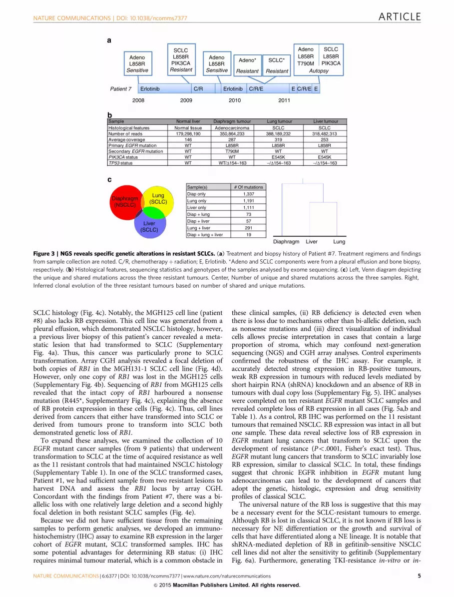
DNA sequencing reveals genetic lesions specific to resistant SCLC.In our previous report7, we described a patient (Patient #7) who hadbeen biopsied multiple times over the course of their disease. In thispatient, both EGFR mutant adenocarcinoma and SCLC had beenobserved at different times. This patient ultimately passed away, andat autopsy, both SCLC and NSCLC were identified (Fig. 3a). Theoscillating pattern of adenocarcinoma and SCLC that was observedsuggested that different clones were selected depending on theselective pressure of the applied treatment (conceptual schematicshown in Supplementary Fig. 3a). The autopsy that was performedidentified two SCLC transformed tumours (one each from the liverand lung) as well as a diaphragmatic tumour that retainedadenocarcinoma histology (Supplementary Fig. 3b). All threelesions contained the original activating EGFR mutation, and thediaphragmatic NSCLC tumour harboured the EGFR resistance
mutation, T790M, whereas the SCLCs did not (Fig. 3b). All samples(along with normal liver tissue) were analysed by WES. The variantallele frequencies of the activating EGFR mutation were 66% and77% in the resistant SCLC samples, consistent with earlier resultsdemonstrating that the EGFR mutation is harboured in thetransformed SCLC cells7. Clonal analyses revealed that the twoSCLC samples had a greater number of mutations in common witheach other (n¼ 291) than either shared with the resistant tumourthat maintained NSCLC morphology ((n¼ 73) shared mutationsbetween the SCLC lung and NSCLC diaphragm, and (n¼ 57)shared mutations between the SCLC liver and NSCLC diaphragm;Fig. 3c). This suggests that the two SCLC resistant lesions are moreclosely related and likely diverged later in the evolution of theresistant disease compared with the adenocarcinoma lesion.
By comparing the genomic variants from these four samples,we were able to look for somatic changes frequently detected inNSCLC and SCLC genomes. Both SCLC transformed samplesharboured an activating mutation in PIK3CA, which wepreviously observed in SCLC transformed cases7 as well as lossof heterozygosity and an inactivating mutation of TP53, which isuniversally altered in classical SCLC24,25 (Fig. 3b). In addition,there was a near absence of reads for a portion of the RB1 gene(o10% of the reads compared with the normal liver andadenocarcinoma), in both resistant SCLC transformed tumours,but not in the resistant adenocarcinoma sample (SupplementaryFig. 3c). This suggests bi-allelic loss of RB1 specifically in theSCLC transformed tumours. This was particularly noteworthy asRB is invariably lost in classical SCLC24–26. Alterations to otherfrequently altered genes in SCLC such as MYC, PTEN and FGFR1were not detected. Thus, analogous to classical SCLC, alterationsto TP53 and RB1 were observed in EGFR mutant NSCLC toSCLC transformed tumours.
MGH131-2(SCLC)
MGH156
(NSCLC)
aH&E SynaptophysinChromogranin
b
*- Patient 6
MGH119 MGH119-R MGH121 MGH125 MGH126 MGH134 MGH141 MGH157 MGH131-1 MGH131-2 NCI-H82 NCI-H446
ASCL1SYP
CHGACHGB
NCAM1NCAM2NeuroD1
c
Row min Row max
Cell line EGFR MutationEGFR TKI
StatusResistance Mechanism
MGH119 Exon 19 del Sensitive NAMGH119-R Exon 19 del Resistant T790MMGH121 Exon 19 del Resistant T790MMGH125 L858R Resistant T790M/EMTMGH126 Exon 19 del Resistant EMTMGH134 L858R Resistant T790MMGH141 Exon 19 del Resistant T790MMGH157 Exon 19 del Resistant T790MMGH131-1* Exon 19 del Resistant NSCLC --> SCLCMGH131-2* Exon 19 del Resistant NSCLC --> SCLCNCI-H82 None Insensitive NANCI-H446 None Insensitive NA
Figure 1 | SCLC transformed cell lines exhibit neuroendocrine (NE) features. (a) Haematoxylin and eosin (H&E) staining and IHC for NE markers
chromogranin and synaptophysin were performed on xenografts derived from EGFR mutant MGH131-2 SCLC and MGH156 NSCLC cells. (b) EGFR mutation
status, TKI sensitivity and resistance mechanism for the patient-derived cell lines analysed in c. (c) Gene expression array data of NE marker expression
across a panel of cell lines derived from TKI-resistant patients (n¼ 10). NCI-H82 and NCI-H446 are classical SCLC cell lines used as controls for NE marker
expression. Red indicates lower expression and blue indicates higher expression.
NATURE COMMUNICATIONS | DOI: 10.1038/ncomms7377 ARTICLE
NATURE COMMUNICATIONS | 6:6377 | DOI: 10.1038/ncomms7377 | www.nature.com/naturecommunications 3
& 2015 Macmillan Publishers Limited. All rights reserved.

In the liver SCLC tumour, comparative genomic hybridization(CGH) array analysis revealed that there was a relatively largedeletion in one copy of RB1 that encompassed the entire gene andthe surrounding region. This was accompanied by a focal deletionin the second copy that spanned only the middle exons of RB1but spared the beginning and end of the gene (Fig. 4a). Thesedeletions were not observed in the resistant cancer with a T790Mmutation and NSCLC histology. These results were confirmed by
quantitative PCR (qPCR) of different exons of RB1, which alsodemonstrated similar focal loss of RB1 in the lung SCLC (Fig. 4b).
RB is universally lost in resistant SCLC patients. The cell linesestablished from biopsies of resistant EGFR mutant lung cancerswere assessed for RB expression. Western blotting revealed loss ofRB expression specifically in resistant EGFR mutant cell lines with
Patient 3
PostPre
Patient 18
NSCLC (T790M)
Post
a b
c d
e f
MG
H12
1 -T
790M
MG
H13
4 -T
790M
MG
H11
9 -N
aive
MG
H14
1 -T
790M
MG
H12
5 -T
790M
MG
H13
1-2
MG
H13
1-1
NC
I-H
196
NC
I-H
82
NSCLC NS
CLC
/SC
LC
SCLCNS
CLC
SC
LC
EGFR
Actin
***P< 0.0001 one-way ANOVA
MGH131-1 MGH131-2 MGH121 MGH134
IC50 (μM) 0.039 0.11 4.2 3.0
0.01
0.1
1
10
100
1,000
IC50
(μM
)
SCLC cell lines
ABT-263 IC50 in SCLC cell lines
MGH131-2
MGH131-1
250
150
37
150
100
50
0–6 –4 –2
[TKI] log (um)0 2
Rel
ativ
e ce
ll nu
mbe
r%
con
trol
SCLC transformed cells
MGH131-1 GEFMGH131-1 WZMGH131-2 GEFMGH131-2 WZMGH121 WZ
400
300
200
100
H-s
core
0Pre Post Post -NSCLC
100
50
0–6 –4 –2
ABT263 log(μM)0 2
Rel
ativ
e ce
ll nu
mbe
r %
cont
rol
ABT-263 in NSCLC->SCLC
MGH131-1 (NSCLC-->SCLC)MGH131-2 (NSCLC-->SCLC)MGH121 (T790M)MGH134 (T790M)
150
NSCLC SCLC
EGFR EGFR EGFR
Figure 2 | Resistant SCLCs respond to ABT-263 and lose EGFR expression. (a) The resistant EGFR mutant SCLC cell lines MGH131-1 and MGH131-2, and
a resistant EGFR mutant NSCLC cell line that harbour T790M, MGH121, were treated with indicated concentrations of Gefitinib (GEF) or the third-
generation EGFR inhibitor WZ4002 (WZ) for 72 h. Cell viability was measured with the CellTiter-Glo assay. Experiments were performed in quadruplicate
and error bars depict the standard error of the mean for each data point. (b) Representative blot of lysates from a panel of patient-derived resistant EGFR
mutant cell lines and classical SCLC cell lines was probed with antibodies specific to total EGFR and actin (MGH119 was derived from a TKI naıve patient).
Lysates from this panel were also probed in Supplementary Fig. 1c. (c) IHC staining for total EGFR on a representative pair of matched pre- and post-
resistant samples from a patient whose resistant EGFR mutant cancer transformed from NSCLC to SCLC (Patient #3, left and middle) and a resistant EGFR
mutant cancer that remained NSCLC (patient #18, right). The yellow circle indicates EGFR-positive endothelial cells in the resistant EGFR mutant SCLC.
(d) Quantification (H-score) of EGFR staining from pair-matched pre (n¼6) and post-resistant (n¼ 7) samples from cancers that transformed into SCLC
upon the development of resistance. Resistant EGFR mutant cancers that maintained NSCLC histology are shown for comparison (n¼ 11). ***Po0.0001
one-way analysis of variance (ANOVA) with Bonferroni post-hoc test. (e) Patient-derived TKI-resistant cell lines from resistant SCLC (MGH131-1 and
MGH131-2), and T790M-positive NSCLC (MGH121 and MGH134) were treated with indicated concentrations of ABT-263 for 72 h and cell viability was
measured with the CellTiter-Glo assay. Each data point was repeated in quadruplicate and error bars represent the standard error of the mean. Bottom—
IC50 values for ABT-263 for each cell line. (f) ABT-263 IC50 values compared with those from a panel of SCLC cell lines37.
ARTICLE NATURE COMMUNICATIONS | DOI: 10.1038/ncomms7377
4 NATURE COMMUNICATIONS | 6:6377 | DOI: 10.1038/ncomms7377 | www.nature.com/naturecommunications
& 2015 Macmillan Publishers Limited. All rights reserved.
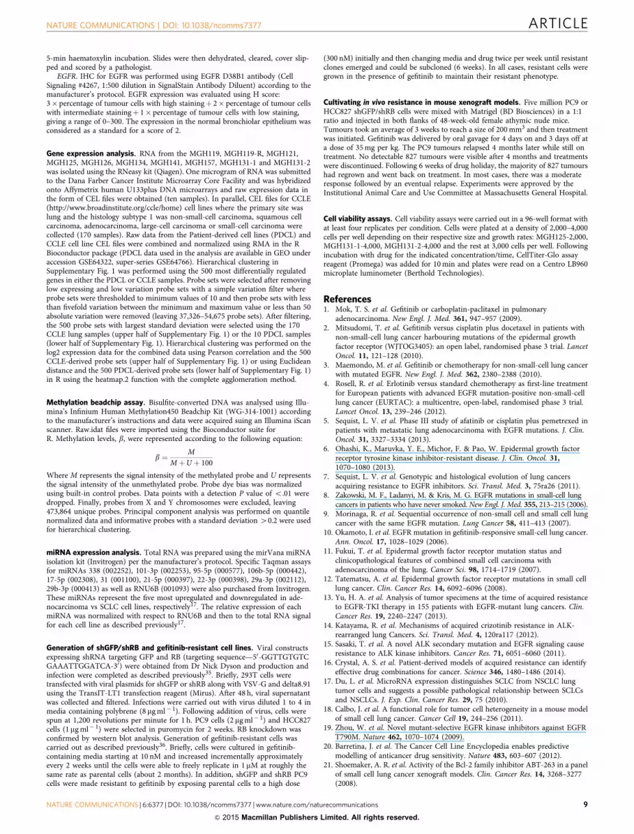
SCLC histology (Fig. 4c). Notably, the MGH125 cell line (patient#8) also lacks RB expression. This cell line was generated from apleural effusion, which demonstrated NSCLC histology, however,a previous liver biopsy of this patient’s cancer revealed a meta-static lesion that had transformed to SCLC (SupplementaryFig. 4a). Thus, this cancer was particularly prone to SCLCtransformation. Array CGH analysis revealed a focal deletion ofboth copies of RB1 in the MGH131-1 SCLC cell line (Fig. 4d).However, only one copy of RB1 was lost in the MGH125 cells(Supplementary Fig. 4b). Sequencing of RB1 from MGH125 cellsrevealed that the intact copy of RB1 harboured a nonsensemutation (R445*, Supplementary Fig. 4c), explaining the absenceof RB protein expression in these cells (Fig. 4c). Thus, cell linesderived from cancers that either have transformed into SCLC orderived from tumours prone to transform into SCLC bothdemonstrated genetic loss of RB1.
To expand these analyses, we examined the collection of 10EGFR mutant cancer samples (from 9 patients) that underwenttransformation to SCLC at the time of acquired resistance as wellas the 11 resistant controls that had maintained NSCLC histology(Supplementary Table 1). In one of the SCLC transformed cases,Patient #1, we had sufficient sample from two resistant lesions toharvest DNA and assess the RB1 locus by array CGH.Concordant with the findings from Patient #7, there was a bi-allelic loss with one relatively large deletion and a second highlyfocal deletion in both resistant SCLC samples (Fig. 4e).
Because we did not have sufficient tissue from the remainingsamples to perform genetic analyses, we developed an immuno-histochemistry (IHC) assay to examine RB expression in the largercohort of EGFR mutant, SCLC transformed samples. IHC hassome potential advantages for determining RB status: (i) IHCrequires minimal tumour material, which is a common obstacle in
these clinical samples, (ii) RB deficiency is detected even whenthere is loss due to mechanisms other than bi-allelic deletion, suchas nonsense mutations and (iii) direct visualization of individualcells allows precise interpretation in cases that contain a largeproportion of stroma, which may confound next-generationsequencing (NGS) and CGH array analyses. Control experimentsconfirmed the robustness of the IHC assay. For example, itaccurately detected strong expression in RB-positive tumours,weak RB expression in tumours with reduced levels mediated byshort hairpin RNA (shRNA) knockdown and an absence of RB intumours with dual copy loss (Supplementary Fig. 5). IHC analyseswere completed on ten resistant EGFR mutant SCLC samples andrevealed complete loss of RB expression in all cases (Fig. 5a,b andTable 1). As a control, RB IHC was performed on the 11 resistanttumours that remained NSCLC. RB expression was intact in all butone sample. These data reveal selective loss of RB expression inEGFR mutant lung cancers that transform to SCLC upon thedevelopment of resistance (Po.0001, Fisher’s exact test). Thus,EGFR mutant lung cancers that transform to SCLC invariably loseRB expression, similar to classical SCLC. In total, these findingssuggest that chronic EGFR inhibition in EGFR mutant lungadenocarcinomas can lead to the development of cancers thatadopt the genetic, histologic, expression and drug sensitivityprofiles of classical SCLC.
The universal nature of the RB loss is suggestive that this maybe a necessary event for the SCLC-resistant tumours to emerge.Although RB is lost in classical SCLC, it is not known if RB loss isnecessary for NE differentiation or the growth and survival ofcells that have differentiated along a NE lineage. It is notable thatshRNA-mediated depletion of RB in gefitinib-sensitive NSCLCcell lines did not alter the sensitivity to gefitinib (SupplementaryFig. 6a). Furthermore, generating TKI-resistance in-vitro or in-
Sample Normal liver Diaphragm tumour Lung tumour Liver tumour
Histological features Normal tissue Adenocarcinoma SCLC SCLCNumber of reads 179,298,190 350,864,233 388,189,232 318,482,313Average coverage 146 287 319 253Primary EGFR mutation WT L858R L858R L858RSecondary EGFR mutation WT T790M WT WTPIK3CA status WT WT E545K E545KTP53 status WT WT/Δ154–163 –/Δ154–163 –/Δ154–163
Diaphragm(NSCLC)
Lung(SCLC)
Liver(SCLC)
Erlotinib C/R Erlotinib C/R/E C/R/E
2008 2009 2010 2011
Adeno SCLCL858R L858RT790M
Autopsy
E E
Adeno*
Resistant
SCLC*
Resistant
Sample(s) # Of mutations
Diap only 1,337
Lung only 1,191
Liver only 1,111
Diap + lung 73
Diap + liver 57
Lung + liver 291
Diap + lung + liver 19
Diaphragm Liver Lung
Patient 7
AdenoL858R
Sensitive
SCLCL858R
PIK3CAResistant
AdenoL858R
SensitivePIK3CA
Figure 3 | NGS reveals specific genetic alterations in resistant SCLCs. (a) Treatment and biopsy history of Patient #7. Treatment regimens and findings
from sample collection are noted. C/R, chemotherapyþ radiation; E, Erlotinib. *Adeno and SCLC components were from a pleural effusion and bone biopsy,
respectively. (b) Histological features, sequencing statistics and genotypes of the samples analysed by exome sequencing. (c) Left, Venn diagram depicting
the unique and shared mutations across the three resistant tumours. Center, Number of unique and shared mutations across the three samples. Right,
Inferred clonal evolution of the three resistant tumours based on number of shared and unique mutations.
NATURE COMMUNICATIONS | DOI: 10.1038/ncomms7377 ARTICLE
NATURE COMMUNICATIONS | 6:6377 | DOI: 10.1038/ncomms7377 | www.nature.com/naturecommunications 5
& 2015 Macmillan Publishers Limited. All rights reserved.

vivo in EGFR mutant cancer cell lines engineered to have loss ofRB expression did not yield resistant cells/tumours withacquisition of NE marker expression or SCLC morphology(Supplementary Fig. 6b,c). These results suggest that loss of RB islikely necessary in order for acquired resistance via transforma-tion to SCLC to develop, but it is not sufficient on its own topromote it. The latter point is further supported by our discoveryof a few examples of RB-deficient adenocarcinomas. Indeed, twoerlotinib-resistant cell lines (MGH125 and MGH141), a resistantpatient sample (Patient #10) and two out of four pre-treatmentadenocarcinoma samples from patients whose cancers trans-formed to SCLC (Patients #2 and #6), were also negative for RB.Although rare, the existence of these RB-deficient adenocarcino-mas serves as further evidence that loss of RB alone is insufficientto promote transformation to SCLC.
DiscussionAcquired resistance is a major problem limiting the clinicalefficacy of targeted therapies. Repeat biopsy studies have led tothe identification of the resistance mechanism in a majority ofEGFR mutant NSCLC patients that have progressed on EGFRTKIs7,13. One unexpected finding from these studies was thediscovery that 5–15% of patient tumours undergo transformationto SCLC histology upon acquisition of resistance. From a historicperspective of lung cancer classification, this observation was asurprise, as differentiation into a NSCLC- or SCLC-type cancerwas thought to occur early in tumorigenesis. Furthermore, thetypical presentation of these diseases were quite distinct, withEGFR-mutant adenocarcinoma occurring primarily in never-smokers and displaying a more indolent natural historycompared with classical SCLC, which occurs almost exclusively
Genome
Chrom. 13q
13q14.2
0
0.2
0.4
0.6
0.8
1
1.2
Exon 3 Exon 13 Exon 25
Nor
mal
ized
RB
1 gD
NA
Healthy liver Diaphragm (NSCLC-T790M)Lung (SCLC) Liver (SCLC)
MG
H12
1 -
T79
0M
MG
H13
4 -
T79
0M
MG
H12
6 -
EM
T
MG
H15
6 -
ME
T A
mp
MG
H12
5 -
T79
0M
MG
H13
1 -
2
MG
H13
1 -
1
NC
I-H
196
NC
I-H
82
NSCLC NS
CLC
/SC
LC
SCLCNS
CLC
SC
LC
MG
H15
7 -
T79
0M
Rb
Actin
NSCLC – (diaphragm) SCLC – (liver)Patient 7
RB1 RB1
RB1
Genome
Chrom. 13q
13q14.2
Genome
Chrom. 13q
13q14.2
Patient 1 - (2 x post-resistant SCLC)
RB1 RB1
100
37
48.5 Mb 48.7 Mb 49.1 Mb 49.3 Mb 48.5 Mb 48.7 Mb 49.1 Mb 49.3 Mb
48.5 Mb 48.7 Mb 49.1 Mb 49.3 Mb
48.5 Mb 48.7 Mb 49.1 Mb 49.3 Mb 48.5 Mb 48.7 Mb 49.1 Mb 49.3 Mb
Figure 4 | Resistant EGFR mutant SCLCs have genetic loss of RB1. (a) CGH array profiles of a resistant NSCLC tumour (left) and SCLC transformed
tumour (right) from Patient #7 at the level of the whole genome (top), chromosome 13q12.12-q32.2 (middle) and the 0.8 Mb region flanking the RB1 gene
(bottom). The RB1 gene locus is depicted and regions of bi-allelic loss are circled. (b) qPCR analysis of RB1 exons 3, 13 and 25 amplified from genomic DNA
from the indicated autopsy specimens from Patient #7. Reactions were carried out in triplicate and error bars representing standard error of the mean are
shown. (c) Representative blot of lysates from resistant EGFR mutant cell lines derived from resistant biopsies along with classical SCLCs was probed with
antibodies specific to RB and actin. (d) CGH array profile of the MGH131-1 cell line of the whole genome (top), chromosome 13q12.12-q32.2 (middle) and
the 0.8 Mb region flanking the RB1 gene (bottom). The RB1 gene locus is depicted and regions of bi-allelic loss are circled. (e) CGH array profiles of two
resistant EGFR mutant SCLCs from Patient #1 with depiction of whole genome (top), chromosome 13q12.12-q32.2 (middle) and the 0.8 Mb region flanking
the RB1 gene (bottom). The RB1 gene locus is depicted and regions of bi-allelic loss are circled.
ARTICLE NATURE COMMUNICATIONS | DOI: 10.1038/ncomms7377
6 NATURE COMMUNICATIONS | 6:6377 | DOI: 10.1038/ncomms7377 | www.nature.com/naturecommunications
& 2015 Macmillan Publishers Limited. All rights reserved.

in heavy smokers and tends to metastasize early and grow rapidly.Indeed, the SCLC transition seen in EGFR-mutant patients isoften accompanied by a change in the clinical behaviour of thedisease, with rapid acceleration in the growth rate, initialresponsiveness to chemotherapy followed by rapid clinicaldeterioration7. However, repeat biopsy studies have consistentlysuggested that the SCLC transformed cancers represent anevolution from the initial adenocarcinomas rather than asecond coincident cancer, because the activating driver EGFRmutations are identical to the original adenocarcinomas in allcases. To date, the mechanistic details regarding this transitionare unknown. This study revealed genetic changes specificallyassociated with the transformation to SCLC, provided insight intowhy these tumours are no longer sensitive to EGFR TKIs, anddetermined a potential therapeutic that could be incorporatedinto future treatment strategies for this subset of resistant cancers.
Assessment of RB status by a combination of next-generationsequencing, array CGH, qPCR and IHC analyses revealed that RBwas lost in 11 out of 11 SCLC transformed samples, a result thatmirrors classical SCLC, in which RB is altered in an over-whelming majority of cases24–26. Interestingly, RB knockdownexperiments in EGFR mutant cell lines suggest that RB loss wasinsufficient to cause resistance or induce NE differentiation. It isnotable that these knockdown studies were performed inestablished EGFR mutant cell lines. Such cell lines may notpossess the pluripotent cells that are present in a tumour in vivothat may have the capacity to differentiate along different lineagesincluding SCLC. We speculate that in these pluripotent cells thatdifferentiation to NSCLC is favoured when EGFR is active, as
EGFR activity has been associated with promoting alveolardifferentiation27 (Supplementary Fig. 7, left panel). Conversely,following treatment with EGFR-TKI, the resistant pluripotentcells, which may have accumulated additional genetic alterations(such as loss of RB1 and TP53) and maintain a differentepigenetic state, are able to differentiate and subsequently expandalong a lineage (SCLC) that does not require EGFR signalling(Supplementary Fig. 7, right panel). It is also interesting to notethat the absence of EGFR signalling induced by the TKI mayremove the impetus to differentiate along the NSCLC lineage,thereby facilitating differentiation along the other lineage. Alongthese lines, there have been case reports of treatment naıve EGFRmutant SCLC12,28, reinforcing the notion that the cell of origin ofEGFR mutant lung cancers may have the potential to differentiatealong a NE lineage. Notably, we assessed one such case (Patient#19, Table 1), and this cancer had loss of RB and EGFRexpression, similar to the cases of EGFR mutant SCLC observedin the setting of acquired resistance to EGFR TKI.
We cannot rule out that EGFR mutant SCLC pre-existed beforetreatment with the EGFR TKI. We have carefully reviewed thehistology of these samples and we do not observe a mix of NSCLCand SCLC histology in the pre-treatment tumours. Of course, thisdoes not rule out the possibility that a very small percentage ofSCLC cells that are below our detection limit do pre-exist(especially, as the biopsies only sample a minute fraction of thepatients’ total cancer burden). However, from a clinical perspective,we feel that it is unlikely that these SCLCs were present from theonset of the disease in a majority of these cases because when theSCLC surfaces in the clinic, it progresses quite rapidly (like classicalSCLC)7. In many of these cases, the TKI-induced remissions lastfor years and then suddenly the patient develops explosive SCLC. Itseems unlikely (but, not impossible) that the same explosive cancerwas present for all of those years while the patients were inremission. In such cases, we favour a model in which the cells thatsurvived treatment undergo further ‘evolution’ to become the bonafide SCLC that ultimately presents in the clinic (as described aboveand shown in Supplementary Fig. 7).
The finding that all EGFR mutant SCLC transformed sampleshave low/absent EGFR expression compared with pre-resistantcontrols provides insight into the explanations for the lack ofsensitivity of these cancers to TKI. We speculate that, upontransformation to SCLC, they take on many of the characteristicsof classical SCLC, which normally do not express EGFR or rely onits activity for growth and survival29. Thus, the treatment strategiesthat will provide the most benefit to this subset of cancers willlikely resemble those that are most effective for classical SCLC.
Our data reveal that EGFR mutant cancers that transform toSCLC also undergo significant epigenetic changes. Hierarchicalclustering analysis of RNA expression data demonstrated that celllines derived from SCLC transformed resistant biopsies sharegene expression profiles more closely related to classical SCLC celllines than other TKI-resistant cell lines that maintained NSCLChistology. Similarly, miRNA analyses revealed that SCLCtransformed cells express miRNAs that are commonly upregu-lated in classical SCLC. It is notable, however, that the SCLCtransformed cells also express a subset of miRNAs that aretypically expressed in adenocarcinoma but not SCLC. Further-more, DNA methylation analysis of resistant SCLC tumours frompatient # 7 revealed a methylation pattern more consistent withadenocarcinoma than SCLC (Supplementary Fig. 8). These resultsindicate that SCLC transformed cancers may retain some featuresconsistent with their adenocarcinoma origins. Importantly,however, from a genetic, mRNA expression profile, and clinicalperspective these cancers behave like classical SCLC.
In summary, this study reveals some of the key molecularchanges associated with EGFR mutant lung adenocarcinomas that
NSCLC SCLC
Post
Pre
Patient 3
Patient 18
NSCLC (T790M)
Rb staining SCLC resistant NSCLC resistantNegative (10/10) – 100% (1/9) – 11%Positive (0/10) – 0% (8/9) – 89%
Fisher’s exact test – P < 0.0001
HE
HE
HE
Rb
Rb
Rb
Figure 5 | RB is invariably absent in resistant EGFR mutant SCLCs.
(a) Haematoxylin and eosin (HE) staining (left) and the corresponding RB
IHC (right) for a representative matched pair of pre-treatment EGFR mutant
NSCLC and the corresponding post-resistant EGFR mutant SCLC (Patient
#3, top, middle). A resistant EGFR mutant cancer that maintained
adenocarcinoma histology and acquired a T790M EGFR mutation is shown
for comparison (Patient #18, bottom). Yellow circles indicate gland
formation of the moderately differentiated adenocarcinomas. Red circles
indicate positive staining in endothelial cells. (b) Results of RB IHC staining
of EGFR mutant-resistant cancers that underwent the transformation from
NSCLC to SCLC (SCLC Resistant, n¼ 10) and those that retained an
adenocarcinoma histology (NSCLC Resistant, n¼9). Resistant EGFR mutant
SCLC is significantly more likely than resistant EGFR mutant NSCLC to have
loss of RB expression (Po0.0001, Fisher’s exact test).
NATURE COMMUNICATIONS | DOI: 10.1038/ncomms7377 ARTICLE
NATURE COMMUNICATIONS | 6:6377 | DOI: 10.1038/ncomms7377 | www.nature.com/naturecommunications 7
& 2015 Macmillan Publishers Limited. All rights reserved.

transform to SCLC upon acquisition of resistance to EGFR TKI. Asnovel therapeutic approaches that inhibit EGFR more efficientlybecome widely implemented30–32, we speculate that the relativefrequency of NSCLC to SCLC transformation in the setting ofacquired resistance may increase moving forward, furtherunderscoring the importance of understanding the basis for thistransformation as well as treatment strategies to overcome it.
MethodsPatients. EGFR mutant NSCLC patients underwent biopsies before and afteracquiring resistance to erlotinib therapy as per standard clinical care over an 8-yearperiod from 2005 to 2013. Standard histology and the SNaPshot assay were carriedout to determine histological subtype and genotype of each sample33. An IRB-approved protocol was followed to review the electronic medical record for relevantclinical information. Patient-derived cell lines were generated under an IRB-approvedprotocol, which required prospective informed consent from participating patients.
Reagents and cell culture. PC9, HCC827, MGH119, MGH119-R, MGH121,MGH134, MGH141, MGH157, NCI-H446, NCI-H196 and NCI-H82 cells werecultured in RPMI 1640 supplemented with 10% fetal bovine serum. MGH125,MGH126, MGH131-1 and MGH131-2 cells were cultured in ACL4 supplementedwith 10% fetal bovine serum. NCI-H446, NCI-H196 and NCI-H82 cells wereobtained from the Center for Molecular Therapeutics at MGH. PC9 and HCC827cells were a gift from Pasi Janne. Gefitinib and WZ4002 were purchased fromSelleck, Abt-263 was purchased from Active Biochem. All compounds were
reconstituted in dimethylsulphoxide for cell culture experiments. Antibodies forRB, actin, NCAM, synaptophysin, NeuroD, pAkt T308, pERK T202/Y204 and totalAkt were purchased from Cell Signaling Technology. pEGFR Y1068 and chro-mogranin A were purchased from Abcam, E-cadherin and Vimentin from BD andtotal EGFR from Santa Cruz Biotechnology. All antibodies were used at a dilutionof 1:1,000. Uncropped scans of the western blots from the main figures can befound in Supplementary Fig. 9.
Generation of patient-derived resistant cell line models. The patient-derivedcell line models MGH119, MGH121, MGH125, MGH126, MGH131-1,MGH131-2, MGH134 and MGH156 were developed on collagen-coated plates inACL4 medium and transferred to RPMI. MGH157 was developed initially inRPMI. The cell line MGH141 was derived using the feeder system with irradiatedfibroblasts (5,000 rad) from normal patient tissue. When a tumour cell majoritywas observed it was passaged off of the feeder layer and later transferred to RPMImedium for experiments. The development of a model was considered completewhen it was independent of the feeder system, free of stromal cells and determinedto maintain known patient tumour mutations. MGH119-R was derived in vitrofrom the treatment naive model, MGH119, through in vitro exposure to gefitinib,escalating from 10 nM to a final concentration of 1 mM.
DNA extraction library construction and WES. Genomic DNA (gDNA) fromnormal liver, diaphragmatic tumour (NSCLC), lung tumour (SCLC) and livertumour (SCLC) from patient #7 was extracted from OCT-embedded frozen tissueblocks using the DNAdvance kit from Agencourt. Three micrograms of gDNAfrom each sample were fragmented to approximately 150–200 bp by sonication andsubjected to exome enrichment using the SureSelectXT Human All Exon TargetEnrichment system. Barcoded deep sequencing libraries for the exome-enrichedgDNA fragments were constructed using Applied Biosystems SOLiD 5500 Frag-ment Library Core Kit. WES was performed with an Applied Biosystems SOLiD5500 deep sequencer to generate paired-end colour space reads (50 nucleotidesforward and 35 nucleotides reverse) by a multiplexed operation. The colour-spacedata were aligned to the human hg19 reference genome sequence by the AppliedBiosystems LifeScope software to generate BAM files. Mutation calls were madeusing the muTect mutation calling software.
Quantification of RB1 gDNA levels by qPCR. RB1 gene copy number wasmeasured via a quantitative PCR assay that has been previously described34. Briefly,reaction samples containing 10 ng of gDNA with SYBR green master mix (Roche)were run on a LightCycler 480 (Roche) for quantification. Primer pairs amplifyingexons 3 (F—50-GAGCTACAGAAAAACATAGAAATCAGG-30, R—50-GGAAAATCCAGAATTCGTTTCC-30), 13 (F—50-GCATCTTTCCAGTTCGTATAAATACTC-30 , R—50-CATAAAGTTACCCATAAATAGCAGCA-30) and 25 (F—50-ACAGCGACCGTGTGCTCAAA-30, R—50-AGCCAGGAGCAGTGCTGAGAC-30)were used to obtain coverage of the beginning, middle and end of the RB1 gene andprimers amplifying long interspersed nuclear element-1 (LINE-1; F—50-AAAGCCGCTCAACTACATGG-30 , R—50-TGCTTTGAATGCGTCCCAGAG-30) wereused for each sample to serve as a loading control. A standard curve with normalfemale genomic DNA was generated for each primer pair in order to comparethe tumour/cell line samples to a normal diploid sample.
DNA extraction and array CGH analysis. DNA for the array CGH studies wasextracted from formalin-fixed, paraffin-embedded tissues with the FormaPure kitfrom Agencourt. Agilent Sureprint G3 Cancer CGHþ SNP 4� 180 k Microarrayswere used to identify genome-wide copy number alterations. Briefly, 1 mg oftumour and control DNA (normal female gDNA, Corriell Institute) were heated to95 �C for 5 min. Random priming was used to label DNA with CY3-dUTP (con-trol) and CY5-dUTP (tumour) dyes from the Agilent SureTag DNA Labeling kit.The labelled DNA was then purified over columns (Agilent) and mixed in equalproportion along with Cot-1 Human DNA (Agilent) for the hybridization steps. Tohybridize the DNA to the array, incubation occurred first at 95 �C for 3 min fordenaturation, followed by a 30-min pre-hybridization step at 37 �C and then ahybridization step for 35–40 h at 65 �C. Slides were then washed with Agilent OligoArrayCGH wash buffer 1 for 5 min at room temperature and wash buffer 2 for1 min at 37 �C. Upon completion of the washes, slides were scanned using theG2505C Microarray Scanner (Agilent). The data were analysed using the AgilentCytoGenomics software v 2.0. CGH array data are available at GEO underaccession number GSE64765 (super-series GSE64766).
Immunohistochemistry. RB. The total RB IHC (Rabbit monoclonal Abcam#E182, 1:500) was performed on formalin-fixed, paraffin-embedded tissue sectionsusing the Leica RX Bond Autostainer (Leica Biosystems). The sections cut 4–5 mmwere baked off-line for 30 min in a 60 �C oven and then loaded onto the machine.The machine then de-waxed and hydrated online. Antigen retrieval was performedin ER 2 (Citrate buffer) for 20 min and stained using the Bond Polymer RefineProtocol under the IHC Modified F Protocol. Steps involved a 5-min PeroxideBlock, a 15-min antibody/marker incubation, an 8-min post primary incubation,an 8-min polymer incubation, a 10-min DAB (diaminobenzidine) incubation and a
Table 1 | RB status of TKI-resistant patients.
Patient Cancertype
Resistance Histology RBstatus
Detection method
1 Lung Pre Adeno Pos IHCLung Post NE Neg IHC/geneticLung Post NE Neg IHC/genetic
2 Lung Pre Adeno Pos IHCLung Pre Adeno Neg IHCLung Post NE Neg IHC
3 Lung Pre Adeno Pos IHCLung Post NE Neg IHC
4 Lung Post NE Neg IHC5 Lung Post NE Neg IHC6 Lung Pre Adeno Neg IHC
Lung Post NE Neg IHC/genetic*7 Lung Post Adeno Pos IHC/genetic
Lung Post NE Neg IHC/geneticLung Post NE Neg Genetic
8 Lung Post Adeno Pos IHCLung Post NE Neg IHC
9 Lung Post NE Neg IHC10 Lung Post Adeno Neg IHC11 Lung Pre Adeno Pos IHC
Lung Post Adeno Pos IHC12 Lung Pre Adeno Pos IHC
Lung Post Adeno Pos IHC13 Lung Post Adeno Pos IHC14 Lung Pre Adeno Pos IHC
Lung Post Adeno Pos IHC15 Lung Post Adeno Pos IHC16 Lung Pre Adeno Pos IHC
Lung Post Adeno Pos IHC17 Lung Pre Adeno Pos IHC
Lung Post Adeno Pos IHC18 Lung Post Adeno Pos IHC19w Lung Intrinsic NE Neg IHC
EGFR, epidermal growth factor receptor; IHC, immunohistochemistry; NE, neuroendocrinecarcinoma; Neg, negative; Pos, positive; TKI, tyrosine kinase inhibitor.RB status in pre/ post-TKI-resistant EGFR mutant lung cancers. EGFR TKI sensitivity, histology,RB expression and the detection method are listed for tumours from nine patients with resistantsmall-cell lung cancer (SCLC) and nine patients with resistant non-small-cell lung cancer. Patient19 presented with classical SCLC with an EGFR mutation that was intrinsically resistant to EGFRTKI.*Genetic data were from a cell line derived from that sample.wPatient presented with EGFR mutant classical NE carcinoma and failed to respond to TKI.
ARTICLE NATURE COMMUNICATIONS | DOI: 10.1038/ncomms7377
8 NATURE COMMUNICATIONS | 6:6377 | DOI: 10.1038/ncomms7377 | www.nature.com/naturecommunications
& 2015 Macmillan Publishers Limited. All rights reserved.
5-min haematoxylin incubation. Slides were then dehydrated, cleared, cover slip-ped and scored by a pathologist.
EGFR. IHC for EGFR was performed using EGFR D38B1 antibody (CellSignaling #4267, 1:500 dilution in SignalStain Antibody Diluent) according to themanufacturer’s protocol. EGFR expression was evaluated using H score:3� percentage of tumour cells with high stainingþ 2� percentage of tumour cellswith intermediate stainingþ 1� percentage of tumour cells with low staining,giving a range of 0–300. The expression in the normal bronchiolar epithelium wasconsidered as a standard for a score of 2.
Gene expression analysis. RNA from the MGH119, MGH119-R, MGH121,MGH125, MGH126, MGH134, MGH141, MGH157, MGH131-1 and MGH131-2was isolated using the RNeasy kit (Qiagen). One microgram of RNA was submittedto the Dana Farber Cancer Institute Microarray Core Facility and was hybridizedonto Affymetrix human U133plus DNA microarrays and raw expression data inthe form of CEL files were obtained (ten samples). In parallel, CEL files for CCLE(http://www.broadinstitute.org/ccle/home) cell lines where the primary site waslung and the histology subtype 1 was non-small-cell carcinoma, squamous cellcarcinoma, adenocarcinoma, large-cell carcinoma or small-cell carcinoma werecollected (170 samples). Raw data from the Patient-derived cell lines (PDCL) andCCLE cell line CEL files were combined and normalized using RMA in the RBioconductor package (PDCL data used in the analysis are available in GEO underaccession GSE64322, super-series GSE64766). Hierarchical clustering inSupplementary Fig. 1 was performed using the 500 most differentially regulatedgenes in either the PDCL or CCLE samples. Probe sets were selected after removinglow expressing and low variation probe sets with a simple variation filter whereprobe sets were thresholded to minimum values of 10 and then probe sets with lessthan fivefold variation between the minimum and maximum value or less than 50absolute variation were removed (leaving 37,326–54,675 probe sets). After filtering,the 500 probe sets with largest standard deviation were selected using the 170CCLE lung samples (upper half of Supplementary Fig. 1) or the 10 PDCL samples(lower half of Supplementary Fig. 1). Hierarchical clustering was performed on thelog2 expression data for the combined data using Pearson correlation and the 500CCLE-derived probe sets (upper half of Supplementary Fig. 1) or using Euclideandistance and the 500 PDCL-derived probe sets (lower half of Supplementary Fig. 1)in R using the heatmap.2 function with the complete agglomeration method.
Methylation beadchip assay. Bisulfite-converted DNA was analysed using Illu-mina’s Infinium Human Methylation450 Beadchip Kit (WG-314-1001) accordingto the manufacturer’s instructions and data were acquired suing an Illumina iScanscanner. Raw.idat files were imported using the Bioconductor suite forR. Methylation levels, b, were represented according to the following equation:
b ¼ MMþU þ 100
Where M represents the signal intensity of the methylated probe and U representsthe signal intensity of the unmethylated probe. Probe dye bias was normalizedusing built-in control probes. Data points with a detection P value of o.01 weredropped. Finally, probes from X and Y chromosomes were excluded, leaving473,864 unique probes. Principal component analysis was performed on quantilenormalized data and informative probes with a standard deviation 40.2 were usedfor hierarchical clustering.
miRNA expression analysis. Total RNA was prepared using the mirVana miRNAisolation kit (Invitrogen) per the manufacturer’s protocol. Specific Taqman assaysfor miRNAs 338 (002252), 101-3p (002253), 95-5p (000577), 106b-5p (000442),17-5p (002308), 31 (001100), 21-5p (000397), 22-3p (000398), 29a-3p (002112),29b-3p (000413) as well as RNU6B (001093) were also purchased from Invitrogen.These miRNAs represent the five most upregulated and downregulated in ade-nocarcinoma vs SCLC cell lines, respectively17. The relative expression of eachmiRNA was normalized with respect to RNU6B and then to the total RNA signalfor each cell line as described previously17.
Generation of shGFP/shRB and gefitinib-resistant cell lines. Viral constructsexpressing shRNA targeting GFP and RB (targeting sequence—50-GGTTGTGTCGAAATTGGATCA-30) were obtained from Dr Nick Dyson and production andinfection were completed as described previously35. Briefly, 293T cells weretransfected with viral plasmids for shGFP or shRB along with VSV-G and delta8.91using the TransIT-LT1 transfection reagent (Mirus). After 48 h, viral supernatantwas collected and filtered. Infections were carried out with virus diluted 1 to 4 inmedia containing polybrene (8 mg ml� 1). Following addition of virus, cells werespun at 1,200 revolutions per minute for 1 h. PC9 cells (2 mg ml� 1) and HCC827cells (1mg ml� 1) were selected in puromycin for 2 weeks. RB knockdown wasconfirmed by western blot analysis. Generation of gefitinib-resistant cells wascarried out as described previously36. Briefly, cells were cultured in gefitinib-containing media starting at 10 nM and increased incrementally approximatelyevery 2 weeks until the cells were able to freely replicate in 1 mM at roughly thesame rate as parental cells (about 2 months). In addition, shGFP and shRB PC9cells were made resistant to gefitinib by exposing parental cells to a high dose
(300 nM) initially and then changing media and drug twice per week until resistantclones emerged and could be subcloned (6 weeks). In all cases, resistant cells weregrown in the presence of gefitinib to maintain their resistant phenotype.
Cultivating in vivo resistance in mouse xenograft models. Five million PC9 orHCC827 shGFP/shRB cells were mixed with Matrigel (BD Biosciences) in a 1:1ratio and injected in both flanks of 48-week-old female athymic nude mice.Tumours took an average of 3 weeks to reach a size of 200 mm3 and then treatmentwas initiated. Gefitinib was delivered by oral gavage for 4 days on and 3 days off ata dose of 35 mg per kg. The PC9 tumours relapsed 4 months later while still ontreatment. No detectable 827 tumours were visible after 4 months and treatmentswere discontinued. Following 6 weeks of drug holiday, the majority of 827 tumourshad regrown and went back on treatment. In most cases, there was a moderateresponse followed by an eventual relapse. Experiments were approved by theInstitutional Animal Care and Use Committee at Massachusetts General Hospital.
Cell viability assays. Cell viability assays were carried out in a 96-well format withat least four replicates per condition. Cells were plated at a density of 2,000–4,000cells per well depending on their respective size and growth rates: MGH125-2,000,MGH131-1-4,000, MGH131-2-4,000 and the rest at 3,000 cells per well. Followingincubation with drug for the indicated concentration/time, CellTiter-Glo assayreagent (Promega) was added for 10 min and plates were read on a Centro LB960microplate luminometer (Berthold Technologies).
References1. Mok, T. S. et al. Gefitinib or carboplatin-paclitaxel in pulmonary
adenocarcinoma. New Engl. J. Med. 361, 947–957 (2009).2. Mitsudomi, T. et al. Gefitinib versus cisplatin plus docetaxel in patients with
non-small-cell lung cancer harbouring mutations of the epidermal growthfactor receptor (WJTOG3405): an open label, randomised phase 3 trial. LancetOncol. 11, 121–128 (2010).
3. Maemondo, M. et al. Gefitinib or chemotherapy for non-small-cell lung cancerwith mutated EGFR. New Engl. J. Med. 362, 2380–2388 (2010).
4. Rosell, R. et al. Erlotinib versus standard chemotherapy as first-line treatmentfor European patients with advanced EGFR mutation-positive non-small-celllung cancer (EURTAC): a multicentre, open-label, randomised phase 3 trial.Lancet Oncol. 13, 239–246 (2012).
5. Sequist, L. V. et al. Phase III study of afatinib or cisplatin plus pemetrexed inpatients with metastatic lung adenocarcinoma with EGFR mutations. J. Clin.Oncol. 31, 3327–3334 (2013).
6. Ohashi, K., Maruvka, Y. E., Michor, F. & Pao, W. Epidermal growth factorreceptor tyrosine kinase inhibitor-resistant disease. J. Clin. Oncol. 31,1070–1080 (2013).
7. Sequist, L. V. et al. Genotypic and histological evolution of lung cancersacquiring resistance to EGFR inhibitors. Sci. Transl. Med. 3, 75ra26 (2011).
8. Zakowski, M. F., Ladanyi, M. & Kris, M. G. EGFR mutations in small-cell lungcancers in patients who have never smoked. New Engl. J. Med. 355, 213–215 (2006).
9. Morinaga, R. et al. Sequential occurrence of non-small cell and small cell lungcancer with the same EGFR mutation. Lung Cancer 58, 411–413 (2007).
10. Okamoto, I. et al. EGFR mutation in gefitinib-responsive small-cell lung cancer.Ann. Oncol. 17, 1028–1029 (2006).
11. Fukui, T. et al. Epidermal growth factor receptor mutation status andclinicopathological features of combined small cell carcinoma withadenocarcinoma of the lung. Cancer Sci. 98, 1714–1719 (2007).
12. Tatematsu, A. et al. Epidermal growth factor receptor mutations in small celllung cancer. Clin. Cancer Res. 14, 6092–6096 (2008).
13. Yu, H. A. et al. Analysis of tumor specimens at the time of acquired resistanceto EGFR-TKI therapy in 155 patients with EGFR-mutant lung cancers. Clin.Cancer Res. 19, 2240–2247 (2013).
14. Katayama, R. et al. Mechanisms of acquired crizotinib resistance in ALK-rearranged lung Cancers. Sci. Transl. Med. 4, 120ra117 (2012).
15. Sasaki, T. et al. A novel ALK secondary mutation and EGFR signaling causeresistance to ALK kinase inhibitors. Cancer Res. 71, 6051–6060 (2011).
16. Crystal, A. S. et al. Patient-derived models of acquired resistance can identifyeffective drug combinations for cancer. Science 346, 1480–1486 (2014).
17. Du, L. et al. MicroRNA expression distinguishes SCLC from NSCLC lungtumor cells and suggests a possible pathological relationship between SCLCsand NSCLCs. J. Exp. Clin. Cancer Res. 29, 75 (2010).
18. Calbo, J. et al. A functional role for tumor cell heterogeneity in a mouse modelof small cell lung cancer. Cancer Cell 19, 244–256 (2011).
19. Zhou, W. et al. Novel mutant-selective EGFR kinase inhibitors against EGFRT790M. Nature 462, 1070–1074 (2009).
20. Barretina, J. et al. The Cancer Cell Line Encyclopedia enables predictivemodelling of anticancer drug sensitivity. Nature 483, 603–607 (2012).
21. Shoemaker, A. R. et al. Activity of the Bcl-2 family inhibitor ABT-263 in a panelof small cell lung cancer xenograft models. Clin. Cancer Res. 14, 3268–3277(2008).
NATURE COMMUNICATIONS | DOI: 10.1038/ncomms7377 ARTICLE
NATURE COMMUNICATIONS | 6:6377 | DOI: 10.1038/ncomms7377 | www.nature.com/naturecommunications 9
& 2015 Macmillan Publishers Limited. All rights reserved.

22. Rudin, C. M. et al. Phase II study of single-agent navitoclax (ABT-263) andbiomarker correlates in patients with relapsed small cell lung cancer. Clin.Cancer Res. 18, 3163–3169 (2012).
23. Gardner, E. E. et al. Rapamycin rescues ABT-737 efficacy in small cell lungcancer. Cancer Res. 74, 2846–2856 (2014).
24. Rudin, C. M. et al. Comprehensive genomic analysis identifies SOX2 as afrequently amplified gene in small-cell lung cancer. Nat. Genet. 44,1111–1116 (2012).
25. Peifer, M. et al. Integrative genome analyses identify key somatic drivermutations of small-cell lung cancer. Nat. Genet. 44, 1104–1110 (2012).
26. van Meerbeeck, J. P., Fennell, D. A. & De Ruysscher, D. K. Small-cell lungcancer. Lancet 378, 1741–1755 (2011).
27. Miettinen, P. J. et al. Epithelial immaturity and multiorgan failure in micelacking epidermal growth factor receptor. Nature 376, 337–341 (1995).
28. Shiao, T. H. et al. Epidermal growth factor receptor mutations in small cell lungcancer: a brief report. J. Thorac. Oncol. 6, 195–198 (2011).
29. Byers, L. A. et al. Proteomic profiling identifies dysregulated pathways in smallcell lung cancer and novel therapeutic targets including PARP1. Cancer Discov.2, 798–811 (2012).
30. Sequist, L. V. et al. First-in-human evalulation of CO-1686, an irreversible,selective, and potent tyrosine kinase inhibitor of EGFR T790M. J. Clin. Oncol.31(Suppl; abstr 2524) (2013).
31. Janjigian, Y. Y. et al. Activity and tolerability of afatinib (BIBW 2992) andcetuximab in NSCLC patients with acquired resistance to erlotinib or gefitinib.J. Clin. Oncol. 29(Suppl; abstr 7525) (2011).
32. Yang, J. C. et al. Updated safety and efficacy from a phase I study of AZD9291in patients (PTS) with EGFR-TKI-Resistant non-small cell lung cancer(NSCLC). EMSO Meet. Abstr. Ann. Oncol 25(Suppl 4): iv149 (2014).
33. Sequist, L. V. et al. Implementing multiplexed genotyping of non-small-celllung cancers into routine clinical practice. Ann. Oncol. 22, 2616–2624 (2011).
34. Corcoran, R. B. et al. BRAF gene amplification can promote acquired resistanceto MEK inhibitors in cancer cells harboring the BRAF V600E mutation. Sci.Signal. 3, ra84 (2010).
35. Morris, E. J. et al. E2F1 represses beta-catenin transcription and is antagonizedby both pRB and CDK8. Nature 455, 552–556 (2008).
36. Engelman, J. A. et al. MET amplification leads to gefitinib resistance in lungcancer by activating ERBB3 signaling. Science 316, 1039–1043 (2007).
37. Garnett, M. J. et al. Systematic identification of genomic markers of drugsensitivity in cancer cells. Nature 483, 570–575 (2012).
AcknowledgementsWe thank Dr Nick Dyson for the constructs targeting GFP and RB and Dr James Roccoand Dr William Michaud for supplying feeder cells and helpful advice. This work wassupported by the Lung Cancer Research Foundation (Scientific Merit Award, M.J.N.),Uniting Against Lung Cancer (L.V.S. and M.J.N.), NIH/National Cancer Institute(R01CA137008, J.A.E., 5R21CA156000, L.V.S. and P50CA090578, D.B.C.), Lungevity(J.A.E. and L.V.S.) Department of Defense (CDMRP LC130190, C.M.R and P.A.J.) and
philanthropic support from Targeting a Cure for Lung Cancer, Be a Piece of the Solution,and the MGH Thoracic Oncology Group.
Author contributionsM.J.N. designed and performed the experiments, analysed data and wrote the manu-script. J.A.E. and L.V.S. collected patient samples, designed experiments, analysed dataand wrote the manuscript. M.M.-K. performed pathological analysis, designed experi-ments and analysed data. J.T.P., E.L.L., A.R.G., R.K., C.C., H.E.M., L.A.B., F.M. and N.M.performed the experiments and analysed data. C.H.M. and K.N.R. analysed data. T.M.,E.H. and L.E.F. coordinated patient sample collection and testing. P.A.V. performedpathological analysis. D.B.C. collected patient samples. P.A.J. and C.M.R. collectedpatient samples and analysed data. D.R.B., S.R., T.S., A.J.I. and G.G. carried out dataanalysis.
Additional informationAccession codes: Accession codes for data sets are as follows: microarray and array CGHare at GEO (GSE64322, GSE64765, super-series GSE64766) and WES is at EuropeanGenomics Association (EGAS00001001102).
Supplementary Information accompanies this paper at http://www.nature.com/naturecommunications
Competing financial interests: J.A.E. is a consultant for Novartis, Sanofi-Aventis,Genentech and Astra Zeneca; owns equity in Gatekeeper Pharmaceuticals, which hasinterest in T790M inhibitors; is a Scientific Advisory Board member for Sanofi-Aventis; has research agreements with Novartis, Sanofi-Aventis and Astra Zeneca.A.J.I. is a consultant for Pfizer and Bioreference Laboratories. P.A.J. is a consultantfor AstraZeneca, Boehringer Ingelheim, Chugai Pharma, Clovis, Genentech,Merrimack Pharmaceuticals, Pfizer and Sanofi; owns stock in Gatekeeper Pharmaceu-tical; receives other renumeration from LabCorp. C.M.R. has been a recent consultantfor AbbVie, Biodesix, Boehringer Ingelheim, Glaxo Smith Kline and Merck regardingcancer drug development. The remaining authors declare no competing financialinterests.
Reprints and permission information is available online at http://npg.nature.com/reprintsandpermissions/
How to cite this article: Niederst, M. J. et al. RB loss in resistant EGFR mutant lungadenocarcinomas that transform to small-cell lung cancer. Nat. Commun. 6:6377doi: 10.1038/ncomms7377 (2015).
This work is licensed under a Creative Commons Attribution 4.0International License. The images or other third party material in this
article are included in the article’s Creative Commons license, unless indicated otherwisein the credit line; if the material is not included under the Creative Commons license,users will need to obtain permission from the license holder to reproduce the material.To view a copy of this license, visit http://creativecommons.org/licenses/by/4.0/
ARTICLE NATURE COMMUNICATIONS | DOI: 10.1038/ncomms7377
10 NATURE COMMUNICATIONS | 6:6377 | DOI: 10.1038/ncomms7377 | www.nature.com/naturecommunications
& 2015 Macmillan Publishers Limited. All rights reserved.


