Embed Size (px)
Citation preview
RESEARCH Open Access
Re-irradiation of recurrent head and neckcarcinomas: comparison of robust intensitymodulated proton therapy treatment planswith helical tomotherapyMartin Stuschke1,2*, Andreas Kaiser2, Jehad Abu-Jawad1, Christoph Pöttgen1, Sabine Levegrün1†
and Jonathan Farr2,3†
Abstract
Background: To test the hypothesis that the therapeutic ratio of intensity-modulated photon therapy using helicaltomotherapy (HT) for retreatment of head and neck carcinomas can be improved by robust intensity-modulatedproton therapy (IMPT).
Methods: Comparative dose planning with robust IMPT was performed for 7 patients retreated with HT.
Results: On average, HT yielded dose gradients steeper in a distance ≤ 7.5 mm outside the target (p<0.0001, F-test)and more conformal high dose regions down to the 50% isodose than IMPT. Both methods proved comparablyrobust against set-up errors of up to 2 mm, and normal tissue exposure was satisfactory. The mean body dose wassmaller with IMPT.
Conclusions: IMPT was found not to be uniformly superior to HT and the steeper average dose fall-off around thetarget volume is an argument pro HT under the methodological implementations used. However, looking at singleorgans at risk, the normal tissue sparing of IMPT can surpass tomotherapy for an individual patient. Therefore,comparative dose planning is recommended, if both methods are available.
Keywords: IMPT, Intensity modulated proton therapy, Robust optimization, Robustness, Plan comparison,Re-irradiation, Helical tomotherapy
BackgroundRe-irradiation of late recurrent head and neck cancerwith concomitant chemotherapy is a curative option forun-resectable recurrences. However, side effects aresignificant. Randomized or prospective uncontrolledtrials with total radiation doses of 60–65 Gy at 1.5 - 2.0 Gyper fraction with or without concomitant chemotherapyshowed a crude incidence of late grade III or worseside effects of 34% - 65% [1-4]. For patients withnon-nasopharyngeal head and neck carcinomas treatedwithin these trials, the predominant late side effects were
trismus, osteoradionecrosis, subcutaneous fibrosis, latemucosal side effects, and pharyngeal, laryngeal, esophagealdysfunctions or carotid ruptures. Highly conformaltechniques are used frequently. In their large retrospectiveseries, Lee et al. [5] found a higher freedom fromloco-regional progression at 2 years with intensitymodulated photon radiotherapy techniques in comparisonto 3D conformal radiotherapy, indicating that thetherapeutic index in the retreatment of head and neckcancer can be improved with more conformal techniques.Recent reviews on proton radiotherapy for head and
neck cancer concluded that protons are able to improvethe therapeutic ratio by significantly decreasing normaltissue dose, while keeping similar or better target coveragethan current photon techniques, and that scanned
* Correspondence: [email protected]†Equal contributors1Department of Radiotherapy, University Duisburg-Essen, 45147 Essen,Germany2Westdeutsches Protonentherapiezentrum Essen, 45147 Essen, GermanyFull list of author information is available at the end of the article
© 2013 Stuschke et al.; licensee BioMed Central Ltd. This is an Open Access article distributed under the terms of the CreativeCommons Attribution License (http://creativecommons.org/licenses/by/2.0), which permits unrestricted use, distribution, andreproduction in any medium, provided the original work is properly cited.
Stuschke et al. Radiation Oncology 2013, 8:93http://www.ro-journal.com/content/8/1/93

intensity modulated proton therapy (IMPT) might provemost advantageous [6,7].In this study, we review a series of re-irradiated
patients with long term follow-up, treated with helicaltomotherapy (HT). We conducted comparative intensitymodulated proton therapy planning using an advancedrobust optimization algorithm. The planning aim was tocreate proton plans reaching or surpassing the goodtarget volume coverage achieved with HT while offeringbetter sparing of the surrounding normal tissues.
MethodsFor comparative planning with IMPT, 7 patients withrecurrent carcinoma in the head and neck were identified,who had received a radical retreatment with HT betweenJanuary 2009 and September 2010. Median and minimumtime between the start of the first and second treatmentseries was 48 months and 37 months, respectively,allowing partial recovery of potential damage in the CNS[8]. The total radiation dose, given in the first radiotherapyseries, was 56 – 63 Gy with conventional fractionation. Allpatients were immobilized in a frameless precisionhead mask system developed for fractionated stereotactictreatments (BrainLAB AG, Feldkirchen, Germany).Planning computed tomograms (CT) were obtained
using a large-bore CT scanner (Somatom Sensation Open,Siemens, Erlangen, Germany). CT slices of 1.5 - 2.0 mmthickness were reconstructed. A CTV margin of 0.5 - 1.0cm was applied around the GTV, respecting non-involvedanatomic borders. An isotropic PTV margin of 0.2 cmwas added to the CTV for the HT treatment planning. Aconstant relative biological effectiveness (RBE) of 1.0 wasassumed for photons and of 1.1 for protons.
Tomotherapy treatment planningHT plans were normalized such that 95% of the PTVwas covered by the prescribed dose in 5 patients and90% of the PTV in two patients. Planning parametercombinations used were field width (FW) of 1.0 cm witha pitch of 0.215 (4 patients), FW 1.0 cm with pitch 0.287(1 patient), and FW 2.5 cm with pitch 0.215 (2 patients),respectively, depending on the cranio-caudal extent ofthe target volume. The dose calculation grid had a sizeof 1.95 mm × 1.95 mm in the axial plane. Modulationfactors between 1.6 and 2.4 were obtained. Dose constraintsdiffered from patient to patient due to the individual natureof re-irradiation. The planning goal was to keep thelifetime dose to the spinal cord and brain stem surfacedose below 60 Gy and 64 Gy, respectively [8]. Two patients,patients 4 and 6, were treated by an integrated boosttechnique, increasing the dose to the boost PTV by 25%and 18.5% in comparison to the larger PTV1, respectively.All treatments were given in a radical intent. The biologic-ally equivalent dose (BED) to the tumor was calculated
according to Ho et al. [9], assuming a fractionationsensitivity of the tumor characterized by an α/β ratio of 10Gy, a time delay to onset of compensatory repopulation of21 d, and a repopulation rate of 0.66 Gy/d thereafter. TheBED for an individual patient was expressed as thetotal dose of a conventional fraction schedule at 5×2Gy / week resulting in the same BED (2 Gy/fractionequivalent scheme). The intended total radiation dosewas equivalent to ≥ 60 Gy with 2 Gy/d fraction schemefor each patient, the recommended dose range forre-irradiation of patients with a response duration of ≥ 6months according to the American College of Radiologyand the National Comprehensive Cancer Network Guide-lines [10,11].
Proton therapy planningComparative IMPT planning was done using a RayStationv2.4.13.31 system, developed by RaySearch Laboratorieswithin a Partnership with the WPE. Robust IMPTplans were obtained applying a minimax optimizationto account for range- as well as setup-uncertainties[12]. The optimizer aims at minimizing the objective ofthe worst case scenario. Set-up errors were simulatedby moving spot weights to corresponding adjacentspot positions within the iso-energy layers for eachbeam. Density errors were approximated by calculatingdose for several density scalings. For IMPT, set-up errorsof 2 mm were considered as well as range uncertaintiesof ±3.5%. We investigated potentials of robust IMPT tocreate plans superior to HT. Optimization criteria forIMPT ordered according to importance were: (1) RobustCTV coverage at least as good as with HT, (2) D1cc to thebrain stem or spinal cord equal or smaller, (3) D2 withinthe ipsilateral optic nerve or the chiasma opticum equalor smaller, (4) D2< 115% within the CTV, (5) D1cc (D5cc)as well as Dmean for the ipsilateral temporal lobe(cerebellum) smaller, (6) conformity around the CTVhigher, (7) Dmean to the parotid glands smaller, and (8)V80 of the lower jaw adjacent to the tumor smaller thanin the HT plan.Quantitative Analysis of Normal Tissue Effects in the
Clinic (QUANTEC) review states Dmax as appropriatedose-volume histogram parameter to assess side effectprobabilities in most CNS structures [13]. But as mostof the underlying empirical data was derived fromstudies before the IMXT era, we used the D2 criterion or,if empirically established, the D1cc - D5cc parametersfor the brain, temporal lobes and brain stem, respectively,to restrict the high dose area in the CNS duringoptimization [13,14].The beam model used for IMPT optimization is made
for the IBA dedicated pencil beam nozzle. The protonpencil beam sigma values were 3.66 and 3.00 mm in airat isocenter at beam energies of 150 and 230 MeV,
Stuschke et al. Radiation Oncology 2013, 8:93 Page 2 of 8http://www.ro-journal.com/content/8/1/93

respectively. Energies below 100 MeV were not used.Additional range shifters were introduced as appropriateto cover the target volume proximally. The proton beamscanning grid was 5 mm in both orthogonal directionswithin an energy layer and the distance between iso-energylayers in water was 5 mm.Multiple field IMPT plans were used for all patients. A 4
fields set up was chosen for patients 3, 5, 6, and 7. A 5, 6,and 7 fields set up was used for patients 2, 4 and 1,respectively. For patient 1, the near central PTV neighboringthe brainstem was tangentially approximated by 3 bi-lateralfields, as well as by a p-a field. Thus, the optimizer hadthe freedom to increase spot weights in tangential fields atthe CTV surfaces near critical organs at risk, i.e. the spinalcord, brain stem, temporal lobes and cerebellum, and toavoid spots stopping right in front of these organs. Formore lateralised target volumes, as in patients 2–7, two orthree ipsi-lateral fields were used with field normalstangential to the major parts of the anterior and posteriorsurfaces of the target volume. Gantry angels between thesefields ranged from 50° to 100°. For most patients, oneadditional field with an intermediary gantry angle betweenthose fields was used. Furthermore, one field from thenear ap- or pa direction or alternatively a contra-lateralfield was used to cover medial parts of the target volumewithout exposure of the spinal cord or brain stem. Doseconformity was measured by the Paddick conformity indexCIX% = Vx*Vx/(BVX*CTV), where Vx is the intersectionvolume between the CTV and BVX, the body volumereceiving x% of the prescribed dose. The CTV was used asreference because the planning aim of both, HT andIMPT plans was to cover the CTV robustly, but IMPTplan robustness did not rely on a PTV. To estimate meandose gradients outside the PTV, 8 adjacent shells of 1 mmwidth were constructed around each PTV1 within thebody volume by isotropic expansion of the PTV1. Forestimation of the dose fall off outside the PTV, the meandose or the D90 in each shell was plotted against themid-distance of the respective shell from PTV.
Statistical analysisPair-wise comparisons of IMPT and HT according toone parameter from the dose volume histogram (DVH)of an organ at risk or target volume were performed forall patients using the signed rank test, Proc Univariate,SAS statistical software Version 9.2 (Cary, NC). Regressionanalyses were performed using the Proc GLM from SAS.
ResultsPatients 2, 4, 5 had a recurrent carcinoma of the auditorycanal, while patient 7, and 3 had a recurrent oropharyngeal,patient 1 a nasopharyngeal, and patient 6 a floor of mouthcarcinoma. The received re-irradiation total 2Gy/fractionequivalent dose was 56 Gy - 68 Gy. Five patients received
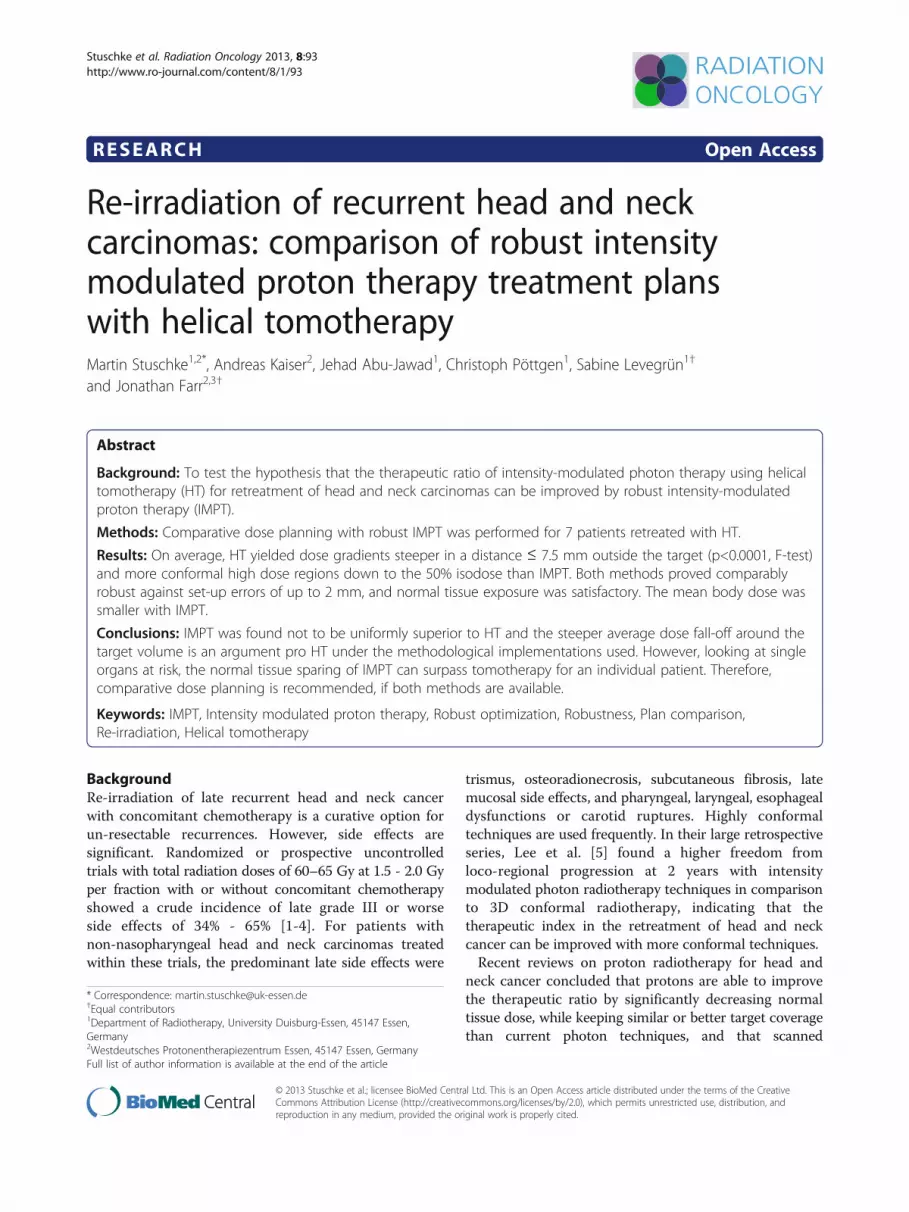
Cisplatin at 30 mg/m2 weekly, the remaining two refusedchemotherapy. With respect to clinical outcome, nopatient experienced grade 3 late toxicities. Medianfollow-up of the seven patients is 39 (31–48) months.Survival at 36 months is 57%. Figure 1 illustrates dosedistributions achieved with HT and IMPT in the first andsecond row, respectively. Dose difference plots are givenin the third row.
Coverage of the CTVFor HT and IMPT, the CTV D95 was equal or largerthan the prescribed dose for all patients except patient 1,in whom it was ≥ 97.5% of the prescribed dose (Table 1).In this patient, the smallest distance between CTV andbrainstem fell below 2 mm so that robustness againstset-up errors and fulfillment of strict brain stem werecompetitive objectives. Patients 4 and 6 received anintegrated boost, and their plans were normalized tothe prescribed dose in the CTVboost. The prescribeddoses to the larger CTV1 were 84.1% and 80.0% ofthe prescribed CTVboost dose, respectively. Robustnessof the IMPT and HT plans was evaluated withinscenarios 1–5, applying a translation of the isocenterof the beam arrangement against the image data setof −2.0/2.0/-2.0/2.0/1.5 mm in the ventral, -2.0/2.0/2.0/-2.0/-1.5 mm in the ipsilateral, and 0.0/ 0.0/ 0.0/0.0/1.5 mm in the cranial direction. In addition, densitychanges by +3%, and −3% were applied to the IMPT plansin scenario 5. The CTV D95 was found stable within 4%and did not fall below 98% of the prescribed dose in6 of the 7 patients for both IMPT and HT. In patient1 however, D95 decreased to 85.5% for HT and to89.1% for IMPT. A pairwise comparison of the D95 valuesof IMPT and HT over all the set-up errors tested did notresult in significant robustness differences between theplans for both radiotherapy methods (p=0.13, F-test). TheD2 values for brainstem or spinal cord increased over thescenarios on average by 3.7% (range, -9.4% - +26%) of theprescribed dose over the different patients for IMPT andby 3.1% (range, -8.3% - +19%) for HT with no significantdifference between HT and IMPT (p=0.58, F-test). Uniqueto IMPT, density changes of +3% and −3% within theset-up scenario 5 led to maximum increases of brain stemD2 by 19% and 15% of the prescription dose in patients 1and 2, respectively, and stayed constant within 4% in theother patients in comparison to scenario 5 without densitychanges. The D95 for the CTV remained constant within2% of the prescribed dose for all patients except patient 1,where it decreased by up to 6.7%.
ConformityThe median CTV volume was 86 cm3 (Table 1). ThePaddick conformity indices (CI) at the 95% isodose werelarger for the HT plans in comparison to robust IMPT
Stuschke et al. Radiation Oncology 2013, 8:93 Page 3 of 8http://www.ro-journal.com/content/8/1/93
in 8 of the 9 target volume comparisons, including theboost volumes given in patients 4 and 6 (p=0.04, signedrank test). Figure 2a shows the Paddick CITomo/ CIprotonratio for 9 target volumes of the evaluated patients atdifferent isodose values between 95% and 20%. The drawnline is a linear quadratic fit as Taylor series expansion tothe last significant term of this ratio on the isodose level.The predicted mean value of CITomo/CIproton and its 95%confidence interval ratio from this fit was >1 fromthe 95% isodose down to the 50% isodose, indicatingconsiderably higher average conformity for HT comparedwith IMPT at these higher isodose levels, while robustIMPT was more conformal for the 30% and lowerisodoses. The shallower average dose fall off in theIMPT-plans is visualized as a red shell around the CTV inthe dose differences plots of Figure 1, third row. Inaddition, we looked at the effect of reducing the rangeuncertainties during optimization from ±3% to ±1.5% onCI95 of the IMPT plans. The Paddick CI95% was foundstable within ±2% without a systematic increase.Figure 3 illustrates the dose fall-off outside PTV1 for
the 7 patients for HT and IMPT normalized by theprescribed dose. Mean doses are shown in adjacent shellsof 1 mm width around the PTV1 within the respectivepatient’s body. The dose fall-offs were adequatelydescribed by a linear dependence on distance from PTV.A quadratic term did not become significant. The slopeswere significantly steeper for HT than for IMPT (−5.94 ±0.26%/mm vs. -3.99 ± 0.26%/mm, p< 0.0001, F-test).
To give an impression of the steepest dose fall off achievedaround the PTV1, the D90 values in the adjacent 1 mmshells were analyzed. Again, gradients with HT weresignificantly steeper than with IMPT (−8.69 ± 0.36%/mmvs. -7.33 ± 0.36%/mm, p< 0.0001, F-test).
Normal tissue exposureSpinal cord or brainstem exposures were compared byD2 and Dmax or D1cm
3 and Dmax as the respective endpoints (Table 1). The paired dose differences betweenIMPT and HT normalized to the prescription dose werenot significantly different from 0 GyRBE over all patientsand all 4 parameters related to spinal cord and brainstemexposure (average difference, -1.6 ± 1.0 GyRBE, p=0.073,signed rank). The normalized dose differences betweenIMPT and HT were smaller for the parameters D2 orD1cm3 (average ΔD2 or ΔD1cm
3 = −3.7%) as for Dmax
(average ΔDmax = +0.4%). All plans fulfilled the clinicalcriteria, i.e. life time accumulated Dmax to the spinal cordand brain stem had to remain below 60 GyRBE and 64GyRBE, respectively.With respect to the ipsilateral optic nerve and the
chiasm, no significant Dmax differences were seen betweenthe IMPT and HT plans (Table 1). The average ΔDmax
between both methods was 0.5 ± 2.4% of the prescriptiondose for the 7 patients (p=0.89).Ipsilateral temporal lobe and cerebellum exposures
were evaluated by Dmean, and one parameter related tothe hot spots in this structure, either D1cm
3 or D5cm3 . The
Figure 1 Dose distribution. Dose distributions for patients 1–7 from left to right. Helical tomotherapy (HT) and intensity modulated protontherapy (IMPT) plans are shown in row 1 and row 2. Corresponding dose difference plots (IMPT minus HT) are given in the third row for therespective patients.
Stuschke et al. Radiation Oncology 2013, 8:93 Page 4 of 8http://www.ro-journal.com/content/8/1/93
Table 1 Dose volume parameters
Patient 1 Patient 2 Patient 3 Patient 4 Patient 5 Patient 6 Patient 7
Proton /Photon
Proton /Photon
Proton /Photon
Proton /Photon
Proton /Photon
Proton /Photon
Proton /Photon
CTV60 33 97
71127
197115
11 66
Paddick CI950.83 / 0.93 0.46 / 0.58 0.57 / 0.66
0.46 / 0.710.69 / 0.79
0.78 / 0.740.69 / 0.71
0.21 / 0.46 0.58 / 0.64
D9886.5 / 91.2 100.6 / 102.0 100.9 / 99.4
86.2 / 85.799.9 / 99.9
79.0 / 82.1100.4 / 101.8
101.3 / 100.5 104.7 / 101.3
D9597.5 / 98.5 102.0 / 102.3 101.1 / 99.7
88.4 / 85.5101.2 / 100.5
80.9 / 82.8100.4 / 102.1
101.4 / 100.8 101.2 / 103.2
D50104.4 / 105.3 105.3 / 103.8 103.8 / 101.3
95.6 / 89.9105.2 / 102.6
91.6 / 88.2104.0 / 103.3
104.2 / 102.0 104.7 / 104.7
D2-D9830.3 / 16.5 13.5 / 4.1 9.2 / 4.2
26.6 / 15.912.2 / 5.0
24.5 / 20.89.9 /3.4
16.2 / 3.2 8.4 / 3.4
Spinal Cord
D2 8.7 / 4.8 9.2 / 14.4 2.6 / 9.7 4.7 / 5.9 5.3 / 13.2 8.5 / 8.7 13,7 / 19.3
Dmax 16.6 / 8.2 13.5 / 18.7 10.2 / 16.0 14.4 / 13.7 13.3 / 18.7 15.4 / 13.3 25.0 / 28.1
Brain stem
D1cc 18.8 /21.6 41.0 / 45.0 11.6 / 11.9 14.3 / 16.5 30.6 / 46.5 3.8 / 5.0 4.7 / 6.2
Dmax 38.5 /40.0 79.5 / 78.7 30.1 / 22.7 33.5 / 25.0 61.6 / 64.5 8.2 / 9.0 13.6 / 10.6
ipsilat. Optic Nerve
Dmax 32.5 / 13.8 22.6 / 26.1 11.9 / 3.8 3.9 / 2.4 2.8 / 18.6 0.0 / 1.6 0.0 / 1.2
Chiasma Opticum
Dmax 18.7 / 15.4 28.1 / 15.1 0.8 / 3.4 6.3 / 2.7 2.0 / 16.0 0.0 / 1.4 0.0 / 1.2
Temporal Lobes
ipsilat. Dmean 17.9 / 21.9 54.4 / 50.9 5.9 / 10.7 28.6 / 15.5 47.3 / 57.1 0.3 / 1.6 0.7 / 1.2
ipsilat. D1cc 72.5 / 77.7 98.3 / 99.8 32.0 / 34.6 86.1 / 76.1 106.8 / 100.8 2.8 / 3.1 0.1 / 2.1
contoured Cerebellum
Dmean 9.0 / 18.4 13.3 / 26.2 0.5 / 9.8 9.1 / 16.4 11.1 / 32.1 0.2 / 2.5 0.1 / 1.4
D5cc 53.9 / 53.7 69.7 / 60.7 3.7 / 21.7 52.0 / 47.2 74.1 / 67.9 2.0 / 9.4 1.2 / 2.8
Parotid Glands
Dmean 37.3 / 26.9 3.0 / 4.5 17.6 / 33.0 1.2 / 7.9 0.0 / 4.8 24.3 /39.0 48.7 / 47.8
contoured Jaw
V80 0.6 / 0.1 4.1 / 2.2 11.2 / 11.5 16.6 / 10.5 7.8 / 6.7 5.7 / 6.0 9.3 / 10.1
Dmean 13.0 / 7.4 8.6 / 8.4 20.9 / 24.4 35.9 / 34.3 12.3 / 15.6 34.2 / 48.0 25.4 / 39.4
scanned Body
Dmean 10.2 / 11.1 6.9 / 9.5 7.2 / 9.7 4.4 / 4.4 3.2 / 5.7 3.7 / 5.3 3.6 / 4.9
V95 89 / 83 73 / 57 171 / 147 156 / 100 186 / 161 254 / 266 167 / 162
V90 108 / 92 87 / 64 193 / 167 178 / 112 205 / 177 281 / 291 190 / 180
V80 143 / 112 114 / 77 234 / 200 221 / 132 247 / 207 337 / 343 231 / 212
PTV 81 49 124 92 152 246 147
17 92
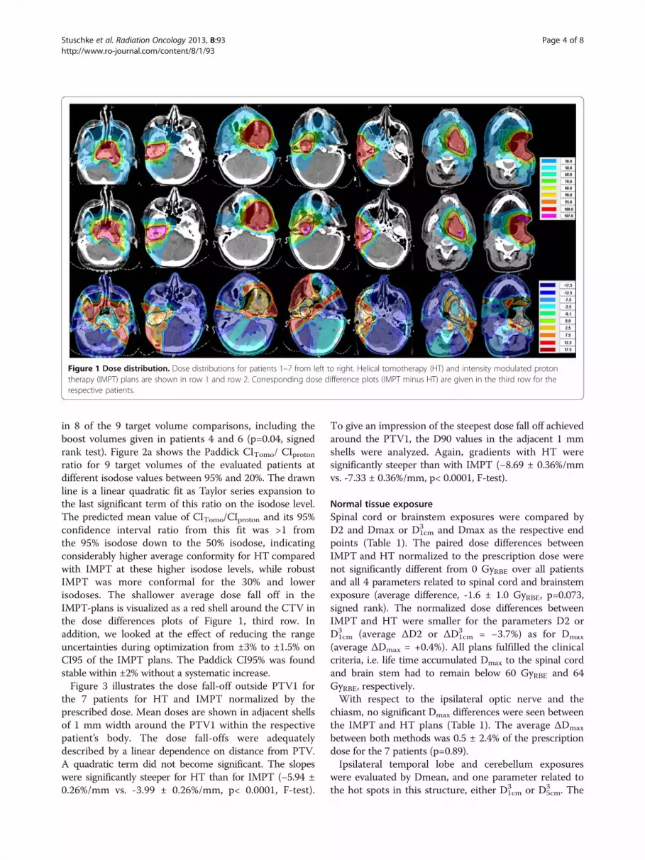
Dose volume parameters related to target coverage, homogeneity and the exposure of normal tissues by helical tomotherapy (Photon) and intensity modulatedproton therapy (Proton). All doses were given in percent of the prescribed dose, all volumes in cm3. Patients 4 and 6 were treated by an integrated boosttechnique and their plans were normalized to the prescribed boost dose. For these two patients, the respective cells for the D2 to D98 values were split and thevalues for the boost clinical target volume (CTV) were given in the lower half and for the larger CTV1 in the upper half of the cell.
Stuschke et al. Radiation Oncology 2013, 8:93 Page 5 of 8http://www.ro-journal.com/content/8/1/93

normalized Dmean differences between IMPT and HTfor ipsilateral temporal lobes were not different from 0%(average ΔDmean = 0.5 ± 2.8%; p=0.58, signed rank test),neither was the D1cm
3 (average ΔD1cm3 = 0.6 ± 2.0%;
p=0.94). The average ΔDmean for the cerebellum was lower
with IMPT (−9.1 ± 2.5%, p=0.02), the average ΔD1cm3 for
the patients of this study was not (1.0 ± 3.5%, p=0.99).In addition, the average Dmean difference of the parotid
gland exposure was −4.5 ± 3.4% (p=0.22). Mean body doseby IMPT was lower in 6 patients and equal in 1 patientcompared with HT, on average by 1.6 ± 0.4% of theprescribed dose (p=0.03).
DiscussionConsidering target coverage, safety margins alone cannotensure dose coverage of the CTV for IMPT plans withmultiple fields and high in-field dose gradients. Deviationsin the position of these dose gradients in the patient fromfield to field due to set-up errors or range uncertaintiescan result in under- or overdosage inside the PTV [15,16].Therefore robust planning was employed to optimize thedose distribution simultaneously for multiple scenariosmimicking set-up errors and range uncertainties andminimizing the penalty of the worst case scenario [12]. Ro-bust optimization can lead to treatment plans considerablyless sensitive to set-up errors and range uncertainties thanIMPT plans optimized using PTV-based conventionalmethods. In addition, robust IMPT optimization wasable to result in higher conformity than margin-basedIMPT optimization methods [17]. HT and IMPT withsmall pencil beams had similar target coverage androbustness against set-up errors within 2 mm. Highlyconstrained IMPT plans showed some residual dependenceof brainstem D2 on range uncertainties. Consideration ofa set-up error of 2 mm seems adequate for small targetsin the head and neck region with daily online navigation[18]. While on average the HT plans in our comparisonshowed a higher conformity around the CTV in the highdose region down to 50% of the prescribed dose or within7.5 mm around the PTV, IMPT plans had a reduced lowdose bath at isodoses below 50%. Similarly, Seco in 2011reported of larger high dose regions with passivelyscattered protons in comparison to photon stereotacticbody radiotherapy for smaller stage I lung cancer [19].IMPT for cancers of the head and neck region was
investigated in previous studies in comparison to fixedfield or rotational IMXT [20-25]. These studies used aPTV margin based concept to consider set-up errors forproton therapy and none used robust optimization. Allof these studies used 2–3 field IMPT plans. Four ofthese studies employed fixed-field IMXT, four HT forcomparison with IMPT [20,23,25,26]. While all of thestudies using fixed-field IMXT found that IMPT had agreater potential to spare adjacent normal tissues, thestudies using HT for comparison found similar conformityand normal tissue exposure. These studies included only1, 1, 3, and 6 patients, respectively. The target volumes inthe latter studies, however, tended to be larger than in thepresent study. The target conformity of the HT plans in
Figure 2 Conformity indices. Ratio of the Paddick conformityindices for helical tomotherapy (CItomo) and intensity modulatedproton therapy (CIproton) plans for all target volumes of this study.The ratios are given for isodose values from 95% to 20% of theprescribed dose. Data points related to the same target volume areconnected by grey lines. The average dependence of the conformityindex ratio on isodose was estimated by linear quadratic Taylorseries expansion (black solid curve). The 95% confidence intervals ofthe predicted mean isodoses by this fit are drawn as blackvertical bars.
Figure 3 Dose fall-off. Dose fall-off outside planning target volumefor helical tomotherapy (HT) (blue) and intensity modulated protontherapy (IMPT) (grey). Mean doses in adjacent shells of 1 mm widtharound the planning target volume (PTV) within the respectivepatient’s body are given normalized to the prescribed dose. Theslopes of the average dose fall-off differed between HT and IMPT(p< 0.0001). In addition, the 95% confidence intervals for thepredicted dose values at a given distance are indicated byvertical bars.
Stuschke et al. Radiation Oncology 2013, 8:93 Page 6 of 8http://www.ro-journal.com/content/8/1/93

the present study compared well with the photon plans inmost of the above studies [21,24,25], and the present studyextends the comparison of IMPT and HT to smallercomplex target volumes and nearby organs at risk, usingrobust IMPT optimization. Four of the above studies gavethe ICRU conformity index at the 95% isodose (CI95%)(ICRU reports 50 and 62) around the PTV for theirIMPT plans [21,22,24,25]. The mean CI95% rangedfrom 1.02 - 1.40. In the present study, the averageCI95% was 1.29 ± 0.24 with IMPT, a value in the middlerange of the above studies. But the median target volumein this study was smaller than in most of the abovestudies. It is well known that the CI decreases andtherefore conformity improves for a considered radio-therapy method with increasing target volumes [27]. Therobustness of the D95 for CTV-coverage against set-uperrors was found similar for robust IMPT and HT. Whileadequate brain, brainstem and spinal cord sparing couldbe achieved with IMPTat the level preset by the HT plans,temporal lobe doses and optic nerve doses were notsubstantially different on average. Dmean but not BitteD1cm3 wie auf der vorhergehenden Seite: cm tief, 3hoch gesetzt. to the cerebellum and the mean body dosewere lower on average with IMPT for the patients of thisstudy. Comparing HT with other rotational IMRT methodssuch as volumetric modulated arc therapy showed a similarconformity that was superior to static field IMRT so thatthe results here in comparison to HT can be generalizedto rotational IMRT methods with photons [28].In the re-irradiation situation, it can be difficult to weight
the better high dose conformity of HT against the reducedlow dose exposure by IMPT. Several of the severe sideeffects that can be consequence of re-irradiation, such astrismus, osteoradionecrosis, subcutaneous fibrosis, brainstem necrosis, and cranial neuropathy and carotid ruptureseem to be related rather to the volume of the highdose region than the low dose bath of radiotherapybelow the 50% isodose [13,29,30]. If steep dose gradientsin the near vicinity of the target volume are of dominantconcern, rotational IMRT is a particularly good radiotherapysolution. The known issue of difficult skin sparing withIMPT, relevant for superficially located target volumessuch as head and neck lymph node regions, was notconsidered in the comparison of the methods.The conformity of both methods, rotational IMXT and
IMPT, has potential to be improved in the future. Avenuesfor possible further improvements with IMPT for shallowdepth head and neck carcinomas include using smallerpencil beam width especially at low energies allowing fortreatments without range shifters, smaller air gaps,and further refinement of robust optimization using e.g.contour-related spot placing and spacing. Conformity withrotational IMXT can be improved for instance by smallerjaw width, smaller pitch and higher modulation factor
in the case of helical therapy and the use of smallpenumbra micromultileaf collimators, use of noncoplanar,multiple arcs in the case of C-arm linear accelerators, orthe use of multiple degrees of freedom robotic systems forbeam delivery.
ConclusionPotentials of intensity modulated proton therapy plansusing robust optimization were investigated under set-upand range uncertainty conditions. While HT showed onaverage a higher conformity down to the 50% isodose andsteeper dose gradients within 7.5 mm outside the PTV inthe high dose region, IMPT had a reduced low dosebath. Because neither of the two methods, IMPT or HT,was found uniformly better in terms of target coverageand organs at risk sparing, comparative planning isrecommended for the individual patient in the clinicalsituation of retreatment of recurrent head and neck cancer,provided both methods are available.
Competing interestsParts of this work were supported by Grant No. STU-151/9-1 from theGerman Research Foundation (Deutsche Forschungsgemeinschaft). Theauthors declare that they have no competing interests.
Authors' contributionsMS carried out the plan comparison, performed statistical analysis anddrafted the manuscript. He also contributed to the optimizations of both,helical tomotherapy and proton therapy plans. AK performed the robustoptimization and evaluation of the IMPT plans. He reviewed and edited draftversions of the manuscript. JAJ and CP conducted clinical treatment of thepatients, generated initial volumes of interest, and approved clinical helicaltomotherapy plans. SL performed the helical tomotherapy optimization andperformed dosimetric analysis of both, tomotherapy and IMPT plans. JF wasinvolved in improving the proton therapy optimization approaches and inthe design of the proton therapy planning system. He also participated inconcept discussions for developing the investigative aims and criticallyreviewed the draft manuscript. All authors read and approved the finalmanuscript.
Author details1Department of Radiotherapy, University Duisburg-Essen, 45147 Essen,Germany. 2Westdeutsches Protonentherapiezentrum Essen, 45147 Essen,Germany. 3Current address: Department of Radiologic Sciences, St. JudeChildren's Research Hospital, Memphis, TN, USA.
Received: 18 March 2013 Accepted: 9 April 2013Published: 20 April 2013
References1. Janot F, de Raucourt D, Benhamou E, Ferron C, Dolivet G, Bensadoun RJ,
Hamoir M, Gery B, Julieron M, Castaing M, et al: Randomized trial ofpostoperative reirradiation combined with chemotherapy after salvagesurgery compared with salvage surgery alone in head and neckcarcinoma. J Clin Oncol 2008, 26:5518–5523.
2. Langendijk JA, Kasperts N, Leemans CR, Doornaert P, Slotman BJ: A phase IIstudy of primary reirradiation in squamous cell carcinoma of head andneck. Radiother Oncol 2006, 78:306–312.
3. Langer CJ, Harris J, Horwitz EM, Nicolaou N, Kies M, Curran W, Wong S,Ang K: Phase II study of low-dose paclitaxel and cisplatin incombination with split-course concomitant twice-daily reirradiation inrecurrent squamous cell carcinoma of the head and neck: results ofRadiation Therapy Oncology Group Protocol 9911. J Clin Oncol 2007,25:4800–4805.
Stuschke et al. Radiation Oncology 2013, 8:93 Page 7 of 8http://www.ro-journal.com/content/8/1/93

4. Tortochaux J, Tao Y, Tournay E, Lapeyre M, Lesaunier F, Bardet E, Janot F,Lusinchi A, Benhamou E, Bontemps P, et al: Randomized phase III trial(GORTEC 98–03) comparing re-irradiation plus chemotherapy versusmethotrexate in patients with recurrent or a second primary head andneck squamous cell carcinoma, treated with a palliative intent. RadiotherOncol 2011, 100:70–75.
5. Lee N, Chan K, Bekelman JE, Zhung J, Mechalakos J, Narayana A, Wolden S,Venkatraman ES, Pfister D, Kraus D, et al: Salvage re-irradiation for recurrenthead and neck cancer. Int J Radiat Oncol Biol Phys 2007, 68:731–740.
6. Mendenhall NP, Malyapa RS, Su Z, Yeung D, Mendenhall WM, Li Z: Protontherapy for head and neck cancer: rationale, potential indications,practical considerations, and current clinical evidence. Acta Oncol 2011,50:763–771.
7. Van de Water TA, Bijl HP, Schilstra C, Pijls-Johannesma M, Langendijk JA:The potential benefit of radiotherapy with protons in head and neckcancer with respect to normal tissue sparing: a systematic review ofliterature. Oncologist 2011, 16:366–377.
8. Kirkpatrick JP, van der Kogel AJ, Schultheiss TE: Radiation dose-volumeeffects in the spinal cord. Int J Radiat Oncol Biol Phys 2010, 76:S42–S49.
9. Ho KF, Fowler JF, Sykes AJ, Yap BK, Lee LW, Slevin NJ: IMRT dosefractionation for head and neck cancer: variation in current approacheswill make standardisation difficult. Acta Oncol 2009, 48:431–439.
10. McDonald MW, Lawson J, Garg MK, Quon H, Ridge JA, Saba N, Salama JK,Smith RV, Yeung AR, Yom SS, Beitler JJ: ACR appropriateness criteriaretreatment of recurrent head and neck cancer after prior definitiveradiation expert panel on radiation oncology-head and neck cancer.Int J Radiat Oncol Biol Phys 2011, 80:1292–1298.
11. National Comprehensive Cancer Network Clinical Practice Guidelines inOncology [http://www.nccn.org], assessed at Feb 1, 2013.
12. Fredriksson A, Forsgren A, Hardemark B: Minimax optimization forhandling range and setup uncertainties in proton therapy. Med Phys2011, 38:1672–1684.
13. Marks LB, Yorke ED, Jackson A, Ten Haken RK, Constine LS, Eisbruch A, BentzenSM, Nam J, Deasy JO: Use of normal tissue complication probability modelsin the clinic. Int J Radiat Oncol Biol Phys 2010, 76:S10–S19.
14. Su SF, Huang Y, Xiao WW, Huang SM, Han F, Xie CM, Lu TX: Clinical anddosimetric characteristics of temporal lobe injury following intensitymodulated radiotherapy of nasopharyngeal carcinoma. Radiother Oncol2012, 104:312–316.
15. Albertini F, Hug EB, Lomax AJ: Is it necessary to plan with safety marginsfor actively scanned proton therapy? Phys Med Biol 2011, 56:4399–4413.
16. Stuschke M, Kaiser A, Pottgen C, Lubcke W, Farr J: Potentials of robustintensity modulated scanning proton plans for locally advanced lungcancer in comparison to intensity modulated photon plans. RadiotherOncol 2012, 104:45–51.
17. Liu W, Zhang X, Li Y, Mohan R: Robust optimization of intensitymodulated proton therapy. Med Phys 2012, 39:1079–1091.
18. Levegrun S, Pottgen C, Abu Jawad J, Berkovic K, Hepp R, Stuschke M:Megavoltage computed tomography image guidance with helicaltomotherapy in patients with vertebral tumors: analysis of factorsinfluencing interobserver variability. Int J Radiat Oncol Biol Phys 2012,85:561–569.
19. Seco J, Panahandeh HR, Westover K, Adams J, Willers H: Treatment of non-small cell lung cancer patients with proton beam-based stereotacticbody radiotherapy: dosimetric comparison with photon plans highlightsimportance of range uncertainty. Int J Radiat Oncol Biol Phys 2012,83:354–361.
20. Muzik J, Soukup M, Alber M: Comparison of fixed-beam IMRT, helicaltomotherapy, and IMPT for selected cases. Med Phys 2008, 35:1580–1592.
21. Simone CB 2nd, Ly D, Dan TD, Ondos J, Ning H, Belard A, O'Connell J, MillerRW, Simone NL: Comparison of intensity-modulated radiotherapy,adaptive radiotherapy, proton radiotherapy, and adaptive protonradiotherapy for treatment of locally advanced head and neck cancer.Radiother Oncol 2011, 101:376–382.
22. Taheri-Kadkhoda Z, Bjork-Eriksson T, Nill S, Wilkens JJ, Oelfke U, JohanssonKA, Huber PE, Munter MW: Intensity-modulated radiotherapy ofnasopharyngeal carcinoma: a comparative treatment planning study ofphotons and protons. Radiat Oncol 2008, 3:4.
23. Thorwarth D, Soukup M, Alber M: Dose painting with IMPT, helicaltomotherapy and IMXT: a dosimetric comparison. Radiother Oncol 2008,86:30–34.
24. Van de Water TA, Lomax AJ, Bijl HP, de Jong ME, Schilstra C, Hug EB,Langendijk JA: Potential benefits of scanned intensity-modulated protontherapy versus advanced photon therapy with regard to sparing of thesalivary glands in oropharyngeal cancer. Int J Radiat Oncol Biol Phys 2011,79:1216–1224.
25. Widesott L, Pierelli A, Fiorino C, Dell'oca I, Broggi S, Cattaneo GM, Di MuzioN, Fazio F, Calandrino R, Schwarz M: Intensity-modulated proton therapyversus helical tomotherapy in nasopharynx cancer: planning comparisonand NTCP evaluation. Int J Radiat Oncol Biol Phys 2008, 72:589–596.
26. Fogliata A, Yartsev S, Nicolini G, Clivio A, Vanetti E, Wyttenbach R, BaumanG, Cozzi L: On the performances of intensity modulated protons, rapidarcand helical tomotherapy for selected paediatric cases. Radiat Oncol 2009,4:2.
27. Nakamura JL, Verhey LJ, Smith V, Petti PL, Lamborn KR, Larson DA, WaraWM, McDermott MW, Sneed PK: Dose conformity of gamma kniferadiosurgery and risk factors for complications. Int J Radiat Oncol Biol Phys2001, 51:1313–1319.
28. Lu SH, Cheng JC, Kuo SH, Lee JJ, Chen LH, Wu JK, Chen YH, Chen WY, WenSY, Chong FC, et al: Volumetric modulated arc therapy fornasopharyngeal carcinoma: a dosimetric comparison with TomoTherapyand step-and-shoot IMRT. Radiother Oncol 2012, 104:324–330.
29. Emami B, Lyman J, Brown A, Coia L, Goitein M, Munzenrider JE, Shank B,Solin LJ, Wesson M: Tolerance of normal tissue to therapeutic irradiation.Int J Radiat Oncol Biol Phys 1991, 21:109–122.
30. McDonald MW, Moore MG, Johnstone PA: Risk of carotid blowout afterreirradiation of the head and neck: a systematic review. Int J Radiat OncolBiol Phys 2012, 82:1083–1089.
doi:10.1186/1748-717X-8-93Cite this article as: Stuschke et al.: Re-irradiation of recurrent head andneck carcinomas: comparison of robust intensity modulated protontherapy treatment plans with helical tomotherapy. Radiation Oncology2013 8:93.
Submit your next manuscript to BioMed Centraland take full advantage of:
• Convenient online submission
• Thorough peer review
• No space constraints or color figure charges
• Immediate publication on acceptance
• Inclusion in PubMed, CAS, Scopus and Google Scholar
• Research which is freely available for redistribution
Submit your manuscript at www.biomedcentral.com/submit
Stuschke et al. Radiation Oncology 2013, 8:93 Page 8 of 8http://www.ro-journal.com/content/8/1/93


