Embed Size (px)
Citation preview

3 Recent Advances in Pressure Ulcer ResearchDan Bader and Cees Oomens
Introduction
The concept of scientific research aimed at both the prevention and treatment of pressure ulcers has been evident in the literature for at least four decades.Indeed in 1975 a seminal conference entitled Bed Sore Biomechanics was organized at Strathclyde University, the proceedings of which were published in abook, Bed Sore Biomechanics,1 which included an impressive list of contributionsfrom a variety of scientific and medical disciplines. The book contained a number of critical messages concerning the factors associated with the absolutelevels of prolonged pressure at the patient–support interface that can cause tissue breakdown. In particular, the time of prolonged pressure2 and the pre-sence of shear forces3,4 were both clearly established as important factors. In addition, the effects of a number of external mechanical stimuli on tissue usinganimal models were described, the damage being assessed using histologicalmethods.
Given this knowledge base it may be worth asking what has been achieved inthe last 25 years as prevalence rates have remained unacceptably high as describedin other chapters. This is, at least, partly due to the limited fundamental knowl-edge related to the etiology of the clinical condition. Thus, the design and appli-cation of preventive aids and risk assessment techniques are still dominated bysubjective measures or, at best, based on a relatively small amount of data focus-ing on skin, which are largely outdated or misinterpreted.
A striking example is the traditionally quoted value for capillary closure pres-sure of 32 mmHg (4.3 kPa) that is still frequently used as a threshold for tissuedamage. This value was based on the measured pressure in the skin capillarieswithin the nail folds5 and thus represents a measure of localized interstitial pres-sure not relevant to areas at risk of pressure-induced damage. Its use is totally inap-propriate as a threshold value for interface pressures at load-bearing sites. Interfacepressures at the contact area between skin and supporting surfaces in excess ofthis value are assumed to produce a degree of ischemia that, if applied for asufficient period of time, may lead to tissue breakdown.6,7 Ignoring factors otherthan pressure-induced ischemia for tissue breakdown in pressure ulcers, capillaryclosure depends on local pressure gradients across the vessel wall and not just on
11
interface pressures at skin level. Hence interface pressures well above capillarypressures can be supported by the soft tissues before blood flow is seriouslyimpaired.8 An interesting observation reported by Husain9 was that localized inter-face pressures obliterated more vessels in the skin and subcutaneous tissue thanin the muscle, while the latter was severely damaged and the skin and subcutiswere not. Later studies also demonstrated that muscle tissue is more susceptibleto mechanical loading than skin.6,10
In order to be able to reduce the prevalence of pressure ulcers it is essential toimprove and expand understanding of the etiology in terms of both basic scienceand clinical experience. A more rigorous analysis of existing data is postulated followed by a hierarchical research approach in which the effects of mechanicalloading on the different functional units of soft tissue are studied. This chapterevaluates the current research achievements and proposes new avenues which canprovide the necessary scientific evidence to enable the development of successfulprevention strategies.
Interface Pressure Measurements
It has long been recognized that the field of bioengineering can play a major rolein the research activity. Perhaps its most established activity in pressure soreresearch has involved the development of a range of pressure monitoring systems,to supersede the previous gold standard, the Talley-Schimedics single cell system(described by Reswick and Rogers2). One such advance was the Oxford Mk I/II,later the Talley Pressure Monitoring system, employing an array of 96 sensors.11
This system has been replaced by other more numerous sensor arrays with asso-ciated elegant software to display pressure profiles, produced by companies suchas Tekscan, FSA, and Novel. Such monitoring systems are clearly valuable in bothresearch and clinical settings, either in assessing the performance of one product(often new) against its competitors or in the comparison of a range of supportproducts with an individual patient. However, it is well recognized that pressuremeasurements alone are not able to either alert the clinician to areas of tissue thatare particularly vulnerable to the initiation of ulcers or provide insight into manyfundamental aspects of the clinical problem, such as etiology or identification ofsusceptible subjects.
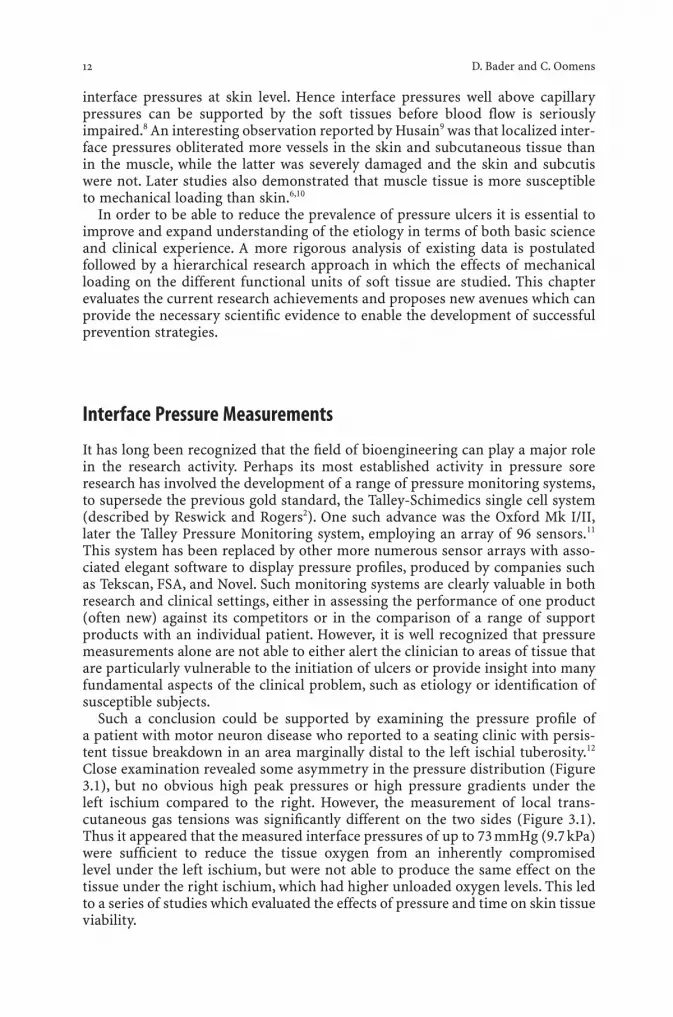
Such a conclusion could be supported by examining the pressure profile ofa patient with motor neuron disease who reported to a seating clinic with persis-tent tissue breakdown in an area marginally distal to the left ischial tuberosity.12
Close examination revealed some asymmetry in the pressure distribution (Figure3.1), but no obvious high peak pressures or high pressure gradients under the left ischium compared to the right. However, the measurement of local trans-cutaneous gas tensions was significantly different on the two sides (Figure 3.1).Thus it appeared that the measured interface pressures of up to 73 mmHg (9.7 kPa)were sufficient to reduce the tissue oxygen from an inherently compromised level under the left ischium, but were not able to produce the same effect on thetissue under the right ischium, which had higher unloaded oxygen levels. This ledto a series of studies which evaluated the effects of pressure and time on skin tissueviability.
12 D. Bader and C. Oomens

Evaluation of Tissue Status under External Loading
Over the last two decades, a number of techniques have been proposed to indicatethe viability, or status, of soft tissues subjected to periods of loading. These tech-niques have been, to date, largely restricted to examining the response of skinlayers to mechanical loading, and include measurements of blood flow in the skinusing laser Doppler fluxmetry13 and reflective spectrophotometry.14,15 Advances inthe latter technique have enabled distinct absorption spectra to be identified foroxygenated and deoxygenated blood in skin. The authors claimed that a numberof other skin biomolecules, such as melanin and collagen, can also be distin-guished.15 However, the most common technique employed to measure skin via-bility involves transcutaneous gas tensions (TcPO2 and TcPCO2), which to ensuremaximum vasodilation have to be measured at elevated skin temperatures.8,16–19
One such study examined the effects of cyclic loading on the tissue viability ofhealthy and debilitated subjects.8 Two distinct responses were observed as shownin Figure 3.2. The normal response yielded a rapid and complete tissue recoveryto unloaded TcPO2 levels and the apparent effect of the applied load diminishedwith successive cycles. By contrast, in some cases recovery was not fully achievedwithin a prescribed period and subsequent loading had a cumulative effect on thediminution of TcPO2 levels. It is this latter group who must be considered to be atparticular risk of developing pressure ulcers.
The technique has also been employed specifically to investigate patients both in the acute phase20 and in the subacute phase of spinal cord injury.21 Thelatter study employed an assessment criterion for tissue viability based on the
Recent Advances in Pressure Ulcer Research 13
Interface pressures (mmHg)
Local transcutaneous oxygen tension (mmHg)
L
31
33
49
15
25
42
39
73
39
41
62
71
31
56
67
29
34
39
39
39
33
45 63
31
22
14
R
Figure 3.1 Interface pressure profile under theischia of a patient with recurrent tissue break-down under the left ischium.(Based on Bader andChase.12)

percentage time at which the TcPO2 and TcPCO2 values were within acceptablelevels when subjects were seated on prescribed support cushions. Clear relation-ships were indicated between depressed levels of TcPO2 and elevated levels ofTcPCO2, at associated high values of interface pressure.21 In addition, it wasreported that changes in tissue viability do occur during a 12-month period,although a subpopulation, involving paraplegic subjects with flaccid paralysis,remain highly susceptible to the development of pressure ulcers.
This research activity spawned the routine use of these objectives measures toassess all patients with spinal cord injury at a specialized seating clinic.18 Thisrecent paper questioned the efficiency of short-term pressure lifts in restoring thetissue oxygen levels following prolonged seated periods. Indeed the authors rec-ommend the use of alternative pressure relief strategies tailored to individualpatients. Although yielding solid practical education for both patients and carers,these and related studies have still yielded no clear guidelines as to the precise rela-tionship between compromised tissue gas levels for a set time period and the onsetof progressive tissue breakdown that will ultimately result in a pressure ulcer.
Tissue Biochemistry
An alternative biochemical approach to assessing tissue status is to examine themetabolite levels in localized soft tissue areas subjected to pressure ischemia andsubsequent reperfusion. These metabolites can be transferred via the sweat glands,which are simple tubular glands, and can be collected at the skin surface. Sweat isa hypotonic solution of sodium and chloride ions in water, together with other con-stituents including lactate, urea, and potassium, these metabolites accounting forabout 95% of the osmotically active substances in sweat.23
In one of the few relevant studies, Hagisawa and colleagues24 used a bulky systemto chemically induce sweat production. By contrast, a series of studies by the authorand colleagues25–28 collected thermally induced sweat by absorption on thin pads,made from filter paper, attached to the skin surface. This collection system
14 D. Bader and C. Oomens
Impaired
Tissueviability
Loadon
Normal
Impaired
Time
Figure 3.2 A schematic rep-resentation of two distinctresponses with respect to theviability of soft tissues sub-jected to repeated loading.Arrows up (down) representstart of applied loading(recovery) period. (Based onBader and Chase.12)

provided minimal distortion and proved ideal for use at a loaded tissue supportinterface. One such study compared sweat collected during periods of loading atthe ischium and sacrum with sweat collected during unloaded periods at adjacenttissue sites.25 The study revealed that tissues subjected to partial ischemia,specifically produced by a uniaxial indenter system, yielded a general increase inconcentrations of sweat lactate, chloride, urea, and urate associated with adecreased sweat rate. Following the removal of loading, the levels of both sweatmetabolites tended to be restored to basal levels.
In a separate study,29 sweat was collected at two adjacent sites, one loaded andone unloaded, at the sacrum of a number of able-bodied subjects. Three distinctpressures were applied. Estimations were made of both the absolute values of sweatmetabolite concentrations and the ratios of the concentration at both loaded andunloaded tissue sites, thus eliminating the wide variation between subjects. As anexample, the ratio for lactate is presented as a function of the three applied pres-sures in Figure 3.3. It is evident that there is a significant increase in sweat lactateratios at applied pressures of 40 mmHg (5.3 kPa) and above. Indeed a linear regres-sion model applied to the lactate data, using the Spearman correlation coefficient,revealed statistical significance at the 5% level. Similar trends were also apparentwith sweat urea, urate, and chloride.28 In addition, the absolute lactate concentra-tions for the three pressures were pooled as loaded data in conjunction withunloaded data to yield two separate relationships with the inverse of sweat rate.The data sets yielded significant linear trends, although both slopes and interceptsof the models associated with the loaded data were higher than those for theunloaded controls.30
Recent Advances in Pressure Ulcer Research 15
p < 0.01p < 0.01
Applied pressure (mmHg)
[Lac
tate
] load
ed /
[Lac
tate
] cont
rol
0
0 20 40 60 80 100 120
0.2
0.4
0.6
0.8
1
1.2
1.4
1.6
1.8
2
Figure 3.3 The effect of applied pressure on the ratio of sweat lactate concentration as a result of sacral loading in a group of able-bodiedsubjects. Linear model y = 0.0046x + 0.975; r = 0.48, p < 0.01.

The study was extended by employing two independent techniques in combi-nation to assess the soft tissue response to applied pressure in a group of able-bodied subjects, to establish baseline data.29 The methods involved thesimultaneous measurement of the local tensions of oxygen and carbon dioxide(TcPO2 and TcPCO2) and the collection and subsequent analysis of metabolite con-centrations of sweat samples. Adjacent loaded and unloaded sites on the sacrumwere tested to allow for between-subject variation. Several parameters wereselected from each of the techniques and their interrelationships were examined.Results indicated that oxygen levels (TcPO2) were lowered in soft tissues subjectedto applied pressures of between 40 mmHg (5.3 kPa) and 120 mmHg (16.0 kPa).29 Atthe higher pressure levels, this decrease was generally associated with an increasein carbon dioxide levels well above the normal basal levels of 45 mmHg (6 kPa). Bycomparing selected parameters, a threshold value for loaded TcPO2 could beidentified, representing a reduction of approximately 60% from unloaded values,as indicated in Figure 3.4a. Above this threshold level there was a significant rela-tionship between this parameter and the loaded/unloaded concentration ratios forboth sweat lactate and urea.29 Given that tissue oxygen and sweat lactate reflect dif-ferent aspects of tissue ischemia, this degree of reduction (60% in median oxygentension) may represent a critical level for the development of tissue damage. Thestudy also related the lactate ratio to the percentage time at which TcPCO2 exceeded50%. Figure 3.4b indicates the presence of two distinct clusters of data. Forexample, when the carbon dioxide parameter exceeded 37%, the lactate ratios werewell in excess of unity. Differences could be attributed to the degree of pressure-induced tissue ischemia. Thus under conditions of mild ischemia elevated levelsof tissue carbon dioxide may be released from loaded areas in a normal manner,resulting in TcPCO2 values below 50 mmHg, whereas in severe conditions, bothsweat lactate and TcPCO2 will be elevated (Figure 3.4b).
Sweat lactate is generally thought to be derived from the sweat gland itself.23,31
During normal metabolism, oxidative phosphorylation is believed to be the mainmetabolic pathway of the eccrine sweat gland.32 However, under conditions ofischemia and/or in anaerobic conditions, glycolysis becomes the main metabolicpathway resulting in the formation of lactate. This explains the elevated lactate con-centrations observed in the sweat collected from the loaded experimental site andsuggests that a sufficient degree of ischemia was induced in the sacral tissue duringthe two loading periods.
Sweat urea is believed to be derived mainly from serum urea by the passive dif-fusion across the glandular wall and cell membrane, although it is still unknownwhether it is also produced by the sweat gland.32 Urea is the main product ofprotein metabolism and can thus be an indicator of tissue damage if elevated levelsare found in bodily fluids, such as urine or blood. Prolonged periods of ischemiacan lead to muscle damage, resulting in an increased serum urea level which, inturn, can result in enhanced concentrations of sweat urea.32 These findings as evi-denced in the published study29 suggest that the tissue was compromised duringthe loading period. It was strongly proposed by the authors that such an approach,using a series of parameters, might prove useful in identifying those subjectswhose soft tissue may be compromised during periods of pressure ischemia.
Current work by the authors suggests that monitoring sweat lactate and ureaalone is not sufficient to give a full indication of the tissue status, particularlyduring reperfusion.30 Sweat purines, specifically uric acid, xanthine, and hypoxan-thine, are undoubtedly useful markers or “finger prints,” as they provide an
16 D. Bader and C. Oomens

Recent Advances in Pressure Ulcer Research 17
2.5
2.0
1.5
1.0
0.5
0.00 20 40 60 80 100
Lact
ate r
atio
(loa
ded/
unlo
aded
)
Percentage reduction in median TcPO2a
b
2.5
2.0
1.5
1.0
0.5
0.00 20 40 60 80 100
Lact
ate r
atio
(loa
ded/
unlo
aded
)
Percentage loading time TcPO2 > 50 mmHg
Figure 3.4 Relationship between ratio of sweat lactate concentration and (a) percentage reduction in transcutaneous gas tension (TcPO2)and (b) percentage of time for which transcutaneous carbon dioxide tension (TcPCO2) exceeded 50 mmHg, as a result of sacral loading onindividual subjects. (Based on Knight et al.29)

indication of the metabolic status of the tissue during both ischemia, when thereis energy depletion, and reperfusion and, as such, may be of significant potentialuse to identify patients at risk of developing pressure ulcers. It is clear that the useof a combination of biochemical markers is required to monitor the status of softtissues.
Internal Mechanical Environment
Although it is well acknowledged that pressure sores are primarily caused by sus-tained mechanical loading of the soft tissues of the body, prevention of the soresby reducing the degree of loading alone remains difficult. This is mainly due to thefact that the underlying pathways whereby mechanical loading leads to tissuebreakdown are poorly understood. It is not clear how global, external loading con-ditions are transferred to local stresses and strains inside the tissues and how theseinternal conditions may ultimately lead to tissue breakdown.
As mentioned in the introduction, surface or interface pressures are not repre-sentative of the internal mechanical conditions inside the tissue,which are most rel-evant for tissue breakdown. This is especially the case when tissue geometry andcomposition are complex and surface pressures result in highly inhomogeneousinternal mechanical conditions, as is the case adjacent to bony prominences.Nonetheless, in order to study the response of various tissue layers to mechanicalloading the local mechanical environment within these layers needs to be known.There are options available to measure the internal mechanical state, although theyinevitably involve invasive techniques such as a wick catheter.33,34 Sangeorzan et al.34
reported that the values for interface and intersitial pressures were not equivalentand were highly dependent on the nature of the intervening soft tissues. Thus thethickness, tone, and mechanical integrity of subcutaneous tissues, and the proxim-ity of bony prominences will influence this relationship. A more recent investiga-tion of elderly subjects during a single surgical procedure, namely the fixation of afractured neck of femur, examined the response of tissues adjacent to the lateralaspect of the proximal thigh. Results indicated that skin interface pressures weredissipated within the depth of the tissues resulting in reduced internal stresses.35
Indeed linear models of the data suggested interstitial stresses ranging between 29%and 40% of the applied interface pressures, as illustrated in Figure 3.5. This high-lights the protective nature of tissues to attenuate the effects of sustained pressure.
An alternative approach to investigate the transition from global external loadsto local internal stresses and strains involves the use of computer models, in par-ticular using finite element analysis (FEA).36–39 This approach, which models thecomplex geometries and material behavior of the human buttocks, is often unfa-miliar to experimentalists and clinical and nursing staff. In the study by Todd andTacker,37 the seated positions were simulated, thereby manipulating boundary con-ditions of the model. These authors concluded that there was no clear correlationbetween interface pressures and the local mechanical conditions. Oomens and co-workers40 created a finite element model of a human subject sitting on a cushion,which incorporated three different tissues, overlaying the human ischial tuberosi-ties, simulated by an undeformable bony indenter. The soft tissues, namely themuscle, fat, and skin, were modeled as nonlinear viscoelastic materials. Figure 3.6clearly shows the inhomogeneous mechanical condition of the various tissue layersand areas of high internal stresses in the deeper fat and muscle layers.
18 D. Bader and C. Oomens

However, any extrapolation of results from these computer analyses to the clin-ical setting must be undertaken with extreme caution. Specifically these modelsare dependent on the lack of reliable material properties for soft tissues, which canbe influenced by many systemic and local factors, such as temperature and nutri-tional status. Thus, although several studies have examined uniaxial and biaxialproperties of skin parallel to its surface, there are few reported studies examiningthe compressive properties of the soft tissue composite. Such studies have been
Recent Advances in Pressure Ulcer Research 19
80
70
60
50
40
30
20
10
00 20 40 60 80 100 120 140
Inte
rstit
ial p
ress
ure (
mm
Hg)
Interface pressure (mmHg)
Figure 3.5 The relationshipbetween interface pressuresand interstitial pressureswithin the soft tissues adja-cent to the greater trochanterof two surgical patientsundergoing hip screw fixa-tion of an intertrochantericfemoral fracture. Slopes oftwo linear models are 0.28and 0.41, r > 0.96 in bothcases. (Based on Bader andWhite.35)
Cushion
Skin
Buttockmodel
BoneMuscle
Fat2.0 [MPa]1.61.20.80.40.0
Figure 3.6 Simplified com-puter model of axisymmetricdeformed buttock (top right)demonstrating the differen-tial response of the separatesoft tissue layers (left) duringsitting of an 80 kg malesubject on a foam cushion.Values indicate Von Misesstresses, representing distor-tional energy. Note the areasof high stress in the subcuta-neous fat and muscle layers(arrows). (Based on Oomenset al.40)

hampered by the lack of appropriate non-invasive techniques that can character-ize material properties of tissue under load. For example, ultrasound has offeredmuch potential for many years but has, as yet, not proved reliable, although moresophisticated systems involving elastography in association with ultrasoundimaging might prove successful in the future. Other imaging technologies involving infrared spectroscopy and magnetic resonance imaging/spectroscopy(MRI/MRS) may also provide valuable data under loading conditions for bothhealthy tissues and where tissue status is compromised. Indeed in recent studiesGefen and colleagues41,42 have determined mechanical stiffness of soft tissuesunder load, using routine MRI scans. An increased mechanical stiffness was alsoreported corresponding to mixed tissue specimens around human ulcers com-pared to control values.43
Mechanisms of Pressure Ulcer Development
Conventional wisdom on the pathogenesis of pressure ulcers has focused on theeffects of pressure-induced ischemia on skin tissues. Although important there areother major considerations, as outlined in a recent viewpoint article,44 involvingthe lymphatic system, interstitial transport, underlying tissues particularly themuscle, ischemia–reperfusion injury, and sustained deformation of cells. Severalseminal papers associated with each of these mechanisms have been highlightedin Table 3.1. Although known for several decades, these mechanisms have not beenfully explored often due to technical reasons. As an example, the obliteration oflymphatic flow due to external pressure was measured in an animal limb, using aradioactive tracer.48 Clearly, this experimental approach could not be adopted in ahuman model. In a similar manner, ischemic and reperfusion damage is tradi-tionally evaluated using histological techniques, which are both time-consumingand do not permit real-time assessment of damage.
Overall the theories focus on different functional units of soft tissue, involvingcells, the interstitial space with extracellular matrix, and blood and lymph vessels.These units are affected by mechanical loading to varying degrees and hence havedifferent relevance for tissue breakdown. Most probably each of them contributesto the causation of pressure ulcers, although their individual and combined role in
20 D. Bader and C. Oomens
Table 3.1. The pathophysiology of pressure ulcers: soft tissue response to mechanical loading
Mechanism Consequences Key papers
1. Localized ischemia Capillary perfusion decreases with Daniel et al.6—animal model mechanical loading Kosiak7—animal model
Lack of local vital nutrients Dinsdale4—animal model Herrman et al.45—animal model
2. Impaired interstitial fluid Accumulation of metabolic waste products Krouskop et al.46—hypothesis flow and lymphatic drainage Reddy et al.47—theoretical model
Miller and Seale48—animal model
3. Reperfusion injury Restoration of blood flow may lead to McCord49—hypothesis toxic levels of oxygen free radicals Peirce et al.50—animal model
Unal et al.51—animal model
4. Sustained deformation of cells Local cell damage and death Ryan52—theoretical modelLandsman et al.53—cell modelBouten et al.54—cell model

tissue breakdown will undoubtedly vary depending on the nature of the mechan-ical insult and patient characteristics such as illness or age,55 which affect softtissue properties and hence the liability to tissue breakdown.
Hierarchical Approach
A hierarchical approach has recently proposed44 in which the effects of loading arestudied using different, yet complementary, model systems with increasing com-plexity and length scale and incorporating one or more functional tissue units.Thus, in vitro models, ranging from the single cell (mm scale) to cell-matrix con-structs (mm scale) and individual tissue layers (mm–cm scale), might be used tostudy the relationship between cell deformation and cell damage as well as theinfluence of the surrounding extracellular matrix and three-dimensional tissuearchitecture on this relationship. The role of tissue (re)perfusion and lymph flowas well as the interaction between tissue layers in bulk tissue might further beassessed using in vivo studies with animal models or human subjects.
The different length scales of these models can be coupled to multiscale computer calculations that enable the prediction of the internal microscopicmechanical environment within a given model from global, macroscopic loadingconditions, such as interface pressures (and vice versa). In this way relationshipsbetween, for instance, cell deformation and cell damage54 can be extrapolated tothe level of bulk tissue to give clinically relevant predictions on tissue breakdown.
Recent Focus on Pressure-Induced Muscle Damage
Muscle tissue is particularly susceptible to sustained compression. Compression-induced muscle breakdown predominantly occurs in muscle layers associated withbony prominences, eventually leading to gross tissue degeneration in the form ofdeep pressure ulcers.8,21,57–59 This breakdown starts at the cellular level with nuclearpyknosis and disintegration of the contractile proteins and the cell membrane, fol-lowed by inflammatory reactions.6,7,10,60,61 Although it is clear that both the magni-tude and the duration of compression affect the cellular breakdown, the underlyingpathways whereby tissue compression leads to injury of the cell remain poorlyunderstood. Moreover, most of the mechanisms detailed in Table 3.1 ignore the direct effects of cellular deformation due to prolonged tissue compression,which have recently been suggested as an important trigger for pressure ulcerdevelopment.52–54
The earlier study52 was extended to study cellular breakdown in response to sus-tained cell deformation, independently of other factors, such as blood perfusion.It utilized a three-dimensional in vitro system, incorporating cultured muscle cellsseeded in an agarose gel construct. The feasibility of this system to induce pro-longed cell deformation during gross construct compression was recently demon-strated by the authors.54 Strain applied to the translucent agarose gel results indeformation of the muscle cells to an elliptical form, which can be quantified usingconfocal laser scanning microscopy. Identical cylindrical cores cut from theagarose/cell suspension were subjected to two separate compressive strains, 10%and 20%. The strain was applied for time periods ranging from 0.5 to 12 hours,using a specially designed loading apparatus.62 After each compression period,
Recent Advances in Pressure Ulcer Research 21

sections taken from the central horizontal plane of the individual constructs werestained using both histological and fluorescent probes, to assess the proportion ofdamage. It was found that constructs subjected to the higher strain values demon-strated significantly higher values of nonviable cells for equivalent time pointscompared to the unstrained constructs, as illustrated in Figure 3.7. These findingsimply a relationship between the duration of applied compression and damage tomuscle cells seeded in the gel. Such an approach might be useful in establishingdamage threshold levels at a cellular level. The model was extended further bydeveloping a more physiological tissue equivalent muscle,63 by suspending pre-mature muscle cells in a collagen scaffold. The muscle cells fused into a branchednetwork of multinucleated, contractile myofibers by the application of appropri-ate biochemical and mechanical cues. Results indicated that cell death was evidentwithin 1–2 hours at clinically relevant straining percentages.
22 D. Bader and C. Oomens
100
10% strain
20% strain
90
80
70
60
50
40
30
20
10
00 2 4 6
Time of compression/h
Perc
enta
ge
diff
eren
ces
in c
ell v
iab
ility
dta
co
mp
ress
ion
8 10 12
Figure 3.7 THE effects of prolonged static compres-sion at two applied strains onthe viability of muscle cellsseeded in agarose constructs,as indicated by histologicalassessment.

In addition, the uniform distribution of dead cells throughout the muscle con-structs suggested that sustained deformation was the principal cause of cell death.A hybrid approach was then adopted by the authors in which these experimentaldata were used in the derivation of a damage law.64 In particular, the evolution ofdamage was predicted in a single microstructural unit, which could be extrapo-lated to the macroscopic scale. A damage evolution parameter, D, was defined,which accumulates with time when the dimensionless strain energy density para-meter, U, in a cell is higher than a cell tolerance parameter, a. The authors pro-posed a damage evolution equation:
where both a and b are material parameters that can be determined from the invitro experiment. Although limited at the present time to qualitative insight intotissue damage, this multilevel finite element approach has future potential as aquantitative predictor of damage in patient-related simulations.
The advent of new technologies that are sensitive to changes throughout the soft tissue composite provide new opportunities for the examination of animalmodels,50,51,60 despite their limitations associated with intrinsic biological variation,ethical issues, and inadequate experimental controls. As an example, Bosboom etal.60 examined the ability of MRI to assess local muscle damage after prolongedtransverse loading. The tibialis anterior muscle (TA) and overlying skin of a ratwere compressed between an indenter and tibia. A very large pressure, equivalentto 1875 mmHg (250 kPa), was applied for 2 hours. Histological examination, usinga semi-automated image-processing program, and in vivo T2-weighted MRI wereperformed 24 hours after the completion of the loading session. Figure 3.8 (seecolor section) illustrates the damage in transverse histological slices (below) andthe associated MR images for three sets of experiments. In each case, the locationof damage coincided well in the two assessment techniques. However, the inter-animal variability in damage is evident. Current work has involved a modified MR-compatible loading apparatus to produce more reproducible tissue damage andlearn more about the influence of deformation of the tissue and the influence ofreperfusion.65
A large variety of imaging techniques have been developed that can be appliedto assess structure, function, and metabolism of skeletal muscle. These includetagging MRI and perfusion MRI, which can be used to measure local tissue defor-mation and tissue perfusion, respectively. In addition, MR spectroscopy could beapplied to examine the biochemical status of the tissue.
Final Comments
After a stagnant period of research on pressure ulcers and their etiology, there isnow real hope of a resurgence of progress, largely associated with the applicabil-ity of new technology allied to the well-established financial implications of thecosts of the clinical problem to the health of individual nations. This can only beachieved by research teams, medical doctors, carers, and organizations such as theEuropean Pressure Ulcer Advisory Panel (EPUAP) lobbying the appropriate agen-cies to release valuable research funds.66
D U dtt
= -( )Úb a ,0
Recent Advances in Pressure Ulcer Research 23

Acknowledgments
The author (DLB) is grateful to a large number of clinical colleagues and students, who have workedwith him in Oxford and London. In addition, since 2000, he has had the wonderful opportunity to col-laborate with his joint author at the Technical University of Eindhoven, with valuable contributions andsupport from Carlijn Bouten and Frank Baaijens, and the team of enthusiastic research students.
References
1. Kenedi RM, Cowden JM, Scales JT. Bed sore biomechanics. Basingstoke: Macmillan; 1976:1–357.
2. Reswick JB, Rogers JE. Experiences at Rancho Los Amigos Hospital with devices and techniquesto prevent pressure sores. In: Kenedi RM, Cowden JM, Scales JT (eds) Bed sore biomechanics.Basingstoke: Macmillan; 1976: 301–310.
3. Reichel S. Shearing force as a factor in decubitus ulcers in paraplegics. JAMA 1958; 116:762.4. Dinsdale SM. Decubitus ulcers: role of pressure and friction in causation. Arch Phys Med Rehabil
1974; 55:147–152.5. Landis EM. Micro-injection studies of capillary blood pressure in human skin. Heart 1930;
15:209–228.6. Daniel RK, Priest DL, Wheatley DC. Etiologic factors in pressure sores: an experimental model.
Arch Phys Med Rehabil 1982; 62:492–498.7. Kosiak M. The etiology of pressure sores. Arch Phys Med Rehabil 1961; 42:19–29.8. Bader DL. The recovery characteristics of soft tissue following repeated loading. J Rehabil Res Dev
1990; 27:141–150.9. Husain T. An experimental study of some pressure effects on tissues, with reference to the bed
sore problem. J Pathol Bacteriol 1953; 66:347–358.10. Nola GT, Vistnes LM. Differential response of skin and muscle in the experimental production of
pressure sores. Plast Reconstr Surg 1980; 66:728–733.11. Bader DL, Hawken MB. Pressure distribution under the ischium of normal subjects. J Biomed Eng
1986; 8(4):353–357.12. Bader DL, Chase AP. The patient-orthosis interface. In: Bowker P, Bader DL, Pratt D, et al. (eds)
Biomechanical basis of orthotic management. Oxford: Butterworth-Heinemann; 1993: 58–69.13. Schubert V, Fagrell B. Post-occlusive reactive hyperaemia and thermal response in the skin micro-
circulation of subjects with spinal cord injury. Scand J Rehabil Med 1991; 23:33–45.14. Hagisawa S, Ferguson-Pell M, Cardi M, Miller SD. Assessment of skin blood content and
oxygenation in spinal injured subjects during reactive hyperaemia. J Rehabil Res Dev 1994;31:1–14.
15. Ferguson-Pell M, Hagisawa S. An empirical technique to compensate for melanin when moni-toring skin microcirculation using reflectance spectrophotometry. Med Eng Phys 1995; 7:104–110.
16. Newson TP, Rolfe P. Skin surface PO2 and blood flow measurements over the ischial tuberosities.Arch Phys Med Rehabil 1982; 63:553–556.
17. Bader DL. Effects of compressive load regimens on tissue viability. In: Bader DL (ed) Pres-sure sores—clinical practice and scientific approach. Basingstoke: Macmillan Press; 1990:191–201.
18. Colin D, Saumet JL. Influence of external pressure on transcutaneous oxygen tension and laserDoppler flowmetry on sacral skin. Clin Physiol 1996; 16:61–72.
19. Colin D, Loyant R, Abraham P, Saumet JL. Changes in sacral transcutaneous oxygen tension in theevaluation of different mattresses in the prevention of pressure ulcers. Adv Wound Care 1996;9:25–28.
20. Bogie KM, Nuseibeh I, Bader DL. Transcutaneous gas tensions in the sacrum during the acutephase of spinal cord injury. Eng Med 1992; 206:1–6.
21. Bogie KM, Nuseibeh I, Bader DL. Early progressive changes in tissue viability in the seated spinalcord injured subject. Paraplegia 1995; 33:1441–1447.
22. Coggrave MJ, Rose LS. A specialist seating assessment clinic: changing pressure relief practice.Spinal Cord 2003; 41:692–695.
23. Van Heyningen R, Weiner JS. The effect of arterial occlusion on sweat composition. Physiology1952; 116:404–413.
24 D. Bader and C. Oomens

24. Hagisawa S, Ferguson-Pell M, Cardi M, Miller SD. Biochemical changes in sweat following pres-sure ischaemia. J Rehabil Res Dev 1988; 25:57–62.
25. Polliack AA, Taylor RP, Bader DL. The analysis of sweat during soft tissue breakdown followingpressure ischaemia. J Rehabil Res Dev 1993; 30(2):250–259.
26. Polliack AA, Taylor RP, Bader DL. Sweat analysis following pressure ischaemia in a group of debil-itated subjects. J Rehabil Res Dev 1997; 34(3):303–308.
27. Taylor RP, Polliack AA, Bader DL. The analysis of metabolites in human sweat: analytical methodsand potential application to investigation of pressure ischaemia of soft tissues. Ann Clin Biochem1994; 31:18–24.
28. Knight SL. Non-invasive techniques for predicting soft tissue status during pressure inducedischaemia. PhD thesis, Queen Mary, University of London; 1997.
29. Knight SL, Taylor RP, Polliack AA, Bader DL. Establishing predictive indicators for the status ofsoft tissues. J Appl Physiol 2001; 90:2231–2237.
30. Bader DL, Wang Y-N, Knight SL, et al. Biochemical status of soft tissues subjected to sustainedpressure. In: Bader DL, Bouten CVC, Colin D, CWJ Oomens (eds) Pressure ulcer research: Currentand future perspectives. Springer-Verlag; 2005 (in press).
31. Sato K. The physiology, pharmacology and biochemistry of the eccrine sweat gland. Rev PhysiolBiochem Pharmacol 1977; 79:51–131.
32. Sato K, Dobson RL. Glucose metabolism of the isolated eccrine sweat gland. J Clin Invest 1973;5:2166–2174.
33. Dodd KT, Gross DR. Three-dimensional tissue deformation in subcutaneous tissues overlyingbony prominences may help to explain external load transfer to the interstitium. J Biomech 1991;24:11–19.
34. Sangeorzan BJ, Harrington RM, Wyss CR, et al. Circulation and mechanical response of skin toloading. J Orthopaed Res 1989; 7:425–431.
35. Bader DL, White SH. The viability of soft tissues in elderly subjects undergoing hip surgery. AgeAgeing 1998; 27:217–221.
36. Chow CC, Odell EI. Deformation and stresses in soft body tissues of a sitting person. J BiomechEng 1978; 100:79–86.
37. Todd BA, Tacker JG. Three dimensional computer model of the human buttocks in vivo. J RehabilRes Dev 1994; 31(2):111–119.
38. Oomens CWJ, Van Campen DH, Grootenboer HJ. A mixture approach to the mechanics of skin. JBiomech 1987; 9:877–885.
39. Zhang JD, Mak AFT, Huang LD. A large deformation biomechanical model for pressure ulcers. JBiomech Eng 1997; 119:406–408.
40. Oomens CWJ, Bressers OFJT, Bosboom EMH, et al. Can loaded interface characteristics influencestrain distributions in muscle adjacent to bony prominences? Comput Methods Biomech BiomedEng 2003; 6:171–180.
41. Gefen A, Megido-Ravid M, Azariah M, et al. Integration of plantar foot stiffness measurements inroutine MRI of the diabetic foot. Clin Biomech 2001; 16:921–925.
42. Linder-Ganz E, Gefen A. Stiffening of muscle tissue under bony compression is a key factor in theformation of pressure sores. In: 25th International Conference of the IEEE Engineering in Medi-cine and Biology Society, Cancun, Mexico; 2003.
43. Edsberg LE, Cutway R, Anain S, Natiella JR. Microstructural and mechanical characterisa-tion of human tissue at and adjacent to pressure ulcers. J Rehabil Res Dev 2000; 37:463–471.
44. Bouten CVC, Oomens CWJ, Baaijens FPT, Bader DL. The aetiology of pressure sores: Skin deep ormuscle bound? Arch Phys Med Rehabil 2003; 84:616–619.
45. Herrman EC, Knapp CF, Donofrio JC, Salcido R. Skin perfusion responses to surface pressureinduced ischemia: Implication for the developing pressure ulcer. J Rehabil Res Dev 1999;36:109–120.
46. Krouskop TA. A synthesis of the factors that contribute to pressure sore formation. Med Hypothe-ses 1983; 11:255–267.
47. Reddy NP, Patel H, Krouskop TA. Interstitial fluid flow as a factor in decubitus ulcer formation. JBiomech 1981; 14:879–881.
48. Miller GE, Seale J. Lymphatic clearance during compressive loading. Lymphology 1981;14:161–166.
49. McCord JM. Oxygen-derived free radicals in postischaemic tissue injury. N Engl J Med 1985;312:159–163.
50. Peirce SM, Skalak TC, Rodeheaver GT. Ischemia-reperfusion injury in chronic pressure ulcer for-mation: a skin model in the rat. Wound Repair Regen 2000; 8:68–76.
Recent Advances in Pressure Ulcer Research 25

51. Unal S, Ozmen S, Demir Y, et al. The effect of gradually increased blood flow on ischaemia-reperfusion injury. Ann Plast Surg 2001; 47(4):412–416.
52. Ryan TJ. Cellular responses to tissue distortion. In: Bader DL (ed) Pressure sores: Clinical prac-tice and scientific approach. Basingstoke: Macmillan Press; 1990: 141–152.
53. Landsman AS, Meaney DF, Cargill RS 2nd, et al. High strain rate tissue deformation. A theory onthe mechanical aetiology of diabetic foot ulcerations. J Am Podiatr Med Assoc 1995; 85:519–527.
54. Bouten CVC, Lee DA, Knight MM, Bader DL. Compressive deformation and damage of muscle cellsub-populations in a model system. J Biomech Eng 2001; 29:153–163.
55. Bliss MR. Aetiology of pressure sores. Rev Clin Gerontol 1993; 3:379–397.56. Crenshaw RP, Vistnes LM. Decade of pressure sore research: 1977–1987. J Rehabil Res Dev 1989;
262:63–74.57. Harman JW. The significance of local vascular phenomena in the production of ischaemic necro-
sis in skeletal muscle. Am J Pathol 1948; 24:625–641.58. Bouten CVC, Stijnen JM, Oomens CWJ, et al. Interstitial fluid pressure measurement during com-
pressive loading of the rat tibialis anterior muscle. ASME Bioengineering Conference, BED-35;1997: 491–492.
59. Makelbust J. Pressure ulcers: Etiology and prevention. Nurs Clin North Am 1987; 22:359–375.60. Bosboom EMH, Bouten CVC, Oomens CWJ, et al. Quantification and localisation of damage in rat
muscles after controlled loading; a new approach to the aetiology of pressure sores. Med Eng Phys2001; 23:195–200.
61. Caplan A, Carlson B, Faulkner J, et al. Skeletal muscle. In: Woo SL-Y, Buckwalter JA (eds) Injuryand repair of the musculoskeletal soft tissues. Park Ridge, IL: American Academy of OrthopedicSurgeons; 1988: 213–291.
62. Lee DA, Bader DL. Compressive strain at physiological frequencies influence the metabolism ofchondrocytes seeded in agarose. J Orthop Res 1997; 15:181–188.
63. Breuls RGM, Bouten CVC, Oomens, et al. Compression induced cell damage in engineered muscletissue: An in vitro model to study pressure ulcer aetiology. Ann Biomed Eng 2003; 31:1357–1364.
64. Breuls RGM, Bouten CVC, Oomens CWJ, et al. A theoretical analysis of damage evolution in skele-tal muscle tissue with reference to pressure ulcer development. J Biomech Eng 2003; 125:902–909.
65. Stekelenburg A, Oomens CWJ, Bader DL. Compression induced tissue damage; animal models. In:Bader DL, Bouten CVC, Colin D, Oomens J (eds) Pressure ulcer research: Current and future per-spectives. Springer-Verlag; 2005 (in press).
66. Bouten CVC, Bosboom EMH, Oomens CWJ. The aetiology of pressure sores: A tissue and cellmechanics approach. In: Van der Woude LHV, Hopman MTE, Van Kemenade CH (eds) Biomed-ical aspects of manual wheelchair propulsion. Amsterdam: IOS Press; 1999: 52–62.
26 D. Bader and C. Oomens


